A Review of Toxins from Cnidaria - Review - MDPI
←
→
Page content transcription
If your browser does not render page correctly, please read the page content below
marine drugs
Review
A Review of Toxins from Cnidaria
Isabella D’Ambra 1, * and Chiara Lauritano 2
1 Integrative Marine Ecology Department, Stazione Zoologica Anton Dohrn, Villa Comunale,
80121 Napoli, Italy
2 Marine Biotechnology Department, Stazione Zoologica Anton Dohrn, Villa Comunale, 80121 Napoli, Italy;
chiara.lauritano@szn.it
* Correspondence: isabella.dambra@szn.it; Tel.: +39-081-5833201
Received: 4 August 2020; Accepted: 30 September 2020; Published: 6 October 2020
Abstract: Cnidarians have been known since ancient times for the painful stings they induce to
humans. The effects of the stings range from skin irritation to cardiotoxicity and can result in death
of human beings. The noxious effects of cnidarian venoms have stimulated the definition of their
composition and their activity. Despite this interest, only a limited number of compounds extracted
from cnidarian venoms have been identified and defined in detail. Venoms extracted from Anthozoa
are likely the most studied, while venoms from Cubozoa attract research interests due to their lethal
effects on humans. The investigation of cnidarian venoms has benefited in very recent times by
the application of omics approaches. In this review, we propose an updated synopsis of the toxins
identified in the venoms of the main classes of Cnidaria (Hydrozoa, Scyphozoa, Cubozoa, Staurozoa
and Anthozoa). We have attempted to consider most of the available information, including a
summary of the most recent results from omics and biotechnological studies, with the aim to define
the state of the art in the field and provide a background for future research.
Keywords: venom; phospholipase; metalloproteinases; ion channels; transcriptomics; proteomics;
biotechnological applications
1. Introduction to the Phylum Cnidaria
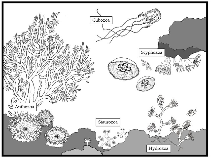
The phylum Cnidaria includes five main classes: Hydrozoa, Scyphozoa, Cubozoa, Staurozoa
and Anthozoa (Table 1; Figure 1). Basically, the unit organism in Cnidaria is the polyp, a sessile,
small (from few millimeters to less than two centimeters) gastrovascular cavity surrounded by three
layers (an internal endodermis, and an external ectodermis with an intermediate matrix, the mesoglea).
The mouth of the polyp is surrounded by tentacles, which facilitate prey capture [1]. Polyps form
different types of colonies in Hydrozoa and Anthozoa. Conversely, they live as single individuals in
Scyphozoa, Cubozoa and Staurozoa. In addition to the polyp stage, Hydrozoa, Scyphozoa, Cubozoa
and Staurozoa have a freely swimming pelagic stage (medusa), which co-exists with the polyp stage.
The fact that Hydrozoa, Scyphozoa and Cubozoa have a metagenic life cycle (Table 1) suggests that
they may have evolutionary separated from Anthozoa, which have only the polyp stage (Table 1).
Phylogenetic analyses confirmed that Anthozoa appeared earlier than the other three classes within
the evolutionary history of Metazoa, because they possess a circular DNA, in contrast with Hydrozoa,
Scyphozoa and Cubozoa, which have a linear DNA [2].
Mar. Drugs 2020, 18, 507; doi:10.3390/md18100507 www.mdpi.com/journal/marinedrugsHydrozoa Alternance polyp/medusa Nematocyst [3,4]
Scyphozoa Alternance polyp/medusa Nematocyst [3,4]
Mar. Drugs 2020, 18, 507 2 of 28
Cubozoa Alternance polyp/medusa Nematocyst [3,4]
Table 1. The main five classes of the phylum Cnidaria with their life stages and cnida traits.
Anthozoa Polyp only Nematocysts [3,4]
Class Life Stage Cnida Type Reference
Hydrozoa Alternance polyp/medusa SpirocystNematocyst
(Zoantharia only) [4] [3,4]
Scyphozoa Alternance polyp/medusa Nematocyst [3,4]
Cubozoa Alternance polyp/medusa Nematocyst [3,4]
Anthozoa Polyp only PtychocytesNematocysts
(Ceriantharia only) [5] [3,4]
Spirocyst (Zoantharia only) [4]
Ptychocytes (Ceriantharia only) [5]
Staurozoa Alternance
Staurozoa Alternance polyp/medusa
polyp/medusa Nematocysts
Nematocysts [6] [6]
Figure 1.1.The
Figure Thediverse organisms
diverse belonging
organisms to thetophylum
belonging CnidariaCnidaria
the phylum within the main classes:
within the main Hydrozoa,
classes:
Scyphozoa, Cubozoa, Staurozoa
Hydrozoa, Scyphozoa, Cubozoa,and Anthozoa
Staurozoa and(drawing
Anthozoaby(drawing
Louise Merquiol).
by Louise Merquiol).
Both
Both pelagic
pelagic and
and benthic
benthic organisms
organisms belonging
belonging toto this
this phylum
phylum possess
possess complex,
complex, specialized
specialized
organelles
organelles (cnidae) made of an amino acid matrix secreted by the Golgi system. TheThe
(cnidae) made of an amino acid matrix secreted by the Golgi system. structure
structure of
of the
the cnidae is common across Cnidaria: a capsule with collagen walls filled with a venom
cnidae is common across Cnidaria: a capsule with collagen walls filled with a venom with a coiled with a
coiled
hollowhollow thread-like
thread-like tubule.
tubule. The The tubule
tubule everts
everts after
after a amechanical
mechanicalstimulation.
stimulation. Most
Most tubules,
tubules,
particularly
particularly in the nematocysts, are able to penetrate the skin and inject the venom contained in
in the nematocysts, are able to penetrate the skin and inject the venom contained in the
the
capsule [3]. The threads have an enlargement at their basal portion (the shaft) and bear
capsule [3]. The threads have an enlargement at their basal portion (the shaft) and bear numerous numerous
and different spines. The classification of cnidae is based on the shape of the shaft and the type
and distribution of the spines [3,4]. The main classification includes nematocysts, spirocysts and
ptychocytes [3–5]. Nematocysts have two walls surrounding the capsule. Conversely, the capsule of
spirocysts has a single wall that is very thin. Hydrozoa, Scyphozoa, Cubozoa and Staurozoa possess
various types of nematocysts, while spirocytes have been found in Zoantharia and ptychocytes appear
to be a characteristic of Ceriantharia tubes (Table 1) [3–5]. Ames et al. [7] described “cassiosomes” in
the upside-down scyphomedusa Cassiopea xamachana using a combination of histology, microscopy,
microfluidics, videography, molecular biology and mass spectrometry-based proteomics. Cassiosomes
are complex stinging-cell structures found in C. xamachana mucus and likely used to kill prey.Mar. Drugs 2020, 18, 507 3 of 28
Cassiosomes are made up by an outer epithelial layer of nematocytes that is filled in the internal part by
endosymbiotic dinoflagellates, which are known to be hosted within the mesoglea of the scyphomedusa.
The mechanism of nematocyst discharge is activated by chemical and physical stimulation.
When the cnidocil is solicited, the thread, which is enveloped inside the capsule, is pushed by
the osmotic pressure outside the cell, and eventually penetrates the object, which stimulated the
cnidocil [1]. In most organisms, the injection induces paralysis, which facilitates the following capture
of the organism. The prey capture mechanism described above is basically the same for all Cnidaria,
polyp and medusa stages alike [8]. However, as reported by Moran and co-workers [9], Cnidaria
toxins may be localized in both nematocysts and ectodermal gland cells, depending on the species. For
example, Nv1 neurotoxin from the sea anemone Nematostella vectensis is confined to the ectodermal
gland cells, while Anemonia viridis Type I toxins are localized in both nematocysts and ectodermal
gland cells [9].
2. Venoms of Cnidaria
The composition of cnidarian venoms is not known in detail, but they appear to contain mainly
a variety of proteinaceous (peptides, proteins, enzymes and proteinase inhibitors) together with
non-proteinaceous compounds (purines, quaternary ammonium compounds, biogenic amines and
betaines) [10–14] (Table 2). The main venom components identified to date have a maximum molecular
mass of 220 kDa. Phopholipase A2 appears to be common across all cnidarian classes [15]. Anthozoans
contain several inhibitors of both sodium and potassium voltage-gated channels [11]. The channel
binding site is not always known. However, as reported by [11,16], receptor site three has been
identified as the binding site for sea anemone Type I and Type III sodium channel inhibitors. Toxins
have been recently reviewed in Scyphozoa [13,14,17] and in sea anemones (Actinaria) [11,14]. In this
review, we provide an updated synopsis of the main compounds identified to present in the venom of
cnidarians and discuss the recent application of omics and biotechnological tools in the field.
Table 2. Toxins identified in cnidarian species, including their molecular mass (kDa) and biological
activity. N/A means not available. TRPV1 stands for Vanilloid Receptor 1, TRPA1 stands for transient
receptor potential ankyrin 1 ion channel and ASIC stands for acid-sensing ion channel.
Species Compound Molecular Mass (kDa) Biological Activity References
Hydrozoa
Hydra magnipapillata CqTX-A ~40 Cardiovascular, hemolytic [18]
HALT-1 N/A Hemolytic, cytolysis [19]
HALT1–HALT7 N/A Cytolytic [20]
H. viridissima Hydralysin 27 Neurotoxic, cytolytic, paralytic [21]
Millepora platyphylla Milleporin-1 30–34 Cytolytic, hemolytic [22]
Cytolytic, hemolytic, myonecrotic, and
Olindias sambaquiensis Oshem1 3.013 [23]
cytotoxic
Oshem2 3.376 Cytolytic [23]
Metalloproteinases N/A Cytolytic, neurotoxic [24]
Physalia physalis Phospholipase A2 N/A N/A [25]
Phospholipase B N/A N/A [25]
Collagenase 25 Cytotoxic, hemolytic [26]
Elastases N/A Musculotoxic, cytolytic, hemolytic [27]
PpV19.3 4.72 Neurotoxic, cardiotoxic [28]
PpV9.4 0.55 Hemolytic [28]
P3 85 Neurotoxic [29]
P1 220 Neurotoxic [30]
Physalitoxin 220 Hemolytic [31]
DNase 75 Cytolytic [32]
Histamine N/A N/A [33]Mar. Drugs 2020, 18, 507 4 of 28
Table 2. Cont.
Species Compound Molecular Mass (kDa) Biological Activity References
Tubularia larynx Phospholipase A2 N/A Cytolytic, hemolytic [15]
Scyphozoa
Aurelia aurita Phospholipase A2 N/A Cytolytic [15,34]
Hemolytic, neurotoxic, myotoxic, local
Proteolytic enzymes N/A [35]
skin irritation
Tetramine and
N/A Dermotoxic, temporary paralysis, edema [36]
unidentified protein
Metalloproteinases N/A Gelatinolytic, caseinolytic, fibrinolytic [37]
Antimicrobial, neurotoxic (voltage-gated
Aurelin 4.30 [38]
potassium channel inhibitor)
Hemolytic, dermonecrotic, local skin
Cassiopea andromeda Phospholipase A2 N/A [34]
irritation
Hemolytic, dermonecrotic, local skin
C. xamancha Phospholipase A2 N/A [34]
irritation
Chrysaora hysoscella Cationic protein N/A Dermotoxic, cytotoxic [39]
C. quinquecirrha DNase 110 Dermonecrotic, cytotoxic [26]
Acid protease 120–150 N/A [26]
Metallopeptidase 100 N/A [26]
Collagenase N/A N/A [26]
Cyanea capillata Basic protein(s) 70 Cardiotoxic, dermonecrotic, musculotoxic [26,40]
CcTX-1 31.173 Cytotoxic [41]
CcNT 8.22 Neurotoxic [42]
Phospholipase A2 N/A Cytolytic, cytotoxic, hemolytic [15,43]
C. lamarckii ClGP-1 27 Cytotoxic [43]
Phospholipase A2 N/A Cytolytic, cytotoxic, hemolytic [43]
C. nozakii Metalloproteinases N/A Gelatinolytic, caseinolytic, fibrinolytic [37]
Metalloproteinases 28–36 Gelatinolytic, caseinolytic, fibrinolytic [37]
Nemopilema nomurai
20–40/10–15 Cytotoxic, hemolytic [44]
Proteinaceous Hemolytic, cytotoxic, dermonecrotic,
Pelagia noctiluca 44–66 [45–49]
macromolecules hemolytic, local tissue damage
Phyllorhiza punctata Phospholipase A2 N/A Neurotoxic [50]
Rhizoprotease 95 Proteolytic, hemolytic [51]
Rhizostoma pulmo
Rhizolysin 260 Hemolytic [52]
N/A Cytotoxic, hemolytic [53]
Metalloproteinases N/A Gelatinolytic, caseinolytic, fibrinolytic [37]
Rhopilema esculentum
Degradation of extracellular matrix
Hyaluronidase 55–95 [37]
components
N/A Proteolytic, cytotoxic, hemolytic [54,55]
R. nomadica Phospholipase A2 N/A Hemolytic [56]
Serine protease N/A Local skin damage [57]
Rhopilema spp. Phospholipase A2 N/A Hemolytic [58]
SmP90 90 Radical scavenging [59]
Stomolophus meleagris
Cytotoxic, cytolytic, hemolytic, local
Phospholipase A2 N/A [59]
tissue damage
218 toxins including
Voltage-gated potassium channel
C-lectin and N/A [59]
inhibitor
metalloprotease
Cubozoa
Alatina moseri CaTX-A 43 Hemolytic [18]
Akatina alata CAH1 42 Hemolytic [60]
CaTX-A 43 Hemolytic [61]
CaTX-B 45 Hemolytic [61]Mar. Drugs 2020, 18, 507 5 of 28
Table 2. Cont.
Species Compound Molecular Mass (kDa) Biological Activity References
C. marsupialis Haemolysin 102–107 Hemolytic [62]
CmHl1 139 Cytolytic [63]
CmHl5 220 Cytolytic [63]
CmH17 139 Cytolytic [63]
CmNt 120 Neurotoxic [63]
C. rastonii Phospholipase A2 N/A Cytolytic [15]
CrTX-I N/A Hemolytic [64]
CrTX-II N/A Hemolytic [64]
CrTX-III N/A Hemolytic [64]
CrTX-A 43 Cutaneous inflammation of human skin [61]
CrTX-B 46 N/A [61]
Phospholipase A2 N/A Cytolytic, hemolytic [15]
Carukia barnesi
CbTX-I 21.67 Neurotoxic [65]
CbTX-II 18.16 Neurotoxic [65]
Chironex fleckeri Phospholipase A2 N/A Cytolytic, hemolytic [15]
Metalloproteinases 17–130 N/A [66]
Cardiotoxic, cytotoxic, dermonecrotic,
CfTX-1 43 [67]
lethal
Cardiotoxic, cytotoxic, dermonecrotic,
CfTX-2 45 [67]
lethal
CfTX-A 40 Hemolytic [18]
CfTX-B 42 Hemolytic [18]
CfTX-Bt 31.29 N/A [18]
Chiropsalmus
CqTX-A 44 Hemolytic, neurotoxic, myotoxic [58,68,69]
quadrigatus
MkTX-A; MkTX-B 43–45 Dermonecrotic, inflammatory [65]
Malo kingi
43–45 Dermonecrotic, inflammatory [49]
Anthozoa (Hexacorallia)
Acropora spp. Phospholipase A2 N/A Catalytic [15]
Actinia australis Phospholipase A2 N/A Catalytic [15]
Lethal activity to crabs, Type I sodium
Actinia equina AeI N/A [70]
channel toxin
AEPI-I, II, III and IV 6.2–7 Kunitz-type toxins [71]
Equinatoxin-I, II and
19 Cytolytic and hemolytic [72]
III
Equinatoxin-IV N/A Hemolytic [73]
Equinatoxin-V N/A N/A [74]
Acrorhagin I and II N/A Lethal activity to crabs [75]
Acrorhagin Ia and IIa N/A N/A [75]
Actinia fragacea Fragaceatoxin C 20 Lytic [76]
Actinia tenebrosa Tenebrosin-A, B and C 19–20 Hemolytic [77]
Actinia villosa Avt-I 19 Hemolytic [78]
Avt-II N/A Cytolysis [78,79]
AvTX-60A 60 Fatal toxicity to mice [80]
Avt120 120 Lethal activity to mice, cytotoxic [81]
Adamsia palliata AcPLA2 13.5 Phospholipase A2 catalytic activity [82]
Adamsia carciniopados Phospholipase A2 N/A Phospholipase A2 catalytic activity [15]
Crab lethality, Type I sodium channel
Anemonia erythraea AETX-I 5 [83]
toxin
AETX-K ~4 Type I potassium channel toxin [84]
AETX-II 6.5 Crab lethality [83]
AETX-III 6.6 Crab lethality [83]Mar. Drugs 2020, 18, 507 6 of 28
Table 2. Cont.
Species Compound Molecular Mass (kDa) Biological Activity References
Anemonia sulcata Toxin I N/A Type I sodium channel toxin [85]
Cardiotoxic action, Type I sodium
ATX-II 5 [62,85]
channel toxin
Toxin III 2.7 N/A [86]
ATX-V N/A Type I sodium channel toxin [87]
SA5 II N/A Kunitz-type proteinase toxin [88]
Type II potassium channel toxin, Kunitz
Kalicludin 1, 2, 3 ~6.7 [89]
inhibitors
BDS-I and BDS-II 4.7 Inactivating Kv3.4 channel [78]
Kaliseptine 3.8 Potassium channel toxin [89]
Anthopleura asiatica Bandaporin 20 Hemolytic [90]
Anthopleura Cardiotoxic to rats, Type I sodium
Anthopleurin-C N/A [91]
elegantissima channel toxin
APE 1-1, 1-2, 2-1, 2-2, 3, Crab paralysis, Type I sodium channel
5 [92]
4 and 5-3 toxin
ASIC3 inhibition (acting on channel
APET x2 4.6 [93]
external side)
Anthopleura fuscoviridis AFT-I and II N/A Type I sodium channel toxins [94]
Anthopleura Cardiotoxic to rats, Type I sodium
Anthopleurin-A and B N/A [91]
xanthogrammica channel toxin
Toxin PCR1-2, 2-1, 2-5,
N/A Type I sodium channel toxin [95]
2-10, 3-6, 3-7, 3-3,4
AXPI-I and II N/A Kunitz-type inhibitor [96]
Hk2a, Hk7a, Hk8a, Rat heart stimulation, Type I sodium
Anthopleura spp. ~5 [97]
Hk16a channel toxins
Bolecera tuedia Phospolipase A2 N/A Phospholipase A2 catalytic activity [15]
Toxic on crustacean nerves, Type I
Bunodosoma caissarum Bc-I, II and III N/A [98]
sodium channel toxin
Weak-paralyzing action in swimming
Bc-IV ~5 [99]
crabs
Renal function and induced insulin
BcPLA(2)1 ~5 secretion in conditions of high glucose [100]
concentration
BcsTx3 5.71 Potassium channel inhibition [101]
Neurotoxic on mice, Type I sodium
Bunodosoma granulifera Bg II and III N/A [102]
channel toxins
Bg toxin ~4 Type I potassium channel toxin [103]
Severe neurotoxic effects such as circular
movements, aggressive behavior,
Granulitoxin (GRX) ~5 [104]
dyspnea, tonic-clonic convulsion and
death in mice
Bunodosoma cangicum Cangitoxin 5 Type I sodium channel toxin [105]
Cangitoxin II and III
(CGTX-II and ~5 Type I sodium channel toxin [106]
CGTX-III)
Type I potassium channel toxin,
Bcg 25.96, 28.19, 30.24 ~4 [105]
Neurotoxic to crabs
Bcg 31.16, 28.78, 25.52, May target different types of ion channels,
4–5 [105]
29.21 Neurotoxic to crabs
Possible new class of potassium channel
Bcg 21.75, 23.41, 21.00 3 [105]
blockers, Neurotoxic to crabs
Increased transmitter release, causing
repetitive firing of the axons in
Calliactis parasitica Calitoxin 1 ~5 [107]
neuromuscular preparation of
crustaceans
Calitoxin 2 N/A N/A [108]
Condylactis gigantea CgNa ~5 Type I sodium channel toxin [109]Mar. Drugs 2020, 18, 507 7 of 28
Table 2. Cont.
Species Compound Molecular Mass (kDa) Biological Activity References
Lethal activity against crabs, Type I
Condylactis passiflora CpI, II and III N/A [110]
sodium channel toxin
Cryptodendrum
Ca I N/A Type II sodium channel toxin [111]
adhaesivum
Dendronephthya spp. Phospholipase A2 N/A Phospholipase A2 catalytic activity [15]
Halcurias carlgreni Halcurin ~5 N/A [112]
Heteractis crispa
(=Radianthus crispus = Neurotoxin I N/A Type II sodium channel toxin [113]
R. macrodactyla)
Neurotoxin II N/A Type II sodium channel toxin [114]
Neurotoxin III N/A Type II sodium channel toxin [115]
Neurotoxin IV and V N/A Type II sodium channel toxins [116]
Lethal against crabs, Type I sodium
Rc I N/A [117]
channel toxin
Kunitz-type Trypsin
N/A Kunitz-type inhibitor [118]
inhibitor IV
Actinoporin RTX-A N/A Hemolytic [119]
Actinoporin RTX-S II 19 Hemolytic [120]
HCIQ2c1 6 Neuroprotective, Kunitz-type inhibitor [121]
Hcr 1b-1 4.5 ASIC3 channel inhibitor [122]
Hcr1b-2, Hcr1b-3,
5 Neurotoxic (ASIC1 channel inhibitors) [123]
Hcr1b-4
Anti-inflammatory, Kunitz-type
HCRG1, HCRG2 6 [124]
inhibitors
InhVJ 6 Kunitz-type inhibitor [125]
Analgesic, inhibitor of TRPV1,
HGRC21 6 [126]
Kunitz-type inhibitor
APHC1, APHC2,
6 Analgesic (TRPV1 modulation) [127–129]
APHC3
HCGS 1.10 6 Analgesic [130]
HCGS 1.20 6 Anti-inflammatory [131]
rHCGS1.19 and Antihistamine, Kunitz-type serine
6 [132]
rHCGS1.36 protease inhibitors
Heteractis magnifica
(=Radianthus magnifica Magnificalysins I and
~19 Hemolytic and lethal activity on mice [133]
= R. paumotensis = II
R. ritteri)
HMGS1 6 Kunitz-type inhibitor [134]
HMgIII Hemolytic and cytolytic [135]
HmK ~4 Potassium channel inhibitor [136]
RpI, RpII, RpIII, and Toxic in mice and crabs, Type II sodium
~5 [137]
RpIV channel toxins
HMIQ3c1 N/A Neuroprotective (Kunitz-type inhibitor) [138]
Magnificamide 4.7 Alpha-amylase inhibitor [139]
δ-TLTX-Hh1a and Lethal on crabs, Type II sodium channel
Heterodactyla hemprichi N/A [111]
δ-TLTX-Hh1c toxin
Metridium senile Metridin 3.97 Hemolytic [140]
Phospholipase A2 N/A Phospholipase A2 catalytic activity [15]
Ms 9a-1 3.6 Analgesic by potentiating TRPA1 [141]
Nv1116.25.1, 116.27.1,
116.28.1, 116.37.1,
Nematostella vectensis N/A Type II sodium channel toxins [142]
116.39.1, 116.40.1,
116.41.1, 116.45.1
Nv4 – Nv8 N/A Sodium channel inhibitors [143]
NEP1 – NEP20 N/A N/A [144]Mar. Drugs 2020, 18, 507 8 of 28
Table 2. Cont.
Species Compound Molecular Mass (kDa) Biological Activity References
Actinoporin Or-A and
Oulactis orientalis ~18 Cytolytic [145]
Or-G
NEP1 to NEP 20 N/A N/A [144]
Parasicyonis
PA-TX N/A N/A [146]
actinostoloides
Phyllodiscus semoni PsTX-115 N/A Renal injury [147]
PsTX-60A; PsTX-60B 60 Cytolytic, hemolytic [148]
Phymanthus crucifer PhcrTx1 3.47 ASIC inhibitor [149]
Pocillopora damicornis Phospholipase A2 N/A Phospholipase A2 catalytic activity [15]
Sagartia rosea Cytolysin Src-I 19.6 Cytolysis [150]
Sarcophyton elegans Phospolipase A2 N/A Phospholipase A2 catalytic activity [15]
Neurotoxic on crabs, Type II sodium
Stichodactyla helianthus Sh1 ~5 [151]
channel inhibitor
Helianthamide 4.7 Alpha-amylase inhibitor [152]
SHPI-1 ~6 Kunitz-type proteinase inhibitor [153]
ShK ~5 Potassium channel inhibitor [154]
Sticholysin-I and II ~19 Hemolytic [155]
Stoichactis sp. Phospholipase A2 N/A Phospholipase A2 catalytic activity [15]
Stichodactyla gigantea Gigantoxins I-III N/A Crab toxicity, Sodium channel inhibitors [156]
Crab-paralyzing activity, SHTX-III is a
Stichodactyla haddoni SHTX I–III ~7 [157]
Kunitz-type proteinase inhibitor
Crab lethality, Type II sodium channel
SHTX IV ~5 [157]
toxin
EGF-like peptide
N/A N/A [157]
SHTX-5
Thalassianthus aster Ta I3,8-7,6 N/A Type II sodium channel toxin [111]
Urticina coriacea U1 N/A Hemolytic, cytotoxic [124]
U2 3–10.5 Analgesic (ASIC1 channel inhibitor) [124]
Urticina crassicornis Uc-I 30 Cytolysis [158]
Antibacterial, analgesic by potentiating
Urticina eques Ueq 12-1 4.79 [159]
TRPA1
Urticina grebenyi UGR9a-1 N/A Analgesic, ASICS channel inhibitor [160]
Urticina piscivora Up-I 28 Hemolytic [161]
Virgularia nidularis Phospholipase A2 N/A Phospholipase A2 catalytic activity [15]
Anthozoa (Octocorallia)
Alcyonum digitatum Phospholipase A2 N/A Phospholipase A2 catalytic activity [15]
Paramuricea spp. Phospholipase A2 N/A Phospholipase A2 catalytic activity [15]
Sinularia flexibilis Phospholipase A2 N/A Phospholipase A2 catalytic activity [15]
2.1. Phospholipase A2
Phospholipase A2 induces the breakdown of glycerophospholipds, which produces
lysophospholipid and fatty acids, such as the arachidonic acid [162]. The metabolites derived from
arachidonic acid (prostaglandins, thromboxanes and leukotrienes) control a variety of cellular functions,
including dietary lipid catabolism, in cell membrane metabolism and inflammatory diseases [163].
Phospholipase A2 is common in mammalians but also across venomous animals. It has been
identified in reptiles (snakes and anguimorph lizard), centipedes, insects (their bristles, proboscises,
and stingers), arachnids (scorpions, spiders, and ticks), cnidarians and cephalopods [15,164].
Toxic functions of phospholipase A2 in cnidarian venoms have been proposed to include defense,
immobilization and digestion of prey [15]. The first cnidarians phospholipase A2 fully sequenced was
published in 2002 for Adamsia carcinoapados [82].Mar. Drugs 2020, 18, 507 9 of 28
2.2. Metalloproteinases
Metalloproteinases include a large variety of proteinase enzymes, which host a metal atom to
perform their catalytic activity. They are found in the venom of terrestrial animals, such as centipedes,
snakes and ticks [164,165]. They induce hemorrhage and necrosis by degrading the extracellular
matrix and preventing blood clot formation [164,166]. These functions are commonly associated
with several symptoms following a sting (skin damage, edemas, blister formation, myonecrosis and
inflammation) [166]. They appear to be common in stinging jellyfish, such as Stomolophus meleagris [59]
and Chironex fleckeri [66].
2.3. Voltage-Gated Sodium and Potassium Channel Toxins
Any ion channel family generally comprises various subtypes with particular physiological,
pharmacological and structural characteristics [167]. The voltage-gated ion channels play a crucial
role in the excitability of cells and neuromuscular transmission of signals. Voltage-gated ion channels
activate non-selective pores within membranes by which the ions can pass using the electrochemical
gradient across the membrane itself [167]. When this mechanism is altered, the transmission of
signals through the neurons and muscles is critically changed too, which can lead to certain disorders,
including paralysis.
Voltage-gated sodium channel toxins have been isolated since the 1970s in Anthozoa [11], and they
account for the most abundant fraction in their venom [16]. These toxins, whose molecular mass
ranges from 3.5 to 6.5 kDa, are able to bind specifically with the receptor site three of the sodium
channel, and regulate their functioning [168]. By controlling the opening and closing of the sodium
channel, the toxins control the electrical signals that encode and propagate vital information across
long distances. The activity of the sodium channel toxins suggests that they may find application as
pain blockers. Conversely, Kalima and co-workers [169] suggested that the sodium channel inhibitors
extracted from Heteractis crispa were not appropriate for pharmacological applications, but might be
used to study the mechanisms beyond sodium transportation and to produce insecticides.
Compared to the voltage-gated sodium channel toxins, voltage-gated potassium channel toxins
were discovered during the 1990s [11]. Recently, toxins acting on the voltage-gated potassium channel
have been investigated for the treatment of multiple sclerosis and other autoimmune diseases [170].
2.4. Kunitz-Type Proteinase Inhibitors
Kunitz-type proteinase inhibitors have been identified in several diverse species of cnidarians,
but exclusively anthozoans (Table 2). Their active domains, also known as Kunitz/BPTI domains, are
relatively small, with a molecular weight of about 6 kDa. Before the discovery of potassium channel
Type II toxins, sea anemone proteinase inhibitors were considered to serve as inhibitors of endogenous
proteinases in the sea anemones themselves and as protectors of the toxins injected into prey and
predators from rapid degradation. However, the identification of potassium channel toxins with
proteinase inhibitory activity suggests that the offensive role of sea anemone proteinase inhibitors,
by paralyzing prey, may be more important than their defensive role [171]. The Kunitz-type IQ-peptide
HMIQ3c1, extracted from the venom of Heteractis magnifica, has shown neuroprotective activity,
which may find application in the treatment of neurodegenerative diseases, such as Alzheimer’s [138].
Another Kunitz-type proteinase inhibitor that may support the treatment of Parkinson disease is
HCIQ2c1 extracted from Heteractis crispa [121]. This study shows for the first time that Kunitz-peptides
significantly increase neuroblastoma cell viability in an in vitro 6- hydroxydopamine-induced
neurotoxicity model of Parkinson’s disease [121].
2.5. TRPV1 Channel Inhibitors
The transient receptor potential vanilloid 1 (TRPV1s) are transmembrane non-selective cation
channels in mammalian peripheral and central neuronal systems. Because they control the response ofMar. Drugs 2020, 18, 507 10 of 28
the neurons during inflammation, they are considered among the most important molecular triggers
of pain stimuli [172]. The first TRPV1 peptide inhibitor from sea anemone venoms was τ-SHTX-Hcr2b
(APHC1) from Heteractis crispa [127]. Research on the TRPV1 inhibitors has continued on this species,
from which the other three homologous peptides, τ-SHTX-Hcr2c (APHC2), τ-SHTX-Hcr2d (APHC3)
and HCRG21, have been isolated [126,129]. These three peptides showed analgesic activity in an
in vivo heat stimulation model [130,139]
2.6. TRPA1 Channel Modulators
The transient receptor potential ankyrin 1 ion channel (TRPA1) is a nociceptor [173] that has been
suggested to serve as the main mechanical and chemical stress sensor. Agonists of TRPA1 activate the
sensory neurons in vivo, which cause acute pain, thermal and mechanical hyperalgesia and neurogenic
inflammation. This receptor is similar to the transient receptor potential vanilloid 1 (TRPV1), with
which it is usually co-expressed [174]. Another TRPA1 modulator, named Ms 9a-1, was isolated from
Metridium senile and showed an analgesic effect [141]. In addition, Logashina and co-workers [159]
isolated Ueq 12-1 from Urticina eques, which belongs to a group of toxins never identified previously.
The antibacterial activity and the moderate enhancement of TRPA1 suggest that this peptide may find
large application as an analgesic with antibacterial properties [159].
2.7. ASICs Channels Modulators
ASICs are sodium-selective, acid-sensing ion channels in the peripheral nervous system,
particularly activated during inflammation and ischemia. Peptides π-AnmTX Ugr 9a-1 from the
venom of the sea anemone Urticina grebelnyi [160] and PhcrTx1 from Phymanthus crucifer [149] target
ASIC channels. These peptides are cross-linked by two disulfide bridges and have no sequence
homology to other sea anemone neurotoxin peptides [10]. In addition, Hcr 1b-1, Hcr 1b-2, Hcr 1b-3 and
Hcr 1b-4 have been also identified from the sea anemone Heteractis crispa and have shown neurotoxic
activity by inhibiting the ASIC channels. In particular, Hcr 1b-1 is a specific ASIC3 inhibitor and
Hcr 1b-2 is able to inhibit both ASIC1a and ASIC3 channels; the others are specific for the ASIC1a
channel [123].
2.8. Beta-Defensin Like Alpha-Amylase Inhibitors
Among animals, amylase inhibitors have been identified only in sea anemones, which are among
the most ancient phyla that appeared on Earth [2]. Helianthamide, a potent inhibitor of human
pancreatic α-amylase produced by the Caribbean Sea anemone Stichodactyla helianthus, is the first
representative of a new group of amylase inhibitors that was isolated in 2016 [175], while Magnificamide
is the second representative of such inhibitors. Both peptides appear to be promising compounds
to manage obesity and type 2 diabetes [139]. The biological relevance of the presence of α-amylase
inhibitors in the Cnidaria venoms remains largely unexplained. It is hypothesized that inhibition of
the α-amylase activity intervenes with the metabolism of carbohydrates, a major source of energy for
many organisms [139].
3. Omics Applied to Cnidaria to Identify Chemical Defences
Recently, omics technologies (genomics, transcriptomics, proteomics and metabolomics) have
helped to better understand the ecology, distribution and defence strategies of marine organisms,
as well as having speeded up studies on their possible biotechnological applications. Available studies
on defence strategies in Cnidaria mainly involve deadly toxins released to capture prey, defend
themselves or fight for territorial acquisition.
Considering the available genomes, there are 6 Hydrozoa, 10 Scyphozoa, 4 Cubozoa, 18 Anthozoa,
1 Staurozoa and 7 Myxozoa genomes deposited in the public database GenBank (Table 3). For many of
them, sequences have been deposited during the last year and the related publications are not available
yet. Several transcriptomic, proteomic and metabolomic studies are available as well, but few of themMar. Drugs 2020, 18, 507 11 of 28
focused on defence activities. Recently, in order to shed light on the molecular basis of coral responses
to environmental changes and reef-building coral strategies for conservation, several genomes and
transcriptomes have been sequenced (Table 3).
Table 3. Cnidaria genome deposited in Genbank, their Assembly Accession Numbers (accessed on
8 July 2020) and references. N.A. stands for not available.
Species Accession Number Reference
Hydrozoa
Clytia hemisphaerica GCA_902728285.1 N.A.
Craspedacusta sowerbii GCA_003687565.1 N.A.
Hydra oligactis GCA_004118135.1 [176]
Hydra viridissima GCA_004118115.1 [176]
Hydra vulgaris GCA_000219015.1 [177]
Hydra vulgaris GCA_000004095.1 [177]
Scyphozoa
Aurelia aurita GCA_004194415.1 N.A.
Aurelia aurita complex sp. pacific GCA_004194395.1 N.A.
Aurelia coerulea GCA_011634815.1 N.A.
Cassiopea xamachana GCA_900291935.1 N.A.
Chrysaora quinquecirrha GCA_012295145.1 N.A.
Chrysaora chesapeakei GCA_011763395.1 N.A.
Chrysaora fuscescens GCA_009936425.1 N.A.
Nemopilema nomurai GCA_003864495.1 N.A.
Rhopilema esculentum GCA_013076305.1 N.A.
Sanderia malayensis GCA_013076295.1 N.A.
Cubozoa
Alatina alata GCA_008930755.1 [178]
Alatinida sp. Z8VKAUB7J3 GCA_010016025.1 N.A.
Carybdea marsupialis auct. non (Linnaeus, 1758) GCA_010016065.1 N.A.
Morbakka virulenta GCA_003991215.1 N.A.
Anthozoa
Actinia equina GCA_011057435.1 [179]
Actinia tenebrosa GCA_009602425.1 [180]
Acropora digitifera GCA_000222465.2 [181]
Acropora millepora GCF_004143615.1 [182]
Anemonia viridis GCA_900234385.1 N.A.
Dendronephthya gigantea GCA_004324835.1 [183]
Exaiptasia diaphana GCA_001417965.1 [184]
Heteractis magnifica GCA_011763375.1 N.A.
Montipora capitata GCA_006542545.1 [185]
Nematostella vectensis GCA_000209225.1 [186]
Orbicella faveolata GCA_002042975.1 N.A.
Orbicella faveolata GCA_001896105.1 [187]
Phymanthus crucifer GCA_009858155.1 N.A.
Pocillopora damicornis GCA_003704095.1 [188]
Porites rus GCA_900290455.1 N.A.
Renilla reniformis GCA_900177555.1 N.A.
Stichodactyla mertensii GCA_011800005.1 N.A.
Stylophora pistillata GCA_002571385.1 [189]
Staurozoa
Calvadosia cruxmelitensis GCA_900245855.1 N.A.
Myxozoa
Enteromyxum leei GCA_001455295.2 [190]
Henneguya salminicola GCA_009887335.1 N.A.
Kudoa iwatai GCA_001407235.2 [190,191]
Kudoa iwatai GCA_001407335.1 [191]
Myxobolus squamalis GCA_010108815.1 N.A.
Sphaeromyxa zaharoni GCA_001455285.1 [191]
Thelohanellus kitauei GCA_000827895.1 [192]Mar. Drugs 2020, 18, 507 12 of 28
Gene families encoding toxins are found in many venomous species, but there is still limited
understanding of their evolution. Genome sequencing and analyses have started to help shed light on
toxins and their synthesis. Sea anemones may produce various toxic compounds (such as peptide
toxins found in their venoms), which can have potential therapeutical applications. For example,
the genome of the sea anemone Nematostella vectensis enabled to study a gene family whose neurotoxin
product, Nv1, affects voltage-gated sodium channels [142]. In particular, the gene family members
identified (116.25.1, 116.27.1, 116.28.1, 116.37.1, 116.39.1, 116.40.1, 116.41.1 and 116.45.1) clustered in
a highly repetitive approximately 30-kb genomic region and encoded the toxin Nv1. Transcriptome
analyses of the sea anemone Nematostella vectensis allowed to identify the sequences encoding precursor
proteins homologous to the Nv1 toxin (named as Nv4, Nv5, Nv6, Nv7 and Nv8). In addition, Sachkova
and co-workers showed that the new toxins, Nv4 and Nv5, were lethal for zebrafish larvae but harmless
to arthropods, and were localized to ectodermal gland cells in larvae [143]. Recently, the genome
of Actinia tenebrosa was sequenced and annotated. Bioinformatic analyses showed that the genes
encoding toxins contributed to a significant proportion of the lineage-specific genes and gene families,
giving new insights into the evolution of toxins and possibly guiding the discovery of novel bioactive
compounds from Cnidaria.
Regarding the transcriptome data, there are 35 BioProjects and 62 sequence read archives (SRA)
deposited in GenBank (accessed on August 25th, 2020). A limited number of them are transcriptomes
related to defence strategies in Cnidaria. An example of a transcriptomic study (using an Illumina
HiSeq 2500 automatic sequencing platform) was performed by Huang et al. [193] for the sea anemone
Protopalythoa variabilis. The transcriptome analyses identified various predicted polypeptides with
canonical venom protein features belonging to various toxin families: neurotoxic peptides, hemostatic
and hemorrhagic toxins, pore-forming proteins, proteinase inhibitors, mixed-function venom enzymes
and venom auxiliary proteins. Two of these predicted toxin products, ShK/Aurelin-like peptide
and a novel anthozoan neurotoxin-like peptide, were further investigated and displayed potent
in vivo neurotoxicity that impaired swimming in larval zebrafish. A complex array of venom-related
transcripts was identified and some of them are reported in Cnidaria for the first time, giving new
insights in toxin distribution among species and their evolution.
Another transcriptome study (using an Illumina HiSeq 2500 Sequencing System) by Ames et
al. [194] focused on sequencing and analysing the transcriptomes of adult and larval tissues of the
cubozoan Alatina alata, which is emerging as a cnidarian model because it forms predictable monthly
nearshore breeding aggregations in tropical to subtropical waters. Differential expression analyses
were performed to identify the candidate genes involved in nematogenesis and venom production,
giving a boost for further investigations on the evolution of distinctive characteristics of cubozoans.
In particular, various candidate genes implicated in predation, defence, vision and phototransduction
pathways, sexual reproduction and embryogenesis were identified, which may be considered for
further studies on cubozoan/cnidarian evolution.
Li and collaborators [59] used a combination of transcriptomic and proteomic approaches in
order to shed light on the toxic jellyfish Stomolophus meleagris. Venom proteomics was performed by
tryptic digestion of the crude venom followed by RP-HPLC separation and MS/MS analysis of the
tryptic peptides. The venom gland transcriptome was analysed using an Illumina sequencing platform
(HiSeq™ 2000) with de novo assembly. A total of 218 toxins were identified, including the C-type lectin,
phospholipase A2 , potassium channel inhibitors, serine proteinase inhibitors, metalloproteinases and
hemolysins [59].
Very recently, Koch and Grimmelikhuijzen [195] used a software to annotate neuropeptides in the
publicly available genomes and transcriptomes from Cubozoa, Scyphozoa and Staurozoa (which all
belong to the subphylum Medusozoa) and compared these results with the neuropeptides present in
Octocorallia (class Anthozoa). Three to six neuropeptide preprohormone genes were identified within
members of the abovementioned cnidarian classes, each coding for several (up to thirty-two) similar or
identical neuropeptide copies [195]. Two of these neuropeptide preprohormone genes were presentMar. Drugs 2020, 18, 507 13 of 28
in all the cnidarian classes investigated, and they were supposed to be among the first neuropeptide
genes evolved in cnidarians.
Ponce et al. [196] used an integrated transcriptomic (using an Illumina HiSeq 2000 system) and
proteomic approach to identify putative toxins and their potential role in the venom of the scyphozoan
Chrysaora fuscescens. The de novo tentacle transcriptome contained more than 23,000 contigs and a
total of 163 proteins were identified in the venom proteome of C. fuscescens. Of these proteins, 27 were
classified as putative toxins and grouped into six protein families: proteinases, venom allergens, C-type
lectins, pore-forming toxins, glycoside hydrolases and enzyme inhibitors. Interestingly, other putative
toxins were identified in the transcriptome, but not in the proteome (they were probably not expressed
in the moment of the experiment), such as other proteinases, lipases and deoxyribonucleases. Sequence
analysis also revealed the presence of ShKT domains (domains found in a group of potent potassium
channels blockers originally isolated from sea anemones) in two putative venom proteins from the
proteome and 15 from the transcriptome, suggesting potential ion channel blockade activities. Analysis
of the venom proteomes has recently become more feasible and is generally used to study which are
the possible proteins responsible for the most severe symptoms of jellyfish stings and also for guiding
the identification of proteins with potential therapeutic applications [196].
Knowledge of toxic peptides in cnidarians is very limited due to the small number of toxins
(mainly from sea anemones) identified to date by traditional protein analyses. Protein analyses of
nematocysts of the hydromedusa Olindias sambaquiensis allowed to identify 29 putative toxins using
a high throughput proteomics platform [24]. The data revealed 29 potential toxins homologous to
toxic proteins from diverse animal phyla, including cone snails, snakes, spiders, scorpions, wasps,
bees, parasitic worms and other Cnidaria. The presence of several toxic enzymes has been observed,
such as sphingomyelin phosphodiesterase B (previously described in some spider venoms) and a
prepro-haystatin P-IIId snake venom metalloproteinase, which is very rare, even within snake venoms.
Similarly, Brinkman and co-workers [197] used a proteomic approach to identify the protein
components of the venom from the cubozoan Chironex fleckeri. Collectively, 61 proteins were identified,
including toxins and proteins important for nematocyte development and nematogenesis. Venom
proteins and their post-translational modifications were also further characterized using toxin-specific
antibodies and phosphoprotein/glycoprotein-specific stains. Data showed that glycosylation is a
common post-translational modification of the toxin family. In addition, a lack of cross-reactivity by
toxin-specific antibodies was observed, suggesting that there is significant divergence in toxin structures.
A proteomic analysis of the most venomous jellyfish in the Mediterranean Sea, Pelagia noctiluca,
was performed to study the jellyfish proteins involved in defence, body constituents and metabolism,
and also explored the potential application of such bioactive molecules [198]. The results allowed to
identify for the first time in P. noctiluca a zinc metalloproteinase, a red fluorescent protein (RFP) and a
peroxiredoxin. Zinc metalloproteinase was previously reported in the venom of other jellyfish species,
it has a ShK toxin domain and therefore should be implicated in P. noctiluca toxicity. The RFP is an
important family of proteins with possible applications as molecular markers, while peroxiredoxin is a
known antioxidant and suggest this scyphozoan species as a potential natural source of antioxidants
and anti-UV radiation agents.
Zaharenko et al. [105] reported the first peptide mass fingerprint of the sea anemone Bunodosoma
cangicum venom and, in particular, of the neurotoxic (on crabs) fraction named FrIII. This proteomic
approach allowed them to identify some novel peptides. FrIII was purified obtaining 41 fractions.
Between the 81 components present in these fractions, three groups of toxic peptides were identified.
Bcg 25.96, Bcg 28.19 and Bcg 30.24 of about 4 kDa were identified as Type I potassium channel toxins.
Bcg 31.16, Bcg 28.78, Bcg 25.52 and Bcg 29.21, with a molecular mass between 4 and 5 kDa, showed
sequence features characteristic of toxins that may target different types of ion channels (e.g., the human
cardiac potassium channel HERG and the human acid-sensing ion channel ASIC3). Finally, Bcg 21.75,
Bcg 23.41 and Bcg 21.00 were characterized as a possible new class of potassium channel blockers
neurotoxic to crabs.Mar. Drugs 2020, 18, 507 14 of 28
Comparative proteomics (by mass spectrometry analyses) was performed to compare the soluble
nematocyst’s proteome from the sea anemone Anemonia viridis, the jellyfish Aurelia aurita and the
hydrozoan Hydra magnipapillata [199]. Even if several protein domains were shared between the three
organisms’ nematocyst content, suggesting common proteome functionalities, only six proteins were
identified as shared by the three organisms. The authors suggested that conserved proteins among
distantly related cnidarians are likely to be important for nematocyst structure or function. The six
common proteins were nematogalectin (structural component of the nematocyst tubule), elongation
factor-1α (involved in translation), dickkopf (a Wnt ligand, multipurpose protein that is suggested
to also serve as a toxin), the chaperone protein heat shock protein 70 or HSP70 (which promotes the
correct protein folding) and two proteins with unknown function. The venoms of Hydra magnipapillata
and Aurelia aurita appeared more similar to each other, composed mainly of cytotoxins and enzymes,
while the venom of Anemonia viridis was characterized by peptide neurotoxins. Altogether, the results
suggested that the protein pools were unique to each organism and potentially to each nematocyst type.
Comparative proteomics was also used to determine the venom composition of the scyphozoan
Chrysaora lacteal, the two cubozoans Tamoya haplonema and Chiropsalmus quadrumanus, and the
other 5 cnidarian venom proteomes available in literature (the anthozoans Anemonia viridis and
Acropora digitifera; the hydrozoans Olindias sambaquiensis and Hydra magnipapillata; and the scyphozoan
Aurelia aurita) [200]. The comparative analysis allowed to identify 28 putative toxin protein families,
many on them identified for the first time in Cnidaria (e.g., a glycosyl hydrolase 56, a lipase,
huwentoxin-1 and latarcin).
Metabolomic studies are very scarce. Metabolomic profiling of three genetically different
individuals of the same coral species, the threatened coral Acropora cervicornis [201], was performed.
The data showed differences in protein synthesis among the genotypes, suggesting that different
genotypes may have different abilities, related to growth and stress tolerance, to persist under present
and future environmental conditions. Research in this field will have the potential to support a guided
selection of robust genotypes for restoration programs.
4. Cnidaria Bioprospecting
The crude venom extracted from cnidarians has a wide range of effects on humans, such as
dermonecrosis, edema, diffused neurotoxicity, motorial and respiratory problems, cardiovascular
symptoms, hypotension and occasionally death [17]. Cytotoxic, cytolytic, hemolytic and neurotoxic
activities are the most common effects observed for crude Cnidaria venom (Table 2). Cnidaria
compounds and toxins have attracted attention, not only for their effects on humans, but also because
they can be very useful molecular probes for the study and analysis of ion channels involved in
electrical signaling and immune responses, which can have biomedical interest [202]. In 1913, Charles
Robert Richet won the Nobel Prize in Physiology or Medicine for his work on anaphylaxis, a potentially
life-threatening immune hypersensitivity reaction to an antigen, discovered during experiments with a
sea anemone (Actinia) toxin [202].
As reported in the previous paragraphs, Cnidaria venoms may contain enzymes, pore-forming
toxins, neurotoxins and enzyme inhibitors. Neuroactive peptides targeting the central nervous system,
due to their affinity with sodium and potassium channels, may provide new lead compounds to treat
neurological diseases caused by ion channel dysfunctions (e.g., neurodegenerative diseases, epilepsy,
as well as acute and chronic pain) [170]. Only one peptide from Cnidaria, named ShK-186 or dalazatide,
is currently in clinical trials for the treatment of autoimmune diseases. In particular, dalazatide is a
potassium channel (Kv1.3) blocker and has successfully completed Phase 1 within clinical trials and is
about to enter Phase 2 of the trials for the treatment of multiple sclerosis and rheumatoid arthritis [203].
Venom components have been shown to possess interesting bioactivities for possible
pharmaceutical applications [14,204]. As reviewed by Mariottini and Pane [14], several compounds/
extracts from Cnidaria have shown cytotoxic and cytolytic activities on various mouse and
human cell lines. Studies also tested cnidarian compounds on normal cells in order to showMar. Drugs 2020, 18, 507 15 of 28
their selectivity. Compounds/extracts from Cnidaria have shown to have activity against
leukemia, colon carcinoma, lymphoma, lung adenocarcinoma, glioblastoma, melanoma, ovarian
adenocarcinoma, breast adenocarcinoma, cervix carcinoma, epidermoid carcinoma, glioblastoma,
hepatoma, pheochromocytoma, prostate and pancreatic carcinoma, fibrosarcoma, renal and central
nervous system cancer. The lowest IC50 of 0.000005 µg/mL was observed for sesquiterpenes isolated
from Isis hippuris (Anthozoa, Octocorallia) and active on human colon adenocarcinoma HT-29 and
mouse lymphoma P388. Five new polyoxygenated marine steroids, named punicinols A, B, C, D
and E, were isolated from the gorgonian Leptogorgia punicea (Anthozoa, Octocorallia) and showed
in vitro cytotoxic activity (evaluated by using the sulforhodamine B assay) against human lung cancer
A549 cells [205]. Punicinols A and B were the most active, with IC50 values of 9.7 µM and 9.6 µM,
respectively. These two compounds were also tested in combination with paclitaxel, a well-known
cytotoxic compound, and showed synergistic effects. Cell-cycle analysis showed that punicinol A
induced the arrest of the Sub G0/G1, while punicinol B the G2M phase. Punicinols A and B also showed
effects on the clonogenic potential of A549 cells, completely inhibiting the growth of cancer cells after
24 h of treatment and 10 days for cell proliferation monitoring. Further studies (e.g., by also using
normal cells) can clarify punicinols A and B activity and may propose them combined with paclitaxel
in tumor chemotherapy.
Pelagia noctiluca (Scyphozoa) venom constituents have shown anticancer and anti-inflammatory
activities [206]. P. noctiluca venom was fractioned using Sephadex G75; four fractions were obtained
and their anti-proliferative activity was tested on three cancer cell lines (human bladder carcinoma
RT112, glioblastoma U87 and human myelogenous leukemia K562) and on human peripheral blood
mononuclear cells (PBMC) obtained from the blood of healthy volunteers. Three fractions (F1, F2 and
F3) exhibited cytotoxic activity, in a dose-dependent manner, but had little effect on PBMC, showing
selective anti-proliferative activity. P. noctiluca also showed a dose-dependent anti-inflammatory
activity, as the nitric oxide (NO) production inhibition activities in IFN-G/LPS stimulated murine
RAW 264.7 macrophages. F1 was the most active, inducing an 84% decrease of NO production.
In addition, molecular analyses showed that F1, F2 and F3 significantly and dose-dependently
inhibited the inducible nitric oxide synthase (iNOS) mRNA expression, showing fraction activity at the
transcriptional level. F4 had no effects.
Considering the increasing human consumption of sea anemones as food, Silva et al. [207]
evaluated the effects of aqueous extracts, in order to mimic the route of ingestion, of two species of sea
anemones, Actinia equina and Anemonia sulcata. They found that the major compound present in the
extracts was the methylpyridinium alkaloid homarine and, considering that it was not commercially
available, also synthetized it. They first tested the cytotoxic effects of the two extracts in murine
RAW 264.7 macrophages. At 24 h, the aqueous extract of A. equina showed the highest toxicity and a
concentration-dependent toxic effect (IC50 = 0.629 mg/mL). At higher incubation periods, the A. equina
extract did not show major differences, while the extract of A. sulcata was more toxic with time,
causing a reduction in cell viability around 62.63% ± 7.01% after 72 h at the highest concentration
tested (1 mg/mL). The synthetized homarine displayed higher cytotoxicity at 48 h (decreasing cell
viability by 41.93% ± 7.35% at 1 mg/mL). In addition, analyses showed that incubation of cells with
A. sulcata (1 mg/mL) and A. equina (0.5 mg/mL) extracts resulted in increased activity of caspase-3,
suggesting a caspase-dependent cell death in macrophages. In vitro anti-inflammatory activity of the
two extracts and homarine was also evaluated in a murine macrophage model of inflammation, using
a lipopolysaccharide (LPS)-induced RAW264.7 cell line. Results showed that they were able to reduce
the LPS-induced levels of NO (extracts of A. sulcata at 0.374 mg/mL, A. equina at 0.125 mg/mL and
homarine at 0.25 mg/mL) and intracellular reactive oxygen species (ROS) (the two extracts were able to
decrease ROS, while homarine could not) in macrophages and that both extracts and homarine were
able to inhibit phospholipase A2 , a pivotal enzyme in the initial steps of the inflammatory process.
Finally, possible cytotoxicity on human gastric adenocarcinoma cells (AGS) was tested. After 24 h,
the cytotoxic effect of both extracts was concentration-dependent in the same concentration rangeMar. Drugs 2020, 18, 507 16 of 28
(the A. equina extract was more cytotoxic, IC50 = 0.365 mg/mL): homarine was cytotoxic as well, causing
decreased cell viability by 27.57% ± 5.42% at the highest concentration tested of 1 mg/mL). At the
48 and 72 h treatments, results were similar to the 24 h effects. Both extracts activated caspase 3,
while homarine did not. On the contrary, caspase-4 activity was increased after incubation with the
aqueous extracts of A. sulcata and homarine, while not by the A. equine extract. Altogether, these
data suggest a non-classical mechanism of apoptosis mediated by caspase-4 and -3 in human gastric
cells. The study gave new insights on the toxicity and biological potential of the two sea anemones,
which are increasingly used in human nutrition.
Anti-inflammatory activity was also observed for other sea anemones. Sea anemones are rich
sources of Kunitz-type polypeptides, which can have proteinase inhibitory, Kv channels toxicity,
analgesic, antihistamine and anti-inflammatory activities. In 2015, Gladkikh and co-workers [125]
isolated two Kunitz-type inhibitors from the sea anemone H. crispa, named HCRG1 and HCRG2.
Both showed anti-inflammatory activity, reducing the secretion of the pro-inflammatory mediators
tumor necrosis factor-α (TNF-α), interleukin 6 (IL-6) and proIL-1β expression in lipopolysaccharide
(LPS)-activated macrophages. TNF-α and IL-6 reduction was evaluated by enzyme-linked
immunosorbent assay (ELISA) and HCRG1 and HCRG2 were active when tested at both 1, 3 and
10 µM. The proIL-1β expression reduction was evaluated by Western blotting and HCRG1 and HCRG2
were active when tested at both 1 and 10 µM. Similarly, the HCGS 1.20 recombinant polypeptide from
the same sea anemone species showed anti-inflammatory activity by inhibiting the histamine-induced
increase in the concentration of calcium ions in mouse bone marrow-derived macrophages and the
lipopolysaccharide-stimulated increase in the concentration of nitric oxide in RAW 264.7 mouse
macrophages [131]. Successively, two recombinant peptides, the Kunitz-type serine protease inhibitors
rHCGS1.19 and rHCGS1.36, from H. crispa showed antihistamine activity at 10 µM, inhibiting an
increase in the calcium ion concentration in murine bone marrow-derived macrophages elicited
by histamine at 62.2 and 84.0%, respectively [125,132]. HCGS1.36 and HCGS1.10 have also shown
analgesic effects in the thermal pain stimulation model (at a concentration of 0.5 mg/kg) [130].
Sea anemones have been shown to be also excellent sources of human pancreatic α-amylase
inhibitors with possible applications in the control of blood sugar levels in the management of
diabetes mellitus patients. In particular, helianthamide was isolated from the Caribbean Sea anemone
Stichodactyla helianthus and showed to adopt a β-defensin fold and bind into and across the amylase active
site (with Ki = 10 pM) [152]. Magnificamide, recently isolated from sea anemone Heteractis magnifica,
shared 84% sequence identity with helianthamide and inhibited porcine pancreatic and human saliva
α-amylases with Ki’s equal to 0.17 ± 0.06 nM and 7.7 ± 1.5 nM, respectively, showing to be another
potential drug candidate for diabetes treatment [139]
Venom proteins identified in the cubomedusa Chironex fleckeri, CfTX-1, CfTX-2, CfTX-A and
CfTX-B, showed possible cardiovascular and cytolytic applications [18]. In particular, CfTX-1/2
(25 µg kg−1 ) had effects on the cardiovascular system of anesthetized rats, while CfTX-A/B were less
active. In addition, CfTX-A/B had a hemolytic activity 30 times strong than CfTX-1/2.
Recently, a new metalloproteinase was identified and partially purified from Rhizostoma pulmo
(Scyphozoa) [51]. This metalloproteinase showed significant hemolytic activity against human red
blood cells and a strong proteolytic activity for substrates like (azo) casein and gelatin [51].
With the urgent need to discover and develop new antibiotics, over the past few decades, several
studies have explored the antimicrobial/antibiotic properties of cnidarian extracts [208]. Mariottini
and Grice [208] recently reviewed the antimicrobial compounds from both marine and freshwater
Cnidaria. Their study highlighted the presence of several active compounds. However, due to
sampling difficulties and extracting low amounts, very few proceeded in pre-clinical evaluation and
no one reached the market.
In addition to these therapeutic possible applications, it is important to remind the reader
that various Cnidaria species, mainly scyphomedusae, are a common ingredient in the Eastern
cooking tradition and suggested in Western countries as an alternative/integrative source of foodYou can also read