Efficacy and safety of adult human bone marrow-derived, cultured, pooled, allogeneic mesenchymal stromal cells (Stempeucel): preclinical and ...
←
→
Page content transcription
If your browser does not render page correctly, please read the page content below
Gupta et al. Arthritis Research & Therapy (2016) 18:301
DOI 10.1186/s13075-016-1195-7

 RESEARCH ARTICLE                                                                                                                                Open Access

Efficacy and safety of adult human bone
marrow-derived, cultured, pooled, allogeneic
mesenchymal stromal cells (Stempeucel®):
preclinical and clinical trial in osteoarthritis
of the knee joint
Pawan Kumar Gupta1*, Anoop Chullikana1, Mathiyazhagan Rengasamy1, Naresh Shetty2, Vivek Pandey3,
Vikas Agarwal4, Shrikant Yeshwant Wagh5, Prasanth Kulapurathu Vellotare1, Devi Damodaran1,
Pachaiyappan Viswanathan1, Charan Thej1,6, Sudha Balasubramanian1 and Anish Sen Majumdar1*

  Abstract
  Background: Osteoarthritis (OA) is a common and debilitating chronic degenerative disease of the joints. Currently,
  cell-based therapy is being explored to address the repair of damaged articular cartilage in the knee joint.
  Methods: The in vitro differentiation potential of adult human bone marrow-derived, cultured, pooled, allogeneic
  mesenchymal stromal cells (Stempeucel®) was determined by differentiating the cells toward the chondrogenic
  lineage and quantifying sulfated glycosaminoglycan (sGAG). The mono-iodoacetate (MIA)-induced preclinical model
  of OA has been used to demonstrate pain reduction and cartilage formation. In the clinical study, 60 OA patients
  were randomized to receive different doses of cells (25, 50, 75, or 150 million cells) or placebo. Stempeucel® was
  administered by intra-articular (IA) injection into the knee joint, followed by 2 ml hyaluronic acid (20 mg). Subjective
  evaluations—visual analog scale (VAS) for pain, intermittent and constant osteoarthritis pain (ICOAP), and Western
  Ontario and McMaster Universities Osteoarthritis (WOMAC-OA) index—were performed at baseline and at 1, 3, 6,
  and 12 months of follow-up. Magnetic resonance imaging of the knee was performed at baseline, and at 6 and
  12 months follow-up for cartilage evaluation.
  Results: Stempeucel® differentiated into the chondrogenic lineage in vitro with downregulation of Sox9 and
  upregulation of Col2A genes. Furthermore, Stempeucel® differentiated into chondrocytes and synthesized a
  significant amount of sGAG (30 ± 1.8 μg/μg GAG/DNA). In the preclinical model of OA, Stempeucel® reduced pain
  significantly and also repaired damaged articular cartilage in rats. In the clinical study, IA administration of
  Stempeucel® was safe, and a trend towards improvement was seen in the 25-million-cell dose group in all
  subjective parameters (VAS, ICOAP, andWOMAC-OA scores), although this was not statistically significant when
  compared to placebo. Adverse events were predominant in the higher dose groups (50, 75, and 150 million cells).
  Knee pain and swelling were the most common adverse events. The whole-organ magnetic resonance imaging
  score of the knee did not reveal any difference from baseline and the placebo group.
  (Continued on next page)

* Correspondence: pawan.gupta@stempeutics.com;
anish.majumdar@stempeutics.com
1
 Stempeutics Research Pvt Ltd, Akshay Tech Park, No. 72 & 73, 2nd Floor,
EPIP Zone, Phase I-Area, Whitefield, Bangalore 560066, India
Full list of author information is available at the end of the article

                                        © The Author(s). 2016 Open Access This article is distributed under the terms of the Creative Commons Attribution 4.0
                                        International License (http://creativecommons.org/licenses/by/4.0/), which permits unrestricted use, distribution, and
                                        reproduction in any medium, provided you give appropriate credit to the original author(s) and the source, provide a link to
                                        the Creative Commons license, and indicate if changes were made. The Creative Commons Public Domain Dedication waiver
                                        (http://creativecommons.org/publicdomain/zero/1.0/) applies to the data made available in this article, unless otherwise stated.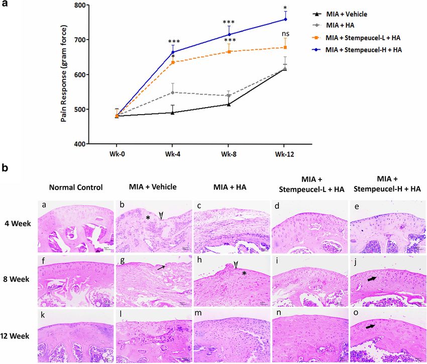
Gupta et al. Arthritis Research & Therapy (2016) 18:301                                                           Page 2 of 18

 (Continued from previous page)
 Conclusion: Intra-articular administration of Stempeucel® is safe. A twenty-five-million-cell dose may be the most
 effective among the doses tested for pain reduction. Clinical studies with a larger patient population are required
 to demonstrate a robust therapeutic efficacy of Stempeucel® in OA.
 Trial registration: Clinicaltrials.gov NCT01453738. Registered 13 October 2011.
 Keywords: Mesenchymal stromal cells, Osteoarthritis, Cell therapy

Background                                                       there is a need for randomized, double-blind, con-
Osteoarthritis (OA) is a common and debilitating chronic         trolled clinical trials. We have carried out in vitro
degenerative disease of large joints, especially the hip and     studies to show the differentiation efficiency of adult
knee, characterized by a loss of articular cartilage, sub-       human bone marrow-derived, cultured, pooled, allo-
chondral sclerosis, and marginal osteophyte formation.           geneic mesenchymal stromal cells (Stempeucel®) into
Worldwide, approximately 9.6% of men and 18% of                  the chondrogenic lineage and the expression of chon-
women aged ≥60 years have symptomatic osteoarthritis             drocyte-specific markers. In order to determine if
[1]. Current treatment in early-stage OA includes weight         Stempeucel® is efficacious in a preclinical model, we
reduction, quadriceps strengthening exercises, non-              have administered these cells intra-articularly into the
steroidal anti-inflammatory drugs, intra-articular (IA)          knee joints of rats with mono-iodoacetate (MIA)-induced
glucocorticoid injections, viscosupplements, and bracing         OA. After completion of these studies, a phase 2 dose-
[2–4]. Total joint arthroplasty is the mainstay treatment        finding clinical study was initiated to evaluate the safety
for end-stage OA of the knee joint, which is often associ-       (primary endpoint), potential efficacy, and appropriate
ated with serious and life-threatening complications in-         dose (secondary endpoints) of IA administration of
cluding increase risk of infection [5].                          Stempeucel® in patients with OA of the knee joint.
  Currently, cell therapy- and tissue engineering-based
approaches are being used to address the issue of repair         Methods
of damaged articular cartilage. This includes autologous         Production and characterization of Stempeucel® and placebo
cultured chondrocytes and mesenchymal stromal cells              Stempeucel® is a bone marrow-derived, ex vivo expanded,
(MSCs) obtained from various tissues that are used for           pooled, allogeneic human MSC population that has been
transplantation into the cartilage lesion. Autologous            characterized previously [16, 17]. The pooled cells were
chondrocyte implantation has inherent disadvantages              manufactured in an approved Good Manufacturing Prac-
such as a two-stage surgical procedure (harvesting healthy       tice (GMP) facility from bone marrow-derived MSCs
cartilage and transplanting culture-expanded chondrocytes        (BMMSCs) of three different healthy volunteers to produce
from that sample) that may cause further cartilage damage        a working cell bank (WCB). The pooled MSCs from the
and degeneration [6, 7], and chondrocyte dedifferentiation       WCB were further expanded to manufacture the investiga-
during culture that might result in fibrocartilage rather than   tional medicinal product, Stempeucel®. The cells expressed
hyaline cartilage formation [6, 8]. Thus, autologous or allo-    all markers characteristic of MSC, were negative for
geneic MSCs are rapidly emerging as an investigational           hematopoietic surface antigens, and also efficiently differ-
product for cartilage repair [9–11]. The anti-inflammatory       entiated into osteocytes, chondrocytes, and adipocytes
and immunomodulatory properties of MSCs suggest that             in vitro [16]. Two hundred million expanded BMMSCs
these cells can reduce inflammation and pain reduction in        were cryopreserved and stored in 15 ml PLASMA-LYTE A
the knee. Concurrently, MSCs may initiate the repair             (Baxter, Deerfield, Illinois) containing 5% human
process of the damaged cartilage by differentiating into         serum albumin and 10% dimethyl sulfoxide (DMSO)
chondrocytes, as well as by inducing proliferation and mat-      in cryobags (MacoPharma, Mouvaux, France). Placebo
uration of the remaining healthy chondrocytes or by indu-        contained 15 ml PLASMA-LYTE A in similar cryo-
cing differentiation of chondroprogenitors [12]. A whole         bags. The investigational medicinal product (IMP)
host of growth factors, biological modulators, and extracel-     specification is given in Table 1.
lular matrix proteins produced by MSCs may play a pivotal
role in enhancing neocartilage formation [12].                   In vitro studies show differentiation of Stempeucel® to
  Several preclinical studies and clinical trials have been      the chondrogenic lineage and quantification of sulfated
conducted using MSCs which have reported the safety and          glycosaminoglycan
therapeutic effect of its administration in patients with OA,    The chondrogenic differentiation potential of six Stempeucel®
although the majority of these studies have been conducted       batches was evaluated in monolayer cultures using chondro-
as single-dose, single-arm pilot studies [13–15]. Hence,         genesis induction medium (catalog no. A10071-01; Gibco).
Gupta et al. Arthritis Research & Therapy (2016) 18:301                                                                               Page 3 of 18

Table 1 Investigational medicinal product (IMP) specification
S. no    Description                                                       Specifications
1        Morphology                                                        Cells are fibroblastic and spindle-shaped in active growing conditions
                                                                           Cells are intact and round in shape after trypsin action
2.       Cell count                                                        180 to 220 million cells per bag
3.       Viability                                                         ≥85%
4.       Cell phenotype                                                    CD 73 >80%                             CD 34  80%                            CD 45 < 5%
                                                                           CD 90 > 80%                            CD 133 < 5%
                                                                           CD 166 > 80%                           CD 14 < 5%
                                                                                                                  CD19 < 5%
                                                                                                                  HLA-DR < 5%
5.       Karyotyping                                                       Normal, 46 XY
6.       Mycoplasma PCR ELISA                                              Not detected
7.       Sterility test                                                    Must comply
8.       Differentiation assay to adiopocyte, osteocyte, and chondrocyte   Confirmation of differentiation

Cells were plated at a density of 1000 cells/cm2 in six-well               Preclinical model of osteoarthritis
plates and cultured in DMEM-KO with 10% fetal bovine                       Ten-week-old male Wistar rats (n = 80) weighing be-
serum (FBS) and 2 ng/mL basic fibroblast growth factor                     tween 175 and 285 g were used for the preclinical study.
(bFGF) until 80% confluency. The cells were induced to dif-                The experimental protocol was approved by the Institu-
ferentiation in chondrogenic induction medium (Gibco) for                  tional Animal Ethics Committee (IAEC), and the experi-
21 days; the medium was replenished every 3 days and un-                   ment was carried out at a CRO animal facility.
induced cells were harvested at 80% confluency and served as                  Bilateral osteoarthritis was induced in rats (n = 74) by in-
the corresponding control for background estimation. After                 jection of MIA (Sigma-Aldrich, USA) into the knee joints
21 days of differentiation, the cells were trypsinized and pel-            according to published methods [18–20]. Briefly, the ani-
leted by centrifugation at 1000 rpm and the chondrogenic dif-              mals were anesthetized with 3% isoflurane; 1 mg MIA dis-
ferentiation was quantified by measuring the amount of                     solved in 50 μl saline was delivered into the articular cavity.
sulfated glycosaminoglycan (sGAG) using a Blyscan kit (cata-               The rats (n = 6) receiving only the saline solution served as
log no. B1000; Bicolor). The final sGAG content was repre-                 sham control animals (group I) throughout the experiment.
sented after normalizing with total DNA content estimated                  The pain sensitivity of the knee joints was measured before
using a Quant-iT Pico green kit (P7589; ThermoFisher, USA).                MIA injection and once every week after MIA injection as
                                                                           described by Di Cesare et al. [21]. The Pressure Application
                                                                           Measurement device (PAM; Ugo Basile, Italy) was used to
Quantitative RT-PCR for chondrogenic-specific gene markers                 measure the mechanical pain threshold of the knee joints.
Total cellular RNA was isolated using an RNeasy mini                       The gram-force (gf) that elicited the limb withdrawal was
kit (Qiagen) from the undifferentiated and the differenti-                 recorded. Three weeks after MIA injection, animals display-
ated BMMSCs for chondroyte lineage (described above).                      ing PAM values that ranged between 272 and 601 gf were
The RNA samples were treated with RNAse free DNase                         considered to have developed OA and were selected for the
I (Ambion), and reverse-transcribed into cDNA using a                      study (n = 60). Prior to performing the experiment described
high-capacity cDNA reverse transcription kit (Applied                      in this paper, we had conducted a validation study in which
Biosystems) according to the manufacturer’s instruc-                       the range of PAM values was evaluated against gross path-
tions. Real-time PCR was carried out using the SYBR                        ology and histological evidence of OA in rats (data not
green kit (catalog no. 4309155; Applied Biosystems)                        shown). The range of PAM values was selected accordingly
using step one plus (Applied Biosystems). Beta actin                       prior to randomizing the animals in the current study. Rats
served as the internal control. The sequences for the                      were divided into four different groups and each group con-
gene-specific primers are as follows: Sox9, forward                        sisted of 15 rats (Table 2). Sham control animals (group I)
TTTCCAAGACACAAACATGA, reverse AAAGTC-                                      showed an average PAM value of 870 ± 138 gf.
CAGTTTCTCGTTGA; Col2A, forward TTTCCCA
GGTCAAGATGGTC, reverse TCACCTGGTTTTC-                                      Stempeucel® administration in MIA-induced rats
CACCTTC. Ct values were normalized to the house-                           At day 0, animals in both groups 1 (sham control) and
keeping gene β-actin.                                                      group 2 (vehicle control) received 60 μl vehicle
Gupta et al. Arthritis Research & Therapy (2016) 18:301 Page 4 of 18
Table 2 Animal grouping and sacrifice schedule
Group Animals sacrificed (no of rats)*
4 weeks 8 weeks 12 weeks
Group 1 Sham control 2 2 2
Group 2 Vehicle control 3 6 6
Group 3 Hyaluronic acid (HA) 3 6 6
Group 4 Stempeucel® low dose + HA (6 × 10⁵ cells/joint) 3 6 6
Group 5 Stempeucel® high dose + HA (1.3 × 10⁶ cells/joint) 3 6 6
*Number of animals sacrificed at each time point after Stempeucel® administration
(Plasmalyte A; Baxter) through the IA route delivered by grading system [23, 24] on the H&E-stained sections. The
a 27-gauge needle. For group 3 animals, 30 μl hyaluronic binding intensity of Safranin-O to the sGAG was quanti-
acid (HA; Hyalgan®; Fidia Pharmaceuticals, Italy) was fied using imageJ software [25]. The region of interest
injected followed by 30 μl vehicle. For group 4 and 5 (ROI) around the cartilage area (n = 3 per section) was se-
rats, freshly-thawed Stempeucel® was used after washing lected, and the intensity of red (R), green (G), and blue (B)
in Plasmalyte A to remove DMSO and resuspending in were measured and the proportion of red staining was cal-
Plasmalyte A. Stempeucel® was administered at two dif- culated using the equation r = R/ (R2 + G2 + B2)1/2 where,
ferent doses: 6 × 10⁵ cells/joint (low dose; human equiva- R is the intensity of red and 'r' refers to the intensity of the
lent dose (HED) of 25 million cells) and 1.3 × 10⁶ cells/ red fraction with respect to other primary colors [26].
joint (high dose; HED of 50 million cells). Both doses The sections were graded and quantified by an independ-
were formulated in 30 μl Plasmalyte A and injected into ent observer blinded to the treatment groups.
group 4 (low-dose group) and group 5 (high-dose group)
animals, respectively. Cell administration was immedi- Clinical study design and enrollment criteria
ately followed by 30 μl HA injection. In order to reduce This was a randomized, double-blind, multicentric,
the xenogeneic rejection of cells, cyclosporine A (CsA; placebo-controlled, phase II study assessing the safety and
Novartis, Switzerland) was injected subcutaneously into efficacy of IA Stempeucel® in patients with OA of knee.
all the experimental animals at a dose of 10 mg/kg daily The study was conducted in accordance with the Good
for an initial 1 week starting at day –3 of cell injection, Clinical Practice (GCP) guidelines as issued by the Inter-
after which daily CsA administration was continued or- national Conference on Harmonization (ICH/135/95, July
ally at the same dose until the end of the study. The pain 2002), Schedule Y of the Drugs and Cosmetics Rules,
response was measured at weekly intervals up to week 1945, Ethical guidelines for biomedical research on human
10. The final measurements were taken at week 12. participants prepared by the Indian Council of Medical
Research in 2006 and the Declaration of Helsinki (64th
Gross and histological evaluation of cartilage repair in WMA General Assembly, Fortaleza, Brazil, October
preclinical model 2013). Approval was obtained from the Central Drugs
The treatment regimen and the number of animals Standard Control Organization (Indian FDA) and the in-
sacrificed after BMMSC administration at various time stitutional ethics committees of the five participating hos-
points are shown in Table 2. Rats from all the groups pitals. The study was registered in the National Institute
were sacrificed at 4, 8, and 12 weeks after vehicle/HA/ of Health registry of clinical trials (https://clinicaltrials.-
Stempeucel® administration. The knee joints were dis- gov/ct2/show/NCT01453738). An independent data safety
sected and the cartilage surface was visualized macro- monitoring board was formed comprising of drug safety
scopically on the exposed joints, and the distal femur physicians and an expert in the therapeutic area to moni-
from the right joints was dissected and processed for tor the safety data at predefined intervals during the pro-
histological analysis. gress of the study. The study was conducted from
All joint specimens were fixed in 10% formalin buffer November 2011 to November 2013. Written informed
and then decalcified in 10% EDTA (RFCL, India) for consent was obtained from all participants before screen-
2 weeks; the decalcification solution was changed twice in ing. Of the 82 patients screened, 62 patients were random-
a week. The joints were embedded in paraffin; serial sagit- ized to the study from five centers. Two patients dropped
tal sections (5-μm thick) were prepared and stained with out from the study after randomization but before IMP
hematoxylin and eosin (H&E) and Safranin-O fast green administration; thus 60 patients received the IMP. Four
staining for proteoglycan visualization and estimation dose levels were studied in this trial: 25, 50, 75, and 150
[22]. The severity of articular damage was evaluated using million cells (25 M, 50 M, 75 M, and 150 M, respectively).
the Osteoarthritis Research Society International (OARSI) At each dose level, 15 patients were randomized into twoGupta et al. Arthritis Research & Therapy (2016) 18:301 Page 5 of 18 groups (Stempeucel® and placebo) in a 2:1 ratio using a amount of PLASMA-LYTE A was loaded to a blinded computer generated randomization. Thus, 10 subjects re- syringe. The IMP (Stempeucel® or placebo) was pre- ceived Stempeucel® and 5 subjects received placebo at sented to the investigators in a blinded syringe (using each dose level (Fig. 1). As this was the first study of IA semitransparent tape) in a temperature-controlled trans- administration of Stempeucel® in OA patients, no formal port box at 2–8 °C. It was not possible to distinguish be- sample size calculation was performed. Eligibility criteria tween Stempeucel® and placebo upon visual inspection of the patients in the trial are given in Table 3. of the blinded syringes. Preparation of IMP at the clinical trial sites Block randomization (block size 5) was performed cen- Injection protocol for the clinical study trally by a biostatistician using PROC PLAN in SAS. Pre-medication (hydrocortisone 100 mg IV and phenira- The IMP (Stempeucel® or placebo) was shipped in a mine maleate 45.5 mg IV) was administered 15–30 min cryoshipper (temperature –185 to –196 °C) to clinical before administration of the IMP to prevent the possibil- trial sites whenever each patient was eligible for the ity of a potential anaphylactic reaction to the allogeneic study. Preparation of IMP for injection was performed cells. The IA injections were performed by qualified and under a validated biosafety cabinet by a trained person experienced investigators (either orthopedician or independent of the investigator’s team. A cryobag con- rheumatologist) using a 2.0-inch (5.1-cm) 20-gauge nee- taining IMP was thawed at 37 °C in a water bath. The dle as a lateral midpatellar injection (an injection into cell suspension was diluted to 100 ml using PLASMA- the patellofemoral joint). IMP injection was followed by LYTE A using two 50-ml centrifuge tubes. The cell pel- injection of 2 ml hyaluronic acid (20 mg; Hyalgan, Fidia let was resuspended in 2 ml PLASMA-LYTE A (for the Farmaceutici S.p.A., Italy). Patients were hospitalized for 25- and 50-million-cell dose groups) and in 4 ml the procedure, and were monitored for 24 h after the PLASMA-LYTE A (for the 75- and 150-million-cell dose injection. Patients were discharged after inspection of groups) based on the viable cell count and was loaded to the target joint, a general physical examination, and vital a blinded syringe. For the placebo preparation, a similar signs evaluation. Fig. 1 CONSORT flow chart showing the number of patients randomized, followed-up, and analyzed. M million cells, Cell Stempeucel®, mITT modified intention to treat
Gupta et al. Arthritis Research & Therapy (2016) 18:301                                                                            Page 6 of 18

Table 3 Subject eligibility criteria                                               improvement in pain from OA graded on a visual analog
Inclusion criteria                                                                 scale (VAS) from 0 to 10 (0 = no pain; 10 = worst pain);
1. Males or females in the age range 40–70 years (both inclusive)                  also, the Western Ontario and McMaster Universities
2. Radiographic evidence of grade 2 to 3 osteoarthritis (OA) based on
   the Kellgren and Lawrence radiographic entry criteria*                          Osteoarthritis (WOMAC-OA) index (total score, pain,
3. History of primary idiopathic OA of the knee characterized by pain              stiffness, and physical function scores) and intermittent
   which required intake of analgesics                                             and constant osteoarthritis pain (ICOAP) was used (con-
4. Self-reported difficulty in at least one of the following activities
   attributed to knee pain: lifting and carrying groceries, walking 400            stant and intermittent pain score) to evaluate pain and
   meters, getting in and out of a chair, or going up and down stairs              function of the joint. X-ray of the knee was carried out
5. Patients who had been on stable medication, including non-steroidal             at baseline, and at 3 and 6 months follow-up. Magnetic
   anti-inflammatory drugs (NSAIDs)/opioid or opiate analgesics, for the
   past 3 months                                                                   resonance imaging (MRI) of the knee was carried out at
6. Female patients of childbearing age who agreed to use accepted                  baseline, and at 6 and 12 months follow-up. MRI of the
   methods of contraception during the course of the study                         knee was performed using a 1.5-T whole body scanner
7. Ability to provide written informed consent
                                                                                   and a circumferential eight-channel knee coil. Proton
Exclusion criteria                                                                 density T2-weighted sequences were captured: sagittal
1. Prior or ongoing medical conditions (e.g., concomitant illness,
   psychiatric condition, alcoholism, drug abuse), medical history, physical       intermediate, axial intermediate, and coronal intermedi-
   findings, electrocardiogram (ECG) findings, or laboratory abnormality           ate for assessing osteophytes and cartilage (for scoring
   that, in the investigator’s opinion, could adversely affect the safety of the   cartilage signal and morphology, marginal osteophytes,
   subject, makes it unlikely that the course of treatment or follow-up
   would be completed or could impair the assessment of study results              subarticular bone marrow abnormality, subarticular cysts,
2. History of surgery or major trauma to the study joint                           and subarticular bone attrition); sagittal PD FS for asses-
3. Arthroscopy on the study joint in the previous 12 months                        sing menisci and cruciate ligaments (for scoring of ACL,
4. Signs of active study joint inflammation including redness, warmth,
   and/or, if qualifying with OA of the knee, a large, bulging effusion of         PCL, medial, and lateral menisci); and sagittal 3D
   the study knee joint with the loss of normal contour of the joint at            FIESTA-C for assessing cartilage surface and superficial
   the screening visit or at the baseline examination                              erosions (for scoring cartilage signal and morphology).
5. Patients who received intra-articular steroids or hyaluronan within the
   last 3 months                                                                   Images were scored to assess the whole-organ magnetic
6. Infections in or around the knee                                                resonance imaging score (WORMS) by two experienced
7. Patients awaiting a replacement knee or hip joint                               radiologists using combined reads. In case of non-
8. Patients with other conditions that caused pain
9. Patients with deformity of the knee joint                                       concurrence, advice was taken from a third independent
10. Significantly incapacitated or disabled and would be categorized as            radiologist whose report was considered final. WORMS
     ACR Functional Class IV (largely or wholly incapacitated) or unable to        scoring used in this study was modified for calculation of
     walk without assistive devices
11. Patients with other known rheumatic or inflammatory disease such               total WORMS score. The parameters included were ar-
     as rheumatoid arthritis                                                       ticular surface features which include cartilage signal and
12. Other pathologic lesions on X-rays of knee                                     morphology, subarticular bone marrow abnormality, sub-
13. Positive hepatitis B surface antigen, hepatitis C antibody test, anti-HIV
     antibody test, or Rapid Plasma Reagin (RPR)                                   articular cysts, subarticular bone attrition, and marginal
14. History of bleeding disorders                                                  osteophytes. They were scored for all 14 compartments of
15. Known hypersensitivity to hyaluronan or animal sera                            the knee joint. The compartment totals were added to ob-
16. For women of child-bearing potential: positive pregnancy test or
     lactating (females who were planning pregnancy within the next                tain the overall knee joint score [27].
     year were excluded)
*If both knees of a patient were eligible to be included in the study, the knee    Statistical analysis
to be included was as per investigator judgment                                    Preclinical data
                                                                                   GraphPad Prism software was used to calculate the stat-
Follow-up for the clinical trial patients                                          istical significance of all preclinical experimental data.
Patients were followed up at 1 week, and 1, 3, and                                 The data are expressed as the mean ± SEM. Quantitative
6 months after injection of IMP. The clinical data were                            RT-PCR data analysis was performed using Student’s
unblinded after 6 months and patients were further                                 t test. Pain threshold differences between various treat-
followed-up for both safety and efficacy until 12 months                           ment groups and the Safranin-O quantification were ex-
after the injection. Safety assessments included monitor-                          amined for statistical significance using two-way analysis
ing of all adverse events (AEs), assessment of electrocar-                         of variance followed by a Bonferroni post-hoc test. The
diogram (ECG) parameters, hematological (complete                                  OARSI grade analysis was performed using one-way ana-
blood count including erythrocyte sedimentation rate)                              lysis followed by Kruskal–Wallis test. P < 0.05 denoted the
and biochemical (liver function tests, kidney function                             presence of a significant difference between groups.
tests, and lipid profile) values, physical examination, and
vital signs measurements. Adverse events were captured                             Clinical trial data
by interviewing the subjects and laboratory data evalua-                           The SAS package (SAS® Institute Inc., USA, version 9.2)
tions during the visits. The efficacy endpoints included                           was used for statistical evaluation. For analysis purpose,
Gupta et al. Arthritis Research & Therapy (2016) 18:301 Page 7 of 18
subjects were grouped as follows: four treatment groups weekly by PAM test. Pain threshold values from various
(25 M, 50 M, 75 M, and 150 M cell dose), and two pla- time points are shown in Fig. 3a. Analysis of PAM scores
cebo groups. Patients who received placebo in 25 M and indicates that animals treated with both low and high
50 M dose levels were grouped into one placebo group doses of Stempeucel® showed improvement in pain
(both received the IMP in 2 ml PLASMA-LYTE A; P1) threshold from week 2 onwards as compared to vehicle or
and those who received placebo corresponding to the HA-treated animals and continued to improve until
75 M and 150 M dose groups (both received the IMP in 8 weeks after cell injection. While the pain improvement
4 ml PLASMA-LYTE A) were grouped into another appeared to reach saturation by 8 weeks with the low dose
group (P2). Thus, there were four treatment groups and of cells, it continued to improve until 12 weeks in the ani-
two placebo groups forming a total of six groups. The mals that received a high dose of Stempeucel® (Fig. 3a).
treatment groups 25 M and 50 M were compared with Animals treated with HA alone showed some improve-
P1 (the three groups collectively called cohort 1) and ment in pain threshold at the initial time period (week 3);
treatment groups 75 M and 150 M were compared with however, this improvement was not found to be consist-
P2 (the three groups collectively called cohort 2). AEs ent. When comparing the PAM scores between the HA-
were coded by the Medical Dictionary for Regulatory Ac- and Stempeucel®-treated groups, animals treated with the
tivities (MedDRA) primary system organ class (SOC) and high dose of cells exhibited a significant pain-reduction ef-
preferred term (PT). AEs were summarized descriptively fect at week 4 (P < 0.001), week 8 (P < 0.001) and week 12
by total number of AE(s) and compared between the (P < 0.05), whereas the low-dose group showed a signifi-
six study groups. Continuous variables are presented cant effect at week 4 (P < 0.05) and week 8 (P < 0.001), but
as mean ± SD. The data distribution was visually ex- not at week 12. These results clearly demonstrate that
amined for normality before applying the statistical intra-articularly injected Stempeucel® is able to reduce the
tool for analysis. Comparisons among groups were pain significantly in the MIA-induced OA rat model.
conducted using Kruskal–Wallis test, with alpha set
at 0.05 for significance. Cartilage repair induced by Stempeucel® administration
in a preclinical model
Results Macroscopic evaluation of articular cartilage was per-
Differentiation efficiency of Stempeucel® into formed in rats at 4, 8, and 12 weeks after cell injection.
chondrogenic lineage in vitro Varying degrees of cartilage damage were observed in
We evaluated the chondrogenic differentiation potential animals injected with MIA and subsequently adminis-
of Stempeucel® batches in monolayer cultures. The extent tered with vehicle. These degenerative changes include
of differentiation was also assessed by mRNA expression of cartilage fibrillation, erosion, and osteophyte formation
Sox9 and Col2A. We observed a significant downregulation (data not shown). The degree of cartilage damage was
of Sox9, which is an early inducer of chondrogenesis in the progressively reduced with both low and high doses of
differentiated cells compared to the control (P < 0.02, n = 6), Stempeucel® + HA treatment, while only marginal
and upregulation of Col2A, the gene that encodes for type change was noticed in a few HA-treated animals.
2 collagen, a major cartilage matrix protein and a mature Based on H&E staining of the cartilage tissue, we
chondrocyte marker (Fig. 2a and b). Chondrogenic differen- scored the femoral condyle section of rats using the
tiation of BMMSCs was quantified by measuring the amal- OARSI grading system. As expected, sham control ani-
gamation of sGAG which is known to play a central role in mals showed a normal histological appearance of the
cartilage homeostasis [28]. All six batches of Stempeucel® cartilage throughout the study (Fig. 3b, panels a, f, and k
differentiated to chondrocytes and synthesized significant for weeks 4, 8, and 12, respectively). In the vehicle-
amount of sGAG (30 ± 1.8 μg/μg GAG/DNA) compared to treated animals, the OA joints showed fibrillation, and
that produced by the undifferentiated cells (12.07 ± 5.6 μg/ appreciable loss of chondrocytes was observed at both
μg GAG/DNA; P < 0.001, n = 6) (Fig. 2c). These data sug- weeks 4 and 8 (Fig. 3b, panels b and g). By week 12, the
gest that the pooled BMMSC samples efficiently differenti- cartilage damage was found to be severe and the hyaline
ated into the chondrogenic lineage, confirming the tissue was replaced with fibrocartilage (Fig. 3b, panel l).
presence of mature chondrocytes after differentiation. The HA-treated animals showed marginal improvement
in cartilage architecture (Fig. 3b, panels c, h, and m). In
Intra-articular administration of Stempeucel® ameliorates contrast, animals receiving both low and high doses of
OA-induced joint pain in a preclinical model Stempeucel® showed almost intact cartilage with a larger
The MIA-induced OA model was the first validated pain number of chondrocytes (Fig. 3b, panels d, i, and n for a
model of OA used to evaluate the analgesic and anti- low dose of cells and panels e, j, and o for a high dose of
inflammatory properties of therapeutic agents [20]. In our cells). The calculated OARSI grades of the different
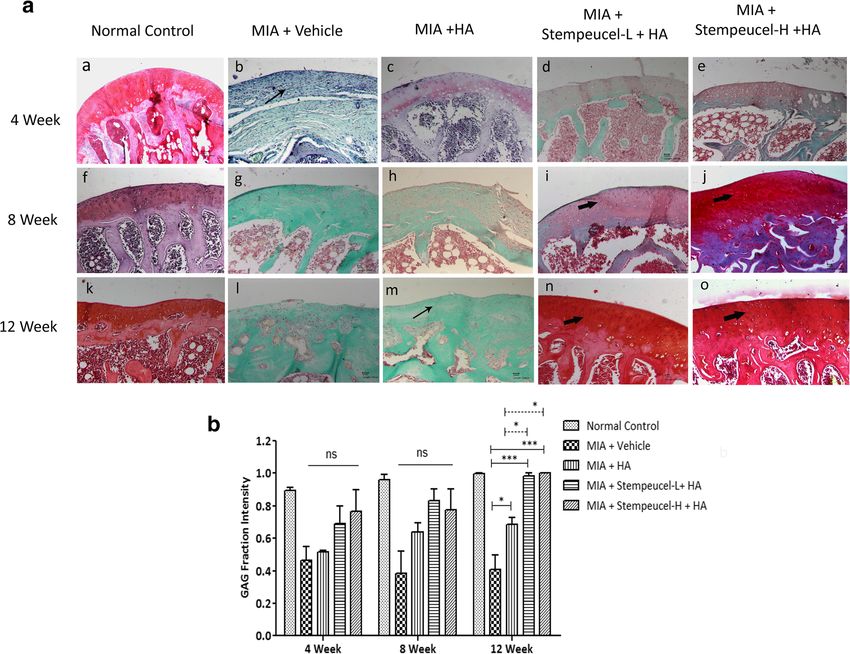
study, the mechanical pain threshold was measured treatment groups were compared at week 12. Although,Gupta et al. Arthritis Research & Therapy (2016) 18:301 Page 8 of 18 Fig. 2 Quantification of gene expression and sGAG. Quantitative mRNA expression of a SOX9 and b Col2A in the control (white bar) and chondrogenically differentiated Stempeucel® (black bar) by real-time PCR analysis (n = 6). c Sulfated glycosaminoglycan (sGAG) content in the control (white bar) and chondrogenically differentiated Stempeucel® (black bar) by DMMB dye-binding assay. The sGAG values were normalized to the DNA content in the control and chondrogenically differentiated Stempeucel® (n = 6). Results are represented as mean with SEM both low- and high-cell doses showed histological improve- Clinical study ment, a statistically significant reduction in OARSI grade Demographics and baseline characteristics of patients was observed only in the high-dose Stempeucel®-treated The demographics and other baseline characteristics of animals (P < 0.05) in comparison to animals in the vehicle the enrolled patients are presented in Table 4. All six group. The Safranin-O stained area of the cartilage was groups were mostly comparable in terms of baseline greater in both the cell-treated groups (Fig. 4a, panels d, i, characteristics. Sixty patients were included in the modi- and n for the low dose and panels e, j, and o for the high fied intention to treat analysis (mITT) group. There was dose). It is important to note that the intensity of Safranin- no premature unblinding of any patient. The patients’ O staining increased progressively with both doses of age, sex, weight, height, and body mass index (BMI) Stempeucel®. In fact, at week 12, Safranin-O staining inten- were balanced across groups. High scores for VAS, sity was found to be comparable between the high- and WOMAC, and ICOAP suggest that these patients had low-cell dose animals and sham controls (Fig. 4a). In severe pain and were balanced across all groups. comparison to the HA- and vehicle-treated groups, sGAG intensity was found to be significantly higher in Stempeu- Procedural safety cel®-treated and in sham control animals at week 12 All patients tolerated the procedure well in cohort 1 (25 M, (Fig. 4b). These findings suggest that intra-articularly 50 M, and P1) and cohort 2 (75 M, 150 M, and P2). Ten injected Stempeucel® + HA repaired MIA-induced articular patients (1 in 50 M, 6 in 75 M, and 3 in 150 M dose cartilage damage in rats with OA of the knee joint. groups) experienced pain and swelling at the injection site.
Gupta et al. Arthritis Research & Therapy (2016) 18:301                                                                                            Page 9 of 18

 Fig. 3 Effect of intra-articular injection of Stempeucel® on pain reduction and cartilage repair in an osteoarthritic rat model. a The effect of Stempeucel® on
 pain reduction at week 0 (before cell injection), and at weeks 4, 8, and 12 after cell injection. Data are presented as mean ± SEM. *P < 0.05, ***P < 0.001
 versus hyaluronic acid (HA)-treated group. b Photomicrographs of representative joint sections of femoral condyle stained with H&E at 4 (a–e), 8 (f–j), and
 12 weeks (k–o) after Stempeucel® treatment. Osteoarthritic changes, such as loss of chondrocytes (*), loss of cartilage (vertical arrow), and
 fibrillation (thin arrow) are evident in vehicle-treated and HA-treated joints. Proliferation of chondrocytes (thick arrow), regeneration, and
 repair of cartilage tissue was evident in Stempeucel®-treated groups. Scale bars = 100 μm, magnification 10×. H high dose of Stempeucel®,
 L Low dose of Stempeucel®, MIA mono-iodoacetate, ns not significant

The events were mild to moderate in severity, assessed as                         Overall evaluation of adverse events
related to the IMP, and recovered upon conservative ther-                         A total of 97 AEs were reported in 40 subjects (Table 5).
apy. Among the 10 patients, one subject (150 M dose                               The distribution of AEs in the different dose groups was
group) had IMP-related synovial effusion requiring                                as follows: 24 (25 M), 13 (50 M), 21 (P1), 17 (75 M), 11
hospitalization for one additional day of observation, thus                       (150 M), and 11 (P2). No patient died or was withdrawn
meeting the criteria of a serious adverse event (SAE). Other                      from the study due to an AE. Most of the AEs were mild
SAEs in the study were determined to be unrelated to the                          to moderate in severity. One severe AE was reported in
IMP and were as follows: hysterectomy for menorrhagia in                          each of 25 M (dyslipidemia), 50 M (anemia), 150 M
one subject in the 25 M group, suture-related complication                        (muscle hemorrhage), and P2 (umbilical hernia) groups.
and varicose vein in one subject in the P1 group, and                             Physical examination and vital signs data were unre-
hemorrhoidal hemorrhage and umbilical hernia, respect-                            markable after the injection. In cohort 1, most of the
ively, in one subject each in the P2 group.                                       AEs observed in the study were related to the SOC
Gupta et al. Arthritis Research & Therapy (2016) 18:301                                                                               Page 10 of 18

 Fig. 4 Histological evaluation of Safranin-O stained joint sections. a Photomicrographs of representative joint specimens of femoral
 condyle stained with Safranin-O at 4 (a–e), 8 (f–j), and 12 weeks (k–o) after Stempeucel® treatment. Loss of articular surface, roughening
 of cartilage and reduced staining of Safranin-O (thin arrow) were observed in vehicle- and HA-treated joints. Strongly stained Safranin-O-
 positive cartilage (thick arrow) with increased numbers of chondrocytes was seen in the Stempeucel®-treated groups. Scale bars = 100 μm,
 magnification 10×. b Sulfated glycosaminoglycan (GAG) fraction intensity was measured from histological images of Safranin-O-stained sections at
 weeks 4, 8, and 12. The intensity of Safranin-O staining is represented graphically, and the data are represented as mean ± SEM. At 12 weeks, the
 Stempeucel®-treated groups (both low (L) and high (H) dose) showed a significant improvement in the sGAG content compared to the
 disease control (mono-iodoacetate; MIA) and hyaluronic acid (HA)-treated groups. *P < 0.05, *** P < 0.001. ns not significant

(musculoskeletal and connective tissue disorders); the                      Hematology, serum chemistry, serology, urine analyses,
most common AE was arthralgia. In cohort 2, most of                         and ECG evaluation did not reveal any significant
the AE(s) observed in the study were related to the SOC                     abnormalities.
(general disorders and administration site conditions).
The most common AEs in the 75 M group were injec-
tion site pain and arthralgia. Three events of arthralgia                   Efficacy results
were experienced by two subjects (one subject had two                       VAS scores decreased over the study period for all the
episodes of arthralgia due to OA) and four events of in-                    treatment groups except for patients in the 150 M
jection site joint pain were experienced by four subjects                   group. The maximum reduction in the VAS score was
in the 75 M dose group. One event of hypersensitivity to                    seen in the 25 M group at 12 months compared to the
IMP (joint swelling) was experienced by a subject in the                    other groups of patients (40.3 ± 17.3, 30.3 ± 31.0, and
75 M dose group. Three events of hypersensitivity to                        21.3 ± 28.3 cm in 25 M, 50 M, and P1, respectively; P =
IMP (joint swelling) were experienced by three subjects                     0.3833). VAS decreased by 67.4% in the 25 M group
in the 150 M group. All events of joint pain and swelling                   compared to 41.4% and 36.0% in the 50 M group and
recovered completely upon symptomatic treatment.                            P1, respectively (P = 0.0587) (Fig. 5).
Gupta et al. Arthritis Research & Therapy (2016) 18:301                                                                                                   Page 11 of 18

Table 4 Summary of Demographic Characteristics at Baseline
                                        Cohort 1                                                       Cohort 2
Parameter                               25 M (n = 10)     50 M (n = 10)     P1 (n = 10)     P value 75 M (n = 10)          150 M (n = 10) P2 (n = 10)               P value
Age (years)                             58.10 ± 8.23      57.30 ± 9.45      54.90 ± 8.27    0.73       55.00 ± 6.72        54.00 ± 6.73          56.70 ± 5.19       0.6
Female (n)                              7                 8                 10              NA         8                   5                     7                  NA
Male (n)                                3                 2                 0               NA         2                   5                     3                  NA
Height (cm)                             156.85 ± 9.64     157.30 ± 12.23 152.25 ± 9.72      0.39       158.40 ± 8.86       158.88 ± 9.30         159.70 ± 10.67 0.9
Weight (kg)                             73.10 ± 15.86     69.00 ± 14.62     66.10 ± 7.67    0.45       71.30 ± 9.09        66.00 ± 9.13          66.90 ± 8.57       0.39
            2
BMI (kg/m )                             29.73 ± 6.09      27.74 ± 4.16      28.84 ± 4.91    0.76       28.38 ± 2.38        26.33 ± 4.48          26.40 ± 3.99       0.3
WOMAC total                             1315.8 ± 444.8 1498.4 ± 407.4 1239.6 ± 472.2 0.28              1470.6 ± 471.0 1388.1 ± 508.8 1382.0 ± 324.7 0.9
ICOAP total                             45.7 ± 19.2       59.3 ± 21.7       49.3 ± 18.7     0.38       58.4 ± 20.7         46.4 ± 22.0           54.8 ± 17.8        0.54
VAS                                     60.9 ± 19.7       73.7 ± 15.2       61.0 ± 23.8     0.24       57.4 ± 29.0         46.6 ± 23.6           65.3 ± 12.2        0.11
WORMS total score                       67.0 ± 19.8       78.8 ± 40.9       76.5 ± 23.5     0.65       71.3 ± 21.4         62.0 ± 17.9           70.8 ± 14.7        0.48
Kellgren and Lawrence Grade 2 (n) 4                       1                 3               NA         1                   3                     2                  NA
Kellgren and Lawrence Grade 3 (n) 6                       9                 7               NA         9                   7                     8                  NA
Values are shown as mean ± SD unless indicated otherwise
25 M, 50 M, 75 M, 150 M = 25, 50, 75, and 150 million cells, respectively
P1, P2 = placebo 1 and placebo 2, respectively
BMI body mass index, ICOAP intermittent and constant osteoarthritis pain, NA statistical comparisons for these groups have not been conducted due to too few
samples, VAS visual analog scale, WOMAC Western Ontario and McMaster Universities

Table 5 Summary of adverse events
                                                              Cohort 1                                            Cohort 2
System organ class                                            25 M (n = 10)      50 M (n = 10)     P1 (n = 10)    75 M (n = 10)          150 M (n = 10)         P2 (n = 10)
Any adverse event                                             24 (7)             13 (7)            21 (7)         17 (7)                 11 (6)                 11 (6)
Blood and lymphatic system disorders                          0                  1 (1)             1 (1)          0                      0                      0
Endocrine disorders                                           0                  0                 0              1 (1)                  0                      0
Eye disorders                                                 0                  1 (1)             0              0                      0                      0
Gastrointestinal disorders                                    1 (1)              0                 2 (2)          1 (1)                  0                      3 (3)
General disorders and administration site conditions          2 (1)              4 (2)             0              9 (6)                  4 (3)                  0
Infections and infestations                                   3 (3)              1 (1)             4 (3)          0                      1 (1)                  1 (1)
Injury, poisoning, and procedural complications               0                  0                 1 (1)          2 (1)                  3 (3)                  0
Investigations                                                2 (2)              1 (1)             1 (1)          0                      1 (1)                  2 (2)
Metabolism and nutrition disorders                            4 (3)              0                 3 (3)          0                      0                      1 (1)
Musculoskeletal and connective tissue disorders               6 (3)              3 (3)             5 (4)          4 (2)                  2 (2)                  3 (2)
Nervous system disorders                                      1 (1)              0                 1 (1)          0                      0                      0
Renal and urinary disorders                                   0                  1 (1)             0              0                      0                      0
Reproductive system and breast disorders                      1 (1)              0                 0              0                      0                      0
Respiratory, thoracic, and mediastinal disorders              0                  0                 0              0                      0                      1 (1)
Skin and subcutaneous tissue disorders                        1 (1)              0                 1 (1)          0                      0                      0
Surgical and medical procedures                               2 (1)              0                 0              0                      0                      0
Vascular disorders                                            1 (1)              1 (1)             2 (1)          0                      0                      0
Values are shown as number of events (number of patients)
25 M, 50 M, 75 M, 150 M = 25, 50, 75, and 150 million cells, respectively
P1, P2 = placebo 1 and placebo 2, respectively
Gupta et al. Arthritis Research & Therapy (2016) 18:301 Page 12 of 18 The WOMAC composite index score decreased over there was no perceptible change in WORMS score in- the study period for all the treatment groups. The max- cluding cartilage signal and morphology from baseline imum reduction in the WOMAC composite score was to follow-up visits in any of the groups of patients seen in the 25 M group at 12 months compared to the (Table 6). other groups (717.8 ± 503.8, 359.9 ± 786.4, and 233.8 ± 641.9 in the 25 M, 50 M, and P1 groups, respectively; Discussion P = 0.2651). The WOMAC composite index decreased The propensity of MSCs to differentiate into chondro- by an mean of 64.8% in the 25 M patients compared cytes in vitro [29] and their ability to repair articular car- to 14.4% and 49.3% in the 50 M and P1, respectively tilage has been shown in various preclinical models of (P = 0.1793). A similar trend was observed in all the OA [30–32]. In several studies, MSCs were prepared WOMAC subscores (WOMAC pain reduced by 145.1 ± and injected with sodium hyaluronan to increase the 105.2, 74.1 ± 167.2, and 57.4 ± 151.3 (P = 0.3484); engraftment and chondrogenic activity [30, 33]. In the WOMAC stiffness reduced by 69.6 ± 44.7, 4.5 ± 87.2, and present study, the efficacy of Stempeucel® was evaluated 25.8 ± 53.4 (P = 0.0324); and WOMAC physical function in a well-validated animal model of OA that was induced reduced by 503.1 ± 375.1, 290.3 ± 559.2, and 150.6 ± 457.8 by MIA injection into the knee joints. Both low and high (P = 0.2939) in the 25 M, 50 M, and P1 groups, respect- doses of Stempeucel® + HA treatment showed significant ively) (Fig. 6). improvement in the pain threshold from week 2 on- ICOAP total scores decreased over the study period wards when compared to animals treated only with HA; for all the treatment groups, except the 150 M group. treatment with only HA provided a short-term benefit The maximum reduction in the ICOAP total score was on pain reduction, which corroborates with an earlier seen in the 25 M group at 12 months (21.4 ± 21.2, publication [34]. We did not observe a significant differ- 12.3 ± 27.4, and 7.5 ± 27.1 in the 25 M, 50 M, and P1 ence between the two Stempeucel® treatment groups of groups, respectively; P = 0.5271) (Fig. 7). ICOAP total animals (low and high dose) on pain reduction. How- decreased by 34.6% in the 25 M group compared to ever, it is important to note that the pain reduction in 29.0% and 22.2% in the 50 M and P1 groups, respectively the high-dose animals continued to improve until the (P = 0.3844). A similar trend was seen in ICOAP subscores end of the study (12 weeks). Although the exact mech- (constant pain reduced by 26.5 ± 25.3, 20.5 ± 30.2, and anism of action of MSCs on pain reduction is not 12 ± 31.8 (P = 0.6140); intermittent pain reduced by known, anti-inflammatory activity has been attributed to 17.1 ± 28.4, 5.4 ± 33.1, and 3.8 ± 26.3 (P = 0.6215) in this effect. To date, some studies have demonstrated the the 25 M, 50 M, and P1 groups, respectively). role of MSCs on OA-induced pain behavior [35–37]. Thus, overall the patients in the 25 M group consist- Van Buul et al. reported improvement of weight-bearing ently showed pain reduction in all subjective parameters joints of the affected limb after intra-articular applica- measured in the study. Due to the small sample size, tion of both rat and human BMMSCs in MIA-induced none of the efficacy parameters were statistically signifi- OA rats [37]. However, unlike the results presented in cant as the study was not powered for establishing effi- this study, the authors did not observe cartilage regener- cacy. There were no clinically meaningful changes in the ation. Furthermore, in several animal studies, it has been X-ray parameters at follow-up visits compared to baseline shown that the increased levels of pro-inflammatory (data not presented). In the MRI evaluation, overall, cytokines might have contributed to pain increase. Intra- Fig. 5 Visual analog scale values. Data are presented as mean ± SD. 1 M, 3 M, 6 M, and 12 M = 1, 3, 6, and 12 months, respectively; C1 cohort 1, C2 cohort 2, M million cells, P placebo, VAS visual analog scale
Gupta et al. Arthritis Research & Therapy (2016) 18:301                                                                                Page 13 of 18

                              a                                          b

                              c                                          d

                              e                                          f

                              g                                          h

 Fig. 6 WOMAC results. WOMAC a, b composite, c, d pain, e, f stiffness, and g, h physical function (PF) results are shown for cohorts 1 (a, c, e
 and g) and 2 (b, d, f, and h). Data are presented as mean ± SD. 1 M, 3 M, 6 M, and 12 M = 1, 3, 6, and 12 months, respectively; C1 cohort 1, C2
 cohort 2, M million cells, P placebo, WOMAC Western Ontario and McMaster Universities

articularly administered MSCs probably play an import-                       treated only with HA. One of the short comings of the
ant role in attenuating the inflammation-induced pain                        preclinical results is that we did not determine the thera-
by secreting a wide range of anti-inflammatory cytokines                     peutic effect of BMMSCs without HA. However, based on
and analgesic peptides [38], and Stempeucel® might have                      the published data it appears that administration of MSCs
also contributed to pain reduction through a similar                         in combination with HA provided better therapeutic
mechanism.                                                                   benefit than either HA or MSC treatment alone in an ex-
  We also demonstrated that the pooled BMMSC popula-                         perimental animal model of OA [30]. The concomitant re-
tion are efficient in differentiating into chondrocytes                      duction in MIA-induced pain followed by an increase in
in vitro, and secrete a significant amount of sGAG                           cartilage regeneration observed in this study suggests that
(Fig. 2c). When these cells were administered intra-                         human bioactive factors synthesized by BMMSCs may be
articularly into OA-affected joints, we observed a progres-                  responsible for both the reduction in inflammation and
sive increase in proteoglycan staining. The improvement                      promotion of endogenous cartilage regeneration via a
in cartilage repair was observed both macroscopically and                    paracrine mechanism [12].
microscopically. The sGAG intensity data revealed that                         This clinical study met its predefined endpoint of
the total proteoglycan content was significantly higher in                   safety of intra-articular administration of Stempeucel® in
both the cell + HA treated groups compared to animals                        osteoarthritis of the knee joint. Adverse events were
Gupta et al. Arthritis Research & Therapy (2016) 18:301 Page 14 of 18
a b
c d
e f
Fig. 7 ICOAP results. ICOAP a, b total, c, d constant pain, and e, f intermittent pain results are shown for cohorts 1 (a, c, and e) and 2 (b, d, and
f). Data are presented as mean ± SD. 1 M, 3 M, 6 M, and 12 M = 1, 3, 6, and 12 months, respectively; C1 cohort 1, C2 cohort 2, ICOAP intermittent
and constant osteoarthritis pain, M million cells, P placebo
predominantly local pain and swelling, particularly seen swelling, as reported earlier [39]. The frequency of these
in patients randomized to the higher dose groups (75 M complications was similar to a report from another study
and 150 M) and they resolved completely upon symp- using culture-expanded bone marrow-derived MSCs
tomatic treatment. There was no evidence of ectopic tis- [40]. In another study using allogeneic non-HLA
sue or tumor formation locally at 1-year follow-up. matched BMMSCs in two different doses (50 and 150
Hematological, biochemical, and serological parameters million cells) which were pre-mixed with hyaluronic acid
were comparable in both the cell and placebo arm in all (5 ml) and administered in partial medial meniscectomy
groups of patients. Limited joint space, higher dose, and patients [10], the adverse events were similar to those
volume of injection (6 ml) may be the reason for in- seen in our study, with the most frequently reported AE
creased joint swelling and pain seen in cohort 2 (75 M by system organ class being musculoskeletal and con-
and 150 M). Furthermore, it can be assumed that a pro- nective tissue disorders [10]; however, the adverse events
portion of the cells injected into the joint space have not did not differ between the two doses tested. Recently,
survived and this phenomenon was more pronounced Vega et al. have conducted a study using IA injection of
with higher cell doses. Probably, such non-viable cells allogeneic BMMSCs (40 million cells suspended in 8 ml
produce an inflammatory reaction causing pain and of Ringer-Lactate) in OA of the knee joint [11]. Post-
Table 6 WORMS scoring of MRI of the knee at each visit
WORMS 25 M 50 M P1 P value 75 M 150 M P2 P value
Baseline 67.0 (19.8) 78.8 (40.9) 76.5 (23.5) – 71.3 (21.4) 62.0 (17.9) 70.8 (14.7) –
6 months 67.5 (20.5) 77.9 (41.2) 74.9 (22.4) 0.5521 71.4 (20.9) 62.0 (17.7) 69.9 (14.3) 0.7360
12 months 66.1 (19.2) 78.0 (41.1) 74.9 (22.5) 0.5310 67.0 (20.9) 60.6 (15.7) 72.3 (15.2) 0.0609
Values are shown as mean (SD)
The range of WORMS score used in this study was 0–314
25 M, 50 M, 75 M, 150 M = 25, 50, 75, 150 million cells, respectively
P1, P2 = Placebo 1 and 2, respectively
MRI magnetic resonance imaging, WORMS whole-organ magnetic resonance imaging scoreGupta et al. Arthritis Research & Therapy (2016) 18:301 Page 15 of 18 implantation pain was observed in 53% to 60% of pa- shown that bone marrow-derived cells are equally effica- tients in both the experimental and control groups. The cious [10, 11, 50–52]. A current focus for knee cartilage pain responded to analgesics and improved within 1 to repair is to use scaffolds that provide a three-dimensional 6 days. Hence, pain and local swelling may be the most environment for guiding and supporting the cells for car- common post-injection complication in patients after IA tilage repair. An advantage for using a scaffold is contain- injection of MSCs which responds within a few days of ment of the implanted cells on the lesion, and these symptomatic treatment. biomaterials may act as barriers for fibroblast invasion of One of the most important factors influencing the the graft [53, 54]. Koh et al. have used PRP as a scaffold as clinical outcome of a study is to determine the optimal it acts as an MSC accelerator for clinical chondrogenesis, treatment dose. In this study, patients in the low-dose is non-immunogeneic and bioabsorbable, and can be eas- group (25 million cells) showed improved outcomes in ily prepared preoperatively [13]. In another study, fibrin the pain measurement scores, whereas those in the glue has been used as a scaffold in MSC implantation to higher dose groups did not. The VAS and WOMAC induce improved cell survival, proliferation, gene expres- composite index scores decreased by 64% and 64.4% in sion, differentiation, and matrix synthesis leading to repair the 25-million-cell arm as compared to 36% and 49.3% of the cartilage lesion [55]. Cartistem® (MEDIPOST Co. in the active controls with HA, respectively, at 12 months Ltd., South Korea) is a combination product of human follow-up. In a proof of concept study, three doses of au- umbilical cord blood-derived mesenchymal stem cells and tologous adipose tissue-derived MSCs (AD-MSCs) were hyaluronic acid [56]. This acts as a biodegradable matrix used: 10 million, 50 million, and 100 million cells. The in MSC implantation as it facilitates the migration and ad- WOMAC score improved at 6 months follow-up in the herence of cells to the damaged cartilage, leading to better high-dose group [14]. In another study using allogeneic healing of the damaged lesion. Hence, more studies are re- BMMSCs at a dose of 40 million cells, improvement in quired to pinpoint the best source of stem cells and the pain, disability, quality of life, and cartilage quality by scaffold to be used to demonstrate both safety and MRI was noted in the cell-treated group [11]. Several efficacy. reasons are hypothesized for this effect in the low-dose The method of delivery of cells—either by direct intra- group of patients as observed in this study. Firstly, a articular injection or by open arthroscopy injection—into dose of 25 M cells may be optimum with the volume of the joint cavity is also important and may be one of the hyaluronic acid (2 ml) used in the study as a supporting factors for deriving efficacy. In one of the initial studies, matrix. Secondly, the 25-million-cell dose maybe optimal Wakitani et al. transplanted cells of bone marrow embed- for the limited IA space in the knee joint. Thirdly, doses ded in collagen gel into the articular cartilage defect at the higher than 25 million might cause cell aggregation due time of high tibial osteotomy [43]. Cartistem®, a combin- to a high cell concentration or insufficient space in the ation product approved by the Korean FDA, has been knee joint and subsequently cause cell death. Fourthly, applied to the damaged area through arthroscopy after the 25-million-cell dose may be lying in the upper range conducting a microfracture [57]. These open surgical of the efficacy dose since numerous studies reports that methods have their disadvantages such as pain, longer doses in the range of 10 to 25 million BMSCs may be ef- hospital stay, and higher cost. Minimally invasive tech- ficacious in OA of the knee joint [15, 41–45]. Finally, niques such as intra-articular injection have been adopted higher doses of MSCs may activate the MSCs to func- by different groups [14, 15, 41, 45, 50]. IA injection is tion as an M1-type cell with a pro-inflammatory re- patient-friendly in terms of being less invasive, with re- sponse [46], whereas the 25 M dose may be the optimal duced hospital stay, and are likely to reach a larger patient concentration of cells which gives rise to an M2-type population as it can be performed in peripheral hospitals. MSC with an anti-inflammatory/immunosuppressive Ultrasound guidance of knee injections could be a better response. Hence, which cell dose will lead to the best option to more precisely deliver the cells intra-articularly. outcome cannot be determined until a series of dose- Berkoff et al. have reported that ultrasound guidance of finding studies are carried out. knee injections resulted in better IA accuracy of needle Various studies are ongoing to determine the optimal placement than anatomical guidance (95.8% versus 77.8%; tissue source of MSCs for therapeutic repair of the car- P < 0.001) [58]. This enhanced injection accuracy achieved tilage tissue. The combination of MSCs with scaffolds, with ultrasound needle guidance directly improves growth factors, platelet-rich plasma (PRP), and genetic patient-related clinical outcomes. However, in developing modification have also been studied. It is not clear which countries, ultrasound-guided intra-articular injection may source of stem cells, or a combination product, will be be a challenge due to limited access to the instrument. the best for the disease condition. Studies have shown The present study, though it has shown good subject- that adipose tissue-derived stem cells are both safe and ive improvement in pain and functional scores, did not efficacious [13, 14, 47–49], whereas other studies have demonstrate improvement in cartilage signal and
You can also read