RECENT ADVANCES IN CAMEL IMMUNOLOGY - TIHO ELIB
←
→
Page content transcription
If your browser does not render page correctly, please read the page content below
REVIEW
                                                                                                                                             published: 25 January 2021
                                                                                                                                       doi: 10.3389/fimmu.2020.614150

                                          Recent Advances in Camel
                                          Immunology
                                          Jamal Hussen 1 and Hans-Joachim Schuberth 2*
                                          1 Department of Microbiology, College of Veterinary Medicine, King Faisal University, Al-Ahsa, Saudi Arabia, 2 Institute of

                                          Immunology, University of Veterinary Medicine Hannover, Foundation, Hannover, Germany

                                          Camels are domesticated animals that are highly adapted to the extreme desert
                                          ecosystem with relatively higher resistance to a wide range of pathogens compared to
                                          many other species from the same geographical region. Recently, there has been
                                          increased interest in the field of camel immunology. As the progress in the analysis of
                                          camel immunoglobulins has previously been covered in many recent reviews, this review
                                          intends to summarize published findings related to camel cellular immunology with a focus
                                          on the phenotype and functionality of camel leukocyte subpopulations. The review also
                           Edited by:
                 Victor PMG Rutten,       describes the impact of different physiological (age and pregnancy) and pathological (e.g.
      Utrecht University, Netherlands     infection) conditions on camel immune cells. Despite the progress achieved in the field of
                        Reviewed by:      camel immunology, there are gaps in our complete understanding of the camel immune
                      Pamela Burger,
    University of Veterinary Medicine,
                                          system. Questions remain regarding innate recognition mechanisms, the functional
                                Austria   characterization of antigen-presenting cells, and the characterization of camel NK and
        Renukaradhya J. Gourapura,
                                          cytotoxic T cells.
           The Ohio State University,
                         United States    Keywords: camel (Camelus dromedarius), immune, overview, review—systematic, leukocytes,
                            Petr Horin,   monocyte subpopulations
         University of Veterinary and
     Pharmaceutical Sciences Brno,
                              Czechia

                *Correspondence:          INTRODUCTION
           Hans-Joachim Schuberth
       hans-joachim.schuberth@tiho-       Camels (Camelus spp.) are essential inhabitants of desert and semi-desert ecosystems (1). Unlike
                       hannover.de        many other domestic species, camels thrive despite extreme temperatures, scarce vegetation, and
                                          very limited food and water resources (2, 3). The family of Camelidae comprises two major
                  Specialty section:      subfamilies, namely Camelinae (Old World camelids) and Laminae (New World camelids). The Old
        This article was submitted to     World camelids include two domesticated species: the dromedary or one humped camel (Camelus
          Comparative Immunology,         dromedarius) and the two humped camel or Bactrian camel (Camelus bactrianus) (4, 5). The wild
              a section of the journal
                                          camel (Camelus ferus) is a third species of Old World camelids, which is a double-humped camel
             Frontiers in Immunology
                                          living in central Asia and closely related to the Bactrian camel (6). The New World camelids, which
        Received: 05 October 2020         live in the high altitudes of South America, comprise four main species including two wild species
      Accepted: 07 December 2020
                                          (guanaco and vicuña) and two domesticated species (llama and alpaca) (7).
       Published: 25 January 2021
                                              In addition to their economic importance as domestic food animals in many regions of the world
                           Citation:
                                          including the Middle East, different parts of Africa, and most regions of Asia (8, 9), camels are also
        Hussen J and Schuberth H-J
            (2021) Recent Advances
                                          found in circus or zoological collections in the northern hemisphere (10). Camels are of zoonotic
               in Camel Immunology.       importance due to many pathogens that can be transmitted to humans. For example, dromedary
         Front. Immunol. 11:614150.       camels are considered as the main reservoir for the lethal zoonotic coronavirus, which is responsible
   doi: 10.3389/fimmu.2020.614150         for Middle East Respiratory Syndrome (MERS) in humans (11).

Frontiers in Immunology | www.frontiersin.org                                      1                                        January 2021 | Volume 11 | Article 614150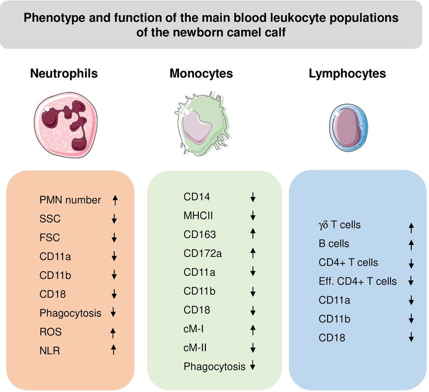
Hussen and Schuberth                                                                                                              Camel Immunology

    The immune system consists of a complex network of cellular              supporting disease diagnosis and guiding therapy and
and non-cellular components, which contribute equally to                     prognosis prediction.
effective immune responses against pathogens. Whereas                            A broad total WBC count range in the healthy dromedary
considerable research has been devoted to studying camel                     camel, from 8.3 to 19.6 cell x103/µl blood, has been reported in
immunoglobulins (12), rather less attention has been paid to                 the literature (30, 31). Lower (32) as well as higher (33) WBC
the cellular compartment of the camel immune system. As the                  counts were reported for the Bactrian camel in comparison to the
progress achieved in the analysis of camel immunoglobulins has               dromedary camel. In general, camels have a higher WBC count
previously been covered in many recent reviews (5, 12–14), the               than domestic ruminants (33, 34). This is mainly due to higher
present review will highlight the most important findings                     numbers of neutrophils in camel blood. The fraction of
concerning camel cellular immunology. The review will                        neutrophils among blood leukocytes accounts for up to 77%
especially focus on recent phenotyping and functional studies                followed by lymphocytes (30% on average) (32, 35–38). This is in
characterizing camel blood leukocyte subpopulations. In                      contrast to domestic ruminants, where lymphocytes outnumber
addition, the impact of different physiological (age and                     the leukocyte population in blood (33). The dominance of
pregnancy) and pathological (e.g. infection) conditions on the               neutrophils among camel blood leukocytes results on average
cellular immune compartment will be discussed.                               in a very high neutrophils to lymphocyte ratio (NLR) of 5:1
    In comparison to other species from the same geographical                compared to a NLR of 1:2 found in domestic ruminants (39, 40).
area, camels show higher resistance to some infectious diseases              The NLR is a novel marker which has been found to be
and environmental stress (15–19). Compared to the severe course              associated with systemic inflammatory responses (41–44). In
of many Middle East Respiratory Syndrome Coronavirus (MERS-                  other species, high NLR has been linked to impaired immune cell
CoV) infections in humans, camels show only mild and transient               function and was indicative of poor patient survival in different
respiratory symptoms with no need for veterinary care (15, 16).              diseases including sepsis (45, 46) and autoimmune diseases (47).
Possible mechanisms for the higher resistance of camels to MERS-             The clinical relevance of the relatively high NLR in camels and its
CoV are discussed below (The camel immune response to MERS-                  impact on the functionality of the camel immune system still
CoV). Dromedary camels also appeared to be resistant to                      needs to be investigated.
infectious doses of foot-and-mouth disease virus (FMDV),                         Historically, there has been a great deal of discrepency in the
which were sufficient to infect sheep in the same experiment                  literature regarding leukocyte composition in camels, depending
(18). This type of resistance is species-dependent, since FMDV is            on the techniques used to analyze the samples. Earlier studies
more infectious for Bactrian than for dromedary camels (18).                 mostly applied hemocytometers to estimate the camel leukogram
FMDV utilizes different integrin heterodimers (avb1, avb3, avb6,             with settings adapted from other species. According to those
and avb8) as cellular receptors (20). Whether these integrins are            studies, lymphocytes represent the most abundant leukocyte
differentially expressed or regulated in the susceptible and resistant       subpopulation in camel blood followed by neutrophils (48–54).
species, and whether the higher resistance of camels to FMDV is              However, recent hemocytometer and flow cytometric studies
determined at the level of virus-integrin interaction, represents an         (55–57) identified neutrophils as the main fraction of camel
important question that has yet to be addressed. Dromedary                   leukocytes (36, 58–62).
camels are also well adapted to extreme levels of heat stress (19).              Numerous studies investigated the changes in the camel
Compared to humans, this may rely on a faster, stronger and                  leukogram pattern under different physiologic (age, sex,
differential induction of heat-shock protein family members and              pregnancy) (63–65) and pathologic (infection) conditions (66,
the higher resistance of general protein synthesis in response to            67). As most camel leukogram studies presented leukocyte
thermal stress (21, 22). Deeper insights into the mechanisms                 composition as relative values (fractions) rather than absolute
behind the higher resistance of camels to some infectious agents             values, it is difficult to compare the results obtained from
and the adaptation of camel immune cells towards thermal stress              different studies.
are still pending.
    Several immunogenomic studies described the genomic                      Impact of Animal Age and Sex
diversity of immunity-related genes in domesticated and wild
camels, including genes encoding for B cell receptors, T cell
                                                                             on the Camel Leukogram
                                                                             The impact of age on the camel leukogram has been described in
receptors, and MHC molecules (6, 23–29).
                                                                             different studies (35, 68). In comparison to adults, young camels
                                                                             show higher WBC counts with higher percentages of
                                                                             lymphocytes and eosinophilic granulocytes but lower
THE LEUKOGRAM PATTERN OF CAMELS                                              percentage of neutrophilic granulocytes (35). The leukogram
IN HEALTH AND DISEASE                                                        pattern of the newborn camel calf is discussed below in detail.
                                                                                 Although some studies reported higher WBC counts in male
The species-specific leukogram, which comprises the total white               camels (69), in general the animal sex shows no impact on total
blood cell (WBC) count and the relative proportions of the main              WBC count or differential leukocyte composition (32, 70, 71).
leukocyte subpopulation including neutrophils, eosinophils,                  Some authors reported a higher proportion of lymphocytes among
basophils, lymphocytes, and monocytes, provides a cost-                      leukocytes in males compared to female camels (70, 72), while the
effective evaluation tool in human and veterinary medicine,                  eosinophil fraction was higher in females compared to males (70).

Frontiers in Immunology | www.frontiersin.org                            2                                 January 2021 | Volume 11 | Article 614150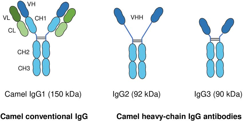
Hussen and Schuberth                                                                                                                       Camel Immunology

The impact of pregnancy on the camel leukogram (60) and the                 TABLE 1 | Camel leukocyte antigen cross-reactive monoclonal antibodies.
detailed immunophenotype of leukocytes in pregnant she-camels               Antigen                     Clone                Isotype             Source
will be discussed in a separate section below.
                                                                            CD4                         GC50A1               mIgM                WSU
Seasonal Effects on the Camel Leukogram                                     CD11a                       G43-25B              mIgG2a              BD
                                                                            CD11a                       HUH73A               mIgG1               WSU
There have been inconsistent observations regarding the impact of           CD11b                       ICRF44               mIgG1               BD
seasonal effects on the camel leukogram (70, 72, 73). For instance,         CD14                        TÜK4                 mIgG2a              Biorad
according to Mehrotra and Gupta (74), the number of leukocytes              CD14                        M5E2                 mIgG2a              BD
tends to decline during the summer season. In contrast, Babeker             CD14                        CAM36A               mIgG1               WSU
                                                                            CD18                        6.7                  mIgG1               BD
et al. reported higher numbers of WBC with increased total
                                                                            CD18                        HUH82A               mIgG2a              WSU
numbers of neutrophils, lymphocytes, eosinophils, and basophils             CD26                        polyclonal           gIgG                R&D systems
in camel blood during the summer season in comparison to the                CD44                        LT41A                mIgG2a              WSU
winter (73).                                                                CD45                        LT12A                mIgG2a              WSU
                                                                            CD62L                       MEL14                mIgG2a              Biolegend
                                                                            CD163                       LND68A               mIgG1               Kingfisher
The Camel Leukogram Pattern During                                          CD172a                      DH59b                mIgG1               WSU
Infectious Diseases                                                         B cells                     GC26A                mIgM                WSU
Changes in leukocytes count and composition during different                MHCI                        H58A                 mIgG2a              WSU
parasitic and bacterial infections have been frequently described.          MHCII                       TH81A5               mIgG2a              WSU
For example, trypanosomiasis in camels induces a marked increase            MHCII                       TH14B                mIgG2a              WSU
                                                                            MHCII                       L243                 mIgG2a              BD
in the number of WBC with increased percentages of neutrophils              WC1                         BAQ128A              mIgG1               Kingfisher
and reduced percentages of lymphocytes (49). Relative eosinophilia          WC1N2                       BAQ4A                mIgG1               WSU
(elevated fraction of eosinophils in blood) has been reported in            Activation marker           LH9A                 mIgM                WSU
camels suffering from different parasitic infestations, including           Activation marker           VPM30                mIgM                Biorad
Trypanosoma evansi (75), gastrointestinal helminths (71), and               MHC, Major Histocompatibility Complex; WSU, Washington State University; BD, Becton
nasal Cephalopina titillator (76). In camels infected with the              Dickinson; mIgM, mouse immunoglobulin M; mIgG, mouse immunoglobulin G; gIgG, goat
blood parasite Theileria anulata, the leukogram pattern was                 IgG (36, 59, 61, 85, 86).

characterized by leukocytosis, neutrophilia, eosinophilia, and
lymphopenia (67). Bacterial pathogens are mainly responsible for            with antibodies against cell adhesion molecules (CD11a, CD11b,
post-partal infections in camels, including mastitis and metritis           CD18, and CD62L) has enabled the characterization of camel
(77–80). The leukogram pattern associated with camel endometritis           monocyte subsets. Monoclonal antibodies specific for CD4 and
has been described for the clinical and subclinical form of the             WC1 molecules allowed for the chracterization of camel CD4-
disease. Clinical endometritis in camels is characterized by a              positive T cells and gd-T cells. The characterization of other
significant rise in the total cell count of blood leukocytes, which is       important lymphocyte subpopulation, especially CD8-positive T
mainly due to higher cell numbers of neutrophils (37, 81). However,         cells and NK cells, still requires the identification of monoclonal
in the case of subclinical endometritis she-camels did not show a           antibodies to camel CD8 and CD335 (NKp46) molecules.
different leukogram in comparison to healthy animals (82).
                                                                            Camel Neutrophilic Granulocytes
                                                                            Flow cytometric analyses identified camel blood neutrophils as
                                                                            highly complex/granular cells (side scatter, SSChigh) expressing
CHARACTERIZATION OF PHENOTYPE                                               CD45 and CD172a (35, 62). The higher green autofluorescence
AND FUNCTION OF CAMEL LEUKOCYTE                                             of eosinophils can be used to differentiate between camel
SUBPOPULATIONS                                                              eosinophils and neutrophils within the granulocyte population
                                                                            (35, 60).
Early attempts to study the cellular immune system in camels were               Neutrophil recruitment is a cascade process organized by a set of
hampered by the limited availability of camel-specific monoclonal            cell adhesion molecules, which mediate their adhesion to endothelial
antibodies (83, 84). As the production of monoclonal antibodies is          cells of blood vessels and the subsequent steps of extravasation (90).
a very costly process, attempts have been made to evaluate the              Compared with human neutrophils, dromedary camel neutrophils
cross-reactivity of commercially available monoclonal antibodies            express similar levels of the integrins LFA1 (CD11b/CD18) and
raised against leukocyte antigens of ruminants, swine, or human,            MAC1 (also known as aMb2; CD11b/CD18) (59).
with camel leukocyte antigens. Using the identified cross-reactive               Similar to bovine neutrophils (91), camel neutrophils show a
antibodies (Table 1) and flow cytometry, several camel myeloid               low but distinct expression level of the LPS co-receptor CD14,
and lymphoid immune cell populations and subpopulations have                suggesting a role in the sensing of gram-negative bacteria (35).
been recently characterized (59, 83–85, 87–89). The antibody                This has been partially proven in whole blood stimulation assays
toolbox for camel leukocyte antigens includes antibodies to                 (Figure 1) where LPS induced the activation and degranulation
several myeloid markers such as CD172a, CD14, and CD163,                    of camel neutrophils. In addition, LPS stimulation reduced the
and major histocompatibility complex (MHC) class I and II                   phagocytosis activity of camel neutrophils, while their ROS
molecules. Using those monoclonal antibodies in combination                 generating potential remained unchanged (62).

Frontiers in Immunology | www.frontiersin.org                           3                                        January 2021 | Volume 11 | Article 614150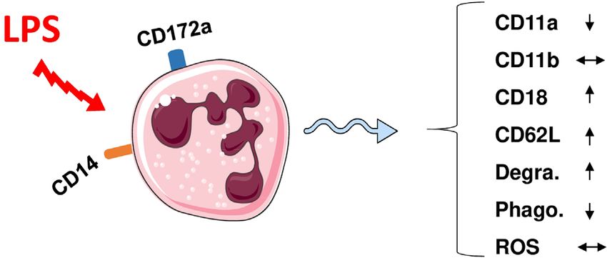
Hussen and Schuberth Camel Immunology
FIGURE 1 | Modulation of phenotype and function of camel neutrophils by LPS stimulation. Camel neutrophils can be identified as CD172ahighSSChigh leukocytes
(CD45+) with low expression of CD14. Stimulation of whole camel blood with LPS induces the modulation of the expression pattern of different adhesion molecules
and different antimicrobial functions (reduced SSC values as an indicator for neutrophil granularity, decreased phagocytosis capacity but no change in ROS
production). Degra, Degranulation; Phago, phagocytosis; ROS, generation of reactive oxygen species.
A series of recent studies on human neutrophils has indicated the porcine and bovine systems (104, 106, 110–112), the signal-
that distinct neutrophil subsets exist within the whole neutrophil regulatory protein alpha (CD172a) has been identified as a pan
population with diverse roles in infection and inflammation (92– monocyte marker for camel monocytes. The most abundant
95). For the camel, the heterogeneity of blood neutrophils remains subset of camel monocytes (87% of total monocytes) expresses
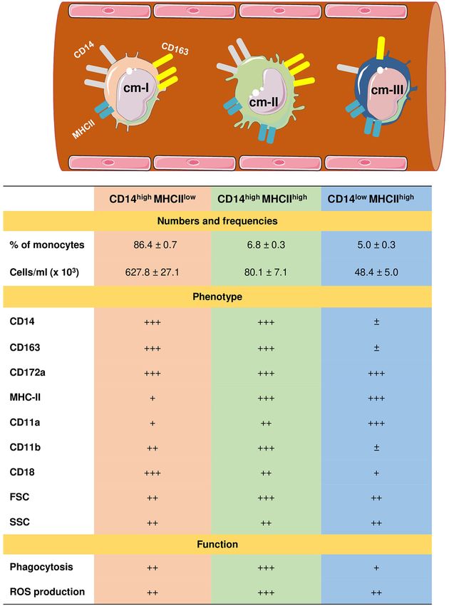
an open issue. In addition, the interplay between camel high levels of CD14 and CD163, but low levels of MHCII
neutrophils and other innate immune cells such as monocyte (CD14 high CD163 high MHCII low ) and is classified as camel
subsets and macrophages (96) has not yet been studied. monocyte (cMo)-I. A small fraction of camel monocytes (6 % of
total monocytes) expresses high levels of CD14, CD163, and
Camel Monocyte Subsets MHCII (CD14highCD163highMHCIIhigh) and is designated as
Monocytes are circulating immune cells with an essential role in the cMo-II. The third minor monocyte subpopulation cMo-III (5 %
innate immune defense against pathogens (97). Upon migration of total monocytes) displays high expression of MHCII but low
into tissues, monocytes are responsible for the replenishment of expression of CD14 and CD163 (CD14lowCD163lowMHCIIhigh)
other immune cells of the mononuclear phagocyte system including (61) (Figure 2).
macrophages and dendritic cells, which bridge the innate and Different monocyte classification systems have been used in
adaptive immune responses (98, 99). For their effective different species (113). Human and bovine monocytes were
antimicrobial functions, monocytes are equipped with several classified into the major population of CD14++ CD16- classical
receptors enabling pathogen sensing, engulfment, and elimination monocytes and two minor populations of CD14++ CD16+
(100, 101). The cell surface cluster of differentiation (CD) antigens intermediate monocytes and CD14+ CD16++ non-classical
CD172a, CD14, CD16, CD163, and MHCII have been proven as monocytes (104, 106, 112, 114, 115). Due to their low expression
reliable markers to describe monocyte heterogeneity, their of CD14, murine monocytes were classified into three subsets based
functional status, and their polarized differentiation into distinct on their expression of the myeloid markers Ly6C and CD43 (116,
macrophage subtypes (102–105). CD172a, which is known as 117). Whereas for the analysis of monocyte heterogeneity in the pig
signal-regulatory protein alpha (SIRPa), is a glycosylated cell (110, 118) and the dog (119), other monocytic markers including
surface receptor expressed on myeloid cells and functions as a CD163, CD172a and MHCII have been used.
regulatory receptor that inhibits cell signaling (106). CD14 is The expression of high levels of CD14 and CD163 on cMo-I
membrane protein mainly expressed on monocytes and functions and the low MHCII expression together with their dominance
with TLR-4 as a bacterial pattern recognition receptor responsible among all blood monocytes suggests close similarity with bovine
for binding lipopolysaccharide (LPS), the cell wall component of and human classical monocytes (104, 115). The phenotypic and
gram-negative bacteria (107). CD163 is a scavenger receptor for functional properties (highest anti-bacterial activity) of cMo-II
haptoglobin–hemoglobin complexes that is mainly expressed on suggests this subset is an equivalent to human and bovine
monocytes and macrophages and is considered as a marker for anti- intermediate monocytes (106, 113). Similarly, high levels of
inflammatory functional subtypes of these cells (108). Major surface MHCII and adhesion molecule leukocyte function-
histocompatibility (MHC) class II molecules are antigen receptors associated antigen (LFA)-1 (a1b2; b2 integrin; CD11a/CD18)
expressed on blood monocytes and B cells and present antigens to T and the low expression density of surface CD14 and CD163
helper cells (109). together with a reduced phagocytic and ROS generation activity,
Due to the lack of monoclonal antibodies cross-reactive with suggest that cMo-III represent the counterparts of bovine non-
camel CD16 (59), three subpopulations of monocytes in dromedary classical monocytes (96, 104, 106).
camels have recently been identified based on the expression In a recent report, the clinical relevance of camel monocyte
profiles of CD172a, CD14, MHCII, and CD163 (61). Similar to subsets in camel clinical endometritis has been investigated (81).
Frontiers in Immunology | www.frontiersin.org 4 January 2021 | Volume 11 | Article 614150Hussen and Schuberth Camel Immunology FIGURE 2 | Heterogeneity of camel monocyte subsets. Camel monocytes are subdivided according to the surface expression of CD14 and MHCII into three monocyte subsets. (1) Camel monocyte I (cM-I) with high expression of CD14 and low expression of MHCII (CD14highMHCIIlow), cM-II with high levels of both CD14 and MHCII (CD14highMHCIIhigh) and cM-III with high expression of MHCII but low expression of CD14 (CD14lowMHCIIhigh). The expression levels of cell surface molecules are presented as ± for very low to no-expression, + for weak expression, ++ for intermediate expression, and +++ for high expression. Functional capacities of monocyte subsets are presented as + for weak, ++ for intermediate, and +++ for strong capacity. In this study, animals with endometritis showed a significant explore the subset-specific function in health and disease for expansion in the fraction of camel inflammatory monocytes camels as for human and bovine monocyte subsets. Their role in (cMo-II). In addition, increased numbers of cMo-II were the pathogenesis of different infectious and non-infectious diseases indicative for the severity of endometritis. The study suggested has been indicated in a series of recent studies (121–124). A camel cMo-II as a disease biomarker for clinical endometritis in further open question is, whether camel monocytes show subset- camels (81). specific potential to differentiate into distinct functional subsets of Camel monocytes appear to be the only leukocyte population macrophages or dendritic cells. that exclusively express CD26, the MersCoV receptor and are therefore suggested to play a key role in either disease pathogenesis Camel Lymphoid Cell Subpopulations or immune response to the virus (86, 120). Whether the Due to the lack of camel-specific antibodies, only selected mentioned camel monocyte subsets differ in their expression subpopulations of camel lymphoid cells are identifiable. Thus, a intensity of CD26 is unknown. More work is needed to further comparison of camel lymphoid cell populations with cells of other Frontiers in Immunology | www.frontiersin.org 5 January 2021 | Volume 11 | Article 614150
Hussen and Schuberth                                                                                                                                               Camel Immunology

domestic animal species and humans is very limited. Notably,                                   naïve CD4+ T cells differentiate into effector T helper cells, which
CD8+ T-cells, cytotoxic T cells and NK cells cannot be identified                               can be distinguished based on the differential expression of cell
in camels, which severely inhibits the analysis of anti-viral and                              surface adhesion molecules such as CD45, CD44, CD62L, and
vaccination responses. Based on the TCR type, T cells are divided                              CD11a (130–134). Similar to their human counterparts (135),
into ab T cells recognizing peptide antigens presented on MHC                                  camel naïve (CD11alow CD44low) and effector (CD11ahi CD44hi) T
molecules and gd T cells recognizing antigen epitopes in an MHC                                helper cells have recently been identified with an elevated
independent manner (125). Using monoclonal antibodies cross-                                   proportion of effector T helper cells in animals with respiratory
reactive to the camel CD4 antigen, the bovine gd T cell marker                                 infections (23.5% of total CD4-positive lymphocytes compared to
WC1, the B cell antigen GC26A together with monoclonal                                         17.1% in healthy camels) (36).
antibodies specific to CD14 to exclude monocytes, it was
possible to identify camel CD4-positive T cells, WC1-positive gd                               Camel Cytokines
T cells, and GC26A-positive B cells in the blood of dromedary                                  Functional properties of camel lymphocyte subpopulations have
camels (Table 2) (36).                                                                         not been investigated so far. Especially the characterization of camel
    In healthy dromedary camels, blood lymphocytes are                                         subsets of helper T cells and the innate signals required for their
composed of a major fraction of B cells (mean percentage of                                    functional polarization into Th1, Th2, or Th17 subsets requires
26.6%) followed by CD4-positive T cells (24.6%) and a minor                                    further investigation. T cell polarization is one of the key factors that
fraction of gd-T cells (7.4%) (36). In comparison to their nearest                             determine the outcome of infectious diseases (136). The
relatives (Lamini), healthy camels show some similarties in their                              characterization of T effector cell subsets is limited by the lack of
lymphocyte composition (126, 127). Similar to their dominance                                  monoclonal antibodies specific for camel Th1, Th2, and Th17
among camel blood lymphocytes, B cells represent the main                                      cytokines. The characterized genes of Th1 (IL-2, IL-12, and
lymphocyte population in blood from healthy alpacas (126, 127).                                IFN-g) and Th2 (IL-4, IL-10 and IL-13) cytokines in the Bactrian
In addition, the fractions of CD4+ T cells and gd -T cells in blood                            camel (137, 138), however, could represent a valuable tool for
from alpacas (126, 127) and dromedary camels (36) are                                          conducting functional studies on T cell polarization in camels. The
comparable. It is unknown, whether camel CD8+ T cells are                                      high homology between Bactrian camels and other species,
present in the same frequency as in blood of alpacas (126, 127).                               including the llama, pig, cow, and horse regarding the nucleotide
The significant expansion of CD8+ T cells in the gut-associated                                 sequences of their cytokine genes (137) also suggests the necessity of
lymphoid tissue (GALT) of alpacas 9 days postinfection with                                    testing monoclonal antibodies specific for cytokines of these species
bovine virus diarrhea virus (BVDV) indicates a key role for this                               for their cross-reactivity with camel cytokines. However, the lack of
lymphocyte subset in the immune response of camelids to viral                                  characterized camel antigen-presenting cells and the establishment
infections (127).                                                                              of in vitro systems for the differentiation of camel monocyte-derived
    The analysis of the expression pattern of the adhesion molecules                           macrophages and monocyte-derived dendritic cells hamper
CD11a, CD11b, CD18, and CD62L, which play essential roles in                                   antigen-specific activation and T-cell polarization studies.
lymphocyte trafficking to peripheral tissues (128), revealed similar                                Studies on cytokine responses in vivo relied on the
expression patterns on camel and bovine CD4+ T cells and gd T                                  measurement of mRNA expression. Bactrian camels vaccinated
cells (36, 129).                                                                               with a live attenuated Brucella abortus S19 vaccine responded with
    T helper cells are key players in the adaptive immune response                             an upregulated expression of the Th-1 cytokine IFNg with low or
through their essential role in managing both humoral and cell-                                no expression of the Th2 cytokines IL-10 and IL-4, indicating the
mediated immune responses. Upon antigen-specific stimulation,                                   activation of a cell-mediated immune response (138). To address

TABLE 2 | Phenotypic properties of T cells and B cells in camel blood.

                                                      CD4+ T cells (CD4+WC1-)                          gd T cells (WC1+CD4-)                       B cells (GC26A+MHC-II+CD14-)

Frequency in blood
% of lymphocytes (Mean ± SEM)                                  24.6 ± 1.7                                      7.4 ± 0.3                                        26.6 ± 1.9
% of lymphocytes                                               14.2–33.1                                       1.0–20.1                                         18.4–42.0
(Min. – Max.)
Phenotype
CD4                                                              +++                                                –                                                –
WC1                                                                –                                              +++                                                –
GC26A                                                              –                                                –                                               +++
MHC-II                                                             –                                                –                                               +++
CD18                                                              ++                                               ++                                                ?
CD11a                                                             ++                                               ++                                                ?
CD11b                                                              +                                               ++                                                ?
CD62L                                                              +                                               ++                                                ?
Effector cells                                             CD11ahighCD44high                                        ?                                                ?
                                                            (17.0 ± 1.2) %

The expression levels of cell surface molecules are presented as - for very low to no-expression, + for weak expression, ++ for intermediate expression, and +++ for high expression. Non
investigated parameters are presented as ? (36).

Frontiers in Immunology | www.frontiersin.org                                              6                                           January 2021 | Volume 11 | Article 614150
Hussen and Schuberth Camel Immunology
the humoral immune response and the production of antigen- species, however, camels show a significantly lower molecular
specific antibodies, a recent immunization study with ovalbumin diversity of both MHC class I and class II genes (23, 24).
proved that the upregulated cytokine expression pattern of Natural killer (NK) cells are innate lymphoid cells with key
Bactrian camel lymphocytes was restricted to Th-2 cytokines roles in innate immune responses against intracellular pathogens
(IL-4, IL-10, and IL-13) (139). At present, these studies basically and tumor cells. These multiple functions are mediated by
indicate a high degree of similarity in the polarized cytokine different activating and inhibitory NK cell surface receptors,
response towards vaccines and antigens in other mammalian which determine the activation status of an individual NK cell
species, namely cattle (140, 141). (151). A recent work by Futas et al. investigated the diversity of
Type 1 interferons represent the most important cytokines in gene families encoding the camel NK cell receptors, including the
innate immunity during infections with viruses in addition to natural killer complex (NKC) and the leukocyte receptor complex
antitumor immune responses. The camel displays a similar (LRC), which mediate their function through the interaction with
broad spectrum of IFN alpha family members as cattle (142) MHC class I molecules (25). Collectively, the study identified a low
and humans (143). For instance, eleven IFN-a subtypes (144) polymorphism of the killer-cell immunoglobulin-like receptors
and one member of the IFN epsilon family were identified (145). (KLR) genes in camels, which is similar to the polymorphism of
The functional properties of type I interferons appear similar to this complex in the domestic pig. The study also revealed
other mammalian species, including the antiviral effect, the important differences in the genomic organization and
induction of interferon-responsive genes, and the tumor cell polymorphism of genes encoding NK cell receptors between
cytotoxicity (144, 145). Studies describing other immune- camels and cattle (25).
modulatory effects of type I interferons are still lacking (145). Recently, high quality genome assemblies have been developed
for domestic and wild camel species (6, 29). Computational
methods were employed for the improvement of genome
assemblies of the three Old World camel species (6). The authors
CAMEL IMMUNOGENETICS used the upgraded genome assemblies to investigate nucleotide
diversity of immune response genes in the three species. The highest
In comparison to other species inhabiting the same geographical
mean nucleotide diversity was identified in the domestic Bactrian
area, camels are more resistant to some pathogens (4, 15, 16, 18,
camel. The comparison between several innate and adaptive
146). The ability to respond to a variety of antigens is affected by
immune response gene groups revealed the highest mean
the diversity of highly specialized antigen receptors (147). This
nucleotide diversity in the major histocompatibility complex (6).
has been addressed in a series of immunogenomic studies which
The overall reduced antigen receptor diversity and MHC
investigated the polymorphism of genes encoding different camel
polymorphism, however, indicates the existence of other
antigenic receptors, including the ab and gd T cell receptor, the
mechanisms responsible for the higher resistance of camels to
NK cell receptor, and the antigen-presenting molecules MHC-
infectious diseases. Whether camel-specific epigenetic regulatory
class I and class II.
mechanisms of adaptive immune responses contribute to the
In comparison to other Artiodactyls, dromedary camels
relatively higher resistance to infections is currently unknown.
display a limited repertoire of T cell receptor delta variable
(TRDV) and T cell receptor gamma variable (TRGV) genes
(26). However, the diversity of the camel dromedary gd T cell
repertoire is significantly expanded by somatic hypermutation of THE IMMUNE SYSTEM OF THE
the TRDV and TRGV genes (27, 28, 148). The diversity of the PREGNANT SHE-CAMEL
variable domains of the ab T cell receptor is formed only by
classical combinatorial and junctional diversity and not by Pregnancy is a physiologic condition, usually associated with
somatic hypermutation (148). modulations in different immune mechanisms, which ensure
Antigen recognition by T cell receptors on CD4+ or CD8+ T protection against pathogens and at the same time prevent
cells requires the presentation of antigenic peptides by MHC class immune mediated destruction of the conceptus (152–154).
II or class I molecules respectively (149, 150). As those Immunomodulation during pregnancy is not restricted to the
polymorphic antigen-presenting molecules display promiscuous local uterine environment but extends also into the periphery
and selective interactions with antigen peptides, diversity in genes (155, 156). Pregnancy-associated immunomodulation has been
and alleles encoding for MHC class I and class II molecules addressed by a large number of studies in pregnant women (157),
contributes directly to the ability of a species to respond towards cows (158, 159), mares (160, 161), and sows (162–165). A recent
a range of different pathogens (150). In a recent report, Plasil and study investigated the impact of pregnancy on the phenotype and
coworkers investigated the localization, organization, and function of she-camel blood leukocytes (60). The observed
sequence of camel MHC genes (23). MHC genes are located on significant leukocytosis of pregnant she-camels is similar to
chromosome 20 in camels and are organized in MHC class II, findings reported for pregnant cows (166) and women (167),
MHC class III, and MHC class I genes, an organization that which is usually linked with an increased cortisol level during
follows the same pattern as in other mammalian species (148). The pregnancy (161). According to the same study, the leukocytosis
camel MHC genomic structure more closely resembles the porcine in pregnant she-camels is characterized by a reduced neutrophil
rather than the bovine MHC. Compared with other mammalian fraction and higher percentages of lymphocytes and monocytes.
Frontiers in Immunology | www.frontiersin.org 7 January 2021 | Volume 11 | Article 614150Hussen and Schuberth Camel Immunology
This leukocyte composition pattern, however, differs from the interest in other species (185–190). However, no studies have yet
pattern reported for pregnant cows (162). In cows, pregnancy is been conducted on the role of colostral immune cells in the
associated with higher fractions of neutrophils, lower fractions of modulation of the camel calf immune system.
lymphocytes but no changes in the fractions of monocytes Age-related changes of innate and adaptive cellular immune
(166, 168). responses have been described for different species (191, 192).
Although not proven, it was suggested that the enhanced The immaturity of newborn immune cells was linked to a higher
neutrophil extravasation and accumulation in the uterine tissue susceptibility to infectious diseases and higher mortality rates
might be responsible for the decreased proportion of neutrophils in during the early weeks after birth (191, 193–200). As in other
the blood, as neutrophils from pregnant animals expressed higher ruminants, camel newborns and adult camels differ significantly
densities of the cell adhesion molecule LFA-1 on their surface than regarding their leukogram pattern, phenotype and functionality
non-pregnant animals (60). Ex vivo functional analyses revealed an of blood leukocyte subpopulations (201). During their first
enhanced antimicrobial activity of neutrophils from pregnant she- month of life, the leukogram pattern of newborn camel calves
camels. This finding is in line with reports of pregnant mares (169), is characterized by higher leukocyte numbers, higher numbers of
but is opposite to the dairy cow, where pregnancy is associated with neutrophils, monocytes, and lymphocytes, but lower numbers of
impaired antimicrobial functions of neutrophils (170, 171). eosinophils in comparison to adult camels (201). The reduced
numbers of eosinophils in newborn camel calves, which play a
major role in parasitic immunity (39), has been related to lower
THE IMMUNE SYSTEM OF THE parasitic manifestation in comparison to adults (202).
NEWBORN CAMEL CALF High neutrophil to lymphocyte ratio (NLR) has been linked
to impaired immune cell function and poor patient survival in
Similar to horses, pigs, and ruminants, the epitheliochorial different inflammatory diseases (45–47). Camel calves are born
placenta of camels does not allow trans-placental passage of with a higher NLR (12.1 in average) than found in adults (5.1 in
maternal immunoglobulins to the fetus (172, 173). Therefore, average) (184). NLRs drop within two months after birth to adult
the newborn camel calf is born without serum immunoglobulins camel values. It was suggested that initially high calf NLRs reflect
and postnatal protection mainly relies on an adequate absorption the pro-inflammatory nature of newborn camel immune
of maternal colostral antibodies until the maturation of the calf’s responses and a shift towards mature and correctly polarized
own immune system (174, 175). The transfer of colostral immune responses takes place in the two-month period after
immunoglobulins to the newborn camel calf has been subject of birth (184).
many investigations (176–182). Several immunoglobulin classes, Similar to other artiodactyls such as sheep, cows and pigs,
including the IgM, IgG, and IgA, have been identified in the camel with higher frequencies of blood gd T cells in younger animals, gd
colostrum (182). However, only the uptake of maternal IgG, T cells account for up to 35% of blood lymphocytes in newborn
representing the most abundant immunoglobulin in camel and young camel calves (36, 70). This indicates that camels
colostrum, into the newborn’s blood has been studied. belong to the gd-high species, in contrast to gd-low mammalian
In addition to the conventional IgG with its heterodimeric species like humans and mice, where gd T cells represent only a
structure, camelids also possess non-conventional single-chain minor subpopulation (< 5%) of circulating lymphocytes (203).
IgG antibodies, which are not found in any other mammalian Compared to adult camels, the fraction of B cells among
species (183). In contrast to conventional IgG structure, which blood lymphoid cells of newborn camels is higher than in adults,
consists of two identical heavy chains (H) and two identical light whereas the fraction of CD4+ T cells is lower than in adults
chains (L), camel single-chain IgG antibodies are devoid of the (200). The authors discussed a link between elevated numbers of
light chain and the first heavy chain constant region CH1. The circulating leukocyte populations in camels and their lower
camel IgG isotype is currently classified into three structurally expression density of cell adhesion molecules (CD11a, CD11b,
different subclasses: camel IgG1 with two light and two heavy CD18) compared to adult leukocytes (201).
chains, camel IgG2 with a long-hinge heavy chain, and camel Monocytes from newborn and adult camels showed different
IgG3 with a short-hinge heavy chain (Figure 3). The camel expression patterns of the monocyte-related surface molecules,
heavy-chain antibodies (HCAbs) IgG2 and IgG3, which lack CD172a, CD14, CD163 and MHCII (61) (Figure 4). Compared
light chains, contribute up to 75% of all serum IgG (13). to adult camels, newborns display higher numbers of cMo-I and
Both classical two-chain antibodies (IgG1) and HCAbs (IgG2 cMo-III, and less numbers of inflammatory cMo-II (61).
and IgG3) are present in camel colostrum (182), and both are Camel calf leukocytes show functional properties that are
involved in the passive transfer of colostral IgG antibodies to the different from adult camel leukocytes. Flow cytometric analysis
newborn calf (178, 179). Some studies investigated the of cell granularity and cell size, which are widely used as
development of IgG (176) and HCAbs (178) in the blood of indicators of the cell activation status (204, 205), revealed a
the newborn camel calf. The rise in serum IgG levels in calf reduced activation potential of calf leukocytes in comparison to
serum two months after birth is indicative of the production of adults (120). Phagocytotic activity of newborn neutrophils and
significant levels of the calf’s own IgG (177, 184). The role of monocytes was found to be lower than in adults, with a lower
maternal colostral cells in neonatal immune system percentage of phagocytosis-positive cells and a reduced number
development, and their responses to vaccination is of growing of bacteria ingested per cell (58).
Frontiers in Immunology | www.frontiersin.org 8 January 2021 | Volume 11 | Article 614150Hussen and Schuberth Camel Immunology
FIGURE 3 | Structure of camel immunoglobulin (Ig) G subclasses. Camel IgGs are currently classified into three structurally different isotypes: Camel IgG1 consists
of two identical heavy chains (H) each composed of three constant domains (CH1–CH3) and a single variable domain (VH). Each heavy chain is covalently bound to
identical light chains (L) with a constant (CL) and a variable domain (VL). Camel IgG2 and IgG3 are composed of only two identical heavy chains (long-hinge heavy
chain in IgG2 and short-hinge heavy chain in IgG3). Camel single-chain IgG subclasses are devoid of the first heavy chain constant region CH1.
FIGURE 4 | Phenotypic and functional properties of neutrophils (PMN), monocytes, and lymphocytes in blood of newborn camel calves. ROS, Reactive Oxygen
Species amount in unstimulated cells; NLR, Neutrophil to lymphocyte ratio; Eff, effector cells. The direction of the arrows indicates higher (up arrow) or lower (down
arrow) values for calves compared to adults.
MUCOSAL IMMUNITY IN CAMELS defense barrier of the body preventing infectious agents from
invading the internal body tissues (206). Therefore, the
Mucosal body surfaces are equipped with specialized mucosa- characterization of mucosal immune mechanisms has essential
associated lymphoid tissue (MALT), which represent the first impact on the understanding of disease pathogenesis of and the
Frontiers in Immunology | www.frontiersin.org 9 January 2021 | Volume 11 | Article 614150Hussen and Schuberth Camel Immunology
development of effective vaccines against mucosal infections, incretins (229), has been identified as a functional receptor for
including mastitis, metritis, respiratory infections, and the MERS-CoV (230–232). The differential expression of DPP4
gastrointestinal infections. Several studies adressed anatomical in the respiratory tracts of humans and camels has been suggested as
structures of the gastrointestinal MALT of Bactrian and dromedary the primary cause of limited MERS-CoV replication in the human
camels (207–211). Detailed and comparative aspects of immune upper respiratory tract and hence restrict transmission between
mechanisms on camel body surfaces are still unkonwn. humans. While DPP4 is only expressed in the human lower
The intestine represents the main surface of interaction between respiratory tract epithelium, only the upper respiratory tract
the immune system and the huge numbers of microorganisms, epithelium of camels show DPP4 expression (233). Whether the
which play a pivotal role in guiding the maturation of the mucosal interaction of MERS-CoV with epithelial cells in the lower (human)
immune system and shaping systemic immunity (212, 213). or upper respiratory tract (camel) results in the secretion of
Morphological studies of the gastrointestinal tract of Bactrian location-specific mediators, which may differently modulate the
camels revealed a distinct structure and distribution of the MALT onset and resolution of subsequent innate and adaptive immune
in this species (209, 214). Distributed along the whole small mechanisms, is still an open question.
intestine, four distinct types of Peyer’s patches, including nodular, In opposite to humans, where DPP4 is mainly found on human
faviform, cup-shaped, and cystic form Peyer’s patches, have been T lymphocytes rather than monocytes (234), dromedary camels
identified in Bactrian camels (214). The nodular and cystic forms of display the highest expression of DPP4 on blood monocytes (86,
Peyer’s patches are unique to this species. The number of Peyer’s 120). This may indicate different roles for innate and adaptive
patches in the small intestine of Bactrian camels increases with age immune responses to MERS-CoV in the two species. This is also
and peaks in 5-year-old camels followed by a subsequent decline supported by recent observations on MERS-CoV-infected human
(214). Peyer’s patches in the large intestine of Bactrian camels are individuals, where gradual increases in blood lymphocyte count
mainly located on the surface of the entire ileocecal orifice, the during MERS progression was observed in all the survivors, whereas
beginning of the cecum, and the first third of the colon. The ileocecal the response in diseased patients was characterized by lymphopenia
orifice has been suggested as the main inductive site for mucosal and increased neutrophils and monocytes counts (235).
immune responses in the Bactrian camel large intestine (209). In One of the potential control strategies of MERS-CoV relies on
the dromedary camel, Peyer’s patches have cup-shaped structures reducing virus transmission from animals to humans through
and are distributed in the anti-mesenteric side of the ileum (211). vaccination of camels (223, 236, 237). The development of
They are not present in the jejunum or duodenum (215). Whether protective MERS-CoV vaccines for dromedary camels, however,
the distinct morphology, structure, and distribution of the MALT requires an in depth understanding of local immune mechanisms
structures in camels are reflected by species-specific functional in the respiratory tract in camels and the identification of
differences of mucosal immune responses, is currently unknown. correlates of protection against the virus. In MERS patients, the
Mucosal immunoglobulins contribute to the immune homeostasis development of neutralizing antibodies was not sufficient for an
at the mucosal interface (216). The distribution of secretory IgA effective clearance of the virus (238). The association between the
(SIgA) and IgG-secreting cells (ISCs) in the lamina propria of the recovery from MERS and the generation of both antibody and T-
small intestine of Bactrian camels suggests their significant cell responses (221, 239) indicates key roles for cell-mediated
contribution to mucosal immunity in this species (210, 217). immune mechanisms against the virus. However, the analysis of
Similar to the age-related changes in the number of PP in the immune responses of camels to MERS-CoV infection was limited
intestine of Bactrian camels, SIgA and IgG ISC numbers increase to the investigation of virus-specific antibodies (179). Studies on
with age with a peak at puberty (210, 217). cell-mediated immune responses are still lacking. The
characterization of camel NK and cytotoxic T cells and their role
The Camel Immune Response to Middle in anti-viral immunity in the context of infection with MERS-CoV
East Respiratory Syndrome Coronavirus is one of the promising lines of research. MERS-CoV naturally
Middle East respiratory syndrome Coronavirus (MERS-CoV) is infected camels are currently discussed as a challenge model in
an emerging zoonotic pathogen that causes the Middle East vaccine efficacy studies (16). The characterization of mucosal
Respiratory Syndrome (MERS) (11, 218–220). Dromedary immune mechanisms in the camel respiratory tract, including
camels are considered the only confirmed animal host for detailed phenotypic and functional analyses of immune cells in
MERS-CoV and the source of zoonotic infection (221–228). In bronchoalveolar lavages and lung parenchymas of MERS-CoV-
humans, MERS-CoV infection is associated with either infected and recovered camels would be a prerequisite for the
hospitalization or death, while MERS-CoV-infected camels elucidation of MERS-CoV pathogenesis in these animals.
show only mild and transient respiratory symptoms with no
need for veterinary care (15). It is unknown whether special host-
pathogen interaction mechanisms in camels contribute to the CONCLUSIONS
higher resistance of this species to MERS.
The very high seroprevalence rates (74–100%) of MERS-CoV in The camel represents a multipurpose domestic animal used for
camel populations in Africa and the Arabian Peninsula indicate meat and milk production, racing, and transportation. Different
high infection and transmission rates of the virus in camels (16). components of the cellular immune system of the dromedary camel
The dipeptidyl peptidase 4 (DPP4; CD26), a type II transmembrane show several species-specific phenotypic and functional properties.
glycoprotein involved in cleavage of dipeptides and degradation of In contrast to other domestic species, the camel leukogram is
Frontiers in Immunology | www.frontiersin.org 10 January 2021 | Volume 11 | Article 614150Hussen and Schuberth Camel Immunology
dominated by the neutrophil fraction resulting in a higher NK and cytotoxic T cells and their role in anti-viral immunity,
neutrophil to lymphocyte ratio. Camel monocytes are classified especially in the context of infection with the zoonotic pathogen
into three phenotypically and functionally different subsets based MERS-CoV is still in its infancy. Promising lines of research would
on the expression of the surface molecules CD14 and MHCII also include host-pathogen interactions on camel mucosal body
with many similarities with the bovine monocyte classification surfaces such as the respiratory tract, the mammary gland, the
system. Camels belong to the gd T cell-high species with uterus, and the intestine.
especially high percentages of gd T cells in newborn and young A deeper characterization of camel infection immunity would
animals. Circulating camel newborn immune cells contain lower help to identify protection-relevant immune mechanisms,
numbers of inflammatory monocytes, show a reduced expression of essential for the design of effective vaccines, the identification
cell adhesion molecules on all leukocytes, and a reduced of disease biomarkers, and the selection of animals with higher
antimicrobial functionality of monocytes and neutrophils. disease resistance.
Despite the progress achieved in the field of camel immunology,
there are still many gaps towards a more profound understanding
of the camel immune system. Open questions cover the innate
recognition mechanisms, the functional characterization of AUTHOR CONTRIBUTIONS
macrophages and dendritic cells, and their signals responsible for T
cell activation and polarization toward distinct functional subtypes JH and HS wrote the manuscript. All authors contributed to the
such as type-1, type-2, or type-17 cells. The characterization of camel article and approved the submitted version.
REFERENCES coronavirus (MERS-CoV) in Najran, Kingdom of Saudi Arabia (KSA); A
retrospective record based study. J Infect Public Health (2020) 13:1342–6.
1. Jirimutu, Wang Z, Ding G, Chen G, Sun Y, Sun Z, et al. Genome sequences doi: 10.1016/j.jiph.2020.04.007
of wild and domestic bactrian camels. Nat Commun (2012) 3:1202. doi: 16. Alharbi NK, Ibrahim OH, Alhafufi A, Kasem S, Aldowerij A, Albrahim R,
10.1038/ncomms2192 et al. Challenge infection model for MERS-CoV based on naturally infected
2. Andersen HT. [Desert, man and camel]. Nord Med (1966) 75:61–3. camels. Virol J (2020) 17:77. doi: 10.1186/s12985-020-01347-5
3. Yousif OK, Babiker SA. The desert camel as a meat animal. Meat Sci (1989) 17. Bitter H. Disease resistance in dromedaries with particular reference to
26:245–54. doi: 10.1016/0309-1740(89)90010-7 Trypanosoma evansi infection. Hanover, Germany: Tierartliche Hochschule
4. Burger PA. The history of Old World camelids in the light of molecular (1986).
genetics. Trop Anim Health Prod (2016) 48:905–13. doi: 10.1007/s11250- 18. Larska M, Wernery U, Kinne J, Schuster R, Alexandersen G, Alexandersen S.
016-1032-7 Differences in the susceptibility of dromedary and Bactrian camels to foot-
5. Ali A, Baby B, Vijayan R. From Desert to Medicine: A Review of Camel and-mouth disease virus. Epidemiol Infect (2009) 137:549–54. doi: 10.1017/
Genomics and Therapeutic Products. Front Genet (2019) 10:17. doi: S0950268808001088
10.3389/fgene.2019.00017 19. Hoter A, Rizk S, Naim HY. Cellular and Molecular Adaptation of Arabian Camel
6. Lado S, Elbers JP, Rogers MF, Melo-Ferreira J, Yadamsuren A, Corander J, to Heat Stress. Front Genet (2019) 10:588. doi: 10.3389/fgene.2019.00588
et al. Nucleotide diversity of functionally different groups of immune 20. Ruiz-Saenz J, Goez Y, Tabares W, Lopez-Herrera A. Cellular receptors for foot
response genes in Old World camels based on newly annotated and and mouth disease virus. Intervirology (2009) 52:201–12. doi: 10.1159/000226121
reference-guided assemblies. BMC Genomics (2020) 21:606. doi: 10.1186/ 21. Ulmasov HA, Karaev KK, Lyashko VN, Evgen’ev MB. Heat-shock response
s12864-020-06990-4 in camel (Camelus dromedarius) blood cells and adaptation to
7. Dorman AE. The camel in health and disease. 2. Aspects of the husbandry hyperthermia. Comp Biochem Physiol B (1993) 106:867–72. doi: 10.1016/
and management of the genus Camelus. Br Vet J (1984) 140:616–33. doi: 0305-0491(93)90043-5
10.1016/0007-1935(84)90013-7 22. Majeed EN. Effects of heat on camel platelet structure and function–a
8. Kebede F, Gelaty E. Studies on major respiratory diseases of Camel comparative study with humans. Platelets (2009) 20:528. doi: 10.3109/
(Camelus dromedarius) in Northeastern Ethiopia. Afr J Microbiol Res 09537100903207513
(2010) 4:1560–4. doi: 10.5897/AJMR.9000669 23. Plasil M, Mohandesan E, Fitak RR, Musilova P, Kubickova S, Burger PA,
9. Al-Ruwaili MA, Khalil OM, Selim SA. Viral and bacterial infections et al. The major histocompatibility complex in Old World camelids and low
associated with camel (Camelus dromedarius) calf diarrhea in North polymorphism of its class II genes. BMC Genomics (2016) 17:167. doi:
Province, Saudi Arabia. Saudi J Biol Sci (2012) 19:35–41. doi: 10.1016/ 10.1186/s12864-016-2500-1
j.sjbs.2011.10.001 24. Plasil M, Wijkmark S, Elbers JP, Oppelt J, Burger PA, Horin P. The major
10. Al-Ani FK. Domestication, distribution and population. In: F Al-Ani, editor. histocompatibility complex of Old World camelids: Class I and class I-
Camel Management and Diseases. Jordan: Al-Sharq printing press (2004). p. 1–24. related genes. HLA (2019) 93:203–15. doi: 10.1111/tan.13510
11. Drosten C, Meyer B, Muller MA, Corman VM, Al-Masri M, Hossain R, et al. 25. Futas J, Oppelt J, Jelinek A, Elbers JP, Wijacki J, Knoll A, et al. Natural Killer
Transmission of MERS-coronavirus in household contacts. N Engl J Med Cell Receptor Genes in Camels: Another Mammalian Model. Front Genet
(2014) 371:828–35. doi: 10.1056/NEJMoa1405858 (2019) 10:620. doi: 10.3389/fgene.2019.00620
12. Muyldermans S. Nanobodies: natural single-domain antibodies. Annu Rev 26. Ciccarese S, Vaccarelli G, Lefranc MP, Tasco G, Consiglio A, Casadio R, et al.
Biochem (2013) 82:775–97. doi: 10.1146/annurev-biochem-063011-092449 Characteristics of the somatic hypermutation in the Camelus dromedarius T
13. Arbabi-Ghahroudi M. Camelid Single-Domain Antibodies: Historical cell receptor gamma (TRG) and delta (TRD) variable domains. Dev Comp
Perspective and Future Outlook. Front Immunol (2017) 8:1589. doi: Immunol (2014) 46:300–13. doi: 10.1016/j.dci.2014.05.001
10.3389/fimmu.2017.01589 27. Antonacci R, Mineccia M, Lefranc MP, Ashmaoui HM, Lanave C, Piccinni B,
14. Jovcevska I, Muyldermans S. The Therapeutic Potential of Nanobodies. et al. Expression and genomic analyses of Camelus dromedarius T cell receptor
BioDrugs (2020) 34:11–26. doi: 10.1007/s40259-019-00392-z delta (TRD) genes reveal a variable domain repertoire enlargement due to
15. Al Sulayyim HJ, Khorshid SM, Al Moummar SH. Demographic, clinical, and CDR3 diversification and somatic mutation. Mol Immunol (2011) 48:1384–96.
outcomes of confirmed cases of Middle East Respiratory Syndrome doi: 10.1016/j.molimm.2011.03.011
Frontiers in Immunology | www.frontiersin.org 11 January 2021 | Volume 11 | Article 614150You can also read