Structural basis for AcrVA4 inhibition of specific CRISPR-Cas12a - eLife
←
→
Page content transcription
If your browser does not render page correctly, please read the page content below
RESEARCH ARTICLE

                                    Structural basis for AcrVA4 inhibition of
                                    specific CRISPR-Cas12a
                                    Gavin J Knott1, Brady F Cress1, Jun-Jie Liu1, Brittney W Thornton1, Rachel J Lew2,
                                    Basem Al-Shayeb3, Daniel J Rosenberg4,5, Michal Hammel4, Benjamin A Adler6,7,
                                    Marco J Lobba8, Michael Xu1, Adam P Arkin7,9, Christof Fellmann2,10,
                                    Jennifer A Doudna1,2,4,8,11,12,13*
                                    1
                                      Department of Molecular and Cell Biology, University of California, Berkeley,
                                    Berkeley, United States; 2Gladstone Institutes, San Francisco, United States;
                                    3
                                      Department of Plant and Microbial Biology, University of California, Berkeley,
                                    Berkeley, United States; 4Molecular Biophysics and Integrated Bioimaging Division,
                                    Lawrence Berkeley National Laboratory, Berkeley, United States; 5Graduate Group
                                    in Biophysics, University of California, Berkeley, Berkeley, United States; 6UC
                                    Berkeley-UCSF Graduate Program in Bioengineering, University of California,
                                    Berkeley, Berkeley, United States; 7Department of Bioengineering, University of
                                    California, Berkeley, Berkeley, United States; 8Department of Chemistry, University
                                    of California, Berkeley, Berkeley, United States; 9Environmental Genomics and
                                    Systems Biology Division, Lawrence Berkeley National Laboratory, Berkeley, United
                                    States; 10Department of Cellular and Molecular Pharmacology, University of
                                    California, San Francisco, San Francisco, United States; 11Innovative Genomics
                                    Institute, University of California, Berkeley, Berkeley, United States; 12Howard
                                    Hughes Medical Institute, University of California, Berkeley, Berkeley, United States;
                                    13
                                      California Institute for Quantitative Biosciences (QB3), University of California,
                                    Berkeley, Berkeley, United States

                                    Abstract CRISPR-Cas systems provide bacteria and archaea with programmable immunity
*For correspondence:                against mobile genetic elements. Evolutionary pressure by CRISPR-Cas has driven bacteriophage to
doudna@berkeley.edu                 evolve small protein inhibitors, anti-CRISPRs (Acrs), that block Cas enzyme function by wide-
Competing interest: See             ranging mechanisms. We show here that the inhibitor AcrVA4 uses a previously undescribed
page 21                             strategy to recognize the L. bacterium Cas12a (LbCas12a) pre-crRNA processing nuclease, forming
                                    a Cas12a dimer, and allosterically inhibiting DNA binding. The Ac. species Cas12a (AsCas12a)
Funding: See page 21
                                    enzyme, widely used for genome editing applications, contains an ancestral helical bundle that
Received: 06 June 2019              blocks AcrVA4 binding and allows it to escape anti-CRISPR recognition. Using biochemical,
Accepted: 09 August 2019            microbiological, and human cell editing experiments, we show that Cas12a orthologs can be
Published: 09 August 2019
                                    rendered either sensitive or resistant to AcrVA4 through rational structural engineering informed
Reviewing editor: Douglas L         by evolution. Together, these findings explain a new mode of CRISPR-Cas inhibition and illustrate
Black, University of California,    how structural variability in Cas effectors can drive opportunistic co-evolution of inhibitors by
Los Angeles, United States          bacteriophage.
   Copyright Knott et al. This      DOI: https://doi.org/10.7554/eLife.49110.001
article is distributed under the
terms of the Creative Commons
Attribution License, which
permits unrestricted use and
                                    Introduction
redistribution provided that the    Biological warfare between microbes and bacteriophage drives the co-evolution of diverse host-
original author and source are      phage interactions. Clustered regularly interspaced short palindromic repeats (CRISPR) and CRISPR-
credited.                           associated (Cas) proteins provide adaptive immunity in which Cas nucleases are deployed together

Knott et al. eLife 2019;8:e49110. DOI: https://doi.org/10.7554/eLife.49110                                                     1 of 25
Research article                                                                            Biochemistry and Chemical Biology

                                    with CRISPR RNAs (crRNAs) to base-pair with foreign genetic material and trigger its destruction
                                    (Knott and Doudna, 2018). The potency of CRISPR-Cas immunity drives mobile genetic elements to
                                    evolve mechanisms that enable escape from CRISPR-Cas targeting. While genetic diversity offers
                                    some advantages when facing innate immune systems (Labrie et al., 2010), nucleotide variation
                                    alone is insufficient to evade the adaptability of CRISPR-Cas (van Houte et al., 2016). To avoid
                                    CRISPR-Cas targeting, many bacteriophage employ small anti-CRISPR (Acr) proteins to inactivate
                                    specific CRISPR-Cas systems (Borges et al., 2017; Stanley and Maxwell, 2018). Since the initial dis-
                                    covery of type I-F CRISPR-Cas inhibitors (Bondy-Denomy et al., 2013), a wide diversity of viral pro-
                                    teins have been found to inhibit CRISPR-Cas enzymes. Furthermore, genomic data analysis suggests
                                    that unique Acrs may exist for the majority of CRISPR-Cas subtypes (Watters et al., 2018).
                                       Cas12a, like Cas9, is a biotechnologically relevant CRISPR-Cas enzyme for which novel Acrs have
                                    been recently identified (Watters et al., 2018). Cas12a (formerly Cpf1) is an RNA-guided Class II
                                    type V-A CRISPR nuclease which cleaves DNA following recognition of a 20-nucleotide DNA
                                    sequence containing a protospacer-adjacent motif (PAM) (Zetsche et al., 2015) (Figure 1A). Unlike
                                    Cas9, Cas12a directly catalyzes precursor crRNA (pre-crRNA) processing to generate a mature sur-
                                    veillance complex whose crRNA bears a hydroxyl on its 5’ terminus (Zetsche et al., 2015;
                                    Fonfara et al., 2016; Swarts et al., 2017) (Figure 1A). Previously identified inhibitors of Cas12a,
                                    known as type V-A Acrs (AcrVA), either reversibly block substrate association through stoichiometric
                                    binding or irreversibly inactivate the enzyme complex through a chemical transformation
                                    (Suresh et al., 2019; Knott et al., 2019; Dong et al., 2019). While exhibiting stoichiometric inhibi-
                                    tion of Cas12a, the inhibitor AcrVA4 appears to be unique in several ways. At 234 amino acids,
                                    AcrVA4 is almost twice as large as other anti-CRISPR proteins and it contains a predicted coiled-coil
                                    domain not observed in other Acrs. In addition, similar to specific Acrs that target Cas9
                                    (Harrington et al., 2017) or Cas10 (He et al., 2018), its mode of action appears to involve dimeriza-
                                    tion of Cas12a (Knott et al., 2019).
                                       We show here that AcrVA4 is a robust inhibitor of some Cas12a homologs but remains incapable
                                    of inhibiting other closely related homologs. Using single particle cryo-electron microscopy (cryo-
                                    EM), we find that AcrVA4 recognizes the conserved pre-crRNA processing nuclease of Cas12a and
                                    the specific chemistry of a mature crRNA bound within the enzyme. This specific association serves
                                    to position AcrVA4 proximal to highly conformationally dynamic domains that are locked by the
                                    inhibitor to cage the enzyme in a state incompatible with dsDNA binding. Structure-guided muta-
                                    genesis revealed that AcrVA4 dimerization is not required for inhibition of Cas12a in vitro and pro-
                                    vided no advantage to bacteriophage targeted by Cas12a in vivo. Finally, we identified the
                                    structural basis for AcrVA4 ortholog specificity and engineered the AsCas12a enzyme to be suscepti-
                                    ble to AcrVA4 in phage assays and human cell editing experiments. These results reveal a new mode
                                    of inhibition by anti-CRISPRs and demonstrate how structural variability in Cas effectors can drive
                                    opportunistic co-evolution of inhibitors by bacteriophage.

                                    Results
                                    The AcrVA4 C-terminal domain binds to LbCas12a-crRNA
                                    Despite its distinction as one of the largest bacteriophage-derived protein inhibitors of CRISPR-Cas,
                                    AcrVA4 has a narrow spectrum of Cas12a inhibition specificity. Previous biochemical experiments
                                    demonstrated that AcrVA4 blocks dsDNA binding to L. bacterium (Lb) and M. bovoculi (Mb) Cas12a
                                    but not Ac. species (As) Cas12a (Watters et al., 2018; Knott et al., 2019), the last of which is com-
                                    monly utilized as a tool for genome editing (Figure 1B). To investigate the mechanism of AcrVA4-
                                    mediated Cas12a inhibition and identify the basis for its ortholog specificity, we determined the
                                    structure of the LbCas12a-crRNA complex bound to AcrVA4 using single-particle cryo-EM
                                    (Figure 1C–D, Table 1, Figure 1—figure supplement 1A). From a subset of 156,979 particle images
                                    identified by three-dimensional classification, we reconstructed a three dimensional cryo-EM density
                                    map at 3.0 Å resolution (State I) (Figure 1—figure supplement 1A). This high-resolution map con-
                                    tained clear density into which the previously determined structure of the LbCas12a-crRNA complex
                                    was docked and refined (Figure 1C–D). With the exception of the PAM-interacting (PI) domain,
                                    which was disordered in our structure, we resolved most of the LbCas12a protein and crRNA to
                                    high-resolution (Figure 1C–D, Figure 1—figure supplement 2A–B). The map contained additional

Knott et al. eLife 2019;8:e49110. DOI: https://doi.org/10.7554/eLife.49110                                                         2 of 25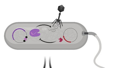
Research article Biochemistry and Chemical Biology
A pre-crRNA dsDNA
B
AcrVA4 L. bacterium
5' 3'
AcrVA4 M. bovoculi
Cas12a
RuvC
AcrVA4 Ac. species
Tree scale: 1
C
L. bacterium Cas12a L. bacterium crRNA
.. 3'
1228
1179
G U G U A G A U A A A G U G
282
521
586
678
808
872
890
997
WED 24
A
1
1 6
RuvC
A U C A U C U U
BH
REC1 REC2 WED PID WED RuvC RuvC Nuc
5' -16
HO A A U
D
136
234
1
REC1 REC1
NDD CBD
REC2 REC2 AcrVA4
AcrVA4
180°
WED
WED Nuc crRNA
crRNA
RuvC RuvC
Figure 1. Overall structure of the LbCas12a-crRNA-AcrVA4 complex. (A) Schematic representation of Cas12a activity. (B) Unrooted maximum likelihood
phylogenetic tree of Type V-A CRISPR-Cas12a. Species known to be susceptible or unsusceptible to phage-derived AcrVA4 are highlighted. The
triangle denotes collapsed branches of Cas12b-e. (C) Schematic representation of LbCas12a and the mature crRNA modeled within the cryo-EM
structures. (D) Two views of the LbCas12a-crRNA complex (cartoon) bound to AcrVA4 (surface) shown related by a 180˚ rotation. The color scheme for
the crRNA, LbCas12a, and AcrVA4 in panels B, C, and D are used throughout the manuscript.
DOI: https://doi.org/10.7554/eLife.49110.002
The following figure supplements are available for figure 1:
Figure supplement 1. Cryo-EM data collection and 3D reconstruction.
DOI: https://doi.org/10.7554/eLife.49110.003
Figure supplement 2. Cryo-EM density for State I.
DOI: https://doi.org/10.7554/eLife.49110.004
Figure supplement 3. Structurally similar proteins to AcrVA4 CBD as determined by Dali search.
DOI: https://doi.org/10.7554/eLife.49110.005
density not attributable to LbCas12a-crRNA that allowed for unambiguous de novo tracing and
sequence register assignment of the C-terminal domain of AcrVA4 (residues 136–233) (Figure 1D,
Figure 1—figure supplement 2C).
Knott et al. eLife 2019;8:e49110. DOI: https://doi.org/10.7554/eLife.49110 3 of 25
Research article                                                                                Biochemistry and Chemical Biology

                                    Table 1. Cryo-EM data collection, reconstruction, and model statistics.
                                                                                   State I                           State II
                                                                                   PDB: 6P7M | EMDB: 20266           PDB: 6P7N | EMDB: 20267
                                    Data Collection
                                    Microscope                                     FEI Titan Krios                   FEI Titan Krios
                                    Voltage (kV)                                   300                               300
                                    Camera                                         Gatan K2 Summit                   Gatan K2 Summit
                                    Defocus range (mm)                             0.7 ~ 2.2                         0.7 ~ 2.2
                                    Pixel Size (Å)                                0.9                               0.9
                                    Magnification                                  135000                            135000
                                                        2
                                    Electron Dose (e/Å )                          47                                47
                                    Total Particles                                324336                            324336
                                    Reconstruction
                                    Software                                       CryoSparc                         CryoSparc
                                    Symmetry Imposed                               C1                                C1
                                    Final number of                                156979                            79787
                                    refined particles
                                    Resolution of polished                         4.2                               8.7
                                     unmasked map (Å)
                                    Resolution of polished masked map (Å)         3                                 5
                                    Map Sharpening B-factor (Å2)                      101.4                             123.5
                                    Refinement
                                    Model Resolution cutoff (Å)                   3                                 5
                                    FSC threshold                                  0.143                             0.143
                                    Map CC (box)                                   0.74                              0.78
                                    Map CC (mask)                                  0.79                              0.51
                                    R..m.s Deviations
                                    Bond lengths (Å)                              0.006                             0.002
                                    Bond angles (˚)                                0.607                             0.524
                                    Molprobity score                               1.77                              1.84
                                    Clashscore                                     7.87                              6.63
                                    Rotamer Outliers (%)                           0                                 1.33
                                    Ramachandran plot
                                    Favored (%)                                    94.43                             94.43
                                    Allowed(%)                                     5.57                              5.57
                                    Outliers (%)                                   0                                 0

                                    DOI: https://doi.org/10.7554/eLife.49110.006

                                       Residues 136–233 of AcrVA4 adopt a compact a1b1-b5a2 fold, defined here as the C-terminal
                                    binding domain (CBD), that forms an extensive interface with LbCas12a (Figure 1D, Figure 1—fig-
                                    ure supplement 2C). The 1479 Å2 of buried surface area between AcrVA4 and LbCas12a accounts
                                    for 23.8% of the total solvent accessible surface area of the CBD. A Dali search (Holm and Rose-
                                    nström, 2010) of the AcrVA4 CBD detected very limited structural similarity to the Pyrobaculum
                                    spherical virus (PDB code: 2X5C), the TrmB archaeal transcriptional regulator (PDB code: 3QPH),
                                    and several PAZ domains from Argonaute proteins (PDB code: 4Z4H) (Figure 1—figure supplement
                                    3). While the CBD of only a single copy of AcrVA4 was well resolved in State I, AcrVA4 exists as an
                                    obligate dimer in solution that binds directly to either one or two copies of the LbCas12a-crRNA
                                    complex (Knott et al., 2019). Examining the EM density of our 3.0 Å reconstruction revealed a
                                    smearing of poorly defined density adjacent to the CBD (Figure 1—figure supplement 1A) most

Knott et al. eLife 2019;8:e49110. DOI: https://doi.org/10.7554/eLife.49110                                                             4 of 25
Research article                                                                                                                  Biochemistry and Chemical Biology

                                    likely attributable to a flexible protomer of AcrVA4 in the absence of another LbCas12a-crRNA
                                    protomer.

                                    AcrVA4 recognizes the Cas12a processing nuclease and crRNA 5’-OH
                                    The CBD of AcrVA4 is nestled on the surface of the LbCas12a-crRNA complex wedge (WED) domain
                                    and abutted against the recognition 1 (REC1), recognition 2 (REC2) and RuvC domains (Figure 2A).
                                    The beta-stranded body of AcrVA4 sits directly on the solvent exposed surface of the LbCas12a
                                    processing nuclease, forming a number of charged or polar contacts. AcrVA4 inserts a glutamate
                                    (VA4:E178) to interact with the 5’-hydroxyl of the processed crRNA and a conserved histidine
                                    (Cas12a:H759) within the WED domain (Figure 2B). These interactions are further stabilized by T201
                                    (Figure 2B) and an extended series of main chain interactions across the WED domain (Figure 2—
                                    figure supplement 1). To test the importance of residues contributed by AcrVA4 in this interaction,
                                    we substituted AcrVA4 residues E184 or T201 for alanine and assayed inhibition of LbCas12a-cata-
                                    lyzed dsDNA cis-cleavage. While the substitutions did not compromise the fold or oligomerization
                                    state of the inhibitor (Figure 2—figure supplement 2A), we observed that both individual AcrVA4
                                    point mutations reduced the inhibition of LbCas12a activity (Figure 2C). The interface between
                                    LbCas12a and the CBD of AcrVA4 extends beyond the conserved crRNA processing nuclease to

   A                                              B                                                  C                               No inhibitor       VA4 (WT)       VA4 (E184A)
       REC2                              AcrVA4                                                                                      VA4 (T201A)        VA4 (K202A)    VA4 (E204A)
                                                       T201     E184
                                                                                                                                     1.0

                                                                                                              Fraction DNA cleaved
       BH                                                                            H759

                                                                                                                                     0.5
                                                                5'-OH                       A(-20)

                                                                                                                                     0.0
                                                                                                                                           0              Time (min)             30
                                                  D                                                  E
                                                             K202          E204                                                       10              No inhibitor     AcrVA4
                                                      N895
                                                                                                         Apparent rate (min -1)

                                                                                         K785
                                         WED
                                                                                                                                       5
                                          crRNA

                                                                             K514
                                           REC1                                                                                        0
       RuvC                                                         E898                                                                                     5'PO4
                                                                                                                                               5'OH                    dT(-21)

Figure 2. AcrVA4 recognizes the crRNA 5’-OH and Cas12a crRNA processing nuclease. (A) AcrVA4 (surface) is shown on the LbCas12a WED domain
(cartoon, yellow) and crRNA (blue, cartoon) braced against the bridge-helix (green, cartoon), RuvC (teal, cartoon), REC1 (cartoon, gray), and REC2
domains (cartoon, dark gray). (B) Detailed atomistic view of AcrVA4 (red) recognition of the WED domain pre-crRNA processing nuclease (yellow) and
crRNA 5’OH (blue). (C) LbCas12a dsDNA cis-cleavage over time measured under single-turnover conditions in the presence or absence of AcrVA4
containing alanine substitutions (mean  s.d., n = 3 independent measurements). Two-phase exponential decay experimental fits are shown as solid or
dashed lines. (D) Detailed atomistic view of the AcrVA4 (red) interface with the LbCas12a RuvC (teal), REC2 (dark gray), and WED domains (yellow). (E)
Bar-graph illustrating the apparent rate of LbCas12a-mediated dsDNA cis-cleavage under single-turnover conditions guided by a crRNA bearing a 5’-
OH, 5’-PO4 or 5’-dT ( 21) in the presence or absence of AcrVA4 (mean  s.d., n = 3 independent measurements).
DOI: https://doi.org/10.7554/eLife.49110.007
The following figure supplements are available for figure 2:
Figure supplement 1. Extended interaction network between the AcrVA4 CBD and the LbCas12a-crRNA complex WED domain.
DOI: https://doi.org/10.7554/eLife.49110.008
Figure supplement 2. Size exclusion chromatography (SEC) traces for AcrVA4 mutants used in this study and the effect of 5’-crRNA chemistry on
AcrVA4 binding to LbCas12a.
DOI: https://doi.org/10.7554/eLife.49110.009

Knott et al. eLife 2019;8:e49110. DOI: https://doi.org/10.7554/eLife.49110                                                                                                        5 of 25
Research article Biochemistry and Chemical Biology
include an elongated b3-b4 loop of AcrVA4 that sits deep in a solvent accessible pocket formed
between the crRNA hairpin, REC2 and RuvC domains (Figure 2A,D). AcrVA4 contributes K202 from
the b3-b4 loop into a negatively charged pocket formed between N895 and E898 of the RuvC
domain (Figure 2D). This interaction is proximal to another charged interaction in which E204 of
AcrVA4 is buried within a positively charged pocket formed by K514 and K785 between Cas12a’s
REC2 and WED interface (Figure 2D). To explore the importance of these interactions, we gener-
ated K202A and E204A mutants of AcrVA4 and found that they displayed a reduced ability to inhibit
LbCas12a-catalyzed dsDNA cis-cleavage (Figure 2C, Figure 2—figure supplement 2A), consistent
with their contributions to the binding interface between Cas12a and AcrVA4 (Figure 2D). Taken
together, these results suggested that the bacteriophage-derived AcrVA4 exploits specific recogni-
tion of the pre-crRNA processing nuclease by effectively mimicking the pre-crRNA substrate with
the contribution of the carboxylates E184 and E204.
Our structure revealed that the CBD of AcrVA4 positions E184 to hydrogen bond with the 5’-
hydroxyl on a mature crRNA, termini chemistry generated by Cas12a-catalyzed pre-crRNA cleavage
(Figure 2B). Given this observation, we wondered if AcrVA4 would be able to block dsDNA cis-
cleavage by LbCas12a in the presence of an unprocessed pre-crRNA substrate. To test this, we com-
plexed LbCas12a with crRNA bearing either a 5’-PO4 or 5’-dT( 21), RNA substrates that are
uncleavable by Cas12a but mimic the structure of an unprocessed pre-crRNA. LbCas12a pre-com-
plexed with a crRNA bearing a 5’-hydroxyl, mimicking the processed mature crRNA, was capable of
robust dsDNA cis-cleavage that was sensitive to inhibition by AcrVA4 (Figure 2E). LbCas12a pre-
complexed with crRNA bearing a 5’-PO4 or 5’-dT( 21) were still effective at catalyzing dsDNA cis-
cleavage; however, AcrVA4-mediated inhibition was compromised in the presence of a 5’-PO4
crRNA and lost in the presence of an unprocessable 5’-dT( 21) pre-crRNA (Figure 2E). To demon-
strate that the loss of AcrVA4 inhibition activity was due to a binding defect, we carried out size
exclusion chromatography (SEC) experiments to assay binding between LbCas12a-crRNA and
AcrVA4. Binding of AcrVA4 to the LbCas12a-crRNA complex was substantially decreased in the
presence of crRNA bearing either a 5’-PO4 or 5’-dT( 21), indicative of a binding defect (Figure 2—
figure supplement 2B). Collectively, these structural and biochemical data demonstrate how the
bacteriophage-derived AcrVA4 exploits recognition of the conserved Cas12a crRNA processing
nuclease through mimicry of the pre-crRNA substrate to bind at the active site and contact the 5’-
hydroxyl of the mature crRNA.
AcrVA4 forms a heterotetrameric complex with LbCas12a-crRNA
Under our experimental conditions, the AcrVA4 homodimer assembles with LbCas12a-crRNA into a
distribution of two complexes at equilibrium: a monomeric LbCas12a-crRNA bound to the AcrVA4
homodimer and a heterotetrameric assembly of two LbCas12a-crRNA complexes bound to an
AcrVA4 homodimer (Knott et al., 2019). To investigate the nature of the heterotetrameric assembly
and what effect it might have on the disposition of the bound Cas12a-crRNA complexes, we reproc-
essed our cryo-EM data to generate a map corresponding to the heterotetrameric arrangement.
Taking a subset of 79,786 particle images identified by 3D classification, we reconstructed a 3D
cryo-EM density map at 4.9 Å resolution representing State II (Table 1, Figure 1—figure supple-
ment 1). Using this reconstructed map, two copies of our high-resolution State I LbCas12a-crRNA-
AcrVA4 structure were rigid-body modeled to generate a butterfly shaped dimer of dimers (Fig-
ure 3). Each copy of the LbCas12a-crRNA complex is held against a CBD of AcrVA4, with a1 of each
AcrVA4 protomer forming the heterotetramer interface (Figure 3A). Assessing the local environment
surrounding the AcrVA4 dimer revealed that the two Cas12a molecules do not make any direct con-
tacts to each other (Figure 3B), suggesting that the interaction between Cas12a and AcrVA4 is due
solely to the CBD contact interface. While lower in resolution, this map of the heterotetrameric com-
plex included additional density not visible in our higher resolution State I reconstruction, enabling
further de novo modeling to extend the CBD a1 helix as a poly-alanine sequence (Figure 3A).
Although EM density for the unmodelled N-terminal domain (residues 1–125) in each AcrVA4 proto-
mer was clearly present (Figure 3A), it was not possible to model this region of the AcrVA4 dimer
due to the limited resolution. Intriguingly, the a1 coiled-coil domain projects away from the interface
with LbCas12a-crRNA and makes no additional contacts with either Cas12a protomer. This analysis
of the heterotetramer architecture suggests that, although visually striking, the contacts stabilizing
Knott et al. eLife 2019;8:e49110. DOI: https://doi.org/10.7554/eLife.49110 6 of 25Research article Biochemistry and Chemical Biology
A B
REC1 CBD CBD REC1
N N
90°
crRNA
REC2 REC2
WED
Nuc RuvC RuvC Nuc
Figure 3. Overall structure of the heterotetrameric LbCas12a-crRNA-AcrVA4 complex. (A–B) Dimeric LbCas12a-crRNA complex (cartoon) bound to
homodimeric AcrVA4 (cartoon, two shades of red) shown related by a 90˚ rotation.
DOI: https://doi.org/10.7554/eLife.49110.010
the interaction between AcrVA4 and Cas12a are limited to those observed and validated through
inspection of our State I structure.
AcrVA4 is an allosteric inhibitor of Cas12a DNA binding
AcrVA4 blocks dsDNA binding to the LbCas12a-crRNA complex (Knott et al., 2019). To recognize
dsDNA, Cas12a binds a TTTV PAM sequence via the PAM-interacting (PI) domain, triggering crRNA
strand invasion and base-pairing with the target strand (Nishimasu et al., 2017; Swarts et al.,
2017; Yamano et al., 2016; Nishimasu et al., 2017). Our cryo-EM structures revealed that AcrVA4
associated at the Cas12a WED domain, far from the site of dsDNA recognition. This structural obser-
vation suggested that AcrVA4 might function through a mode of allosteric, rather than competitive,
inhibition. To identify the nature of AcrVA4 allosteric control, we superimposed the LbCas12a-crRNA
complex (PDB code: 5ID6) on our AcrVA4-bound state revealing that inhibitor binding did not
change the overall architecture of the complex (r.m.s.d = 1.17 Å, Figure 4—figure supplement 1A).
Careful inspection of the superimposed states revealed a set of subtle distortions to the LbCas12a
bridge-helix in the AcrVA4 bound state (Figure 4—figure supplement 1B). To form a stable R-loop
structure upon DNA binding, LbCas12a must undergo a large conformational change (Figure 4—
video 1). Significant among these domain motions is the movement of the LbCas12a bridge-helix
which drives a conserved arginine residue, R887, towards the cRNA: DNA heteroduplex (Figure 4—
video 2) (Yamano et al., 2016; Yamano et al., 2017). In our structures, the bridge-helix is braced
against AcrVA4 (Figure 4A) where AcrVA1 W178 is stacked against the conserved arginine
(LbCas12a: R887) with its indole amino proton capping the bridge-helix main chain carbonyl oxygen
of Cas12a E885 (Figure 4B). The architecture of this interaction suggested that AcrVA4 might use
W178 to allosterically lock the bridge-helix to prevent the propagation of conformational changes
required to stabilize R-loop formation (Figure 4—video 3) (Swarts et al., 2017; Stella et al., 2017;
Stella et al., 2018).
To explore the role of W178 we generated AcrVA4-W178A and observed that this substition
almost completely restored LbCas12a-mediated dsDNA cis-cleavage (Figure 4C). To verify that the
loss of inhibition by AcrVA4-W178A was not due to misfolding, we assayed the ability of AcrVA4-
W178A to form a complex with LbCas12a-crRNA. Using size-exclusion chromatography coupled to
di-angle light scattering (SEC-DALS) we determined that, like wild-type AcrVA4, AcrVA4-W178A was
itself a dimer (Figure 4—figure supplement 2A–B) able to form a monomeric or heterotetrameric
assembly (Figure 4D, Figure 4—figure supplement 2C–E). Collectively, these data support a model
of allosteric inhibition by AcrVA4 where conformational locking of the LbCas12a bridge-helix pre-
vents the dynamics required for R-loop formation.
Knott et al. eLife 2019;8:e49110. DOI: https://doi.org/10.7554/eLife.49110 7 of 25Research article Biochemistry and Chemical Biology
A B C No inhibitor
D
BH AcrVA4 REC2 E176 W178
AcrVA4 AcrVA4 (W178A) 0.4 4
Molecular Weight (Da,105)
1.0
Abs 260|280 nm (AU)
2[Lb12a-crRNA-AcrVA4]
Fraction DNA cleaved
Lb12a-crRNA-AcrVA42
0.5 0.2 2
AcrVA42
R887
E885 0.0 0 0
0 15 30 5 10 15 20
Time (min) Elution volume (mL)
Figure 4. AcrVA4 locks the Cas12a bridge-helix to prevent DNA binding. (A) AcrVA4 (surface) is shown braced against the bridge-helix (BH, green,
cartoon) near the RuvC (teal, cartoon). The conformation of the LbCas12a bridge-helix when bound to DNA is shown semi-transparent with a green
arrow denoting the direction of helix motion upon DNA binding. (B) Detailed atomistic view of AcrVA4 (red) recognition of the Cas12a bridge-helix
(green). (C) LbCas12a dsDNA cis-cleavage over time measured under single-turnover conditions in the presence or absence of AcrVA4 containing
alanine substitutions (mean s.d., n = 3 independent measurements). Two-phase exponential decay experimental fits are shown as solid or dashed
lines. (D) Size-exclusion chromatography coupled di-angle light scattering (SEC-DALS) trace for AcrVA4 W178A in the presence of LbCas12a-crRNA.
The absorbance at 280 nm (blue) and 260 nm (gray) are shown (left axis) with the linear region for the mass estimate corresponding to the relevant
peaks (black lines, right axis).
DOI: https://doi.org/10.7554/eLife.49110.011
The following video and figure supplements are available for figure 4:
Figure supplement 1. Superposition of LbCas12a-crRNA (PDB code: 5ID6) on our State I LbCas12a-crRNA-AcrVA4 complex.
DOI: https://doi.org/10.7554/eLife.49110.012
Figure supplement 2. Size exclusion chromatography coupled di-angle light scattering (SEC-DALS) for AcrVA4 and LbCas12a-crRNA.
DOI: https://doi.org/10.7554/eLife.49110.013
Figure 4—video 1. LbCas12a undergoes a large conformational change from the crRNA-bound state to a DNA-bound state.
DOI: https://doi.org/10.7554/eLife.49110.014
Figure 4—video 2. The LbCas12a bridge-helix undergoes a large conformational change upon DNA binding.
DOI: https://doi.org/10.7554/eLife.49110.015
Figure 4—video 3. AcrVA4 binding prevents LbCas12a bridge-helix conformational dynamics upon DNA binding.
DOI: https://doi.org/10.7554/eLife.49110.016
The C-terminal binding domain of AcrVA4 is sufficient to inhibit Cas12a
Our cryo-EM structures and accompanying biochemistry revealed that the CBD of AcrVA4 binds the
pre-crRNA processing nuclease and allosterically gates key Cas12a conformational changes to inhibit
DNA binding. Intriguingly, both wild-type and AcrVA4-W178A form obligate homodimers that asso-
ciate with one or two copies of LbCas12a-crRNA (Figure 4D, Figure 4—figure supplement 2). How-
ever, LbCas12a-crRNA complexes formed with AcrVA4-W178A can catalyze dsDNA cis-cleavage
(Figure 4C). These data suggested that higher order assembly alone was insufficient to block
Cas12a dsDNA targeting. To test this hypothesis, we generated a dimerization incompetent AcrVA4
truncation (AcrVA4 D1–134) bearing only the CBD as resolved in our higher resolution cryo-EM struc-
ture (Figure 1D). We reasoned that this construct would exist as a monomer in solution and bind
with 1:1 stoichiometry to an LbCas12a-crRNA complex. To determine the solution behavior of
AcrVA4 D1–134, we carried out size-exclusion chromatography coupled to small-angle X-ray scatter-
ing (SEC-SAXS) experiments (Figure 5A–B). Using SEC-SAXS analysis, we observed that the full-
length AcrVA4 was considerably larger than the truncated AcrVA4 D1–134 as indicated by its greater
radius of gyration (Rg), maximum dimension (Dmax), and calculated MALS and SAXS molecular
weights (Table 2, Figure 5—figure supplement 1A). Furthermore, normalized Kratky analysis sug-
gested that each construct was well ordered (Figure 5—figure supplement 1C). Using atomistic
models (Figure 5A), we generated theoretical SAXS profiles which accurately fit the experimental
SAXS data, suggesting that full-length AcrVA4 was dimeric while AcrVA4 D1–134 was monomeric in
solution (Figure 5B and Table 2). To assay the stoichiometry of AcrVA4 D1–134 binding to
Knott et al. eLife 2019;8:e49110. DOI: https://doi.org/10.7554/eLife.49110 8 of 25Research article Biochemistry and Chemical Biology
A B C
2[AcrVA4] AcrVA4 1-134 Lb12a-crRNA Lb12a-AcrVA4 1-134
Lb12a-crRNA-2[AcrVA4] 2[Lb12a-crRNA]-2[AcrVA4]
D No inhibitor AcrVA4 AcrVA4 1-134
E F Controls AcrVA4
λ Phage
1.0 E. coli Acr + Acr - WT Δ1-134 W178A
Fraction DNA cleaved
guide: -
gui + - + - + - + - +
λ
0.5
0.0
0 Time (min) 30 Phage death Phage survival
Figure 5. The C-terminal binding domain is sufficient for Cas12a inhibition. (A) Atomistic models for the AcrVA4, AcrVA4 D1–134, LbCas12a-crRNA,
LbCas12a-crRNA-AcrVA4 D1–134 and LbCas12a-crRNA-AcrVA4 that were used to match experimental small-angle X-ray scattering (SAXS) data shown in
panel B and panel C. (B) Experimental data for AcrVA4, AcrVA4 D1–134 (red and gray) and theoretical (red line) SAXS profiles for the solution state
models shown in the panel A. SAXS fits are shown together with the fit-residuals. (C) Experimental data for LbCas12a-crRNA, LbCas12a-crRNA-AcrVA4
D1–134 and LbCas12a-crRNA-AcrVA4 (teal, gray and red) and theoretical (red line) SAXS profiles for the solution state models shown in the panel A.
SAXS fits are shown together with the fit-residuals. (B/C-insets) Normalized P(r) determined from the experimental SAXS curves. The area of each P(r) is
normalized relative to the SAXS calculated molecular weights (Table 2). (D) LbCas12a dsDNA cis-cleavage over time measured under single-turnover
conditions in the presence or absence of AcrVA4 or AcrVA4 D1–134 (mean s.d., n = 3 independent measurements). Two-phase exponential decay
experimental fits are shown as solid lines. (E) Schematic representation of phage lambda plaque assay in E. coli. All strains harbor both a CRISPR-Cas
plasmid (purple) and anti-CRISPR plasmid (red). Cas12a confers immunity to phage lambda, while anti-CRISPR inhibition of Cas12a restores plaquing.
(F) Phage plaque assay to compare inhibition of LbCas12a by wild-type AcrVA4 and mutants relative to positive (AcrVA1: Acr +) and negative
(AcrIIA4: Acr -) control anti-CRISPRs in E. coli. Ten-fold serial dilutions of heat-inducible phage lambda spotted on lawns of E. coli strains expressing the
specified anti-CRISPR protein and a non-targeting guide (-) or lambda-targeting guide (+). Images shown are representative of the effect seen in
replicates (n = 3 independent measurements).
DOI: https://doi.org/10.7554/eLife.49110.017
The following figure supplements are available for figure 5:
Figure supplement 1. Small-angle X-ray scattering (SAXS) and multi-angle light scattering (MALS) data.
DOI: https://doi.org/10.7554/eLife.49110.018
Figure supplement 2. Phage lambda plaque assays.
DOI: https://doi.org/10.7554/eLife.49110.019
LbCas12a-crRNA, we collected SEC-SAXS data for the uninhibited complex, the AcrVA4 bound com-
plex, and the AcrVA4 D1–134 bound complex. SEC-SAXS analysis revealed that the solution scatter-
ing of the LbCas12a-crRNA complex agreed well with the published X-ray crystal structure (Table 2,
Figure 5C, Figure 5—figure supplement 1B and D). Analysis of the solution scattering from
LbCas12a-crRNA-AcrVA4 revealed a mixture of monomeric and heterotetrameric arrangements,
consistent with our structures describing States I and II (Table 2, Figure 5C, Figure 5—figure sup-
plement 1B and D). Finally, SEC-SAXS analysis for LbCas12a-crRNA in the presence of AcrVA4 D1–
Knott et al. eLife 2019;8:e49110. DOI: https://doi.org/10.7554/eLife.49110 9 of 25Research article Biochemistry and Chemical Biology
Table 2. SEC-SAXS-MALS-UV-vis data.
Sample Theoretical Mwt (kDa) SAXS Mwt (kDa) MALS Mwt (kDa) Rg (Å) Dmax (Å) Px Fit c2
AcrVA4 55.2 ~55 55.9 (±0.092%) 27.71 (±0.27) 85 4 1.68
AcrVA4 (D1–134) 12.1 ~12 12.6 (±0.628%) 14.72 (±0.16) 42 4 1.71
Lb12a-crRNA 156 ~140 160.2 (±0.041%) 36.15 (±0.46) 103 4 1.39
Lb12a-crRNA-AcrVA4 212 (369) ~265 415.1 (±0.012%) 52.76 (±2.55) 183 3.9 1.75
Lb12a-crRNA-AcrVA4 (D1–134) 169 ~155 176.5 (±0.044%) 36.90 (±0.42) 105 4 1.31
DOI: https://doi.org/10.7554/eLife.49110.020
134 revealed a subtle but significant increase in particle size (Figure 5C) and calculated mass
(Table 2), reflecting a single bound AcrVA4 D1–134. Furthermore, an atomistic model of a 1:1
LbCas12a-crRNA-AcrVA4 D1–134 complex accurately described the experimental scattering data
(Table 2, Figure 5C). Collectively, these results indicated that AcrVA4 D1–134 was monomeric and
formed complexes with LbCas12a-crRNA that have 1:1 stoichiometry. This stoichiometric arrange-
ment contrasts with the apparently obligatory dimerization of full-length AcrVA4 and its monomeric-
heterotetrameric equilibrium with LbCas12a-crRNA.
We next tested whether AcrVA4 D1–134 could prevent dsDNA cis-cleavage by pre-incubating
LbCas12a-crRNA with inhibitor prior to the addition of radiolabeled dsDNA substrate. We observed
that AcrVA4 D1–134 alone was sufficient to inhibit dsDNA cis-cleavage by LbCas12-crRNA
(Figure 5D). To directly assess if this truncation of AcrVA4 was capable of countering Cas12a-medi-
ated immunity, we assayed anti-CRISPR restoration of phage activity during infection in an E. coli
phage lambda plaque assay (Figure 5E). Whereas LbCas12a-crRNA targeting the phage genome
confers immunity from lambda infection in E. coli, expression of either AcrVA4 or AcrVA4 CBD effi-
ciently inhibits LbCas12a and restores plaquing by the targeted phage to levels comparable to that
observed in the absence of a targeting spacer (Figure 5F, Figure 5—figure supplement 2). Specifi-
cally, no difference in efficiency of plaquing between full-length and truncated AcrVA4 was
observed, although cells expressing the truncated variant formed smaller plaques (Figure 5F, Fig-
ure 5—figure supplement 2). Collectively, our in vitro biochemical and in vivo plaque assays indi-
cated that the AcrVA4 CBD was sufficient for inhibition of LbCas12a-mediated phage interference.
Furthermore, these data underscore that the N-terminal domain of AcrVA4 is apparently dispensable
for inhibition of LbCas12a both in vitro and in vivo.
AsCas12a evades AcrVA4 by concealing the pre-crRNA processing
nuclease
In contrast to the broad spectrum inhibition mediated by AcrVA1 (Knott et al., 2019;
Watters et al., 2018), AcrVA4 is selective for specific Cas12a enzymes and has no effect on the
Cas12a ortholog AsCas12a. Due to the low sequence identity between Cas12a orthologs (~30%), it
was not possible to attribute any single difference at the sequence level to AcrVA4 susceptibility.
Careful inspection of the LbCas12a-crRNA-AcrVA4 binding interface superimposed onto the avail-
able AsCas12a ternary structure revealed the presence of a helical bundle in the AsCas12a WED
domain that forms a compact arrangement proximal to the pre-crRNA processing nuclease
(Figure 6A, Figure 6—figure supplement 1). In contrast, the same region in LbCas12a forms a short
hairpin-turn above the pre-crRNA processing nuclease, leading back into the core of the WED
domain (Figure 2A, Figure 6—figure supplement 1). These structural observations suggested that
AcrVA4 might be sterically occluded from binding to the AsCas12a WED domain due to the helical
bundle. Consistent with this, superimposing the AcrVA4 bound state of LbCas12a-crRNA onto the
AsCas12a ternary structure revealed that the WED domain helical bundle would sterically clash with
AcrVA4 (Figure 6A).
To determine if the helical bundle governs AsCas12a resistance to AcrVA4, we generated recipro-
cal swaps of the helical bundle between LbCas12a and AsCas12a to create Cas12a chimeras
(Figure 6B). Both chimeric enzymes (As*Cas12a and Lb*Cas12a) exhibited modest (~five fold) reduc-
tions in the rate of pre-crRNA processing (Figure 6—figure supplement 2A and B) but maintained
near wild-type dsDNA cis-cleavage activity in the absence of AcrVA4 (Figure 6C, Figure 6—figure
Knott et al. eLife 2019;8:e49110. DOI: https://doi.org/10.7554/eLife.49110 10 of 25Research article Biochemistry and Chemical Biology
A C E
REC2 AcrVA4 No inhibitor AcrVA4
2 10 +/- Acr Flow
+/- Cas RNP
(transduction) (editing)
Apparent rate (min-1)
HEK-RC1
1 5 (mCherry reporter)
HEK-RC1-Acr
70
0
WED 60
As12a As*12a Lb12a Lb*12a p=0.01 ns
50 p=0.01 ns
B D
Editing (%)
Wild-type Cas12a 40
As12a As*12a Lb12a Lb#12a
As12a Lb12a 30
Acr: _ _ _
VA4 _
+
VA4 VA4 VA4
+
+
+
20
λ
10
Chimeric Cas12a
0
GFP
VA1
VA4
VA5
IIA4
∆VA4
GFP
VA1
VA4
VA5
IIA4
∆VA4
GFP
VA1
VA4
VA5
IIA4
∆VA4
As*12a Lb*12a
As*12a As12a SpCas9
Figure 6. AsCas12a evades AcrVA4 by concealing its pre-crRNA processing nuclease. (A) The C-terminal binding domain of AcrVA4 (surface) is shown
superposed on the AsCas12a structure where it clashes with the WED domain (yellow, cartoon). (B) Schematic representation of the wild-type (Lb and
As) and engineered chimeric (Lb* and As*) Cas12a constructs. (C) Bar-graph illustrating the apparent rate of wild-type or chimeric Cas12a-mediated
dsDNA cis-cleavage under single-turnover conditions in the presence or absence of AcrVA4 (mean s.d., n = 3 independent measurements). Single-
phase exponential fits to the cleavage kinetics from which the rate was derived can be found in Figure 6—figure supplement 2C. (D) Phage plaque
assays to determine susceptibility of chimeric Cas12a to AcrVA4 in E. coli. Ten-fold serial dilutions of heat-inducible phage lambda spotted on lawns of
E. coli strains expressing lambda-targeting guide, wild-type or chimeric (*/#) Cas12a, and AcrVA4 or the indicated control anti-CRISPR protein. Images
shown are representative of the effect seen in replicates (n = 3 independent measurements). (E) CRISPR-Cas12a inhibition specificity in human cells.
Schematic (top panel) showing human cells stably expressing a fluorescence reporter and doxycycline-inducible anti-CRISPR (Acr) constructs. Acr
expression blocks genome editing upon transfection of susceptible Cas ribonucleoprotein (RNP) complexes, quantifiable by flow cytometry of mCherry
fluorescence. Assessment of editing efficiency in HEK-RC1 reporter cells (bottom panel) expressing GFP or GFP-Acr polycistronic constructs (AcrVA1,
AcrVA4, AcrVA5, AcrIIA4, AcrVA4 D1–134) and transfected with various Cas9 and Cas12a RNPs targeting the reporter. Note, in contrast to wild-type
AsCas12a (As12a), editing by the AsCas12a-chimera (As*12a) was moderately susceptible to AcrVA4 and AcrVA4 D1–134 inhibition.
DOI: https://doi.org/10.7554/eLife.49110.021
The following figure supplements are available for figure 6:
Figure supplement 1. Multiple sequence alignment of Cas12a orthologs.
DOI: https://doi.org/10.7554/eLife.49110.022
Figure supplement 2. Wild-type and chimeric Cas12a pre-crRNA processing and dsDNA cleavage.
DOI: https://doi.org/10.7554/eLife.49110.023
Figure supplement 3. Optimizing induction of Cas12a chimeras in phage lambda plaque assay.
DOI: https://doi.org/10.7554/eLife.49110.024
Figure supplement 4. Human CRISPR-Cas and anti-CRISPR (Acr) genome editing assay.
DOI: https://doi.org/10.7554/eLife.49110.025
Figure supplement 5. Phylogenetic reconstruction for Cas12a orthologs.
DOI: https://doi.org/10.7554/eLife.49110.026
supplement 2C). In the presence of AcrVA4, wild-type AsCas12a evades AcrVA4 whereas wild-type
LbCas12a is robustly inhibited (Figure 6C). In contrast, the chimeras exhibited a complete pheno-
type swap where As*Cas12a was robustly inhibited by AcrVA4 while Lb*Cas12a maintained a near
wild-type level of dsDNA cis-cleavage (Figure 6C). We next investigated if the exposed vulnerability
to AcrVA4 in engineered As*Cas12a would support anti-CRISPR rescue of phage lambda infection.
While the chimeric effector maintained the capacity to confer immunity to phage lambda, removal of
the helical bundle rendered the enzyme sensitive to AcrVA4, efficiently restoring plaquing of
Knott et al. eLife 2019;8:e49110. DOI: https://doi.org/10.7554/eLife.49110 11 of 25Research article Biochemistry and Chemical Biology
targeted phage to levels consistent with the positive control anti-CRISPR protein (Figure 6D, Fig-
ure 5—figure supplement 2, Figure 6—figure supplement 3). In contrast, the Lb*Cas12a chimera
was sensitive to AcrVA4 inhibition during phage lambda infection, despite efficient phage interfer-
ence in the absence of Type V-A anti-CRISPRs (Figure 5—figure supplement 2, Figure 6—figure
supplement 3). This result suggested that the AsCas12a helical bundle might somehow destabilize
the Lb*Cas12a chimera in E. coli. To circumvent this, we created an alternative chimera, Lb#Cas12a,
by inserting a putative helical bundle from a closely related L. bacterium strain (OF09-6) (Figure 6—
figure supplement 1). In contrast to Lb*Cas12a, Lb#Cas12a achieved complete immunity against
phage lambda, even in the presence of AcrVA4 (Figure 6C).
Given that AcrVA4-susceptible As*Cas12a maintained DNA targeting activity, we wondered if it
might be susceptible to AcrVA4 in the context of mammalian genome editing. To test this, we gen-
erated C-terminal NLS-tagged constructs of the wild-type AsCas12a and the AcrVA4 sensitive chi-
mera for ribonucleoprotein delivery into HEK293T-based human reporter cells stably expressing
mCherry and one of several anti-CRISPRs (Figure 6E, Figure 6—figure supplement 4). Consistent
with our biochemical data, wild-type AsCas12a was not susceptible to AcrVA4 whereas the chimeric
enzyme was modestly inhibited from inducing genome edits in the presence of either AcrVA4 or the
C-terminal truncated form AcrVA4 D1–134 (Figure 6E). Taken together, these data suggest that the
limited spectrum of inhibition available to some anti-CRISPRs might be expanded by protein engi-
neering, effectively reprogramming anti-CRISPR susceptibilities.
Our experimental exchange of the two-helix bundle between LbCas12a and AsCas12a suggested
that this structural feature might represent an evolutionary path to escape AcrVA4-like activity
through WED domain insertion. To explore this possibility, we generated a phylogenetic reconstruc-
tion of Cas12a orthologs rooted to the proposed ancestral transposon-encoded nuclease TnpB
(Shmakov et al., 2015). We found that Cas12a orthologs harboring the helical bundle appeared
more closely related to TnpB than Cas12a variants lacking the bundle (Figure 6—figure supplement
5A). This was further supported by a phylogenetic tree constructed from the RuvC domains of
Cas12a and TnpB, an inference independent from the presence or absence of the helical bundle
(Figure 6—figure supplement 5B). These observations suggest that AsCas12a containing the helical
bundle is likely more closely related to a Cas12a common ancestor than LbCas12a which lacks the
helical bundle. In summary, these results reveal an apparent evolutionary trajectory describing the
loss of an ancestral two-helix bundle which may have driven opportunistic co-evolution of the
AcrVA4 inhibitor by bacteriophage.
Discussion
Anti-CRISPRs (Acr) have co-evolved with CRISPR-Cas proteins to provide bacteriophage with broad-
spectrum, or in some cases, highly selective protection from RNA-guided destruction (Pawluk et al.,
2018; Hwang and Maxwell, 2019). Inhibitors of Cas12a, AcrVA1 and AcrVA5, each utilize a distinct
mechanism to enzymatically inhibit DNA targeting (Suresh et al., 2019; Knott et al., 2019;
Dong et al., 2019). Here, we present structural and functional data demonstrating that the inhibition
mechanism of AcrVA4 is also unique.
CRISPR-Cas12a, like Cas13a, are systems that carry out autonomous CRISPR array processing
(Zetsche et al., 2015; Fonfara et al., 2016; East-Seletsky et al., 2016). We demonstrated that
AcrVA4 specifically recognizes Cas12a by binding directly to this pre-crRNA processing nuclease
and the mature crRNA 5’-end. While a number of Acrs have been described that effectively mimic
target DNA to block DNA binding, AcrVA4 is the first example of an anti-CRISPR that exploits rec-
ognition of a pre-crRNA processing nuclease. Binding at the site of pre-crRNA processing positions
AcrVA4 to allosterically lock the bridge-helix of Cas12a to prevent dsDNA target recognition and
subsequent interference, consistent with a recent study (Zhang et al., 2019). The existence of such
bacteriophage-derived inhibitors targeting the pre-crRNA processing nuclease highlights a unique
vulnerability in CRISPR-Cas adaptive immunity. Other single component effectors that directly cata-
lyze pre-crRNA processing, including Cas13, might also be susceptible to a similar mode of inhibi-
tion, although such Acrs have not yet been identified. It also raises the possibility that bacteriophage
might evolve inhibitors targeted to alternative pre-crRNA processing machinery such as Csy4
(Przybilski et al., 2011) or Cas6 (Carte et al., 2008). Finally, the specific recognition of the crRNA
Knott et al. eLife 2019;8:e49110. DOI: https://doi.org/10.7554/eLife.49110 12 of 25Research article Biochemistry and Chemical Biology
5’-end chemistry by AcrVA4 provides opportunities to create orthogonal enzymes bearing modified
crRNA that do or do not respond to AcrVA4 in genome editing applications.
Unlike other Acrs that prevent DNA binding by Cas9 or Cas12a, AcrVA4 is unusually large (234
amino acids) and forms a dimer whose binding to Cas12a does not competitively inhibit the DNA
binding site in Cas12a. Although dimerization of AcrVA4 and formation of a heterotetrameric inter-
action with Cas12a is structurally striking, our biochemical data, phage interference assays, and
human gene editing data demonstrated that dimerization is not necessary for inhibition. However,
we did observe subtle phenotypic differences between AcrVA4 and AcrVA4 D1–134, observations
that could be attributed to distinct rates of protein translation and turnover which can vary indepen-
dently of transcriptional inducer concentration. It is possible that the N-terminal domain of AcrVA4
serves an additional function in an endogenous context to provide a selective advantage against
activated and trans-cleaving Cas12a. For example, the dimerization of Cas12a may elicit a cellular
response influencing Cas12a half-life or cellular localization. However, further experiments are
needed to elucidate the true function of the apparently dispensable N-terminal domain of AcrVA4.
Our data also suggest that structural variability in divergent Cas12a effectors may be a driver for
opportunistic co-evolution of specific bacteriophage inhibitors. The narrow spectrum of inhibition for
AcrVA4 results from the presence of a structural feature in some Cas12a orthologs that occludes
inhibitor binding. Although such a structural elaboration might have been expected to evolve as an
insertion to protect Cas12a from Acr targeting, phylogenetic analysis suggested that this AcrVA4
shield is instead ancestral and was lost in a group of Cas12a orthologs after divergence from a com-
mon ancestor (Figure 7). These data suggest that the fitness advantage driving helical-bundle dele-
tion created the co-evolutionary opportunity by exposing an exploitable site on Cas12a. This raises
the possibility that anti-CRISPR surveillance may drive selection in bacteria for Cas enzymes bearing
local insertions or deletions that have little to no effect on the activity of functionally constrained ele-
ments such as the pre-crRNA processing nuclease or RuvC nuclease. In this way, bacteria might
avoid anti-CRISPR activity by accommodating indels that do not interfere with activity but provide
steric protection from Acr association. Such a process could help explain the prevalence of large
indels seemingly scattered at random throughout CRISPR-Cas effectors and the diversification of
CRISPR systems leading to anti-CRISPR resistence (Pausch et al., 2017). Understanding the modes
TnpB AsCas12a
AcrVA
Acr
Ancestral Cas12a LbCas12a / MbCas12a
?
AcrVA AcrVA4
Figure 7. Model for the co-evolution of AcrVA4 susceptibility. The ancestral Cas12a likely possessed a helical
bundle, hindering any exploitation of the processing nuclease and bridge-helix (bottom left). While AsCas12a
maintained the ancestral helical bundle and resistance to Acr activity at that site (top right), LbCas12a and related
orthologs lost the helical bundle, providing a co-evolutionary opportunity for AcrVA4 allosteric inhibition.
DOI: https://doi.org/10.7554/eLife.49110.027
Knott et al. eLife 2019;8:e49110. DOI: https://doi.org/10.7554/eLife.49110 13 of 25Research article Biochemistry and Chemical Biology
of anti-CRISPR inhibition will continue to reveal new aspects of CRISPR biology and provide opportu-
nities to harness these inhibitors to control Cas activities in biotechnological applications.
Materials and methods
Key resources table
Reagent type
(species) or Source or Additional
resource Designation reference Identifiers information
Peptide, AcrVA4 Watters et al., 2018 Addgene
recombinant #115656
protein
Peptide, LbCas12a Watters et al., 2018 Addgene
recombinant #115656
protein
Peptide, AsCas12a Watters et al., 2018 Addgene
recombinant #113430
protein
Strain, NEB 10-beta New England
strain Biolabs
background
(E. coli)
Strain, MG1655 l+ DOI: 10.1073/pnas.
strain (cI857 bor::kanR) 0808831105
background
(E. coli)
Strain, Rosetta 2 (DE3) Novagen
strain
background
(E. coli)
Strain, l phage This paper cI857 bor::kanR
strain
background
(lambda)
Sequence- LbCas12a IDT lab archive: rArGrArUrUrArArArUrArAr
based reagent pre-crRNA rGJK_006 UrUrUrCrUrArCrUrArArGr
UrGrUrArGrArUrArArArGr
UrGrCrUrCrArUrCrArUrUr
GrGrArArArArCrGrU
Sequence- AsCas12a IDT lab archive: rGrArCrCrUrUrUrUrUrArAr
based reagent pre-crRNA rGJK_008 UrUrUrCrUrArCrUrCrUrUr
GrUrArGrArUrArArArGrUr
GrCrUrCrArUrCrArUrUrGr
GrArArArArCrGrU
Sequence- LbCas12a crRNA IDT lab archive: rArArUrUrUrCrUrArCrUr
based reagent rGJK_017 ArArGrUrGrUrArGrArUr
GrArUrCrGrUrUrArCrGr
CrUrArArCrUrArUrGrA
Sequence- AsCas12a crRNA IDT lab archive: rArArUrUrUrCrUrArCr
based reagent rGJK_018 UrCrUrUrGrUrArGrAr
UrGrArUrCrGrUrUrAr
CrGrCrUrArArCrUrArUrGrA
Sequence- Non-target DNA strand IDT lab archive: GAC GAC AAA ACT TTA
based reagent dGJK_006 GAT CGT TAC GCT AAC
TAT GAG GGC TGT CTG
TGG AAT GCT A
Sequence- Target DNA strand IDT lab archive: TAG CAT TCC ACA GAC
based reagent dGJK_007 AGC CCT CAT AGT TAG
CGT AAC GAT CTA AAG
TTT TGT CGT C
Continued on next page
Knott et al. eLife 2019;8:e49110. DOI: https://doi.org/10.7554/eLife.49110 14 of 25Research article Biochemistry and Chemical Biology
Continued
Reagent type
(species) or Source or Additional
resource Designation reference Identifiers information
Sequence- LbCas12a IDT lab archive: rArGrArUrUrArArATrArAr
based reagent crRNA (dT 21) rGJK_051 UrUrUrCrUrArCrUrArArGr
UrGrUrArGrArUrArArArGr
UrGrCrUrCrArUrCrArUrUr
GrGrArArArArCrGrU
Sequence- LbCas12a IDT lab archive: /5Phos/rArArUrUrUrCrUrAr
based reagent crRNA (5’-PO4) rGJK_053 CrUrArArGrUrGrUrArGrAr
UrArArArGrUrGrCrUrCrAr
UrCrArUrUrGrGrArArArAr
CrGrU
Software, Prism7 GraphPad
algorithm
Software, Image GE
algorithm QuantTL Healthcare
Software, ScÅtter 3.0 BioIsis
algorithm
Protein expression and purification
Plasmids encoding Lachnospiraceae bacterium (ND2006) Cas12a (Addgene #113431), Acidamino-
coccus sp. (BV3L6) Cas12a (Addgene #113430), and AcrVA4 (Addgene #115656) were generated
from a custom pET-based expression vector as described previously (Watters et al., 2018). Cas12a
or AcrVA4 point mutations, truncations, and chimeras were generated by either around-the-horn
PCR or Gibson Assembly verified by Sanger DNA sequencing. Proteins were purified as described
previously (Watters et al., 2018; Knott et al., 2019). Briefly, E. coli Rosetta 2 (DE3) containing
Cas12a or AcrVA expression plasmids were grown in Lysogeny Broth overnight with ampicillin (100
mg mL 1). Overnight cultures were subcultured in Terrific Broth to an OD600 of 0.6–0.8, after which
the cultures were cooled on ice for 15 min before induction with 0.5 mM IPTG and incubated over-
night at 16˚C for 16 hr. Cells were harvested by centrifugation and resuspended in wash buffer (20
mM Tris-Cl, (pH 7.5), 500 mM NaCl, 1 mM TCEP, 5% (v/v) glycerol) supplemented with 0.5 mM
PMSF and cOmplete protease inhibitor (Roche), lysed by sonication, and purified over Ni-NTA
Superflow resin (Qiagen) in wash buffer supplemented with either 10 mM imidazole (wash) or 300
mM imidazole (elution). Eluted proteins were digested overnight with TEV protease at 4˚C in a Slide-
A-Lyzer (10 kDa MWCO, Thermofisher) against dialysis buffer (20 mM Tris-Cl (pH 7.5), 125 mM
NaCl, 1 mM TCEP, 5% (v/v) glycerol). Digested proteins were loaded onto an MBP-Trap (GE Health-
care) upstream of a Heparin HiTrap (GE Healthcare, Cas12a) or a HiTrap Q (GE Healthcare, AcrVA4)
and eluted over a salt gradient (20 mM Tris-Cl, (pH 7.5), 1 mM TCEP, 5% (v/v) glycerol, 125 mM – 1
M KCl). The eluted protein was concentrated before injection to a Superdex 75 or 200 10/300
Increase (GE Healthcare) developed in 20 mM HEPES-K (pH 7.5), 200 mM KCl, 1 mM TCEP, 5% (v/v)
glycerol). Purified proteins were concentrated and snap frozen in LN2 for storage at 80˚C. The
purity and integrity of proteins used in this study were assessed by SDS-PAGE (Coomassie blue
staining) (Supplementary file 1).
Electron microscopy sample preparation, data collection, and 3D
reconstruction
LbCas12a complexes were prepared for cryo-EM in a buffer containing 20 mM HEPES-K (pH 7.5),
200 mM KCl, 1 mM TCEP, 1 mM MgCl2 and 0.25% glycerol. Immediately after glow-discharging the
grid for 14 s using a Solaris plasma cleaner, 3.6 ml droplets of the sample (~3 mM) were placed onto
C-flat grids with 2 mm holes and 2 mm spacing between holes (Protochips). The grids were rapidly
plunged into liquid ethane using a FEI Vitrobot MarkIV maintained at 8˚C and 100% humidity, after
being blotted for 4.5 s with a blot force of 8. Data were acquired using an FEI Titan Krios transmis-
sion electron microscope operated at 300 keV with a GIF energy filter, at a nominal magnification of
135,000X (0.9 Å pixel size), with defocus ranging from 0.7 to 2.1 mm. Micrographs were recorded
using SerialEM (Mastronarde, 2003) on a Gatan K2 Summit direct electron detector operated in
Knott et al. eLife 2019;8:e49110. DOI: https://doi.org/10.7554/eLife.49110 15 of 25Research article Biochemistry and Chemical Biology
super-resolution mode. We collected a 4.8 s exposure fractionated into 32, 150 ms frames with a
dose of 9.6 e Å 2s 1. For single particle analysis, 33350 movies were collected. The 30 frames (the
first two frames were skipped) of each image stack in super-resolution mode were aligned, deci-
mated, summed and dose-weighted using Motioncor2 (Zheng et al., 2017). CTF values of the
summed-micrographs were determined using Gctf (Zhang, 2016). Approximately 980,000 particles
were picked by Gautomatch and then imported into CryoSparc (Punjani et al., 2017) for 2D analysis.
Approximately 324,336 particles which contributed to good 2D class averages were selected for fur-
ther ab initio reconstruction generating four classes. Class 2 (20.1% particles) and Class 3 (28.3% par-
ticles) displayed similar architecture and were combined for homogenous refinement producing a
3D cryo-EM reconstruction at the resolution of 2.99 Angstroms (State I). The particles in Class 3
(24.6%) displayed a dimeric architecture and were used for homogenous reconstruction of a 3D
cryo-EM map at the resolution of 4.91 Angstroms (State II).
Model building, refinement, and validation
For State I, the published structure of the LbCas12a-crRNA complex (PDB code: 5ID6) (Dong et al.,
2016) was used as an input model after correcting the protein sequence, removing heteroatoms,
and resetting temperature factors. The resulting model was refined against the final overall recon-
struction at 2.99 Å resolution using the real space refinement program in PHENIX (Afonine et al.,
2018; Adams et al., 2010) with validation using MOLPROBITY (Chen et al., 2010). Initially, the input
model describing LbCas12a-crRNA was rigid body refined before simulated annealing and morphing
with gradient-driven minimization. After manual inspection of the model in COOT (Emsley et al.,
2010), we noted that the PI domain was largely disordered as were segments of the REC1/REC2
domains and these regions were removed from the model. Subsequent refinement was carried out
using global minimization, morphing, local grid search, and temperature factor refinement with
appropriate restraints (secondary structure, CB, rotamer, and ramachandran restraints). The structure
of residues 135–233 of AcrVA4 was then traced and built completely de novo based off the known
amino acid sequence and subjected to iterative refinement as described above. For State II, the
refined model from State I was duplicated and a C2 rotation applied centered on AcrVA4 a1 for
rigid body refinement against the final overall reconstruction at 4.91 Å. The a1 helix of AcrVA4 was
extended as an ideal poly-alanine a-helix to from residues 126–134. Because of the overall lower res-
olution of the State II 3D reconstruction, the heterotetrameric model was also restrained by refer-
ence restraints generated from the completed model of State I.
Figure generation and data deposition
Figures and conformational morphs were created using PyMol (The PyMol Molecular Graphics Sys-
tem, Version 1.8 Schrödinger, LLC). Molecular contacts and the total surface area buried in interface
was determined by jsPISA (Krissinel, 2015). The cryo-EM map of the LbCas12a-crRNA-AcrVA4 com-
plex at 2.99 Å resolution (State I) and the refined coordinates model have been deposited to the
EMDB and PDB with accession codes EMD-20266 and PDB-6P7M, respectively. The cryo-EM map of
the 2[LbCas12a-crRNA-AcrVA4] complex at 4.91 Å resolution (State II) and the refined coordinates
have been deposited to the EMDB and PDB with accession codes EMDB-20267 and PDB-6P7N,
respectively.
RNA and DNA preparation
RNA used in this study were ordered from Integrated DNA Technologies (IDT). RNA substrates were
purified by gel extraction from 12% (v/v) urea-denaturing PAGE (0.5X TBE) and ethanol precipita-
tion. All DNA substrates were synthesized by IDT and purified as described above. Radiolabeled
RNA substrates were prepared by 5’-end-labeling with T4 PNK (NEB) in the presence of gamma 32P-
ATP. Radiolabeled DNA substrates were prepared by 5’-end-labeling with T4 PNK (NEB) in the pres-
ence of gamma 32P-ATP. For dsDNA substrates, non-target strand or target-strand was first 5’-end-
labeled before annealing a 2-fold molar excess of the complementary strand at 95˚C for 3 min in 1X
hybridization buffer (20 mM Tris-Cl, pH 7.5, 150 mM KCl, 5 mM MgCl2, 1 mM DTT) followed by
slow-cooling to room temperature.
Knott et al. eLife 2019;8:e49110. DOI: https://doi.org/10.7554/eLife.49110 16 of 25You can also read