Crossm - American Society for ...
←
→
Page content transcription
If your browser does not render page correctly, please read the page content below
PATHOGENESIS AND IMMUNITY

                                                                                                                                                           crossm

Nidovirus-Associated Proliferative
Pneumonia in the Green Tree Python
(Morelia viridis)
Eva Dervas,a Jussi Hepojoki,a,b Andrea Laimbacher,c Fernando Romero-Palomo,a

                                                                                                                                                                          Downloaded from http://jvi.asm.org/ on January 29, 2021 by guest
Christine Jelinek,a Saskia Keller,a Teemu Smura,b Satu Hepojoki,a Anja Kipar,a
Udo Hetzela
Institute of Veterinary Pathology, Vetsuisse Faculty, University of Zurich, Zurich, Switzerlanda; University of
Helsinki, Medicum, Department of Virology, Helsinki, Finlandb; Institute of Virology, Vetsuisse Faculty,
University of Zurich, Zurich, Switzerlandc

ABSTRACT      In 2014 we observed a noticeable increase in the number of sudden
                                                                                                                  Received 7 May 2017 Accepted 24 July 2017
deaths among green tree pythons (Morelia viridis). Pathological examination re-
                                                                                                                  Accepted manuscript posted online 9
vealed the accumulation of mucoid material within the airways and lungs in as-                                    August 2017
sociation with enlargement of the entire lung. We performed a full necropsy and                                   Citation Dervas E, Hepojoki J, Laimbacher A,
histological examination on 12 affected green tree pythons from 7 different                                       Romero-Palomo F, Jelinek C, Keller S, Smura T,
                                                                                                                  Hepojoki S, Kipar A, Hetzel U. 2017. Nidovirus-
breeders to characterize the pathogenesis of this mucinous pneumonia. By his-
                                                                                                                  associated proliferative pneumonia in the
tology we could show a marked hyperplasia of the airway epithelium and of                                         green tree python (Morelia viridis). J Virol
faveolar type II pneumocytes. Since routine microbiological tests failed to iden-                                 91:e00718-17. https://doi.org/10.1128/JVI
                                                                                                                  .00718-17.
tify a causative agent, we studied lung tissue samples from a few diseased
                                                                                                                  Editor Julie K. Pfeiffer, University of Texas
snakes by next-generation sequencing (NGS). From the NGS data we could as-                                        Southwestern Medical Center
semble a piece of RNA genome whose sequence was ⬍85% identical to that of                                         Copyright © 2017 American Society for
nidoviruses previously identified in ball pythons and Indian pythons. We then                                      Microbiology. All Rights Reserved.

employed reverse transcription-PCR to demonstrate the presence of the novel ni-                                   Address correspondence to Anja Kipar,
                                                                                                                  anja.kipar@uzh.ch.
dovirus in all diseased snakes. To attempt virus isolation, we established primary
cultures of Morelia viridis liver and brain cells, which we inoculated with homog-
enates of lung tissue from infected individuals. Ultrastructural examination of
concentrated cell culture supernatants showed the presence of nidovirus parti-
cles, and subsequent NGS analysis yielded the full genome of the novel virus
Morelia viridis nidovirus (MVNV). We then generated an antibody against MVNV
nucleoprotein, which we used alongside RNA in situ hybridization to demon-
strate viral antigen and RNA in the affected lungs. This suggests that in natural
infection MVNV damages the respiratory tract epithelium, which then results in
epithelial hyperplasia, most likely as an exaggerated regenerative attempt in as-
sociation with increased epithelial turnover.
IMPORTANCE Novel nidoviruses associated with severe respiratory disease were
fairly recently identified in ball pythons and Indian pythons. Herein we report on the
isolation and identification of a further nidovirus from green tree pythons (Morelia
viridis) with fatal pneumonia. We thoroughly characterized the pathological changes
in the infected individuals and show that nidovirus infection is associated with
marked epithelial proliferation in the respiratory tract. We speculate that this and
the associated excess mucus production can lead to the animals’ death by inhibiting
normal gas exchange in the lungs. The virus was predominantly detected in the re-
spiratory tract, which renders transmission via the respiratory route likely. Nidovi-
ruses cause sudden outbreaks with high rates of mortality in breeding collections,
and most affected snakes die without prior clinical signs. These findings, together
with those of other groups, indicate that nidoviruses are a likely cause of severe
pneumonia in pythons.

November 2017 Volume 91 Issue 21 e00718-17                                    Journal of Virology                                                         jvi.asm.org 1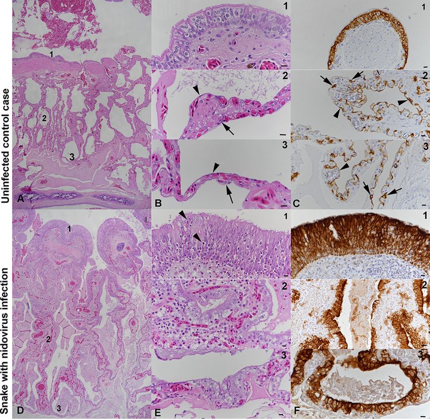
Dervas et al.                                                                                Journal of Virology

KEYWORDS Morelia viridis, NGS, epithelial hyperplasia, green tree python, nidovirus,
pathogenesis, proliferative pneumonia, type II pneumocyte hyperplasia

T    he green tree python, or Southern green python, Morelia viridis (Schlegel, 1872), is
     an oviparous boid constrictor snake with natural habitats in several Indonesian
islands, Florida, Papua New Guinea, and northern Australia (1). In recent years, large
numbers of M. viridis green tree pythons have been exported from Indonesia to Europe
and the United States (2), and they have become increasingly popular in both private
and zoological collections (3, 4). So far, knowledge about the infectious diseases that
occur in M. viridis is limited (5).
    Recently, fatal pneumonias have been reported in two other python species, ball
pythons (Python regius) (6) and Indian pythons (Python molurus) (7); infection with novel

                                                                                                                   Downloaded from http://jvi.asm.org/ on January 29, 2021 by guest
nidoviruses was found to be the common denominator (8, 9). The described viruses are
approximately equidistant from the two current genera, Torovirus and Bafinivirus, of the
Torovirinae subfamily in the family Coronaviridae, order Nidovirales (8–15), in which they
will likely form a novel genus (9).
    The order Nidovirales comprises four families of complex positive-sense single-
stranded RNA (ssRNA⫹) viruses, Arteriviridae, Mesoniviridae, Roniviridae, and Coronaviri-
dae, which are also distinguished by their genome size: roniviruses (⬃26 kb) and
coronaviruses (26 to 33 kb) are known as “large nidoviruses” (16–18), arteriviruses are
known as “small nidoviruses” (13 to 16 kb), and mesoniviruses have genomes with sizes
intermediate between those of the large and small nidoviruses (16, 17, 19, 20). While
roniviruses and mesoniviruses are known to infect crustaceans and insects (19, 21),
arteriviruses have been associated with an acute respiratory syndrome and abortion in
pigs (porcine reproductive and respiratory syndrome virus [PRRSV]) or a lethal hemor-
rhagic disease in nonhuman primates (simian hemorrhagic fever virus [SHFV]) (22). The
Coronavirinae subfamily of the Coronaviridae includes several pathogens of mammals
(feline coronavirus, transmissible gastroenteritis virus, and equine coronavirus, to men-
tion a few), birds, and fish and has provided a steady supply of emerging threats to
human health over the past 15 years, including severe acute respiratory syndrome
(SARS) and Middle Eastern respiratory syndrome (MERS) (23). The members of the
second subfamily of the Coronaviridae, Torovirinae, have so far not generated similar
threats. They include bafiniviruses, which infect ray-finned fish and induce renal tubular
necrosis and necrotizing hepatitis (24), and toroviruses, which infect mammals (includ-
ing humans, cattle, horses, and pigs) (10, 11, 25–27). Toroviruses have a tropism for
epithelial cells of both the respiratory and alimentary tracts (28–30); in cattle, an
association with pneumonia has been reported (30, 31). Toroviruses exhibit a unique
morphology: the viral particles are kidney and/or rod shaped, and toroviruses have a
tubular, torus-shaped ribonucleoprotein (RNP) enveloped by a membrane decorated
with spikes composed of S protein (32). The RNP comprises the nucleoprotein (N
protein) and the ssRNA⫹ genome (33, 34).
    In 2014, a Swiss breeder submitted two adult green tree pythons for diagnostic
postmortem examination. Both animals had died with signs of a mucinous pneumonia.
In the following year, the same breeder submitted another two individuals. All of these
green tree pythons, as well as a further five green tree pythons from three additional
breeders, exhibited similar pathological findings. In 2016, additional cases involving
two more breeding collections were observed. As all routine diagnostic tests under-
taken failed to identify potential causative viruses or specific bacterial agents, we
initiated an investigation into the cause of this apparently emerging disease of green
tree pythons.

RESULTS
   Animals, clinical signs, macroscopic and histological features, and results of
screening for infectious agents. From 2014 to 2016, multiple Morelia viridis green tree
pythons affected by a similar disease arrived for postmortem examination: four animals
from one breeder (animals A1 and A2 in 2014 and animals A3 and A4 in 2015), five

November 2017 Volume 91 Issue 21 e00718-17                                                       jvi.asm.org 2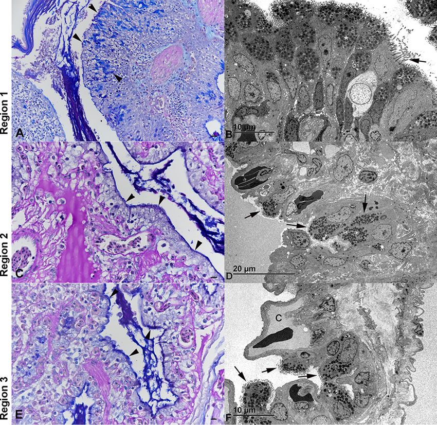
Morelia viridis Nidovirus                                                                                                                       Journal of Virology

TABLE 1 Animals tested in this study
Animala                 Age (yr) Sexb Clinical signs                               Diagnostic testsc                  Diagnosis
MVNV-positive
    animals
  A1                    1         M       No history                               Bacteriology
  A2                    5         F       Sudden death                             NGS
  A3                    8         M       Sudden death, mucus in oral cavity       Bacteriology
  A4                    npd       F       Acute respiratory distress (1 day)
  B1                    6         M       Respiratory distress (3–4 days),         Virology, virus isolation, NGS
                                            expulsion of mucus
   B2                   7         F       Respiratory distress (3–4 days),         Virology
                                            expulsion of mucus
   C1                   2.5       M       Chronic anorexia, acute respiratory      Virology
                                            distress
  C2                    8         M       Respiratory distress                     Virus isolation

                                                                                                                                                                      Downloaded from http://jvi.asm.org/ on January 29, 2021 by guest
  D1                    6         F       Respiratory distress (ⱖ1 wk)             NGS
  E1                    np        F       Respiratory distress (ⱖ1 wk)
  F1                    2         F       Sudden death
  G1                    6         F       Sudden death
Uninfected control
     animals
  G2                    9         M       Sudden death                                                                Mild colitis
  G3                    6         F       Emaciation, death                                                           Mild enteritis and nephritis
  G4                    2         F       Sudden death                                                                BIBD,e severe purulent vasculitis around
                                                                                                                        esophagus and trachea
   G5                   1         M       Sudden death                                                                Focal suppurative pneumonia in one
                                                                                                                        lobe
aAll animals were from breeding collections. MVNV-positive animals tested positive for MVNV by RT-PCR, RNA-ISH, and IH. MVNV-negative animals were negative for
 MVNV by RT-PCR.
bM, male; F, female.

cTests performed prior to or alongside MVNV RT-PCR, IH, and RNA-ISH: bacteriology, routine bacteriological examination performed on lungs; virology, lung tissue

 screened for viruses (reovirus, paramyxovirus, Sunshine virus, nidovirus) in a commercial laboratory; NGS, next-generation sequencing of lung homogenates.
dnp, not provided (adult).

eBIBD, boid inclusion body disease.

animals from three additional breeders (animals B1, B2, C1, C2, and D1 in 2015), and
three animals from three additional breeding collections (animals E1, F1, and G1 in
2016). Of these 12 snakes, 4 had died suddenly without any obvious clinical signs and
7 had exhibited respiratory distress and expulsion of mucus for a period of a few hours
to more than 1 week before death; no clinical history was available for 1 animal. All
animals were adult and ranged from 1 to 8 years of age (Table 1). Six snakes were
female, and six were male. Upon gross postmortem examination, all snakes exhibited
a variable amount of mucoid material in the airways, and the mucoid material was most
abundant in the faveolar spaces of the lungs. In some animals, the entire trachea, the
internal choanae, and the caudal air sacs were obliterated by mucoid material (Fig. 1),
and the lung parenchyma appeared thickened. The histological examination revealed

FIG 1 Gross findings in a green tree python (Morelia viridis) with nidovirus-associated proliferative pneumonia (animal E1). Both trachea (A) and
lungs (B) are filled with abundant mucoid material (arrows). H, heart.

November 2017 Volume 91 Issue 21 e00718-17                                                                                                          jvi.asm.org 3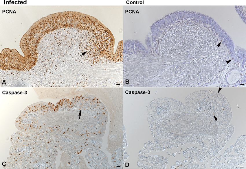
Dervas et al. Journal of Virology
Downloaded from http://jvi.asm.org/ on January 29, 2021 by guest
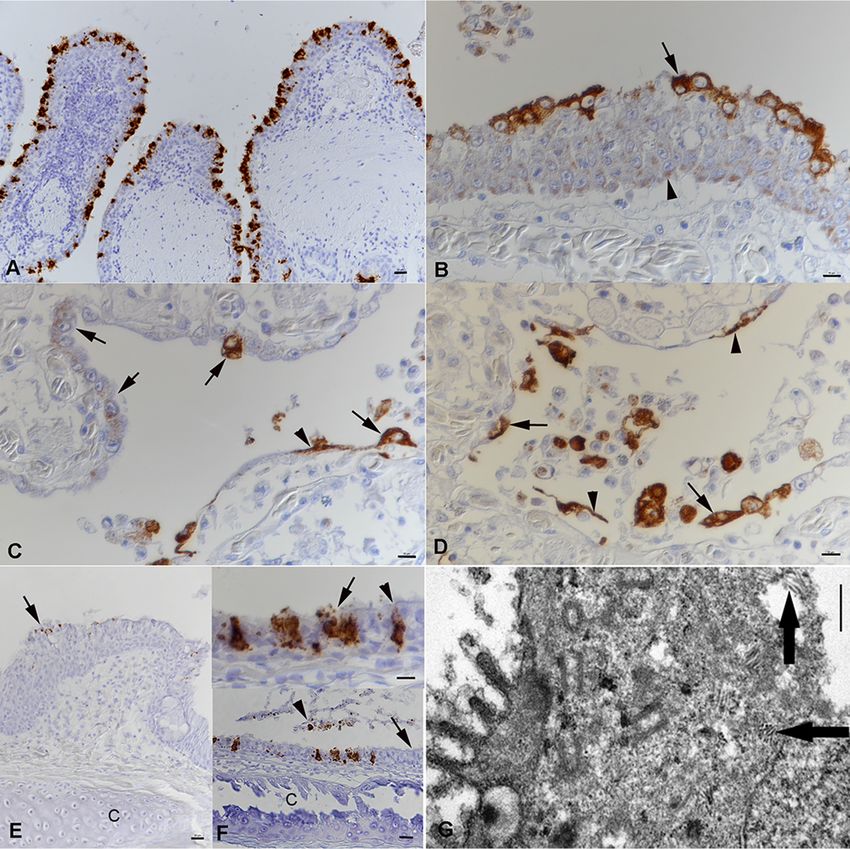
FIG 2 Histological findings in Morelia viridis with nidovirus-associated proliferative pneumonia. (A to C)
Uninfected control case (animal G2). Representative photomicrographs of a healthy lung (cross section)
are shown. Thin pulmonary septa form the faveolar spaces. At the luminal end, the septa exhibit bundles
of smooth muscle cells (myoelastic bundles) that form contractile trabeculae. For detailed assessments,
three regions (regions 1, 2, and 3) were identified. The trabeculae are covered by a multilayered
pseudostratified bronchus-type epithelium, dominated by ciliated cells (panels B1 and C1). In regions 2
(panels B2 and C2) and 3 (panels B3 and C3), the faveolae are covered by a single-layered gas exchange
epithelium, comprised of flat type I pneumocytes (arrowheads); surfactant-producing type II pneumo-
cytes (arrows) are less abundant. (D to F) Snake with nidovirus infection (animal B4). Representative
photomicrographs of a diseased lung (cross section) are shown. In the entire lung, the epithelial layer is
thickened due to hyperplasia. The faveolar space (region 3) is filled with proteinaceous material, and the
interstitium is broadened due to a mixed inflammatory infiltrate (panels E1 and E2). In region 1 (panels
E1 and F1), the pseudostratified epithelium covering the trabeculae exhibits increased cellularity with an
increase in cell layers and an irregular arrangement (hyperplasia), as well as a loss of cilia and several
apoptotic epithelial cells (arrowheads). In regions 2 (panels E2 and F2) and 3 (panels E3 and F3), type I
pneumocytes are almost entirely replaced by large, columnar type II pneumocytes. H&E stain (A, B, D, and
E); IH for cytokeratin, hematoxylin counterstain (C and F). Bars ⫽ 100 m (A, D) and 10 m (B, C, E, F).
a variable degree of epithelial thickening in the trachea and lungs and a mild to
moderate interstitial lymphoplasmacellular and heterophilic infiltration of the lung
parenchyma (Fig. 2). In one animal (animal E1), a moderate multifocal granulomatous-
necrotizing nephritis was additionally observed. The four control snakes (animals G2 to
G6), i.e., animals that had been euthanized due to nonrespiratory diseases (Table 1), did
not exhibit similar gross and/or histological changes (Fig. 2).
We suspected an infectious cause and had routine bacteriological examinations
performed on the lungs of selected snakes from the 2014 and 2015 cohort (animals A1,
A3, and D1). This yielded only a nonspecific bacterial flora (i.e., no primary pathogens,
Pseudomonas aeruginosa, a Proteus sp., Citrobacter braakii, Achromobacter xylosoxidans,
Stenotrophomonas maltophilia, and Providencia rettgeri). A further three animals, from
the 2015 cohort (animals B1, B2, and C1), were then screened in a commercial
laboratory for a range of potentially pathogenic viruses (i.e., reovirus, paramyxovirus,
Sunshine virus, nidovirus); these tests yielded negative results.
November 2017 Volume 91 Issue 21 e00718-17 jvi.asm.org 4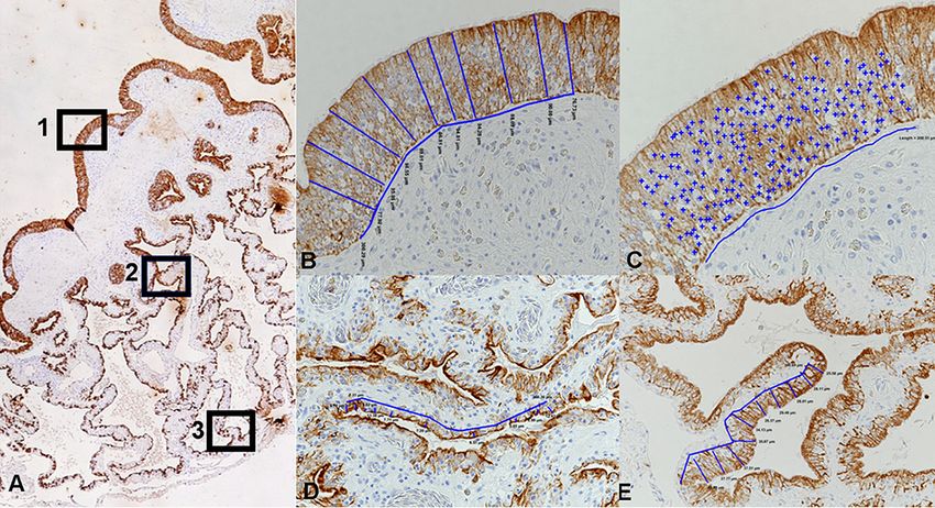
Morelia viridis Nidovirus Journal of Virology
Downloaded from http://jvi.asm.org/ on January 29, 2021 by guest
FIG 3 Virus isolation in a primary Morelia viridis fetal brain cell culture. (A) Uninfected control cells. (B, C)
Cells after inoculation with homogenate of lung tissue from a diseased snake (animal C2). The first
evidence of a cytopathic effect is seen after 2 days (B) as enlargement, rounding, and cytoplasmic
vacuolization as well as a loss of adherence (arrows). After 6 days (C), only a few intact cells have
remained. Most cells are detached and necrotic. (D, E) Ultramicrographs of a cell pellet prepared at 1 dpi.
There are abundant tubular structures arranged in stacks within the cytoplasm of infected cells (arrows).
(F) Concentrated supernatant of the infected cells, negative staining. Rod- and kidney-shaped virions are
approximately 120 nm long. Bars ⫽ 1 m (D) and 100 nm (E).
Virus isolation in tissue cultures. As the routine bacteriological and virological
tests failed to identify a common denominator for the diseased snakes, we decided to
attempt virus isolation. For this purpose, we prepared primary cultures of green tree
python fetal liver and brain cells (Fig. 3A). After four passages, the cells began to
proliferate more rapidly and we could expand the cultures enough to attempt isolation
of the unknown pathogen. When we used homogenates of lung tissue from the
diseased snakes (Table 1) to inoculate both cultures, we observed the first evidence of
a cytopathic effect (CPE) (enlargement, rounding, and cytoplasmic vacuolization of
cells) and a loss of adherence (Fig. 3B) at approximately 3 days postinfection (dpi). At
6 dpi, almost the entire monolayer was affected and most cells had rounded and/or
detached (Fig. 3C). The ultrastructural examination of a cell pellet prepared from the
cultures at 1 dpi identified abundant tubular viral structures arranged in stacks within
the cytoplasm of infected cells (Fig. 3D and E). We concentrated the supernatants of the
infected cells by ultracentrifugation and examined the pelleted material by transmis-
sion electron microscopy (TEM) under negative staining. TEM analysis demonstrated
rod- and kidney-shaped virions of approximately 120 nm in length (Fig. 3F), consistent
with torovirus particles (29, 35).
Identification of nidovirus by NGS and confirmation of the findings by real-
time RT-PCR. In parallel with our attempts at virus isolation, we decided to employ
next-generation sequencing (NGS) for the identification of the causative agent(s) of the
pneumonia. We isolated RNA from homogenates of lung tissue from three animals
(animals A2, B1, and D1) and performed RNA sequencing. For two snakes (animals B1
and D1), only contiguous sequences (contigs) that matched the sequences of bacterial
genomes were identified. We interpreted these bacterial sequences to most likely
represent either contamination during postmortem sample collection or, more likely, a
secondary bacterial infection, as both had also shown moderate diffuse heterophil
infiltration in the lungs. From the third snake (animal A2), however, we obtained a
single ⬃21,000-nucleotide (nt) contig and several shorter contigs that matched the
previously identified sequences of ball python nidovirus (BPNV) and python nidovirus
(PNV) (the 21,000-nt contig was ⬃85% identical to the sequences of both viruses) (8, 9).
In total, we obtained ⬃28,000 nt (⬃75 to 80%) of the genome from the lung tissue
November 2017 Volume 91 Issue 21 e00718-17 jvi.asm.org 5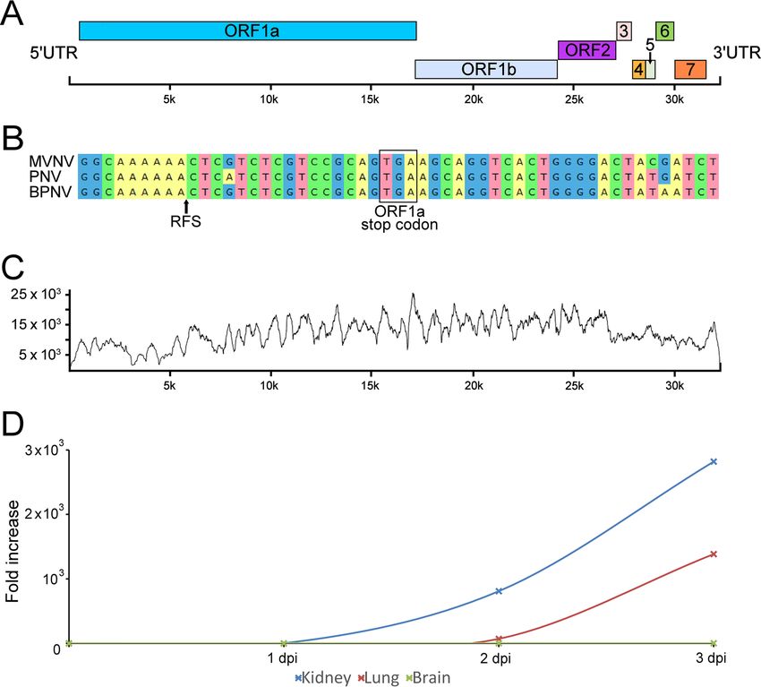
Dervas et al.                                                                                    Journal of Virology

sample. In addition to the novel nidovirus isolate, we identified a contig that matched
the sequences of endogenous retrovirus genes with very high coverage; we will
describe this contig in a separate report (unpublished data). However, the retrovirus
was also present in the supernatants of M. viridis cell cultures inoculated with homog-
enates of lung tissue. We decided to attempt to produce a nidovirus preparation devoid
of the contaminating retrovirus. To do this, we inoculated boid kidney cells with the
supernatant collected from the Morelia viridis liver cell culture inoculated with homog-
enates of lung tissue, since Huder et al. previously reported species-restricted growth
of an endogenous python retrovirus (36). Using this approach, we obtained a pure
nidovirus isolate, as confirmed by retrovirus-specific reverse transcription-PCR (RT-PCR).
We then performed endpoint titration to quantify the number of infectious units in the
Morelia viridis nidovirus (MVNV) stock on M. viridis brain cells and could detect 2.25 ⫻

                                                                                                                       Downloaded from http://jvi.asm.org/ on January 29, 2021 by guest
1010 focus-forming units per 1 ml of cell culture supernatant. RNA extracted from the
pure nidovirus preparation was subjected to another NGS run, which yielded almost a
full-length genome in a single contig. We then performed a reference assembly using
BPNV as the template to recover the missing genome ends (some 150 nt in total) for
the novel virus, MVNV. The overall genome structure of MVNV (GenBank accession
number MF351889) was similar to that of BPNV and PNV (Fig. 4A). The identified contig
contains eight partially overlapping open reading frames (ORFs) flanked by the 5=
untranslated region (UTR) (⬃650 nt) and the 3= UTR (⬃920 nt). We used the HMMER3
web server to look for functional domains in the detected ORFs, and these are
described in Table 2. We also identified a putative ribosomal frameshift signal (RFS)
sequence near the end of ORF1a. To further study the RFS, we aligned the sequences
of other nidoviruses identified in snakes around the RFS and used the Mfold web server
to predict the structure around this region in the MVNV genome (Fig. 4B). The NGS data
coverage for the MVNV genome (raw data are available at http://www.ncbi.nlm.nih
.gov/biosample/7248312) is shown in Fig. 4C. The identities between the nucleotide
and amino acid sequences of the novel nidovirus isolate and those of other python
nidoviruses are indicated in Table 3.
    We then tested whether the isolated MVNV could infect other cell lines. For this
purpose, we selected brain, kidney, and lung cell lines of Boa constrictor and used M.
viridis brain cells as the positive control. We also designed a TaqMan real-time RT-PCR
to be able to quantify and monitor the nidovirus RNA in the cell culture supernatants
and lung tissue samples from the diseased snakes. Analysis of the cell culture super-
natants collected from different cell lines inoculated with MVNV showed clear ampli-
fication in B. constrictor kidney and lung cells (Fig. 4D). The cytopathic effect on M. viridis
brain cells was extremely severe, and therefore, a similar quantification could not be
performed. The infected cells were stained using anti-MVNV N protein antiserum at 3
dpi, and all cell lines, except B. constrictor brain cells (only a few infected cells were
detected), were shown to be extremely permissive for MVNV (Fig. 5).
    As ORF1b is the most conserved ORF among nidoviruses (17), we selected this
region for the phylogenetic analysis. The phylogenetic tree suggests that MVNV forms
an outgroup to BPNV and PNV (Fig. 6). Further, the python nidoviruses clustered
together with shingleback nidovirus (GenBank accession number KX184715) (37),
Xinzhou nematode virus 6 (GenBank accession number KX883637), Xinzhou toro-like
virus (NCBI reference sequence accession number NC_033700), and bovine nidovirus
(NCBI reference sequence accession number NC_027199) (31). Consistent with the
previous reports, these form a clade that is separate from both the genus Bafinivirus and
the genus Torovirus (Fig. 6).
    Finally, we used RNA isolated from cell cultures inoculated with an homogenate of
lung tissue from an infected snake as the positive control and RNA isolated from a Boa
constrictor snake as the negative control in the real-time RT-PCR. All diseased snakes
tested positive for nidovirus RNA in the lungs. In contrast, the four snakes without gross
and/or histological evidence of pneumonia were negative.

November 2017 Volume 91 Issue 21 e00718-17                                                           jvi.asm.org 6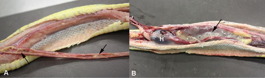
Morelia viridis Nidovirus                                                                                                                     Journal of Virology

                                                                                                                                                                     Downloaded from http://jvi.asm.org/ on January 29, 2021 by guest
FIG 4 Morelia viridis nidovirus (MVNV) genome organization, NGS coverage, and phylogenetic relationship to related viruses. (A) A schematic
representation of the untranslated regions (UTRs) and open reading frames (ORFs) of the 32,414-nt ssRNA⫹ genome of MVNV. (B) Alignment
around the ribosomal frameshift signal (RFS; indicated by an arrow in the alignment and structure prediction) in nidoviruses identified in pythons.
The ORF1a stop codon is highlighted. A prediction for the RNA structure around the RFS in MVNV is shown. The arrow highlights a difference
from the structure described by Bodewes et al. (8). (C) Contig coverage. The y axis shows the number of reads matching each nucleotide position
(x axis) of the contig. The Bowtie2 program was used to map the reads to the contig generated by de novo assembly. (D) Virus growth in different
cell lines. The cell culture supernatant of the indicated cell lines inoculated with MVNV was analyzed by qRT-PCR at the indicated time points.
The fold increase indicates the increase in the amount of viral RNA in the cell culture supernatant compared to the amount detected at 0 dpi.

    Nidoviruses are associated with pneumonia in M. viridis. The histological exam-
ination confirmed that all diseased snakes had suffered from a chronic pneumonia with
epithelial thickening in the trachea and lungs (Fig. 2) and excess mucus in the lumen
of the lungs and airway. The inflammatory component was represented by mild to
moderate multifocal interstitial infiltration of lymphocytes, plasma cells, and/or hetero-
phils (Fig. 2E). The mucus filling the faveolar space often contained heterophils and cell
debris. In three of the nine tracheas examined, we also observed a variable degree of
infiltration with a mixture of inflammatory cells (heterophils, macrophages, lympho-
cytes). Over its entire length, the lung epithelium exhibited numerous epithelial cells
that contained mucus, as indicated by periodic acid-Schiff (PAS)–alcian blue staining
(Fig. 7A, C, and E). In region 1, at the trabeculae, the mucus-containing cells had the
ultrastructural features of secretory cells (38) (Fig. 7B). In regions 2 and 3 (faveolar
epithelium), where the increase in the amounts of these cells was the most striking,

November 2017 Volume 91 Issue 21 e00718-17                                                                                                           jvi.asm.org 7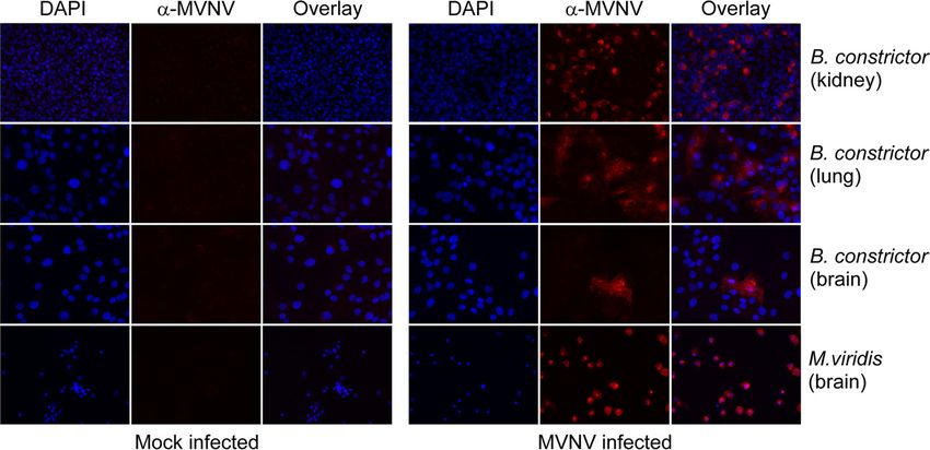
Dervas et al.                                                                                                                            Journal of Virology

TABLE 2 HMMER3 analysis of functional domains in the ORFs detected in the MVNV genome
                                                   Alignment                           E value
           Family             Pfam accession       Start       End
ORF        identifier          no.a                 position    position    Bit score   Independent      Conditional   Description
ORF1a      Methyltransf_25    PF13649.5            3075        3150        15.08       0.027            4.80E⫺06      Methyltransferase domain
           zf-CCCH            PF00642.23           1052        1072        12.09       0.13             2.40E⫺05      Zinc finger C-X8-C-X5-C-X3-H type
           zf-CCCH            PF00642.23           1098        1117        9.91        0.64             0.00011       Zinc finger C-X8-C-X5-C-X3-H type

ORF1b      NSP13              PF06460.11           2077        2239        50.2        1.80E⫺13         1.20E⫺16      Coronavirus NSP13
           Viral_helicase1    PF01443.17           1144        1380        45.91       5.20E⫺12         3.40E⫺15      Viral (superfamily 1) RNA helicase
           AAA_30             PF13604.5            1101        1231        42.93       3.90E⫺11         2.60E⫺14      AAA domain
           RdRP_1             PF00680.19           479         651         40.69       1.00E⫺10         6.60E⫺14      RNA-dependent RNA polymerase
           AAA_19             PF13245.5            1100        1229        33.98       3.10E⫺08         2.00E⫺11      AAA domain

                                                                                                                                                               Downloaded from http://jvi.asm.org/ on January 29, 2021 by guest
ORF5       Arteri_nucleo      PF01481.15           32          132         23.35       5.50E⫺05         9.90E⫺09      Arterivirus nucleocapsid protein
ahttp://pfam.xfam.org/.

they exhibited the morphology of type II pneumocytes (Fig. 7D and F) (39); their
numbers varied in affected snakes and ranged from occasional patchy aggregates to a
diffuse lining of the entire faveolae in association with an almost complete absence of
type I pneumocytes (Fig. 2E). Ultrastructurally, these cells exhibited serous/mucous
granules instead of the lamellar bodies that are characteristic of type II pneumocytes
(Fig. 7B, D, and F); they have recently been described to be transformed type II
pneumocytes (40).
    Having detected nidoviral RNA in the affected lungs, we aimed to identify the viral
target cells. For this purpose, we employed both RNA in situ hybridization (RNA-ISH)
and immunohistology (IH). Viral RNA and antigen (N protein) were detected in the
cytoplasm of the trabecular pseudostratified epithelium (region 1) and the cytoplasm
of both type I and type II pneumocytes lining the faveolar space (regions 2 and 3) in all
affected snakes (Fig. 8A to D). Intact and degenerated cells shed into the mucus were
found to be infected, and occasionally, we also detected cell-free viral RNA and antigen
(Fig. 8B). Infected cells also varied in number between animals and in different areas of
the lung in individual snakes. In the hyperplastic epithelium of region 1, most cells
exhibited a weak, focal cytoplasmic reaction, and only the superficial cell layer con-
tained abundant viral antigen (Fig. 8B). The new type II pneumocytes that seemed to
progressively replace the type I cells exhibited a similarly weak focal reaction, whereas
the fully differentiated pneumocytes were strongly positive for viral antigen (Fig. 8C
and D), suggesting more efficient virus replication. When examined, the trachea (n ⫽ 9)

TABLE 3 Nucleotide and amino acid sequence identities between MVNV and the nidoviruses identified in Python molurus (Indian python)
and Python regius (ball python)
                                % identity
                                Nucleotide sequence                                              Amino acid sequence
                                Python nidovirus S1536/13             Ball python nidovirus      Python nidovirus S1536/13        Ball python nidovirus
ORF                             (KJ935003)a                           07-53 (KJ541759)           (KJ935003)                       07-53 (KJ541759)
5= UTR                          85.1                                  87.0
ORF1a                           72.8                                  72.8                       69.5                             69.6
ORF1b                           86.5                                  86.3                       91.0                             90.5
ORF2                            80.9                                  80.0                       85.0                             84.8
ORF3                            69.5                                  67.0                       64.7                             65.4
ORF4                            76.5                                  79.2                       83.3                             82.9
ORF5                            75.1                                  74.0                       74.2                             74.8
ORF6                            65.6                                  66.9                       59.7                             59.2
ORF7                            77.1                                  78.6                       68.8                             71.9
3= UTR                          95.2                                  93.9
Complete genome/CDSb            77.3                                  77.3                       77.2                             77.1
aGenBank  accession numbers are given in parentheses.
bCDS, coding sequence.

November 2017 Volume 91 Issue 21 e00718-17                                                                                                   jvi.asm.org 8
Morelia viridis Nidovirus Journal of Virology
Downloaded from http://jvi.asm.org/ on January 29, 2021 by guest
FIG 5 Immunofluorescence staining of M. viridis and B. constrictor cell lines with anti-MVNV N protein
antiserum at a 1:2,000 dilution. MVNV- or mock-infected cells were stained at 3 dpi, and images were
recorded at a magnification of ⫻40 under an inverted fluorescence microscope. (Right) Mock-infected cells;
(left) MVNV-infected cells. Blue, DAPI; red, MVNV-specific staining.
and the nasopharyngeal epithelium (n ⫽ 1) also exhibited infected, degenerating
epithelial cells (Fig. 8E and F). Viral antigen was detected in few epithelial cells of the
esophagus in one animal with acute pneumonia (animal G1). Interestingly, viral antigen
was also found cell free and within a few macrophages in the focal granulomatous-
necrotizing nephritis in another affected animal (animal E1). We did not detect viral
RNA or antigen in other tissues of the infected snakes, including the stomach and
intestines, or in the lungs of the control snakes (animals G2 to G6).
We could demonstrate cytoplasmic accumulations of tubular structures that ranged
from 150 nm to 250 nm in length in rare epithelial cells that had the morphology of
type II pneumocytes by ultrastructural examination (Fig. 8G).
The epithelial thickening was the most striking feature in the lungs of the nidovirus-
positive snakes. We therefore performed a more detailed histological examination in
the attempt to identify the processes underlying this phenomenon. The multilayered
epithelium covering the trabeculae (region 1) displayed increased cellularity with an
increase in the number of cell layers and an irregular arrangement, indicating hyper-
plasia (Fig. 2E). The epithelial hyperplasia extended to the upper respiratory tract and
was also observed in the trachea, larynx, and nasal cavity. The middle and basal areas
of the faveolae (regions 2 and 3), where the epithelium is unilayered, showed nuclear
crowding, which also suggested hyperplasia (Fig. 2E). The thickening appeared to result
from an increase in individual cell height, associated with a more columnar appearance
of the cells (Fig. 2E and F). Additionally, we noted an increase in septal connective tissue
(interstitial fibrosis). The described changes resulted in thickening of the septa and
narrowing of the faveolar lumen (Fig. 2D).
In the attempt to quantify the epithelial hyperplasia, we measured the average total
epithelial height in the three defined regions (Fig. 2 and 9) in all diseased, i.e.,
RT-PCR-positive, animals and compared it to that in the RT-PCR-negative animals
without pneumonia (controls). In all locations, the epithelium was significantly (P ⬍
0.05) higher in the diseased animals than in the control snakes (Fig. 10A; Table 4).
Hyperplasia was confirmed in both the lungs (regions 1, 2, and 3) and the trachea,
where the average number of nuclei in the epithelial layer was significantly higher in
the diseased snakes (Fig. 10B; Table 4).
Epithelial hyperplasia was associated with increased proliferative activity, as up to
90% of epithelial cells in all lung regions and in the trachea of the diseased animals
were found to express PCNA (Fig. 11A). Alongside this, a moderate number of epithelial
cells were found to undergo apoptosis, based on cleaved caspase-3 staining (Fig. 11C).
In comparison, control animals exhibited rare PCNA-positive type II pneumocytes in all
November 2017 Volume 91 Issue 21 e00718-17 jvi.asm.org 9Dervas et al. Journal of Virology
Downloaded from http://jvi.asm.org/ on January 29, 2021 by guest
FIG 6 Maximum clade credibility tree constructed from the ORF1b amino acid sequences of representatives of the subfamily Torovirinae. The phylogenetic tree
was constructed using the Bayesian MCMC method with the LG⫹G⫹I model of substitution. Posterior probabilities are shown at each node.
layers (Fig. 11B) as well as scattered apoptotic (cleaved caspase-3-positive) type I and
type II pneumocytes (Fig. 11D), likely representing the physiological turnover of the
epithelium.
DISCUSSION
We initiated the present study by the urge to identify the causative agent of a fatal
pneumonia observed in green tree pythons (Morelia viridis), characterized by the
accumulation of mucoid material in the airways and a histologically notable thickening
of the lung epithelium. NGS revealed the presence of a novel nidovirus, MVNV, which
by phylogenetic analysis groups with toroviruses. RT-PCR, immunohistology, and RNA-
ISH served to confirm its association with the disease and to identify the viral target
cells, i.e., epithelial cells in the airways and the luminal trabeculae, as well as faveolar
type I and II pneumocytes. We were able to isolate and grow MVNV in both M. viridis
and B. constrictor cell cultures. MVNV induced a cytopathic effect both in vitro and in
vivo. However, we also found nidovirus infection to associate with generalized hyper-
plasia of the airway and lung epithelium, which exhibited a distinct proliferative activity
and a degree of apoptotic cell death. Together these findings would suggest that
nidovirus infection increases the turnover of the epithelium. The mucus accumulation
November 2017 Volume 91 Issue 21 e00718-17 jvi.asm.org 10Morelia viridis Nidovirus Journal of Virology
Downloaded from http://jvi.asm.org/ on January 29, 2021 by guest
FIG 7 Changes in the lung epithelium of Morelia viridis green tree pythons with nidovirus-associated
proliferative pneumonia. Representative photomicrographs of cross sections of the diseased lungs of
animal B4 (A, C, E) and ultramicrographs of cross sections of the diseased lungs of animal A1 (B, D, and
F) are shown. (A, B) Region 1. (A) The PAS-alcian blue stain highlights positive secretory epithelial cells
(arrowheads) in all cell layers of the hyperplastic multilayered trabecular epithelium. Abundant mucus
fills the lumen. (B) The ultrastructural features of most cells (cytoplasmic mucous granules and short
microvilli) show that they are secretory cells (38). Only a few ciliated cells are seen (arrow). (C to F) In
regions 2 (C, D) and 3 (E, F), the faveolae are diffusely lined by cuboidal cells containing serous/mucous
granules (arrowheads, C and E), consistent with type II pneumocytes. Ultrastructurally, these cells
resemble so-called transformed type II pneumocytes, as they contain serous/mucous granules (arrows)
and lack the lamellar bodies characteristic for normal type II pneumocytes (39). C, capillary.
in the air-conducting space was accompanied by a significant increase in secretory
epithelial cells at the trabeculae and in (transformed) type II pneumocytes in the
faveolae, indicating increased mucus and/or surfactant production.
These results indicate that MVNV infects and damages the differentiated respiratory
and faveolar epithelium but then persists and induces increased turnover of the
infected epithelial cells. The observed type II pneumocyte hyperplasia is obviously not
specific to nidovirus infection in the python, since it has been described in snakes as a
consequence of pneumocyte injury in a range of infectious diseases of viral, bacterial,
and mycotic origin (39, 40). It might therefore represent an exaggerated regenerative
attempt. In mammals, type II pneumocyte hyperplasia is also seen to be part of a lung
defense mechanism against various insults (41–43). In our cases, the hyperplastic
epithelial cells exhibited abundant cytoplasmic serous/mucous granules instead of the
lamellar bodies that represent surfactant (38). This suggests excess mucus production
and release and reduced surfactant production and release and would explain the
clinical findings (38, 40). Previous studies have shown that the surfactant of snakes, due
to its phospholipid composition, which is different from that in other animals, is likely
less important for airway stabilization but, rather, functions as an antiadherent (also
known as an “antiglue”) factor and an antiedemic factor in the faveolar space (44); the
reduction in the amount of surfactant could therefore have added to the respiratory
distress observed in the affected snakes.
November 2017 Volume 91 Issue 21 e00718-17 jvi.asm.org 11Dervas et al. Journal of Virology
Downloaded from http://jvi.asm.org/ on January 29, 2021 by guest
FIG 8 Viral target cells in lungs and airways of Morelia viridis green tree pythons with nidovirus-associated proliferative pneumonia. (A to D) The
lungs of animals A1 (A) and A2 (B to D). (A) The pseudostratified trabelular epithelium (region 1) exhibits abundant viral RNA mainly in superficial
epithelial cell layers. (B) Viral antigen expression is seen in all cell layers. In the basal and middle layers, it is represented by a focal cytoplasmic
reaction (arrowhead). The fully differentiated superficial epithelial cells exhibit abundant viral antigen (arrow) and a loss of cilia. Infected cells are
also found to be shed into the lumen. (C) Region 2. In areas with intact faveolar epithelium, both type I pneumocytes (arrowhead) and
differentiated type II pneumocytes (arrows) exhibit strong viral antigen expression. The new, hyperplastic type II pneumocytes in other areas
display a weaker focal cytoplasmic reaction (arrows). (D) At the base of the faveolae, type I pneumocytes (arrowheads) and type II pneumocytes
(arrows) are also found to be infected. They are also shed into the faveolar lumen. (E) Nasal cavity of animal A1. The epithelium is thickened due
to hyperplasia and exhibits nidovirus infection mainly of the fully differentiated superficial epithelial cells (arrow). C, cartilage. (F) Trachea of animal
A1. (Bottom) The ciliated epithelium is largely intact (arrow), but there are scattered individual cells or small groups of infected cells. Sloughed,
degenerate epithelial cells are also found to carry viral RNA (arrowhead). (Top) Infected cells are intact (arrowhead) or degenerate, shedding virus
(arrow). C, tracheal ring cartilage. RNA-ISH (RNAscope technology) (A, E, F) and IH (HRP method) for the detection of MVNV N protein (B to D)
and hematoxylin counterstain were used. (G) Ultramicrograph of a type II pneumocyte with staples of tubular structures of 100 to 150 nm in
length (arrows). Bars ⫽ 20 m (A, E, and F [bottom]), 10 m (B to D and F [top]), and 1 m (G).
Increased turnover and/or hyperplasia of the epithelium has been described for
several coronaviral diseases, such as infectious bronchitis in chickens, Breda virus
infection in calves, and coronavirus infection in rats (45–47). So far, however, the
mechanisms underlying this process have not been explained. Members of the sub-
November 2017 Volume 91 Issue 21 e00718-17 jvi.asm.org 12Morelia viridis Nidovirus Journal of Virology
Downloaded from http://jvi.asm.org/ on January 29, 2021 by guest
FIG 9 Illustration of the morphometric approach taken to measure the epithelial height and number of
epithelial cell nuclei in cytokeratin-stained cross sections of the lungs. (A) Three regions were defined in
the proximal, bronchiolar lung: (i) the luminal part of the primary trabeculae, covered by a multilayered
pseudostratified ciliated epithelium (region 1), and (ii) the faveolae at the middle level (region 2) and at
the base (region 3) covered by a single-layered epithelium. (B) Measurement of epithelial height in region
1. The blue lines identify the locations of the 10 measurements taken over a 300-m-wide epithelial
segment, determining the distance between the apical epithelial cell border and the basal membrane.
Magnification, ⫻200. (C) Determination of cell number in region 1. The total amount of epithelial nuclei
(⫹) counted in a 200-m-wide epithelial segment. Magnification, ⫻400. (D, E) Measurement of epithelial
height in regions 2 (D) and 3 (E). The blue lines identify the locations of the 10 measurements taken over
a 300-m-wide epithelial segment, determining the distance between the apical epithelial cell border
and the basal membrane. Magnification, ⫻200.
family Torovirinae are also known to induce epithelial cell apoptosis (35, 48, 49);
however, though our findings point toward this, further studies are required to eluci-
date whether this also applies to MVNV and other snake nidoviruses.
In our study, only fatal cases were examined. The pathological findings suggest that
death was mainly due to impaired gas exchange as a consequence of type I pneumo-
FIG 10 The average epithelial height measured in three lung regions and the trachea of infected animals was significantly higher than that in noninfected
(control) animals. Box plots illustrate the third quartile (Q3) and first quartile (Q1) range of the data, and the open circle represents a data outlier (⬎1.5 times
the interquartile range). Box plots were created with SPSS Statistics software. A Mann-Whitney U test for independent samples (t test) was used for statistical
analysis, and a P value of less than 0.05 was considered significant.
November 2017 Volume 91 Issue 21 e00718-17 jvi.asm.org 13Dervas et al. Journal of Virology
TABLE 4 Results of quantitative assessment of epithelial height and cellularity in lungs
and tracheas of MVNV-positive animals in comparison to uninfected control animalsa
MVNV-positive animals Uninfected control animals
Tissue and region b Epithelial ht (m) No. of nuclei Epithelial ht (m) No. of nuclei
Lung
Region 1 62.97 ⫾ 25.39 62.97 ⫾ 25.39 24.96 ⫾ 26.13 6.54 ⫾ 0.81
Region 2 24.4 ⫾ 14.16 14.97 ⫾ 3.57 6.61 ⫾ 14.59 8.83 ⫾ 0.76
Region 3 17.43 ⫾ 8.1 15.22 ⫾ 2.66 6.46 ⫾ 8.41 10.33 ⫾ 0.85
Trachea 61.5 ⫾ 14.06 82.33 ⫾ 39.15 28.21 ⫾ 5.74 44.25 ⫾ 12.09
aMVNV-positive animals (n ⫽ 12) tested positive by RT-PCR, RNA-ISH, and IH, and uninfected control animals
(n ⫽ 4) were negative for MVNV by RT-PCR and IH. Data represent means ⫾ standard deviations. For all
regions and the trachea, the values were significantly different between infected and control animals (P ⬍
0.05).
Downloaded from http://jvi.asm.org/ on January 29, 2021 by guest
bRegion 1, multilayered pseudostratified epithelium covering the luminal part of the primary trabeculae;
region 2, single layer of epithelium at the middle level of the faveolae; region 3, single layer of epithelium
at the base of the faveolae.
cyte loss. In the healthy snake lung, thin cytoplasmic extensions of type I pneumocytes
cover the capillary walls, forming the gas-blood barrier (38). Their replacement by
mucus/surfactant-secreting type II pneumocytes with their excessive height due to cell
crowding is unlikely to allow effective gas exchange. With an average thickness of 24.40
m (region 2) and 17.43 m (region 3), the barrier was more than 1.7 ⫻ 104 times
thicker than the normal blood-gas barrier in the reptilian lung, which ranges from 0.4
to 1 nm (38).
Further anatomical peculiarities of the Morelia viridis lung (or the boid snake lung in
general) could have contributed to the fatal outcome of the disease (39, 50–52). Boidae
have a well-developed right lung and a rudimental left lung. The right lung displays two
FIG 11 Increased epithelial turnover in the lungs of Morelia viridis green tree pythons with nidovirus-
associated proliferative pneumonia. (A, C) Trabeculae (region 1) of diseased animal C1. The hyperplastic
pseudostratified epithelium is proliferating, as confirmed by the expression of PCNA in almost all epithelial
cells. Infiltrating lymphocytes (arrow, A) are also positive. There are also abundant, cleaved caspase
3-positive apoptotic epithelial cells (C). A proportion of infiltrating leukocytes is also found to undergo
apoptosis (arrow, C). (B, D) Trabeculae (region 1) of control animal G3. In the control animal, only scattered
individual basal epithelial cells are found to express PCNA (arrowhead, B). Similarly, only rare epithelial cells
are found to undergo apoptosis (arrowhead, D). Apoptosis (i.e., cleaved caspase-3 expression) is also seen
in occasional lymphocytes (arrow) of the lymphoid aggregates. IH (HRP method) was used to detect PCNA
(A, B) and cleaved caspase-3 (C, D). Hematoxylin counterstain was used. Bars ⫽ 20 m.
November 2017 Volume 91 Issue 21 e00718-17 jvi.asm.org 14Morelia viridis Nidovirus Journal of Virology
anatomically distinct regions: the anterior region, which contains the profusely com-
partmented gas exchange tissue, and the posterior saccular region, which is devoid of
respiratory tissue and has therefore been referred to as the “air sac” (53, 54). The
combination of elongated lungs and caudal air sacs may contribute considerably to the
outcome of the disease, as they create a cul-de-sac that impairs removal of the mucus
and thereby significantly reduces the amount of air-filled space and the gas exchange
capacity.
Though toroviruses mainly associate with enteric diseases, recent studies have
shown that they can be both entero- and pneumotropic (31, 55, 56). New nidoviruses
were recently identified in the lungs of cattle and wild shingleback lizards with
pneumonia, though their direct association with disease has so far not been examined
(31, 37). We found MVNV-associated lesions almost exclusively in the airways and lungs,
similar to previous reports on nidovirus infections in other python species (8, 9, 15). The
Downloaded from http://jvi.asm.org/ on January 29, 2021 by guest
detection of viral RNA by PCR in other tissues, such as the liver, spleen, kidney, and
intestine, however, indicated that the viruses spread systemically. We found further
evidence of that and of its pathogenicity, as we detected nidovirus N protein within a
focal granulomatous-necrotizing nephritis in one animal. We also detected viral antigen
in epithelial cells of the cranial esophagus in one affected animal; however, in M. viridis
the esophagus carries a ciliated epithelium (data not shown), a feature also known for
other snake species (57, 58); infection could therefore be due to an overspill from the
trachea and nasal cavity. We did not detect viral antigen in any cells of the stomach or
intestine, and a previous study also did not find viral RNA by RNA-ISH, suggesting that
the python nidoviruses are primarily respiratory (8). Viral spread via the expelling of
mucus from the nasal cavity would be a likely route of transmission.
MATERIALS AND METHODS
Animals. The study was performed on 16 green tree pythons (Morelia viridis) from six breeding
collections in Switzerland and one collection in Germany (Table 1). The collections varied in both size (i.e.,
the number of breeding animals) and species range, from a single snake up to a collection of 50 snakes
of various species.
All animals were submitted to the Institute of Veterinary Pathology, Vetsuisse Faculty, University of
Zurich, for diagnostic purposes. Fifteen snakes had died spontaneously, and one was euthanized
following an Animals Scientific Procedures Act 1986 (ASPA) schedule 1 procedure (appropriate methods
of humane killing [http://www.legislation.gov.uk/ukpga/1986/14/schedule/1]). In each case, a full diag-
nostic postmortem examination was performed with the owner’s consent. For these diagnosis-motivated
necropsies, no ethical permission is required at the University of Zurich.
The initial study population was represented by nine snakes, of which four (animals A1 to A4) were
from one breeder (breeder A), two each were from a second breeder (breeder B; animals B1 and B2) and
a third breeder (breeder C; animals C1 and C2), and one (animal D1) was from a fourth breeder (breeder
D) (Table 1). These animals were submitted between September 2014 and November 2015. After
completion of the next-generation sequencing (NGS) study, another seven snakes were submitted
(animals E1, F1, and G1 to G5) by another three breeders (breeders E, F, and G). All initial snakes and
another three snakes of the second cohort exhibited one common gross feature: the airways contained
a variable amount of mucoid material which was most abundant in the faveolar lumen (Table 1). The
remaining four snakes (animals G2 to G5) did not show any changes in the airways or lungs (Table 1) and
later served as controls.
Sample collection and screening for infectious agents. During postmortem examinations, sam-
ples from all organs were collected and fixed in 10% buffered formalin for histological examination.
Additional samples from the brain, lung, liver, and kidney were stored at ⫺80°C for further analysis. Also,
lung tissue samples from the freshly euthanized snake (animal A2) and from two snakes that were
necropsied within a few hours after death (animals B1 and D1) were fixed in glutaraldehyde-
paraformaldehyde and processed for transmission electron microscopy (TEM) as described previously
(59).
Prior to the performance of NGS, six of the initial cases were screened for infectious agents: the
cultivation of bacteria from the lungs of three snakes (animals A1, A3, and D1) was attempted at the
Institute of Veterinary Bacteriology, Vetsuisse Faculty, University of Zurich, and lung tissue samples from
another three snakes (animals B1, B2, and C1) were submitted to a commercial lab (Laboklin, Basel,
Switzerland) for virus detection (reovirus, paramyxovirus, Sunshine virus, nidovirus) (Table 1).
Virus isolation and ultrastructural characterization. Cultures of primary tissues from the Morelia
viridis snakes were established as described previously (59), using brain and liver material from a fetus
(from a clutch of animals B1 and B2). The tissue was trimmed into blocks (1 mm), suspended in 5 ml of
minimal essential medium (MEM; Thermo Fisher Scientific, Gibco) supplemented with HEPES (25 mM),
10% fetal bovine serum (FBS; Biochrom), gentamicin (0.05 mg/ml), L-glutamine (2 mM, Biochrom), 10%
tryptose phosphate broth (Difco), and 20 l ␣-D-glucose (in 90 g/liter phosphate-buffered saline [PBS])
November 2017 Volume 91 Issue 21 e00718-17 jvi.asm.org 15Dervas et al. Journal of Virology
in sterile cell culture dishes (diameter, 5 cm), and incubated at 30°C in 5% CO2. Primary Boa constrictor
snake lung (V/4Lu) and brain (V/4Br) cell lines were established as described above.
Cell cultures were used for inoculations at passages 8 to 15 for the M. viridis cell lines and passages
30 to 35 for the V/4Lu and V/4Br cell lines. Briefly, after initial trimming of the lung tissue into blocks (⬎1
mm), the pieces were mechanically homogenized in 1 ml of trypsin-EDTA solution (0.25%; Thermo Fisher
Scientific, Gibco), the cell debris was pelleted by centrifugation (5 min at 1,000 ⫻ g), and the remaining
supernatant was diluted in 5 ml of MEM supplemented with 25 mM HEPES (Thermo Fisher Scientific) and
15% FBS (Biochrom) and filtered through a 0.45-m-pore-size filter. One milliliter of the filtered
homogenate of lung tissue was further diluted 1:10 in growth medium and was used to inoculate 75-cm2
flasks of both brain and liver cells. The medium was changed at 1- to 2-day intervals until most cells
detached or died. The supernatants were frozen at ⫺20°C, pooled, and filtered through a 0.45-m-pore-
size filter. The cleared supernatant was loaded onto a cushion of 30% (wt/vol) sucrose in PBS, concen-
trated by centrifugation at 100,000 ⫻ g for 2 h at 4°C, and solubilized in PBS. For protection, protease
inhibitor (protease inhibitor cocktail tablets [cOmplete; mini; EDTA free; Roche Diagnostics, Mannheim,
Germany]) was added to the supernatant. For negative staining, samples were adsorbed onto carbon-
coated Parlodion films mounted on 300-mesh/inch copper grids (EMS, Fort Washington, PA, USA) for 10
Downloaded from http://jvi.asm.org/ on January 29, 2021 by guest
min, washed once with H2O, and stained with 2% phosphotungstic acid (PTA; pH 7.0; Aldrich, Steinheim,
Germany) for 1 min. Specimens were analyzed in a transmission electron microscope (CM12; Philips,
Eindhoven, The Netherlands) equipped with a charge-coupled-device camera (Ultrascan 1000; Gatan,
Pleasanton, CA, USA) at an acceleration voltage of 100 kV.
M. viridis brain and liver cells were harvested at 3, 4, and 5 days postinfection (dpi) and pelleted by
centrifugation at 5,000 rpm for 5 min at room temperature (Eppendorf centrifuge 5415C; NIST). The
pellets were fixed with 2.5% glutaraldehyde in PBS (pH 7.4) and embedded in resin by routine procedures
for TEM (59). To obtain a retrovirus-free virus preparation, the supernatant collected from M. viridis liver
cell cultures inoculated with an homogenate of lung tissue was used to inoculate a B. constrictor kidney
cell line, I/1Ki (59). The supernatant was collected from the infected I/1Ki cells at 2-day intervals until 8
dpi. The supernatants were filtered through a 0.45-m-pore-size filter, pooled, aliquoted, and stored at
⫺80°C.
Next-generation sequencing. RNA was isolated from the lungs of three diseased snakes (animals
A2, B1, and D1, Table 1) with the TRIzol reagent (Life Technologies) according to the manufacturer’s
protocol, using 5 g of RNA-grade glycogen (Thermo Fisher Scientific) as the carrier. The RNA samples
were initially treated with DNase I (Fermentas), followed by repurification with a GeneJET RNA purifica-
tion kit (Thermo Fisher Scientific). A Ribo-Zero Gold rRNA removal kit for epidemiology (Illumina) was
used according to the manufacturer’s protocol to further clean the RNA. In the case of sequencing of the
virus from the cell culture supernatants, RNA isolation was done using a QIAamp viral RNA minikit
(Qiagen) following the manufacturer’s protocol without the addition of carrier RNA. For RNA isolated
from cell culture supernatants, the rRNA was removed by use of a NEBNext rRNA depletion kit (New
England BioLabs). Indexing and NGS library preparation were accomplished with a NEBNext Ultra RNA
library preparation kit (New England BioLabs) according to the manufacturer’s protocol. The libraries
were quantified using a NEBNext Library Quant kit for Illumina (New England BioLabs). Pooled libraries
were sequenced on an Illumina MiSeq sequencer (Illumina) using an MiSeq reagent kit (version 3;
Illumina) with 291-bp reads (for lung tissue samples) and 300-bp reads (for RNA isolated from the cell
culture supernatant) from both ends (paired-end reads). After the removal of reads matching the host
genome, de novo sequence assembly of both reads was performed with the MIRA sequence assembler
(version 4.9.5; http://mira-assembler.sourceforge.net/) on a CSC Taito supercluster server (IT Center for
Science Ltd., Espoo, Finland). The generated contiguous sequences (contigs) from the lung tissue sample
run were initially screened by nucleotide sequence analysis with the BLAST program (blastn; https://
blast.ncbi.nlm.nih.gov/Blast.cgi), and several contigs matching the sequences of ball python nidovirus
(BPNV; NCBI reference sequence accession number NC_024709.1) and python nidovirus (PNV; GenBank
accession number KJ935003.1) were identified. In attempt to obtain a full-length genome, the contigs
were mapped to BPNV and PNV genomes using the BWA-SW tool (60) in the Unipro UGENE bioinfor-
matics tool kit (61). However, a contig with an almost full-length genome for a novel nidovirus
subsequently named Morelia viridis nidovirus (MVNV) was obtained via de novo assembly from RNA
isolated from the cell culture supernatant. The genome open reading frames (ORFs) were detected using
the Unipro UGENE bioinformatics tool kit. The contig coverage was determined using the reference
alignment of the entire NGS data determined by use of the Bowtie2 program in the Unipro UGENE
bioinformatics tool kit.
Phylogenetic analysis and bioinformatics. Since the BLASTN search of the contig obtained by de
novo assembly from purified virus material suggested that python nidoviruses (BPNV, PNV, a proposed
new genus in the Torovirinae subfamily of nidoviruses) were the group most homologous to MVNV,
representative sequences of this subfamily (i.e., genus Torovirus, genus Bafinivirus, and python nidovi-
ruses) were downloaded from GenBank. The amino acid sequences of conserved ORF1b were aligned
using the ClustalW algorithm implemented in the MEGA (version 6.06) program (62), followed by manual
refinement. The best-fit substitution model was sought using the maximum likelihood method imple-
mented in MEGA (version 6.06). Phylogenetic trees were constructed using the Bayesian Monte Carlo
Markov chain (MCMC) method implemented in the BEAST (version 1.8.0) program (63). The analyses were
performed with the LG⫹G⫹I model of substitution, a strict clock, and a constant-size demographic
model. The Bayesian analyses were run for 10 million states and sampled every 1,000 states. The analyses
were carried out on the CSC server (IT Center for Science Ltd., Espoo, Finland). Posterior probabilities
were calculated with a burn-in of 1 million states and checked for convergence using the Tracer (version
November 2017 Volume 91 Issue 21 e00718-17 jvi.asm.org 16Morelia viridis Nidovirus Journal of Virology
1.6) program (64). The Mfold web server (available at http://unafold.rna.albany.edu/?q⫽mfold) was
utilized for determining the RNA folding (65) around the ribosomal frameshift signal (RFS). The Unipro
UGENE bioinformatics tool kit (61) was utilized for alignment of the nucleotides (by use of the MUSCLE
program [66]) around the RFS. Biosequence analysis with the HMMER web server by the use of profile
hidden Markov models (available at http://hmmer.org/) (67) was used to identify conserved domains in
the identified ORFs.
Quantitative reverse transcription-PCR (qRT-PCR). RNA was extracted from lung tissue samples
with the TRIzol reagent (Life Technologies) and mechanical homogenization with a MagNA Lyser
homogenizer (Roche). After addition of chloroform and separation of the RNA-containing phase by
centrifugation (15 min, 12,000 ⫻ g, 4°C), the RNA was purified with a Qiagen RNeasy minikit (Qiagen)
following the manufacturer’s protocol for RNA cleanup.
The amount of RNA in the samples was measured with a NanoDrop 2000c spectrophotometer
(Thermo Fisher Scientific), and the samples were subsequently diluted with RNase-free water to a
concentration of 150 ng/l to allow comparative assessment.
A TaqMan qRT-PCR assay was set up using the following primers (Microsynth AG, Switzerland):
Nido-fwd (5=-AGTCATCTGTCTCGACCACCT-3=) and Nido-rev (5=-ACATGTAGAGCACTTTGACTGGTT-3=).
Downloaded from http://jvi.asm.org/ on January 29, 2021 by guest
The sequence for the Nido-probe probe (Microsynth AG, Switzerland) was FAM-CGACAACTG
GGTCATCAGACGC-TAMRA, where FAM represents 6-carboxyfluorescein and TAMRA represents
6-carboxytetramethylrhodamine.
The qRT-PCR was performed on an Applied Biosystems 7500 fast real-time PCR system using 96-well
plates. The reaction volume (25 l) consisted of 12.5 l of One-Step qRT-PCR master mix (Eurogentec),
1 l each of the forward and reverse primers (10 M), 1 l of probe (10 M), 0.125 l of reverse
transcriptase (Euroscript RT and RNase inhibitor mix; Eurogentec), 5 l (750 ng) of template RNA, and 4.5
l of diethyl pyrocarbonate-treated H2O.
The qRT-PCR was set up with the following cycling conditions: (i) 30 min at 48°C, (ii) 10 min at 95°C,
(iii) 15 s at 95°C, and (iv) 1 min at 60°C, with 40 cycles being performed between the third and fourth
steps. The data were collected during the fourth step of the RT-PCR program.
Recombinant protein expression and generation of anti-MVNV N protein antiserum. To identify
the ORF for the nucleoprotein (N protein) gene in the genome of the novel virus isolate, the de novo
assembled genome was aligned with the BPNV (NCBI reference sequence NC_024709.1) and PNV
(GenBank accession number KJ935003.1) genomes using the MUSCLE program (66) in the Unipro UGENE
bioinformatics tool kit (61). The N protein-coding region was obtained from cDNA transcribed with
random primers using RNA isolated from the supernatants of cultured cells inoculated with lung tissue
homogenate (see above). cDNA transcription was done using RevertAid transcriptase (Thermo Fisher
Scientific) following the protocol for random hexamer amplification. PCR amplification of the whole N
protein-coding region was done with primers designed according to the Champion pET directional TOPO
expression kit’s manual (Thermo Fisher Scientific) using Phusion Flash high-fidelity PCR master mix
(Thermo Fisher Scientific). The PCR product was purified from the agarose gel using a GeneJET gel
extraction kit (Thermo Fisher Scientific) and ligated to the pET101/D-TOPO vector with an N-terminal V5
epitope and His tags (Thermo Fisher Scientific). The ligated vector was transformed into One Shot TOP10
competent cells (Thermo Fisher Scientific), and clones with the desired insert were screened by plating
on Luria broth (LB) agar plates with 100 g/ml ampicillin. Individual colonies were grown in 5 ml of LB
supplemented with 100 g/ml ampicillin overnight, and the plasmids were purified using a GeneJET
plasmid miniprep kit (Thermo Fisher Scientific) following the manufacturer’s recommendation. The
plasmids were sequenced (Microsynth AG, Switzerland) using T7 forward and reverse primers. The N
protein-containing plasmid was transformed into BL21 Star(DE3) One Shot chemically competent
Escherichia coli cells (Thermo Fisher Scientific), and protein expression was done in LB supplemented with
100 g/ml carbenicillin (Sigma-Aldrich) and 1% glucose (Sigma-Aldrich) according to the manufacturer’s
recommendation. The recombinant N protein expressed in inclusion bodies was purified using chelating
Sepharose fast-flow medium (GE Healthcare Life Sciences) with cobalt as the immobilized metal ion and
8 M urea in all buffers. The protein in the elution buffer (50 mM Tris, 500 mM NaCl, 300 mM imidazole,
8 M urea, pH 7.5) was concentrated using a 10-kDa-cutoff Amicon Ultra-15 centrifugal filter unit (EMD
Millipore), and buffer exchange (to 25 mM Tris, 75 mM NaCl, pH 7.5) was achieved by decreasing the urea
concentration slowly using a Slide-A-Lyzer dialysis cassette (molecular weight cutoff, 3.5; Thermo Fisher
Scientific). The purified recombinant protein was used to immunize a rabbit by applying the following
scheme: initial immunization on day 0, first booster on day 7, second booster on day 14, third booster
on day 42, and a final bleeding on day 49 (BioGenes GmbH, Berlin, Germany; BioGenes adheres to EU and
global animal welfare regulations). The generated antiserum was cleaned by affinity purification by a
method similar to the methods described previously (68, 69). Briefly, 500 g of purified recombinant N
protein was dialyzed in PBS, followed by coupling to CNBr-activated Sepharose 4B (GE Healthcare Life
Sciences) following the manufacturer’s protocol. The affinity matrix was packed into Econo-Pac chro-
matography columns (Bio-Rad), and antiserum was passed through the column by gravity flow. After the
antibodies were washed with several column volumes of PBS, the bound antibodies were eluted with 0.1
M glycine (pH 2.5), and the fractions were neutralized by addition of 1 M Tris, pH 8.5. The affinity-purified
antibody was dialyzed against PBS and concentrated using a 10-kDa-cutoff Amicon Ultra-15 centrifugal
filter unit (EMD Millipore). The purified antibody was mixed with glycerol (50%) and stored in aliquots at
⫺20°C.
Immunofluorescence staining, qRT-PCR of cell culture supernatants, and estimation of virus
titer. For immunofluorescence staining, M. viridis cells (brain) and B. constrictor cells (kidney, lung, brain)
were detached by the use of trypsin, washed with growth medium, and seeded on glass-bottom 24-well
November 2017 Volume 91 Issue 21 e00718-17 jvi.asm.org 17You can also read