Robo recruitment of the Wave regulatory complex plays an essential and conserved role in midline repulsion
←
→
Page content transcription
If your browser does not render page correctly, please read the page content below
RESEARCH ARTICLE

                                     Robo recruitment of the Wave regulatory
                                     complex plays an essential and conserved
                                     role in midline repulsion
                                     Karina Chaudhari1, Madhavi Gorla1, Chao Chang2,3, Artur Kania2,3,
                                     Greg J Bashaw1*
                                     1
                                      Department of Neuroscience, Perelman School of Medicine, University of
                                     Pennsylvania, Philadelphia, United States; 2Institut de recherches cliniques de
                                     Montréal (IRCM), Montréal, Canada; 3Department of Anatomy and Cell Biology and
                                     Division of Experimental Medicine, McGill University, Montréal, Canada

                                     Abstract The Roundabout (Robo) guidance receptor family induces axon repulsion in response
                                     to its ligand Slit by inducing local cytoskeletal changes; however, the link to the cytoskeleton and
                                     the nature of these cytoskeletal changes are poorly understood. Here, we show that the
                                     heteropentameric Scar/Wave Regulatory Complex (WRC), which drives Arp2/3-induced branched
                                     actin polymerization, is a direct effector of Robo signaling. Biochemical evidence shows that Slit
                                     triggers WRC recruitment to the Robo receptor’s WRC-interacting receptor sequence (WIRS) motif.
                                     In Drosophila embryos, mutants of the WRC enhance Robo1-dependent midline crossing defects.
                                     Additionally, mutating Robo1’s WIRS motif significantly reduces receptor activity in rescue assays in
                                     vivo, and CRISPR-Cas9 mutagenesis shows that the WIRS motif is essential for endogenous Robo1
                                     function. Finally, axon guidance assays in mouse dorsal spinal commissural axons and gain-of-
                                     function experiments in chick embryos demonstrate that the WIRS motif is also required for Robo1
                                     repulsion in mammals. Together, our data support an essential conserved role for the WIRS-WRC
                                     interaction in Robo1-mediated axon repulsion.

*For correspondence:
gbashaw@pennmedicine.upenn.
edu                                  Introduction
                                     The brain is the most complex organ in the body, with trillions of specific synapses whose formation
Competing interests: The
                                     depends on the precise targeting of axons and dendrites during nervous system development.
authors declare that no
                                     Axons are guided to their appropriate targets by a number of conserved guidance cues and their
competing interests exist.
                                     receptors, which enable neurons to form specific connections and establish functional neural circuits.
Funding: See page 31                 The axon guidance receptors that mediate axonal guidance and targeting are tightly regulated to
Received: 29 October 2020            achieve a controlled balance between attractive and repulsive signaling, and disruption of this pro-
Accepted: 06 April 2021              cess results in a number of movement disorders and other neurological deficits (Bosley et al., 2005;
Published: 12 April 2021             Depienne et al., 2011; Jen et al., 2004). Specifically, the Roundabout (Robo) family of repulsive
Reviewing editor: Paola
                                     axon guidance receptors has been implicated in many neurodevelopmental disorders like autism
Bovolenta, CSIC-UAM, Spain           spectrum disorder, dyslexia, horizontal gaze palsy, and others (Anitha et al., 2008; Hannula-
                                     Jouppi et al., 2005; Jen et al., 2004; Suda et al., 2011). Elucidating the mechanisms by which these
   Copyright Chaudhari et al.
                                     guidance receptors function is crucial for understanding the formation of neural circuits both during
This article is distributed under
                                     development and in disease pathogenesis.
the terms of the Creative
Commons Attribution License,            The Drosophila midline is analogous to the vertebrate spinal cord and serves as an intermediate
which permits unrestricted use       target for commissural axons that cross from one side of the body to the other (Klämbt et al., 1991;
and redistribution provided that     Seeger et al., 1993). The Drosophila ventral nerve cord has a ladder-like structure consisting of 13
the original author and source are   repeated segments, each containing an anterior commissure and a posterior commissure into which
credited.                            commissural neurons extend their axons to cross the midline. Midline glial cells secrete a number of

Chaudhari et al. eLife 2021;10:e64474. DOI: https://doi.org/10.7554/eLife.64474                                                     1 of 35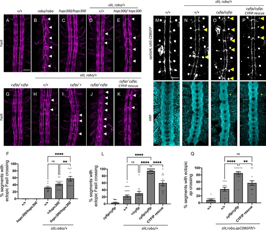
Research article                                                                            Developmental Biology Neuroscience

                                   eLife digest The brain is the most complex organ in the body. It contains billions of nerve cells,
                                   also known as neurons, with trillions of precise and specific connections, but how do these neurons
                                   know where to go and which connections to make as the brain grows? Neurons contain a small set
                                   of proteins known as guidance receptors. These receptors respond to external signals that can be
                                   attractive or repulsive. They instruct neurons to turn towards, or away from, the source of a signal.
                                   During embryonic development, neurons use these signals as guideposts to find their way to their
                                   destination.
                                       One such guidance receptor-signal pair consists of a receptor called Roundabout, also known as
                                   Robo, and its cue, Slit. Robo, which is located on the neuron’s surface, responds to the presence of
                                   Slit in the environment, by initiating a set of signalling events that instruct neurons to turn away.
                                   Neurons make the turn by rearranging their internal scaffolding, a network of proteins called the
                                   actin cytoskeleton. How Robo triggers this rearrangement is unclear. One possibility relies on a
                                   group of proteins called the WAVE regulatory complex, or the WRC for short. Researchers have
                                   already linked the WRC to nerve cell guidance, showing that it can trigger the growth of new
                                   filaments in the actin cytoskeleton. Proteins can activate the WRC by binding to it using a set of
                                   amino acids called a WRC-interacting receptor sequence, or WIRS for short, which Robo has.
                                       Chaudhari et al. used fruit flies to find out how Robo and the WRC interact. The experiments
                                   revealed that when Slit binds to Robo on the outside of a nerve cell, the WRC binds to Robo via its
                                   WIRS sequence on the inside of the cell. This attracts proteins inside the cell involved in rearranging
                                   the actin cytoskeleton. Disrupting this interaction by mutating either WRC or WIRS leads to severe
                                   errors in pathfinding, because when the WRC cannot connect to Robo, neurons cannot find their
                                   way. Experiments in mouse and chicken embryos showed that vertebrates use the WIRS sequence
                                   too, indicating that evolution has conserved this method of passing signals from Robo to the
                                   cytoskeleton.
                                       The fact that Slit and Robo work in the same way across fruit flies and vertebrates has
                                   implications for future medical research. Further work could explain how the brain and nervous
                                   system develop, and what happens when development goes wrong, but Slit and Robo control more
                                   than just nerve cell pathfinding. Research has linked disruptions in both proteins to many types of
                                   cancer, so a better understanding of how Robo interacts with the WRC could lead to new
                                   developments in different fields.

                                   guidance cues that act on their cognate receptors present on axon growth cones to induce attrac-
                                   tion toward or repulsion away from the midline. Slit is secreted by midline glia and acts as a repulsive
                                   ligand for the Robo family of receptors (Brose et al., 1999; Kidd et al., 1999; Kidd et al., 1998).
                                   There are three Robo receptors in Drosophila and four in vertebrates. The Robo receptors are trans-
                                   membrane proteins with an ectodomain consisting of five immunoglobulin-like domains and three
                                   fibronectin repeats, and an intracellular domain containing short, highly conserved cytoplasmic (CC)
                                   motifs (Bashaw et al., 2000; Kidd et al., 1998). Robo1 induces repulsion in growth cones of navi-
                                   gating axons primarily by modulating the actin cytoskeletal network. Previous work has identified
                                   some downstream effectors for Robo1 including Ena, an uncapping protein for actin filaments
                                   (Bashaw et al., 2000), and Son of Sevenless (SOS), a GEF for Rac1 (Yang and Bashaw, 2006). How-
                                   ever, downstream signaling of Robo1 is not completely understood, especially in relation to effectors
                                   that directly link Robo1 to the actin cytoskeleton and the nature of cytoskeletal changes orches-
                                   trated by Robo1. While it seems intuitive for repulsive signaling to induce depolymerization of the
                                   actin network, a recent study reports that dorsal root ganglion axons first extend actin-rich filopodia
                                   toward a source of Slit before retracting away from it McConnell et al., 2016. This challenges the
                                   prevailing notion that repulsive signaling primarily relies on actin depolymerization and suggests
                                   that the actin rearrangements occurring downstream of Robo1 are more nuanced and complex than
                                   previously thought. Indeed, several of the well-known downstream effectors of Robo1 signaling,
                                   namely Ena and Rac1, are documented enhancers of actin polymerization (Barzik et al., 2005;
                                   Ridley et al., 1992).

Chaudhari et al. eLife 2021;10:e64474. DOI: https://doi.org/10.7554/eLife.64474                                                     2 of 35
Research article                                                                           Developmental Biology Neuroscience

                                       The Scar or WAVE regulatory complex (WRC) is a heteropentameric complex consisting of five
                                   different proteins: Scar/WAVE, CYFIP/Sra1, Kette/Nap1, HSPC300/Brick1, and Abi (Eden et al.,
                                   2002). Scar or WAVE contains a VCA (verprolin homology, cofilin homology, acidic) region and
                                   serves as a nucleation-promoting factor for Arp2/3, thereby driving branched actin polymerization.
                                   While mammals have multiple orthologs of these proteins, Drosophila has single homologs of all five
                                   members of the complex, making it a simpler, more tractable model system for studying the WRC.
                                   The WRC has been previously implicated in axon guidance and targeting in Drosophila and Caeno-
                                   rhabditis elegans (Shakir et al., 2008; Stephan et al., 2011; Xu and Quinn, 2012); however, if and
                                   how it is recruited and activated downstream of guidance receptors is not known. Recent work iden-
                                   tified a unique binding site for the WRC known as the WRC-interacting receptor sequence (WIRS)
                                   motif (Chen et al., 2014a). The WIRS motif is a short six amino acid peptide sequence characterized
                                   by a bulky hydrophobic residue at position 1 and a threonine or a serine at position 3, followed by a
                                   phenylalanine at position 4. The WIRS motif is present in a number of transmembrane proteins
                                   including Robo1 (Chen et al., 2014a). Robo1 has a WIRS motif between its CC0 and CC1 domains
                                   that is conserved across species, including humans. Previously, the WIRS motif has been shown to
                                   be important for recruitment of the WRC by neuroligins and SYG-1 in synapse formation (Chia et al.,
                                   2014; Xing et al., 2018) and for neogenin function in maintaining the stability of adherens junctions
                                   (Lee et al., 2016). To our knowledge, this study is the first to demonstrate a role for the WIRS-WRC
                                   interaction in axon guidance.
                                       Here, we show that the WRC is required for Slit-Robo1 repulsive signaling at the Drosophila mid-
                                   line. We present evidence that Robo1 interacts with the WRC partially via its WIRS motif and that
                                   this interaction is enhanced in the presence of Slit. We show that the WIRS motif in Robo1 is impor-
                                   tant for its ability to induce ectopic repulsion in vivo. Using rescue assays, we show that Robo1 also
                                   requires its WIRS motif to mediate repulsion in ipsilateral axons in vivo. In addition, using CRISPR-
                                   Cas9-mediated mutagenesis, we show that the WIRS motif is important for endogenous Robo1 func-
                                   tion as mutating the endogenous WIRS motif results in the complete loss of Robo1 repulsion at the
                                   midline. Finally, we use mouse dorsal spinal cord explants and growth cone collapse assays in mouse
                                   commissural neurons, together with gain-of-function experiments in chick embryos, to demonstrate
                                   that the WIRS motif is also important for vertebrate Robo1 repulsive signaling. We propose a model
                                   in which Slit binding induces recruitment of the WRC to the WIRS motif of Robo1 where it functions
                                   in Robo1-mediated repulsion at the midline.

                                   Results
                                   The WRC interacts genetically with Slit, Robo1, and SOS
                                   WRC members are enriched in the Drosophila ventral nerve cord during embryonic stages 12–17,
                                   encompassing the developmental window when midline crossing decisions are being made
                                   (Schenck et al., 2004). To confirm these previously published observations, we examined the
                                   expression of Scar by immunofluorescence and observed strong axonal staining throughout embry-
                                   onic stages when midline axon guidance occurs (Figure 1—figure supplement 1A). To investigate
                                   the potential role of the WRC in Slit-Robo repulsion at the midline, we tested for genetic interactions
                                   between cyfip and hspc300, two members of the WRC, and the Slit-Robo signaling pathway. In wild-
                                   type embryos, FasII-positive ipsilateral axons project longitudinally and never cross the midline
                                   (Figure 1A). In robo1 mutants, axons in the medial most Fas-II bundle frequently cross and re-cross
                                   the midline, resulting in a very strong ectopic crossing phenotype (Kidd et al., 1998; Figure 1B). In
                                   slit, robo1/+ embryos, where the slit and robo1 gene dosage is reduced by half, the phenotype is
                                   milder (Figure 1D). This represents a sensitized background in which we can detect enhancers or
                                   suppressors of the Slit-Robo pathway (Chance and Bashaw, 2015; Coleman et al., 2010; Fan et al.,
                                   2003; Hsouna et al., 2003). While we see no crossing errors in FasII-positive axons in hspc300
                                   mutants alone (Figure 1C), in the slit, robo1/+ sensitized background, hspc300 mutants exhibit a sig-
                                   nificant enhancement of the ectopic crossing defects (Figure 1E). These interactions are dosage sen-
                                   sitive as removing one copy of hspc300 results in a moderate enhancement of crossing errors while
                                   removing both copies of hspc300 results in a much stronger phenotype (Figure 1F). Similarly, we
                                   see almost no crossing errors in FasII-positive axons in cyfip mutants alone (Figure 1G); however, in
                                   the slit, robo1/+ sensitized background (Figure 1H), cyfip mutants show a strong dose-dependent

Chaudhari et al. eLife 2021;10:e64474. DOI: https://doi.org/10.7554/eLife.64474                                                    3 of 35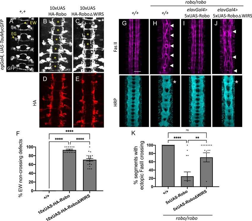
Research article                                                                                     Developmental Biology Neuroscience

Figure 1. The wave regulatory complex genetically interacts with slit and robo. (A–E, G–K) Stage 17 Drosophila embryos stained with anti-FasII to label
ipsilateral axons. (A, C) Wild-type and hspc300 homozygous mutant embryos show three FasII-positive tracts that do not cross the midline. (B) Robo
homozygous mutants show severe ectopic FasII crossing defects in 100% of segments (white arrowheads). (D) Slit, robo transheterozygous embryos
show a mild loss-of-repulsion phenotype with ectopic FasII crossing in 31% of nerve cord segments. (E) Hscp300 homozygous mutants that are
simultaneously heterozygous for slit and robo show ectopic FasII crossing in significantly more segments of the nerve cord (58%). (G) Cyfip embryos
have almost no ectopic crossing defects and appear like wild-type embryos. (H) Double heterozygous slit, robo embryos show a mild loss-of-repulsion
phenotype with ectopic FasII crossing in 22% of nerve cord segments. Removing (I) one and (J) two copies of cyfip in a slit, robo background results in
a dose-dependent enhancement of the ectopic FasII crossing defects (30 and 95%, respectively). (K) Driving UAS-CYFIP expression in neurons using the
pan-neuronal elav-Gal4 driver results in a partial rescue of the ectopic FasII crossing defects (60%). (F, L) Quantitation shows the percentage of
segments in which FasII axons ectopically cross the midline. Data are presented as mean ± SEM, number of embryos, n = 15, 10, 15, 15, 12 (for F) and
17, 27, 13, 21, 12 (for L). Significance was assessed using one-way ANOVA with Tukey’s multiple comparisons test. (M–P) Stage 17 embryos carrying
apGal4 and UAS-CD8GFP transgenes stained with anti-GFP, which labels the apterous (ap) cell bodies and axons, and anti-HRP, which labels all central
nervous system (CNS) axons. (M) Wild-type embryos show ap axons that normally project ipsilaterally without crossing the midline. (N) Double
heterozygous slit, robo embryos show a mild ectopic ap crossing phenotype of 39% (yellow arrowheads) while HRP depicts a wild type arrangement of
longitudinal and commissural axon pathways. (O) Cyfip homozygous mutants in a slit, robo background show a strong enhancement of the ectopic ap
crossing defects to 85% and HRP shows abnormal thickening and fusion of the commissures (asterisk). (P) Ap-specific expression of UAS-CYFIP
Figure 1 continued on next page

Chaudhari et al. eLife 2021;10:e64474. DOI: https://doi.org/10.7554/eLife.64474                                                                 4 of 35
Research article                                                                                  Developmental Biology Neuroscience

Figure 1 continued
significantly rescues the ectopic ap crossing defects (57%) but not the pan-neuronal HRP defects. (Q) Quantitation shows percentage of segments with
ectopic apterous crossing defects. Data are presented as mean ± SEM, number of embryos, n = 12, 13, 15, 13. Significance was assessed using one-way
ANOVA with Tukey’s multiple comparisons test. Scale bars in (A) and (M) represent 20 mm.
The online version of this article includes the following figure supplement(s) for figure 1:
Figure supplement 1. Scar expression in wild-type and scar mutant embryos.

                                    enhancement of the ectopic crossing defects (Figure 1I, J). Strikingly, removing both copies of cyfip
                                    in this background results in a very strong phenotype with ectopic crossing defects in nearly 100% of
                                    segments, similar to the robo1 mutant phenotype (Figure 1B, J). These ectopic crossing defects can
                                    be significantly rescued by the transgenic expression of UAS-CYFIP using the pan-neuronal elav-
                                    Gal4 driver (Figure 1K, L). This suggests that the neuronal function of CYFIP is important for Slit-
                                    Robo-mediated repulsion at the midline. It is important to note that zygotic hspc300 and cyfip
                                    mutants, like mutants for all other members of the WRC, still have significant amounts of the protein
                                    remaining due to maternal deposition (Schenck et al., 2004; Zallen et al., 2002). This likely explains
                                    why these zygotic mutants have no phenotype on their own. This can be seen in scar zygotic mutants
                                    where the overall Scar protein level is significantly reduced but there is still a considerable amount of
                                    Scar protein remaining in central nervous system (CNS) axons (Figure 1—figure supplement 1B, C).
                                        To determine whether CYFIP is required cell-autonomously, we examined a more restricted sub-
                                    set of ipsilateral axons, the apterous (ap) axons. Just like FasII axons, ap axons are sensitive to a par-
                                    tial loss of repulsion. Reducing the slit and robo1 gene dosage by half in slit, robo1/+ embryos
                                    results in a mild phenotype where ectopic midline crossing of ap axons is seen in approximately 40%
                                    of segments (Figure 1N, Q). Homozygous cyfip mutants in this sensitized background show a strong
                                    enhancement of the ectopic ap crossing defects with 85% of segments exhibiting ectopic crossing
                                    (Figure 1O). We also visualized all CNS axons using HRP and observed abnormal thickening and
                                    fusion of the commissures, a phenotype that bears strong resemblance to robo1 mutants. Impor-
                                    tantly, ap-specific expression of UAS-CYFIP significantly rescues the ectopic ap crossing defects but
                                    not the pan-neuronal HRP defects (Figure 1P, Q) providing strong support for a cell-autonomous
                                    role for CYFIP in Slit-Robo1 signaling. Together, these genetic data suggest that the WRC functions
                                    in the Slit-Robo1 pathway at the Drosophila midline.
                                        Previous work has identified Rac1 as an important effector of Robo1 signaling in both Drosophila
                                    and mouse (Fan et al., 2003; Wong et al., 2001). SOS is a Rac-GEF that activates Rac1 downstream
                                    of Robo1 and is required for Robo1-mediated midline repulsion (Chance and Bashaw, 2015;
                                    Yang and Bashaw, 2006). Since Rac1 is a well-known activator of the WRC (Chen et al., 2017;
                                    Chen et al., 2010; Eden et al., 2002; Ismail et al., 2009), we reasoned that Rac1 might be responsi-
                                    ble for activating the WRC downstream of Robo1, and that Rac1 and the WRC would function coop-
                                    eratively in the same pathway to regulate Robo1-mediated repulsion. Thus, we predicted that the
                                    simultaneous reduction of CYFIP and the Rac-GEF, SOS, would greatly impair Robo1-mediated
                                    repulsion, resulting in axons ectopically crossing the midline. As SOS is also maternally deposited
                                    (Yang and Bashaw, 2006), zygotic sos mutants show very mild ectopic crossing defects in approxi-
                                    mately 15% of segments (Figure 2A). In contrast, double mutants for sos and cyfip show a striking
                                    phenotype in which FasII-positive axons ectopically cross the midline in over 80% of segments
                                    (Figure 2B, C), a phenotype that bears strong resemblance to the robo1 mutant phenotype. In addi-
                                    tion to examining the phenotype with FasII immunostaining, we also visualized all CNS axons using
                                    HRP and observed frequent thickening and fusion of the anterior and posterior commissures, which
                                    again bears strong resemblance to robo1 mutants (Figure 2B). Thus, cyfip genetically interacts with
                                    sos to give a strong ectopic crossing phenotype very similar to that seen in robo1 mutants, support-
                                    ing the idea that Rac1 and the WRC act cooperatively to regulate midline repulsion.
                                        In Drosophila embryos, both Robo1 and, to a lesser extent, Robo2 contribute to midline repulsion
                                    in response to Slit (Rajagopalan et al., 2000; Simpson et al., 2000). Indeed, on their own robo2
                                    mutants exhibit only mild phenotypes; however, robo1, robo2 double mutants exhibit a complete
                                    collapse of all CNS axons at the midline, phenocopying the slit mutant phenotype. Therefore, muta-
                                    tions in genes that contribute to robo1 repulsion would be expected to strongly enhance the mild
                                    phenotype observed in robo2 mutants. In robo2 mutant embryos, FasII-positive axons ectopically

Chaudhari et al. eLife 2021;10:e64474. DOI: https://doi.org/10.7554/eLife.64474                                                              5 of 35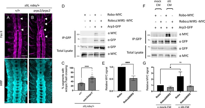
Research article                                                                                      Developmental Biology Neuroscience

Figure 2. The wave regulatory complex genetically interacts with sos and robo2. (A, B, D, E) Stage 17 embryos stained with anti-FasII and anti-HRP. (A)
Sos embryos show mild ectopic crossing defects of 15% in FasII axons (arrowheads) and no phenotype in HRP. (B) Simultaneous removal of sos and
cyfip results in a very strong enhancement of the ectopic FasII crossing defects to 82% and a strong HRP phenotype with thickening and fusion of
commissures (asterisk). Similarly, (D) robo2 mutants show mild ectopic crossing defects of 17% in FasII axons and a mildly disorganized axon scaffold in
Figure 2 continued on next page

Chaudhari et al. eLife 2021;10:e64474. DOI: https://doi.org/10.7554/eLife.64474                                                                  6 of 35
Research article                                                                                   Developmental Biology Neuroscience

Figure 2 continued
HRP while (E) double mutants for robo2 and cyfip show strong ectopic FasII crossing defects of 77% and thickening and fusion of commissures in HRP.
(C, F) Quantitation shows the percentage of segments in which FasII axons ectopically cross the midline. Data are presented as mean ± SEM, number
of embryos, n = 15 and 16 (for E) and 20 and 9 (for F). Significance was assessed using Student’s t-test. Scale bars in (A) and (D) represent 20 mm.

                                    cross the midline in approximately 17% of segments (Figure 2D). In robo2, cyfip double mutant
                                    embryos, ectopic crossing defects are greatly increased to approximately 75% of segments
                                    (Figure 2E, F) and axon commissures are thicker and frequently fused, providing additional support
                                    for a role for the WRC in midline repulsion. Taken together, these genetic interaction results strongly
                                    suggest that the WRC functions in Slit-Robo1-mediated repulsive signaling at the midline.

                                    The WIRS motif in Robo1 is important for its interaction with the WRC
                                    The cytoplasmic tail of Robo1 contains a WIRS motif, which is conserved in vertebrates (Figure 3A).
                                    The purified cytoplasmic tail of human Robo1 directly interacts with the WRC in pulldown assays via
                                    its WIRS motif (Chen et al., 2014a). To determine if this WIRS-dependent interaction with the WRC
                                    is conserved in Drosophila Robo1, we performed co-immunoprecipitation assays in Drosophila
                                    embryonic S2R+ cells (DGRC, #150) using tagged constructs of Robo1 and HSPC300. The relatively
                                    small size of HSPC300 facilitated consistent levels of expression and reduced trial-to-trial variability.
                                    We found that Robo1 immunoprecipitated with HSPC300, indicating that Drosophila Robo1, like
                                    human Robo1, can also interact with the WRC (Figure 3C). Next, we introduced point mutations
                                    into the WIRS motif of Robo1 (Robo1DWIRS; Figure 3B) and found a significant decrease in the
                                    amount of Robo1 that immunoprecipitated with HSPC300 (Figure 3C, E). Thus, mutating the WIRS
                                    motif substantially disrupts the binding of Robo1 to the WRC, indicating that Robo1 interacts with
                                    the WRC partly via the WIRS motif. In contrast, the previously published interaction data for human
                                    Robo1 (Chen et al., 2014a) showed that mutating the WIRS motif completely abolishes binding to
                                    the WRC. We speculate that there may be a small amount of indirect binding of Robo1 to the WRC
                                    via Ena or DOCK, which are known interactors of Robo1 (Bashaw et al., 2000; Fan et al., 2003).
                                    Previous work has identified interactions between Ena and Abi (Chen et al., 2014b) and between
                                    the DOCK homolog Nck and Nap1 (Kitamura et al., 1996). Both Abi and Nap1 are members of the
                                    WRC. As the pulldown assay with human Robo1 was done using purified proteins, any indirect bind-
                                    ing will not be detected. Support for this notion comes from our co-immunoprecipitation results of
                                    Robo2 and HSPC300. Drosophila Robo2 is structurally similar to Robo1 except that it lacks the CC
                                    motifs CC2 and CC3 present in Robo1 that serve as the interaction sites for Ena and DOCK
                                    (Bashaw et al., 2000; Fan et al., 2003; Figure 3—figure supplement 1A). Indeed, we find that
                                    Robo2 can also interact with HSPC300 though mutating the WIRS motif of Robo2 almost completely
                                    abolishes this interaction (Figure 3—figure supplement 1B, C). This result is consistent with the
                                    idea that there might be indirect binding of the WRC to Robo1 via its interaction with other WRC
                                    partners but not to Robo2 that lacks any such interactions.
                                        Next, we wanted to test whether the Robo1-WRC interaction is regulated by the Robo ligand
                                    Slit. We treated S2R+ cells with bath application of Slit-conditioned media (Slit-CM) and found a
                                    substantial increase in the interaction between Robo1 and HSPC300 as compared to cells treated
                                    with mock-CM (Figure 3D, F). By contrast, Robo1DWIRS shows no significant increase in binding to
                                    HSPC300 upon Slit-CM treatment. As there is significant variability in the activity of Slit-CM with
                                    each preparation, we see different levels of enhancement in binding obtained with each Slit treat-
                                    ment. Nevertheless, Slit application consistently increases the interaction between Robo1 and
                                    HSPC300. These results suggest that upon Slit binding the WRC is recruited to Robo1 via its WIRS
                                    motif.
                                        Finally, to test whether this interaction occurs in vivo, we performed co-immunoprecipitation
                                    assays using Drosophila embryonic protein lysates. We generated transgenic flies using the GFP-
                                    tagged HSPC300 construct and HA-tagged Robo1 constructs. The pan-neuronal elav-Gal4 driver
                                    was used to drive expression of UAS-HSPC300-GFP either alone or with the wild-type UAS-HA-
                                    Robo1 or UAS-HA-Robo1DWIRS transgenes in Drosophila embryos. While wild-type Robo1 co-
                                    immunoprecipitates with HSPC300, mutating the WIRS motif results in a significant decrease in this
                                    binding (Figure 3G, H). These results indicate that Robo1 interacts with the WRC in vivo as well and
                                    that this interaction is partly dependent on the WIRS motif.

Chaudhari et al. eLife 2021;10:e64474. DOI: https://doi.org/10.7554/eLife.64474                                                               7 of 35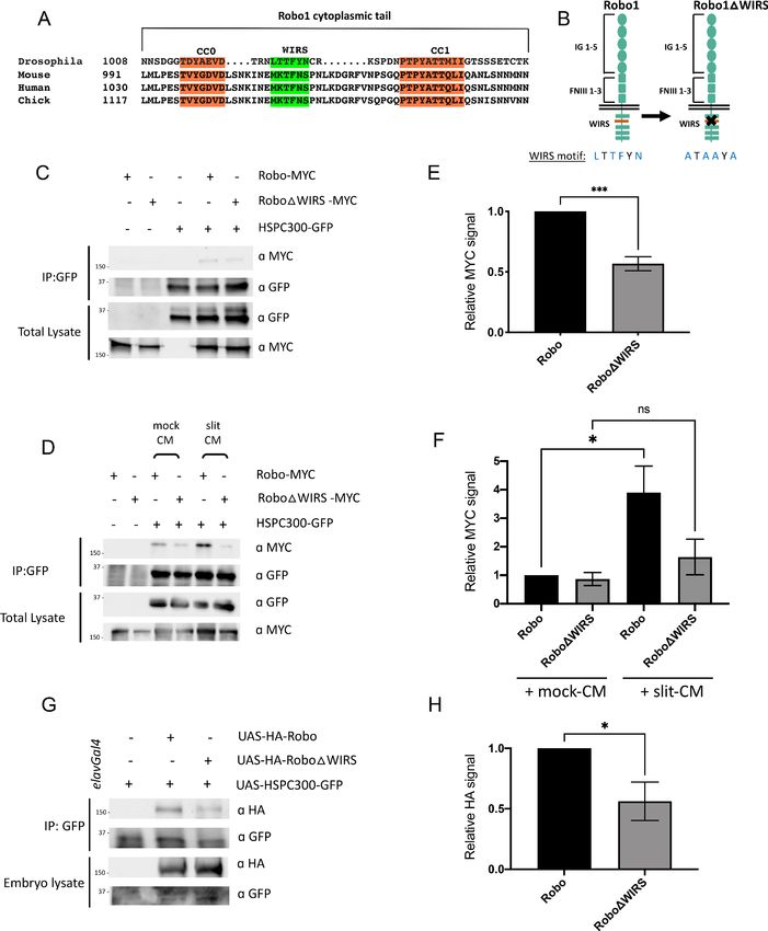
Research article                                                                                  Developmental Biology Neuroscience

Figure 3. Slit-dependent recruitment of the WAVE regulatory complex (WRC) to Robo1 requires the WRC-interacting receptor sequence (WIRS) motif.
(A) Sequence alignments of the cytoplasmic tail of Robo1 showing that the WIRS motif is conserved across species. (B) Schematic depicting the
residues of the WIRS motif that are mutated in the Robo1DWIRS variant. (C) Drosophila S2R+ cell lysates co-expressing HSPC300-GFP with either wild-
type Robo1-MYC or Robo1DWIRS-MYC were immunoprecipitated with an anti-GFP antibody. The first three lanes show the individual proteins
Figure 3 continued on next page

Chaudhari et al. eLife 2021;10:e64474. DOI: https://doi.org/10.7554/eLife.64474                                                              8 of 35
Research article                                                                                     Developmental Biology Neuroscience

Figure 3 continued
expressed alone. The fourth lane shows wild-type Robo1 co-immunoprecipitating with HSPC300 while the fifth lane shows that mutating the WIRS motif
decreases this binding. (D) Cell lysates were immunoprecipitated with anti-GFP following a 12 min bath application of mock conditioned media or
conditioned media obtained from Slit-expressing cells. The interaction between wild-type Robo1 and HSPC300 is increased in the presence of Slit;
however, no significant increase is noted with Robo1DWIRS. (E, F) Quantitation of band intensities of the MYC-tagged Robo1 variants in the
immunoprecipitates normalized to wild-type Robo1-MYC. Data were normalized to lysate levels of the Robo1 variants and HSPC300 levels in the
immunoprecipitates. Error bars represent SEM. Number of trials, n = 4. Significance was assessed using Student’s t-test (for E) and one-way ANOVA
with Tukey’s multiple comparisons test (for F). (G) Lysates from Drosophila embryos with elavGal4 pan-neuronally driving the expression of HSPC300-
GFP alone (lane 1), with wild-type HA-Robo1 (lane 2) or with HA-Robo1DWIRS (lane 3), were immunoprecipitated with anti-GFP. Wild-type Robo1 co-
immunoprecipitates with HSPC300 and mutating the WIRS motif decreases this binding. (H) Quantitation of band intensities of the HA-tagged Robo1
variants in the imunnoprecipitates normalized to wild-type HA-Robo1. Data were normalized to the lysate levels of the Robo1 variants and HSPC300
levels in the immunoprecipitates. Error bars represent SEM. Number of trials, n = 5. Significance was assessed using Student’s t-test. Normalized values
for the co-immunoprecipitation data are provided in Figure 3—source data 1.
The online version of this article includes the following source data and figure supplement(s) for figure 3:
Source data 1. Normalized values of co-immunoprecipitation data.
Figure supplement 1. Robo2 interaction with the WAVE regulatory complex (WRC) is entirely dependent on its WRC-interacting receptor
sequence (WIRS) motif.

                                     The WIRS motif is essential for Robo1 function in vivo
                                     To test whether this interaction with the WRC is required for Robo1 function in vivo, we compared
                                     the gain-of-function and rescue phenotypes of wild-type Robo1 and Robo1DWIRS in specific neuro-
                                     nal subsets in the Drosophila ventral nerve cord. We generated transgenic flies with wild-type UAS-
                                     Robo1 or UAS-Robo1DWIRS constructs. Both the transgenes are tagged with an HA epitope and
                                     inserted into the same genomic locus. Immunostaining for HA shows that both transgenes are
                                     expressed at comparable levels (Figure 4D, E). Using the eg-Gal4 driver, we expressed these trans-
                                     genes in eagle neurons, a subset of commissural neurons. Eagle neurons, visualized here using a
                                     GFP reporter, consist of two populations: the EG population, which extends its axons in the anterior
                                     commissure of a segment, and the EW population, which extends axons in the posterior commissure
                                     (Figure 4A). Overexpression of wild-type Robo1 in these neurons causes ectopic repulsion from the
                                     midline, resulting in a strong gain-of-function phenotype where almost all EW axons fail to cross the
                                     midline (Figure 4B). In contrast, overexpression of Robo1DWIRS results in a significantly weaker
                                     gain-of-function phenotype where EW axons in approximately 70% of segments fail to cross the mid-
                                     line (Figure 4C, F). Thus, mutating the WRC interaction site on Robo1 hampers its ability to induce
                                     ectopic repulsion in vivo.
                                         Next, we assessed the ability of Robo1DWIRS to rescue the ectopic crossing defects of FasII-posi-
                                     tive axons seen in robo1 mutant embryos. Unlike in wild-type embryos, where FasII axons never
                                     cross the midline (Figure 4G), in robo1 mutants, axons in the medial most fascicle freely cross and
                                     recross the midline in 100% of segments (Figure 4H). Re-expressing wild-type Robo1 with the pan-
                                     neuronal driver elav-Gal4 restores the ipsilateral projection pattern in most of the segments, lower-
                                     ing the frequency of ectopic crossing to 25% of segments (Figure 4I). In contrast, re-expression of
                                     Robo1DWIRS fails to rescue the crossing defects in 70% of segments (Figure 4J, K). This indicates
                                     that in the absence of a functional WIRS motif Robo1 is not nearly as effective at restoring repulsive
                                     signaling in ipsilateral axons in vivo. Altogether, these results suggest a role for the WIRS motif in
                                     Robo1 repulsive signaling at the midline.

                                     Mutating the endogenous WIRS motif disrupts Robo1 function in vivo
                                     Our in vivo results obtained so far have relied on misexpression or overexpression of Robo1 that
                                     likely is not subject to the adequate spatial and temporal regulation that is critical for guidance
                                     receptor function. Further, such unregulated high levels of Robo1 expression on the cell surface
                                     could potentially mask dysfunction in receptor activity. We see this especially for the rescue experi-
                                     ments with our UAS-Robo1 transgenes. While the difference in rescue activity between 5XUAS-
                                     Robo1 and 5XUAS-Robo1DWIRS is around 50% (Figure 4K), performing this rescue assay with
                                     10XUAS-Robo1 and 10XUAS-Robo1DWIRS transgenes, which have double the number of UAS
                                     enhancer sites and express much higher levels of the Robo1 variants, gives a much more modest dif-
                                     ference of 13% (Figure 4—figure supplement 1A–E). Indeed, in rescue experiments using 10XUAS-

Chaudhari et al. eLife 2021;10:e64474. DOI: https://doi.org/10.7554/eLife.64474                                                                  9 of 35
Research article Developmental Biology Neuroscience Figure 4. The WRC-interacting receptor sequence (WIRS) motif is essential for Robo1 function in vivo. (A–C) Stage 16 Drosophila embryos carrying egGal4 and UAS-TauMycGFP transgenes stained with anti-GFP, which labels cell bodies and axons of the eagle neurons (EG and EW). EG neurons project through the anterior commissure of each segment while EW neurons project through the posterior commissure. (A) EW neurons cross in 100% of segments in wild-type embryos. (B) Misexpression of wild-type HA-tagged Robo1 in eagle neurons results in a strong disruption of midline crossing where EW axons fail to cross in almost all segments of the nerve cord (93%; asterisk). (C) Misexpressing HA-tagged Robo1DWIRS results in a significantly milder disruption with fewer segments showing EW non-crossing defects (71%). (D, E) Embryos stained with anti-HA show comparable expression of the HA-tagged Robo1 variants that were inserted into the same genomic locus. (F) Quantitation shows the percentage of segments in which EW axons fail to cross the midline. Data are presented as mean ± SEM, number of embryos, n = 17, 13, 23. Significance was assessed using one- way ANOVA with Tukey’s multiple comparisons test. (G–J) Stage 17 embryos stained with anti-FasII and anti-HRP. (G) Wild-type embryos show no ectopic FasII crossing defects and no phenotype in HRP. (H) Robo mutants show severe ectopic FasII crossing defects in 100% of segments (arrowheads) and a strong HRP phenotype with thickening and fusion of commissures (asterisk). (I) Pan-neuronal expression of wild-type 5XUAS-Robo1 significantly rescues the robo mutant phenotype in FasII (to 25%) as well as HRP; however, (J) 5XUAS-Robo1DWIRS fails to rescue the robo mutant phenotype as efficiently as wild-type Robo1 with frequent ectopic crossing in FasII (71%) and thickened commissures in HRP still evident in these embryos. (K) Quantitation shows the percentage of segments in which FasII axons ectopically cross the midline. Data are presented as mean ± SEM, number of embryos, n = 11, 14, 15. Significance was assessed using one-way ANOVA with Tukey’s multiple comparisons test. Scale bars in (A) and (G) represent 20 mm. Figure 4 continued on next page Chaudhari et al. eLife 2021;10:e64474. DOI: https://doi.org/10.7554/eLife.64474 10 of 35
Research article                                                                            Developmental Biology Neuroscience

Figure 4 continued
The online version of this article includes the following figure supplement(s) for figure 4:
Figure supplement 1. 10XUAS-Robo1 rescue of the robo mutant phenotype.

                                      Robo1 transgenes, we see strong gain-of-function effects that lead to both rescue of abnormal
                                      crossing of FasII-positive axons, as well as ectopic repulsion of commissural axons (Figure 4—figure
                                      supplement 1F–J). Notably, the ectopic repulsion of commissural axons induced by the 10XUAS-
                                      Robo1DWIRS transgene is significantly weaker than the ectopic repulsion induced by the wild-type
                                      receptor (Figure 4—figure supplement 1H–J). Given these caveats, we sought to analyze the func-
                                      tion of the WIRS motif in Robo1 signaling in a more endogenous context. First, we performed a res-
                                      cue assay with an HA-tagged genomic rescue construct of robo1 that contains upstream and
                                      downstream regulatory regions of Robo1 in addition to the Robo1 coding sequence (Brown et al.,
                                      2015). Transgenics created with this construct show a Robo1 expression pattern that closely resem-
                                      bles that of endogenous Robo1 (Brown et al., 2015). We mutated the WIRS motif in this robo1
                                      genomic rescue construct and inserted the transgene into the same genomic site as the wild-type
                                      construct. Both transgenes show comparable levels of Robo1 expression upon HA immunostaining
                                      (Figure 5—figure supplement 1A, B). We tested the ability of these transgenes to rescue the robo1
                                      mutant phenotype in FasII-positive axons (Figure 5B). One copy of the wild-type robo1 genomic res-
                                      cue construct (genRobo) was able to rescue ectopic crossing of FasII-positive axons in almost all seg-
                                      ments with only 6% still showing defects (Figure 5C) while robo1DWIRS genomic rescue construct
                                      (genRoboDWIRS) was unable to rescue ectopic crossing defects in over 70% of segments
                                      (Figure 5D, E). Similarly, for HRP stained axons, the frequent thickening and fusion of the anterior
                                      and posterior commissures in robo1 mutants (Figure 5B) can be rescued with the wild-type genRobo
                                      but not with genRoboDWIRS (Figure 5C, D). These results, in more physiologically relevant contexts,
                                      demonstrate a marked decline in Robo1 function upon disruption of the WRC binding site.
                                          Finally, using the CRISPR-Cas9 system, we mutated the WIRS motif in the endogenous robo1
                                      locus. We used a single-guide RNA that targets the endogenous WIRS motif and a single-stranded
                                      oligonucleotide template to introduce point mutations in the WIRS motif (Figure 5—figure supple-
                                      ment 2A). We sequenced the regions surrounding the WIRS motif to verify that we had successfully
                                      mutated the WIRS motif without introducing any unwanted frameshift mutations or deletions. While
                                      we found no frameshifts, we did notice that our strategy had resulted in an unexplained loss of the
                                      smaller intron 16 (Figure 5—figure supplement 2A). Since the genRobo constructs and the previ-
                                      ously used robo swap alleles (Spitzweck et al., 2010) that can restore Robo1 function fully do not
                                      contain any intronic sequences, we believe that it is extremely unlikely that the loss of this intron
                                      affects Robo1 function. Next, we analyzed the phenotypes of both HRP and FasII-positive axons in
                                      these roboDWIRS CRISPR embryos. We see a surprisingly strong ectopic crossing phenotype in
                                      these embryos with defects in almost 100% of segments, showing that they fully phenocopy the
                                      robo mutant embryos (Figure 5B, F). We were able to achieve a near perfect rescue with the intro-
                                      duction of one copy of genRobo, indicating that this phenotype is not a result of any off-target
                                      effects arising from Cas9-mediated cleavage (Figure 5G, H). This result also supports our interpreta-
                                      tion that the loss of intron 16 in our CRISPR allele has no effect on Robo1 function since the gen-
                                      Robo construct does not include any introns. As an additional control, we also tested whether the
                                      roboDWIRS CRISPR mutations disrupt normal Robo1 expression. To investigate this, we immunos-
                                      tained for Robo1 expression using a monoclonal Robo1 antibody. Unlike the robo mutants in which
                                      no Robo1 protein can be detected (Figure 5—figure supplement 2C, F), we see substantial Robo1
                                      staining in the roboDWIRS CRISPR mutants, suggesting that the phenotype is not due to a failure in
                                      protein production (Figure 5—figure supplement 2D, G). Unlike in wild-type embryos where Robo1
                                      expression is seen primarily on longitudinal tracts and is downregulated on commissures (Figure 5—
                                      figure supplement 2B, E), in the roboDWIRS CRISPR mutant embryos, we see Robo1 also being
                                      expressed on commissures (Figure 5—figure supplement 2D, G). While interesting, this observation
                                      is not necessarily surprising to us as this altered Robo1 localization on commissures has also been
                                      noted in previous studies when Robo1 signaling is disrupted (Coleman et al., 2010). Altogether, our
                                      genomic Robo rescue assays and roboDWIRS CRISPR mutant phenotypes strongly suggest an impor-
                                      tant role for the WIRS motif in Robo1 repulsive function in vivo.

Chaudhari et al. eLife 2021;10:e64474. DOI: https://doi.org/10.7554/eLife.64474                                                     11 of 35
Research article Developmental Biology Neuroscience
Figure 5. Mutating the endogenous WRC-interacting receptor sequence (WIRS) motif disrupts Robo1 function in vivo. (A–D) Stage 17 embryos stained
with anti-FasII and anti-HRP. (A) Wild-type embryos showing no phenotype in FasII or HRP. (B) Robo mutants show severe ectopic FasII crossing defects
in 100% of segments (arrowheads) and a strong HRP phenotype with thickening and fusion of commissures (asterisk). (C) The strong FasII and HRP
phenotypes seen in robo mutant embryos can be completely rescued with one copy of a wild-type genomic Robo1 rescue construct (genRobo) that
contains additional upstream and downstream regulatory regions of robo1, more closely mimicking the endogenous Robo1 expression pattern (8%). (D)
In contrast, the genomic Robo1 rescue construct containing mutations in the WIRS motif of Robo1 (genRoboDWIRS) fails to rescue the robo mutant
phenotype in both FasII (77%) and HRP. (E) Quantitation shows the percentage of segments in which FasII axons ectopically cross the midline. Data are
presented as mean ± SEM, number of embryos, n = 14, 11, 16, 16. Significance was assessed using one-way ANOVA with Tukey’s multiple comparisons
test. (F, G) Stage 17 embryos stained with anti-FasII and anti-HRP. (F) CRISPR embryos with mutations in the endogenous WIRS motif of robo1 show
severe phenotypes in FasII and HRP bearing strong resemblance to robo mutants. (G) The phenotypes seen in these CRISPR roboDWIRS embryos can
be completely rescued with one copy of the wild-type genomic Robo1 rescue construct (8%). (H) Quantitation shows the percentage of segments in
which FasII axons ectopically cross the midline. Data are presented as mean ± SEM, number of embryos, n = 14, 11, 14, 20. Significance was assessed
using one-way ANOVA with Tukey’s multiple comparisons test. Scale bars in (A) and (F) represent 20 mm.
The online version of this article includes the following figure supplement(s) for figure 5:
Figure supplement 1. Comparable expression of the genomic rescue transgenes.
Figure supplement 2. Schematic for CRISPR-Cas9 mutagenesis and Robo1 staining in CRIPR roboDWIRS embryos.
The Arp2/3 complex interacts genetically and physically with the Slit-
Robo pathway
We have shown that the WRC is an important component of the Slit-Robo1 repulsive pathway at the
midline. But what happens after the WRC is recruited to Robo1? Is the WRC acting via Arp2/3 to
promote branched actin polymerization downstream of Robo1? To address this question, we tested
for genetic interactions between arpc2, a member of the Arp2/3 complex and the Slit-Robo path-
way. Similar to members of the WRC, arpc2 mutants alone have no ectopic crossing phenotype in
FasII axons; however, when introduced into the slit, robo/+ sensitized background, arpc2 homozy-
gous mutants show a significant enhancement of the ectopic FasII crossing defects (Figure 6A–C),
suggesting that the Arp2/3 complex functions in the Slit-Robo repulsive pathway. Additionally, when
we remove one copy of arpc2 together with one copy of cyfip, we again observe a significant
enhancement of the slit, robo/+ ectopic crossing defects (Figure 6—figure supplement 1A–C). This
genetic interaction between arpc2 and cyfip suggests a cooperative effect of the WRC and Arp2/3
in the Slit-Robo1 signaling pathway at the midline. Next, we overexpressed Robo1 in eagle neurons,
Chaudhari et al. eLife 2021;10:e64474. DOI: https://doi.org/10.7554/eLife.64474 12 of 35Research article Developmental Biology Neuroscience
Figure 6. The Arp2/3 complex interacts genetically and physically with the Slit-Robo pathway. (A, B) Stage 17 Drosophila embryos stained with anti-
FasII and anti-HRP. (A) Slit, robo transheterozygous embryos show a mild loss-of-repulsion phenotype with ectopic FasII crossing in 31% of nerve cord
segments (arrowheads). (B) Arpc2 homozygous mutants that are simultaneously heterozygous for slit and robo show ectopic FasII crossing in
significantly more segments of the nerve cord (55%). (C) Quantitation shows the percentage of segments in which FasII axons ectopically cross the
midline. Data are presented as mean ± SEM, number of embryos, n = 15 and 20. Significance was assessed using Student’s t-test. Scale bar in (A)
represents 20 mm. (D) Drosophila S2R+ cell lysates co-expressing Arp3-GFP with either wild-type Robo1-MYC or Robo1DWIRS-MYC were
immunoprecipitated with an anti-GFP antibody. The first two lanes show the individual Robo1 variants expressed alone. The third lane shows wild-type
Robo1 co-immunoprecipitating with Arp3 while the fourth lane shows that mutating the WIRS motif decreases this binding. Asterisk indicates non-
specific bands. (F) Cell lysates were immunoprecipitated with anti-GFP following a 12 min bath application of mock conditioned media or conditioned
media obtained from Slit-expressing cells. The interaction between wild-type Robo1 and Arp3 is increased in the presence of Slit; however, no
significant increase is noted with Robo1DWIRS. (E, G) Quantitation of band intensities of the MYC-tagged Robo1 variants in the immunoprecipitates
normalized to wild-type Robo1-MYC. Data were normalized to lysate levels of the Robo1 variants and Arp3 levels in the immunoprecipitates. Error bars
represent SEM. Number of trials, n = 7. Significance was assessed using Student’s t-test (for E) and one-way ANOVA with Tukey’s multiple comparisons
test (for G). Normalized values for the co-immunoprecipitation data are provided in Figure 6—source data 1.
The online version of this article includes the following source data and figure supplement(s) for figure 6:
Source data 1. Normalized values of co-immunoprecipitation data.
Figure supplement 1. arpc2 mutants genetically interact with the Slit-Robo pathway.
Figure supplement 2. Comparable surface expression of the wild-type and WRC-interacting receptor sequence (WIRS) mutant forms of Robo1.
which results in a strong gain-of-function phenotype where almost all EW neurons fail to cross the
midline. In contrast, overexpressing Robo1 in arpc2 mutants results in a small but significant sup-
pression of this phenotype (Figure 6—figure supplement 1D–F) that is similar to the suppression
seen in cyfip mutants (Figure 6—figure supplement 1G–I), demonstrating a reduction in Robo1’s
ability to induce ectopic repulsion. Together, these genetic data strongly suggest that the Arp2/3
complex functions in the Slit-Robo1 repulsive pathway.
To determine whether the Arp2/3 complex can physically interact with Robo, we performed co-
immunoprecipitation assays in Drosophila embryonic S2R+ cells using tagged constructs of Robo1
and Arp3, another component of the Arp2/3 complex. We found that Robo immunoprecipitated
with Arp3, suggesting that the Arp2/3 complex can physically interact with Robo (Figure 6D). We
Chaudhari et al. eLife 2021;10:e64474. DOI: https://doi.org/10.7554/eLife.64474 13 of 35Research article Developmental Biology Neuroscience
reasoned that if the Arp2/3 complex was being recruited by the WRC to Robo, we would expect
that mutating the WIRS motif would disrupt this interaction between Arp2/3 and Robo. Indeed, we
found a significant decrease in the amount of RoboDWIRS that immunoprecipitated with Arp3 as
compared to wild-type Robo (Figure 6D, E). Furthermore, we can detect an increase in the interac-
tion between Robo and Arp3 in the presence of Slit-CM as compared to mock-CM, suggesting that
similar to the WRC, the Arp2/3 complex is also recruited to Robo in response to Slit. By contrast,
RoboDWIRS shows no significant increase in binding to Arp3 in the presence of Slit, demonstrating
that the WIRS motif is important for this Slit-dependent recruitment of the Arp2/3 complex to Robo.
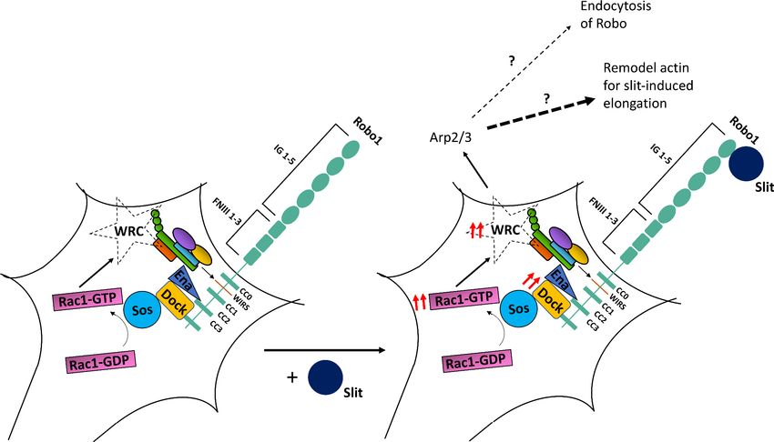
Together, these observations support the model that upon Slit binding the WRC is recruited to the
WIRS motif of Robo and activated, which is in turn responsible for the recruitment of the Arp2/3
complex to facilitate cytoskeletal remodeling downstream of Robo. One possible outcome of initiat-
ing localized actin polymerization is the endocytosis and recycling of transmembrane receptors.
Indeed, both Drosophila Robo as well vertebrate Robo1 have been previously shown to undergo
endocytosis following Slit stimulation (Chance and Bashaw, 2015; Kinoshita-Kawada et al., 2019).
Furthermore, the WRC has been shown to play a role in initiating receptor endocytosis
(Basquin et al., 2015; Xu et al., 2016). Thus, to further evaluate the mechanism of WRC function in
Slit-Robo signaling, we investigated whether mutating the WIRS motif in Robo could disrupt signal-
ing by preventing Robo endocytosis. To address this question, we tested whether RoboDWIRS dis-
plays increased surface localization compared to wild-type Robo in both Drosophila embryonic
neurons and in cultured dorsal commissural neurons from mice. First, we tested whether Drosophila
embryos expressing the genomic HA-tagged Robo rescue transgenes display any difference in sur-
face localization. We dissected embryos live and visualized surface expression of Robo by staining
the N-terminal HA tag before fixation and permeabilization (Figure 6—figure supplement 2A). Sur-
face Robo was quantified as the mean fluorescence intensity of HA normalized to HRP. We observed
no significant difference in the surface expression of Robo and RoboDWIRS (Figure 6—figure sup-
plement 2B). We next cultured E12 mouse dorsal commissural neurons that were electroporated
with either wild-type MYC-tagged human Robo1 (hRobo1) or MYC-tagged hRobo1DWIRS. Following
a 30 min bath application with Slit, we visualized surface expression of hRobo1 by staining the N-ter-
minal MYC tag before fixation and permeabilization (Figure 6—figure supplement 2C). Surface
hRobo1 was quantified as the mean fluorescence intensity of MYC, and the analysis was limited to
Robo3-positive commissural neurons. Here again, we observed no significant difference in the sur-
face localization of hRobo1 and hRobo1DWIRS (Figure 6—figure supplement 2D), suggesting that
the WIRS motif has no detectable effect on Robo1 surface levels. Together, these observations point
to a non-endocytic role for the WRC in promoting Robo repulsion.
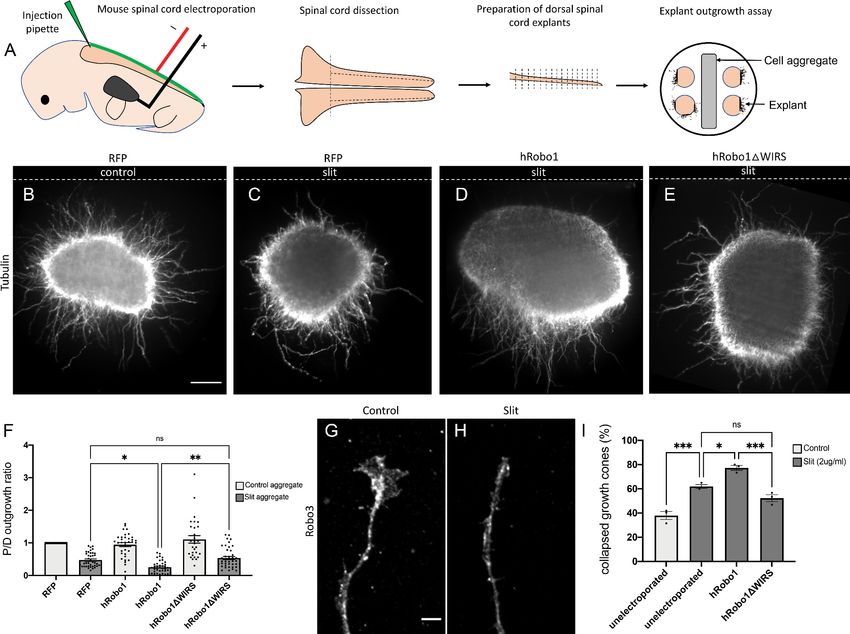
The WIRS motif is required for Slit-dependent repulsion in mouse spinal
commissural axons
The WIRS motif in the Robo1 receptor is conserved in vertebrates, raising the possibility for a poten-
tial role in vertebrate Robo1 signaling. Indeed, the cytoplasmic tail of human Robo1 can bind to the
WRC via its WIRS motif (Chen et al., 2014a). Thus, to address the question of whether the WIRS
motif is important for vertebrate Robo1 signaling, we introduced point mutations into the WIRS
motif of hRobo1 and performed gain-of-function experiments with wild-type hRobo1 and hRo-
bo1DWIRS constructs. We electroporated E12 mouse spinal cords with wild-type hRobo1 or hRo-
bo1DWIRS, along with RFP as a reporter for efficiency of electroporation and cultured dorsal spinal
cord explants next to mock 293 T cell aggregates or cell aggregates expressing Slit (Figure 7A). We
observe poor penetration of the anti-MYC antibody in explants and hence use RFP as a measure of
electroporation efficiency. We observe comparable levels of RFP staining in explants (Figure 7—fig-
ure supplement 1A). Explants cultured adjacent to mock cell aggregates show uniform outgrowth
on all sides of the explant (Figure 7B). In contrast, explants cultured adjacent to Slit-expressing
aggregates show decreased outgrowth on the side proximal to the Slit-expressing aggregate as
compared to the distal side (Figure 7C). Explants electroporated with wild-type hRobo1 show an
increased repulsive response to Slit with even less outgrowth on the proximal side and a significantly
lower proximal/distal outgrowth ratio (Figure 7D, F). In contrast, explants electroporated with hRo-
bo1DWIRS show no such gain-of-function response to Slit and have a proximal/distal outgrowth ratio
similar to that seen for RFP electroporated explants (Figure 7E, F), suggesting that the WIRS motif
is important for the Slit-induced repulsive response of vertebrate Robo1. Next, to assess whether
Chaudhari et al. eLife 2021;10:e64474. DOI: https://doi.org/10.7554/eLife.64474 14 of 35Research article Developmental Biology Neuroscience Figure 7. The WRC-interacting receptor sequence (WIRS) motif is required for Slit-dependent Robo1 repulsion in mouse spinal commissural axons. (A) Schematic of electroporation and culture of spinal cord explants. Dotted lines show cut sites to obtain dorsal spinal cord explants. The image on the right depicts the arrangement of explants cultured around a 293 T cell aggregate (control or Slit-expressing) embedded in collagen. (B–E) E12 dorsal spinal cord explants labeled with anti-tubulin to visualize axon outgrowth. Dotted lines indicate the position of the cell aggregate. (B) RFP electroporated explant cultured next to a mock cell aggregate shows uniform outgrowth on all sides of the explant. (C) RFP electroporated explant cultured next to a Slit-expressing cell aggregate shows decreased outgrowth on the quadrant proximal to the aggregate as compared to the quadrant distal to it (0.47). (D) Explant electroporated with wild-type hRobo1 cultured next to a Slit-expressing cell aggregate shows even less outgrowth on the proximal quadrant demonstrating increased responsiveness to Slit (0.14). (E) Explant electroporated with hRobo1DWIRS cultured next to a Slit- expressing cell aggregate shows no such increase in Slit responsiveness as the proximal: distal outgrowth ratio is similar to that seen for RFP electroporated explants (0.54). (F) Quantification shows the proximal:distal outgrowth ratio for explants cultured next to control cell aggregates (white) and Slit-expressing cell aggregates (gray). Data are presented as mean ± SEM, number of explants, n = 29, 39, 33, 39, 29, 41 (from three independent experiments). Significance was assessed using one-way ANOVA with Tukey’s multiple comparisons test. (G, H) Growth cone collapse in response to Slit in E12-dissociated commissural axons. Growth cone morphology was examined by staining for the commissural marker Robo3. (I) Quantification shows percentage of axons with collapsed growth cones. Unelectroporated neurons show an increased level of collapse when treated with Slit (from 38% without Slit to 62% with bath application of Slit). Neurons electroporated with wild-type hRobo1 show a gain-of-function response to Slit with an even higher collapse level (77%). In contrast, neurons electroporated with hRobo1DWIRS show no gain-of-function and a collapse level similar to unelectroporated neurons (52%). For neurons electroporated with the MYC-tagged hRobo1 variants, only Robo3- and MYC-positive axons were analyzed. Data are presented as mean ± SEM, number of trials, n = 3 (over 30 neurons for each condition/trial). Significance was assessed using one- way ANOVA with Tukey’s multiple comparisons test. Scale bars represent 100 mm in (B) and 5 mm in (G). The online version of this article includes the following figure supplement(s) for figure 7: Figure supplement 1. Expression of hRobo1 variants electroporated into dorsal spinal commissural neurons. Chaudhari et al. eLife 2021;10:e64474. DOI: https://doi.org/10.7554/eLife.64474 15 of 35
Research article Developmental Biology Neuroscience
the WIRS motif is also important for the collapsing activity of Robo1 in response to Slit, we per-
formed Slit-induced collapse assays using dissociated E12 mouse dorsal spinal commissural neurons
(Figure 7G, H). In our control cultures, 38% of Robo3-positive commissural axons show collapsed
growth cones (Figure 7I). Following a 30 min treatment with recombinant Slit2, we see an increase
in the collapse rate to 62%. Neurons electroporated with wild-type MYC-tagged hRobo1 show a fur-
ther increase in collapse rate with 77% of Robo3- and MYC-positive axons ending in collapsed
growth cones. In contrast, we saw no increase in the number of collapsed growth cones in neurons
electroporated with MYC-tagged hRobo1DWIRS (Figure 7I), suggesting that the WIRS motif is also
important for the Slit-induced collapsing activity of Robo1. The hRobo1 variants show comparable
levels of MYC staining in neurons (Figure 7—figure supplement 1B, C).
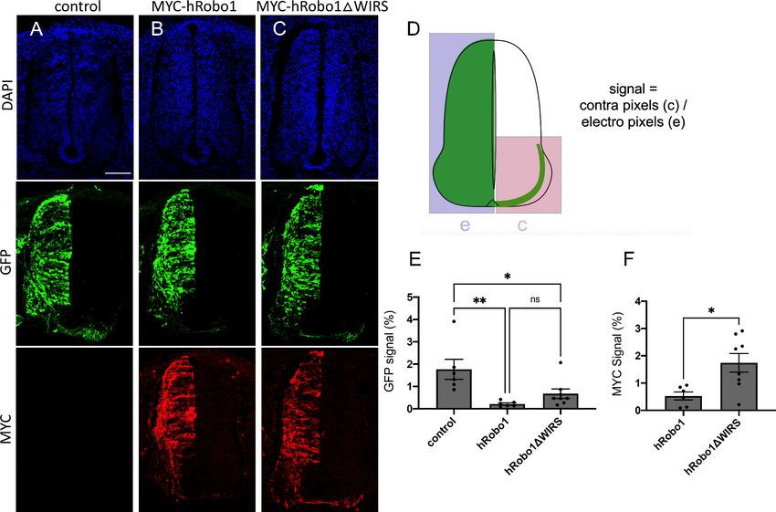
To study the function of the Robo1 WIRS motif in an in vivo context, we examined its role in com-
missural axon guidance in the embryonic chicken spinal cord. We reasoned that unilateral expression
of Robo1 in pre-crossing commissural neurons would prevent their axons from crossing the floor
plate by inducing a premature responsiveness to midline-secreted Slits (Brose et al., 1999;
Long et al., 2004). To do this, we used in ovo electroporation to introduce a GFP expression plas-
mid either alone (Control) or with MYC-tagged wild-type human Robo1 or human Robo1DWIRS
expression constructs into pre-crossing commissural neurons at Hamburger–Hamilton (HH) stage 14
(Hamburger and Hamilton, 1951). At HH stage 22–23, a ‘crossing index’ was calculated by measur-
ing GFP and MYC signal in the contralateral side of the spinal cord as a fraction of GFP and MYC
signal on the electroporated side (Figure 8D). We found that ectopic expression of wild-type Robo1
and GFP resulted in a GFP crossing index of 0.21 ± 0.13% (mean ± SD, n = 6), which was significantly
less than that of GFP alone (Control), with a crossing index of 1.8 ± 1.1% (n = 6, p=0.004), indicating
that Robo1 expression was sufficient to block commissural crossing (Figure 8A, B, E). Robo1DWIRS
and GFP overexpression resulted in a GFP crossing index of 0.68 ± 0.60% (n = 8), which was not sig-
nificantly different from that of wild-type Robo1 (p=0.472; Figure 8C, E). However, quantification of
the crossing index based on the MYC tag fused to the wild-type Robo1 and Robo1DWIRS constructs
resulted in a significantly higher MYC crossing index of Robo1DWIRS-expressing neurons
(1.7 ± 0.97%, n = 8) compared to that of wild-type Robo1-expressing neurons (0.53 ± 0.36%, n = 6,
p=0.013; Figure 8F). The disparity between the effects of the WIRS mutation calculated using GFP
and MYC-based quantification may reflect a greater efficiency of GFP plasmid transduction and
expression compared to the Robo1 expression constructs. These data demonstrate a significant
reduction in Robo1’s ability to prevent spinal commissural crossing in the absence of the WIRS
motif.
Altogether, the results from mouse dorsal spinal cord explants and dissociated neuron cultures
along with the in vivo experiments in chick embryos show that while overexpression of wild-type
hRobo1 is able to enhance the repulsive response to Slit, mutating the WIRS motif in hRobo1 abol-
ishes this gain-of-function response. These observations indicate that the WIRS motif is important for
vertebrate Robo1 signaling and suggest an evolutionarily conserved role for the WIRS motif in
Robo1 repulsive signaling.
Discussion
In this article, we have documented a conserved role for the WRC in Slit-mediated Robo1 repulsive
signaling. Using the developing Drosophila embryonic CNS, we demonstrate a series of dose-
dependent genetic interactions between components of the WRC and Slit-Robo1 signaling, which
show that the WRC functions in vivo to regulate Robo1 repulsive signaling at the midline. Biochemi-
cal experiments in cultured cells show that Robo1 can bind to the WRC partially via its WIRS motif
and that Slit stimulation can induce recruitment of the WRC to Robo1. Further, we present several
lines of evidence to demonstrate that the WIRS motif is important for Robo1 function in vivo. First,
mutating the WIRS motif results in a significantly weaker gain-of-function phenotype when Robo1 is
misexpressed in commissural axons. Second, the Robo1 variant with mutations in its WIRS motif fails
to rescue the robo1 mutant phenotype as effectively as wild-type Robo1. Finally, mutating the WIRS
motif in the endogenous robo1 locus using the CRISPR-Cas9 system results in embryos with severe
ectopic crossing defects that phenocopy robo1 mutants. These data demonstrate a severe decline in
Robo1 function upon disruption of the WRC binding site. Together, our observations support the
model that Slit stimulation results in recruitment of the WRC to the WIRS motif in Robo1, which is
Chaudhari et al. eLife 2021;10:e64474. DOI: https://doi.org/10.7554/eLife.64474 16 of 35You can also read