The Emerging Roles of Axonemal Glutamylation in Regulation of Cilia Architecture and Functions - Frontiers
←
→
Page content transcription
If your browser does not render page correctly, please read the page content below

REVIEW
                                                                                                                                             published: 04 March 2021
                                                                                                                                       doi: 10.3389/fcell.2021.622302

                                            The Emerging Roles of Axonemal
                                            Glutamylation in Regulation of Cilia
                                            Architecture and Functions
                                            Wen-Ting Yang 1† , Shi-Rong Hong 1† , Kai He 2† , Kun Ling 2 , Kritika Shaiv 1 , JingHua Hu 2,3,4*
                                            and Yu-Chun Lin 1,5*
                                            1
                                             Institute of Molecular Medicine, National Tsing Hua University, HsinChu City, Taiwan, 2 Department of Biochemistry
                                            and Molecular Biology, Mayo Clinic, Rochester, MN, United States, 3 Division of Nephrology and Hypertension, Mayo Clinic,
                                            Rochester, MN, United States, 4 Mayo Clinic Robert and Arlene Kogod Center on Aging, Mayo Clinic, Rochester, MN,
                                            United States, 5 Department of Medical Science, National Tsing Hua University, HsinChu City, Taiwan

                          Edited by:
                      Helena Soares,        Cilia, which either generate coordinated motion or sense environmental cues and
            Faculdade de Ciências da        transmit corresponding signals to the cell body, are highly conserved hair-like structures
     Universidade de Lisboa, Portugal
                                            that protrude from the cell surface among diverse species. Disruption of ciliary
                       Reviewed by:
                        Maureen Barr,       functions leads to numerous human disorders, collectively referred to as ciliopathies.
        Rutgers, The State University       Cilia are mechanically supported by axonemes, which are composed of microtubule
        of New Jersey, United States
                                            doublets. It has been recognized for several decades that tubulins in axonemes
                  Jyothi Shilpa Akella,
  Rutgers University, United States, in     undergo glutamylation, a post-translational polymodification, that conjugates glutamic
      collaboration with reviewer MB        acid chains onto the C-terminal tail of tubulins. However, the physiological roles of
                          Koji Ikegami,
          Hiroshima University, Japan       axonemal glutamylation were not uncovered until recently. This review will focus on
                  *Correspondence:          how cells modulate glutamylation on ciliary axonemes and how axonemal glutamylation
                           Yu-Chun Lin      regulates cilia architecture and functions, as well as its physiological importance in
                   ycl@life.nthu.edu.tw
                                            human health. We will also discuss the conventional and emerging new strategies used
                            JingHua Hu
                Hu.Jinghua@mayo.edu         to manipulate glutamylation in cilia.
     † These   authors have contributed
                                            Keywords: primary cilia, motile cilia, tubulin glutamylation, ciliopathies, chemically inducible dimerization
                    equally to this work

                    Specialty section:
         This article was submitted to
                                            CILIA AND CILIOPATHIES
         Cell Adhesion and Migration,
                a section of the journal    The Architecture of Cilia
  Frontiers in Cell and Developmental       The cilium is a hair-like organelle ubiquitously found on the surface of eukaryotic cells, each of
                                Biology     which has a core formed by a microtubule-based axoneme and a basal body (transformed from the
         Received: 28 October 2020          mother centriole) that anchors the cilium (Figure 1). Functionally, there are two different types of
        Accepted: 11 February 2021          cilia: motile cilia (or termed as flagella in some eukaryotic cells) or non-motile cilia (or primary
          Published: 04 March 2021          cilia) (Figure 2). In general, the shaft of the cilium is supported by a ring of nine outer microtubule
                               Citation:    doublets, with an extra central pair of doublets in the motile cilium (termed 9 + 2 arrangement),
           Yang W-T, Hong S-R, He K,        but not in the primary cilium (termed 9 + 0 arrangement) (Figure 2). Other than the central pair of
    Ling K, Shaiv K, Hu J and Lin Y-C       microtubule doublets, motile cilia also possess unique structures such as dynein arms, radial spokes,
            (2021) The Emerging Roles
                                            and nexin-dynein regulatory complex (N-DRC), which attach to outer doublets and act together
             of Axonemal Glutamylation
     in Regulation of Cilia Architecture
                                            to produce ATP-driven beating or waving motion of motile cilia. With this kinetic capability,
                        and Functions.      motile cilia can propel the movement of the ciliated cells/organisms, or generate fluid flow on
       Front. Cell Dev. Biol. 9:622302.     the surface of the ciliated cells. In contrast to the force-generating motile cilium, the primary
       doi: 10.3389/fcell.2021.622302       cilium is recognized as a sensory organelle, which acts like a cell’s antenna to recognize, integrate,

Frontiers in Cell and Developmental Biology | www.frontiersin.org                 1                                            March 2021 | Volume 9 | Article 622302
Yang et al.                                                                                                                                   The Roles of Ciliary Glutamylation

  FIGURE 1 | Structure of the primary cilium. The core primary cilium is a solitary organelle which can be segregated into upper and lower half. The upper half is
  comprised of axonemes which are typically formed by nine peripheral microtubule doublets arranged radially, enclosed within ciliary membrane. The axoneme
  comprises doublets built by A-tubule (gray) and glutamylated B-tubule (green) at the proximal part of cilia. The doublets transit to non-glutamylated A-tubule singlet
  at the middle and distal parts of cilia. The ciliary base further forms two sub-regions namely, transition zone and transition fibers, respectively. The transition zone lies
  between axoneme and transition fiber. The transition fibers emerge from the basal body in a spoked fashion, serving as a link between basal body and ciliary
  membrane. The Y-shaped linkers reside in transition zone and connect axoneme to the ciliary necklace. Dynein moves IFT-A complex and its cargo toward the cell
  body whereas, kinesin2 moves IFT-B complex and its cargo toward the ciliary tip, along the axoneme.

and transform extracellular cues into internal signal transduction                          in human genetics, at least 187 causal loci have been cloned
cascades that allows the cell to perceive and respond properly                              in 35 rare human disorders, such as polycystic kidney disease
to its microenvironment (Goetz and Anderson, 2010; Patel and                                (PKD), Bardet-Biedl syndrome, Joubert syndrome, and Meckel-
Honoré, 2010; Louvi and Grove, 2011).                                                       Gruber syndrome, which are now collectively termed as cilia-
    In general, the structures of both motile cilia and primary cilia                       related diseases, or ciliopathies (Badano et al., 2006; Adams et al.,
are highly conserved during evolution. Evidence from electron                               2008; Reiter and Leroux, 2017). Of note, since all ciliopathies
microscopy and super-resolution microscopy shows that each                                  are rare genetic diseases, a significant share of causal loci have
cilium can be divided into distinct and conserved domains: a                                not been cloned yet due to their extremely low incidence.
microtubule-based axoneme as the ciliary shaft, a basal body                                Consistent with the fact that cilia are ubiquitously present in the
supporting the protrusion of the axoneme, and microdomains                                  human body, ciliopathies usually occur as syndromic disorders
on the proximal end of the axoneme including pinwheel-like                                  that share common manifestations (such as brain anomalies,
transition fibers that connect the distal end of the basal body to                          retinal degeneration, kidney and liver dysfunction, skeletal
the ciliary membrane, the transition zone with Y-shaped linkers                             abnormalities, obesity/diabetes, infertility, and situs inversus)
connecting the axoneme to a specialized membrane domain                                     (Hildebrandt et al., 2011). Despite the importance of cilia in both
known as the ciliary necklace (Figure 1; Reiter et al., 2012;                               cell biology and human health, many central questions in the
Gonçalves and Pelletier, 2017).                                                             context of cilia, especially primary cilia; including how cilia in
                                                                                            different cell types are modified to execute sensory functions, how
                                                                                            cilia signalings convert into specific cellular behaviors, and most
Ciliopathies                                                                                importantly, the molecular function of most identified ciliopathy
Primary cilium acts as a central hub for a wide spectrum                                    proteins, remain poorly understood.
of signaling pathways required for embryonic development
and tissue homeostasis, such as hedgehog (Hh), canonical and
non-canonical Wingless (WNT), transforming growth factor-                                   Intraflagellar Transport Builds and
β (TGF-β), platelet-derived growth factor receptor (PDGFR),                                 Maintains All Cilia
and various G protein-coupled receptor (GPCR) signalings                                    Inside the cilium, the axoneme is closely covered by the ciliary
(Goetz and Anderson, 2010; Nishimura et al., 2019). With rapid                              membrane and thus possesses limited cytosolic space. Extensive
advancements in next-generation sequencing and its application                              electron microscopy studies of cilia across various organisms

Frontiers in Cell and Developmental Biology | www.frontiersin.org                       2                                              March 2021 | Volume 9 | Article 622302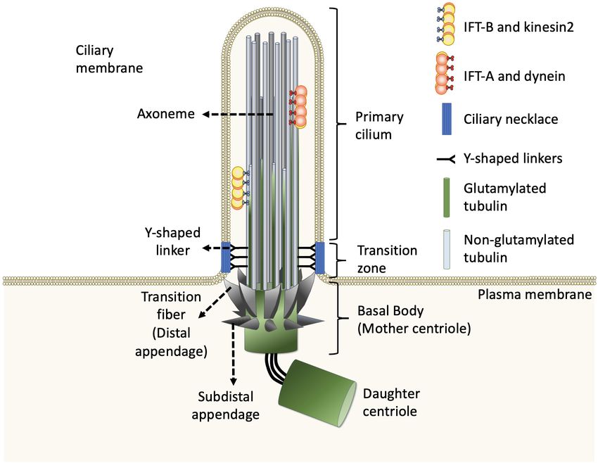
Yang et al.                                                                                                           The Roles of Ciliary Glutamylation

in the last few decades conclude that there is no presence of                 indicates that tubulin PTMs usually are present on long-lived
the ribosome inside cilia. Thus, all ciliogenic proteins required             and stabilized subsets of microtubule polymers. However, the
for cilia biogenesis, maintenance, and function need to be                    debate about whether PTMs contribute to microtubule stability
synthesized in the cytoplasm and then sorted into the cilium via              or, in contrast, whether only older polymers gain access to
intracellular trafficking routes. All cilia and eukaryotic flagella are       PTM enzymes has so far been inconclusive. The molecular
built and maintained by phylogenetically conserved intraflagellar             localization and unstructured property of the C-terminal
transport (IFT) machinery (Rosenbaum and Witman, 2002). IFT                   region of tubulins naturally provides a flexible interface that
consists of bidirectional movement along the axoneme (Figure 1;               potentially influences the association between microtubules and
Satir and Christensen, 2007; Pedersen and Rosenbaum, 2008; Hao                microtubule-binding proteins such as, microtubule-associated
et al., 2009; Pedersen and Christensen, 2012; Lechtreck, 2015;                proteins (MAPs), kinesin, and dynein motors that transport IFT
Prevo et al., 2017). Anterograde movement of particles away from              machinery. As MAPs regulate the properties of microtubules
the ciliary base is mediated by kinesin-2 motor proteins, whereas             and IFT builds and maintains all cilia, it is widely believed
retrograde movement away from the ciliary tip is powered by                   that axonemal PTMs are important for regulation of cilia
cytoplasmic dynein motor proteins. The particles transported                  structure and functions (Gaertig and Wloga, 2008; Janke and
by IFT are composed of at least 20 protein subunits, that form                Bulinski, 2011). In this review, we specifically focus on the
two distinct complexes known as the IFT-A complex and IFT-                    emerging role of microtubule glutamylation, one of the most
B complex (Hao et al., 2009; Pedersen and Christensen, 2012;                  abundant PTMs in ciliary axonemes, which results in the covalent
Broekhuis et al., 2013; Lechtreck, 2015; Prevo et al., 2017). In              conjugation of one (through the γ-carboxyl group) or more
general, kinesin-2 and cytoplasmic dynein bind to the IFT-B                   (through the subsequent α-carboxyl groups) glutamate residues
complex and IFT-A complex, respectively. Depletion or mutation                onto the C-terminal tail of axonemal tubulins (Gaertig and
of most of the IFT components affects cilia biogenesis and ciliary            Wloga, 2008). We describe how cells modulate glutamylation on
signalings in almost all ciliated organisms, revealing that the               ciliary axonemes and how axonemal glutamylation regulates cilia
role of IFT machinery is fundamentally conserved (Hao et al.,                 architecture and functions.
2009; Pedersen and Christensen, 2012; Broekhuis et al., 2013;
Lechtreck, 2015; Prevo et al., 2017).
                                                                              MODULATION OF GLUTAMYLATION ON
Tubulin Post-translational Modifications                                      CILIARY AXONEMES
of the Ciliary Axoneme
The axoneme serves as the skeleton of the primary cilium, giving              Tubulin Glutamylation-Catalyzing
support to its structure and, most importantly, providing a track             Enzymes
for IFT-dependent movement (Figure 1; Singla and Reiter, 2006).               The enzymes required for tubulin glutamylation belong to the
Like other larger structures formed by microtubules, such as the              tubulin tyrosine ligase-like (TTLL) family, of which the members
mitotic spindle, the cilium consists of microtubules assembled                share a conserved tubulin tyrosine ligase (TTL) core and a
from heterodimers of α- and β-tubulin into long, and polarized                cationic microtubule-binding domain (c-MTBD) (Figure 3;
hollow polymers, including the ciliary tip (a plus-end) with                  Van Dijk et al., 2007; Garnham et al., 2015). TTLLs adopt a
exposed β-tubulin and the proximal end of the basal body (a                   tripartite strategy for substrate recognition: TTLLs attach to
minus-end) with exposed α-tubulin. The doublets of the axoneme                the ionic C-terminal tail of tubulin via their TTL core, bind
are formed by a full-circle A-tubule (with 13 protofilaments)                 to microtubules with the c-MTBD, and eventually position
attached by an incomplete B-tubule (with 10 protofilaments)                   themselves for subsequent modifications (Garnham et al., 2015).
(Figure 2; Ichikawa et al., 2017; Ma et al., 2019). To adapt to a             There are 13 TTLL family members in mammals. Nine of
large diversity of functions as well as to generate spatiatomporal            these are characterized as glutamylases, including TTLL1, 2, 4,
specialized identities, the microtubules could be comprised of                5, 6, 7, 9, 11, and 13, whereas the rest act as glycylases or
different tubulin isotypes or undergo many highly conserved                   tyrosinases (Table 1; Van Dijk et al., 2007). According to the
post-translational modifications (PTMs), including acetylation,               architectural differences in their core domains and affinities for
detyrosination, 12/13 modification, glutamylation, glycylation,               substrate binding, TTLLs exhibit enzymatic preferences for either
and phosphorylation, as well as poorly studied ones including                 α- or β-tubulin and specificities with respect to the type of
palmitoylation, glycosylation, arginylation, methylation, and                 glutamylation reactions, such as chain initiation or elongation
SUMOylation, which are collectively referred to as the “tubulin               (Table 1; Natarajan et al., 2017; Mahalingan et al., 2020). A recent
code” (Gaertig and Wloga, 2008; Wloga and Gaertig, 2010;                      work revealed that the corresponding residues Q180 and H362 of
Janke and Bulinski, 2011; Konno et al., 2012; Janke, 2014).                   TTLL6 determine the reaction specificity for elongation instead
For tubulin PTMs with known modifications sites, many of                      of initiation. The substitution with Arg at the position Q180
them occur in the C-terminal region of tubulins that protrudes                allows the formation of hydrogen bond with acceptor glutamate,
from the outer surface of microtubules (Figure 2; L’Hernault                  which thereby favors the γ-linked glutamylation. H362, on the
and Rosenbaum, 1985; LeDizet and Piperno, 1987; Janke and                     other hand, stabilizes the intermediate with van der Waals for
Bulinski, 2011; Soppina et al., 2012; Gadadhar et al., 2017a).                the initiation reaction. The substitution with Ile does not only
Evidence from various organisms and mammalian cell types                      disrupt this interaction but also neutralize the carboxylate of the

Frontiers in Cell and Developmental Biology | www.frontiersin.org         3                                      March 2021 | Volume 9 | Article 622302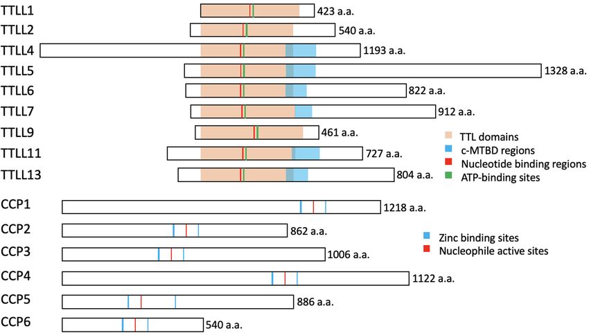
Yang et al.                                                                                                                               The Roles of Ciliary Glutamylation

  FIGURE 2 | Potential tubulin PTMs along the axoneme. Upper left: Schematic drawings of cross-section of the axoneme in primary cilia or motile cilia. Lower left: the
  doublets of the axoneme are hollow tubes composed of 13 (A-tubule) or 10 (B-tubule) protofilaments. Lower right: the wall of each protofilament is assembled from
  the globular domain of α-tubulin–β-tubulin dimers, with unstructured carboxy-terminal segment facing outer surface of the axoneme. PTMs with known molecular
  location on the globular part of tubulins include acetylation, phosphorylation, polyamination, ubiquitination, arginylation, and methylation. Globular PTMs usually
  occur at fixed molecular locations. Some PTMs preferentially choose certain tubulin subtypes, such as K40 acetylation specifically occurs on α-tubulins at the luminal
  surface of microtubules, while the Q15 polyamination appears to present on β-tubulins. Detyrosination/tyrosination and D2/D3-tubulin modifications are limited to the
  very end of the carboxy-terminal tail of a-tubulins. For tyrosination, a modified nitrotyrosine (NO2 Tyr) could be incorporated into the extreme carboxyl terminus of
  α-tubulin via the same tubulin tyrosination enzyme (Eiserich et al., 1999). (Poly)glycylation and (poly)glutamylation are found along the glutamate-rich carboxy-terminal
  tails of both α- and β-tubulins. Several other PTMs, including palmitoylation, glycosylation and SUMOylation, have been identified on tubulins, but with little
  knowledge regarding to molecular location and physiological importance. Ac: Acetylation. Am: Polyamination. Ag: Agination. Me: methylation. P: Phosphorylation.
  Ub: Polyubiquitination. E: Glutamate. G: Glycine.

acceptor glutamate as what other elongases do (Mahalingan et al.,                        and 7 are localized to the basal body, and at the cilia shaft
2020). The flexibility of the catalytic core in mammalian TTLL7                          (Table 1; Van Dijk et al., 2007; He et al., 2018). Therefore,
allows microtubule docking for execution of both chain initiation                        it is likely that axonemal glutamylation is regulated by these
and elongation reactions on β-tubulin, whereas others function                           cilium-preferring TTLL proteins. Indeed, hypoglutamylation of
only as initiates or elongases (Table 1; Van Dijk et al., 2007;                          axonemal microtubules induced by the depletion of TTLL4, 5, or
Mukai et al., 2009; Garnham et al., 2015; Natarajan et al., 2017;                        6 in primary cilia or motile cilia has suggested their key roles in
Mahalingan et al., 2020). Some TTLLs function autonomously,                              regulating ciliary glutamylation. TTLL1, 9, and 11 in the basal
whereas others may require the assistance of associated activators                       body are also involved in axonemal glutamylation because of the
for their functions, such as TTLL1, 2, and 9 (Figure 3 and Table 1;                      critical relationship between basal body and cilia (Table 2; Wloga
Janke et al., 2005; Van Dijk et al., 2007).                                              et al., 2008; Ikegami et al., 2010; Suryavanshi et al., 2010; Vogel
    In addition to their different enzymatic preferences, TTLL                           et al., 2010; O’Hagan et al., 2011, 2017; Lee et al., 2012, 2013; Grau
glutamylases also exhibit distinct subcellular distributions.                            et al., 2013; Kubo et al., 2014; He et al., 2018; Kimura et al., 2018;
Although there is no antibody that is specific for a particular                          Ki et al., 2020). Moreover, the localization of TTLL7 in cilia of
TTLL subtype, fluorescent protein–labeling or tag-labeling                               transfected MDCK cells also implies that it has a role in ciliary
experiments have demonstrated that TTLL1, 9, and 11 are                                  glutamylation, although more studies are required to confirm this
mainly localized to the basal body, whereas TTLL4, 5, 6,                                 (Van Dijk et al., 2007).

Frontiers in Cell and Developmental Biology | www.frontiersin.org                    4                                              March 2021 | Volume 9 | Article 622302
Frontiers in Cell and Developmental Biology | www.frontiersin.org
Yang et al.
TABLE 1 | Members of the TTLL family that act as glutamylases.
Enzyme and PTM Substrate Preference Reaction Autonomy Subcellular/Ciliary Distribution References
Homologs Specificity
TTLL1 Ttll1p Glutamylation α-Tubulin Non-Tubulin Initiation One subunit of a Basal Bodies Contractile Vacuole Pores Oral Janke et al., 2005; Van Dijk et al., 2007;
(Tetrahymena Protein(s): Klf4 complex Deep Fibers Cell Body (excluded from the Wloga et al., 2008; Ikegami et al., 2010;
thermophila) LmTTLL1 nucleus) Vogel et al., 2010; Pathak et al., 2011; Ye
(Leishmania major) et al., 2018; Lan et al., 2020
*TTLL2 Glutamylation Unknown Unknown One subunit of a Unknown Van Dijk et al., 2007
complex
TTLL4 LmTTLL4A Glutamylation β-Tubulin Non-Tubulin Initiation Autonomous Basal Bodies Cilia Cell Body Nucleus Van Dijk et al., 2007; Kashiwaya et al.,
(Leishmania major) Protein(s): PELP1, Mad2, Mitochondria Mitotic Spindles Mid-Bodies 2010; Kimura et al., 2010; Lacroix et al.,
LmTTLL4B (Leishmania NAPs (NAP1 an NAP2) 2010; Rogowski et al., 2010; O’Hagan
major) LmTTLL4C et al., 2011; Pathak et al., 2011; Ye et al.,
(Leishmania major) 2014; Casanova et al., 2015; Chawla et al.,
2016; Ijaz et al., 2017; Kimura et al., 2018;
Ye et al., 2018; Mahalingan et al., 2020
TTLL5 Glutamylation α-Tubulin Non-Tubulin Initiation Autonomous Basal Bodies Cilia Van Dijk et al., 2007; Ghosh-Roy et al.,
Protein(s): RGPR 2012; Lee et al., 2013; Chawla et al., 2016;
Sun et al., 2016; Devambez et al., 2017;
Bompard et al., 2018; He et al., 2018
5
TTLL6 Ttll6Ap Glutamylation β-Tubulin α-Tubulin (under Elongation Autonomous Basal bodies Cilia B-tubules of the outer doublets Janke et al., 2005; Van Dijk et al., 2007;
(Tetrahymena the overexpression §LmTTLL6B: Cell Body (excluded from the nucleus) §Also Wloga et al., 2008; Lacroix et al., 2010;
thermophila) condition in HeLa cell) specific to found in the nucleus of low ploidy Suryavanshi et al., 2010; Pathak et al.,
LmTTLL6A (Leishmania Non-Tubulin Protein(s): initiation megakaryocytes §Also found in tau missorting 2011; Grau et al., 2013; Zempel et al.,
major) LmTTLL6B Mad2 dendrites §LmTTLL6B: Also found as an 2013; Casanova et al., 2015; Ye et al.,
(Leishmania major) additional intense dot at the posterior end of L. 2014; He et al., 2018; Mahalingan et al.,
major 2020
TTLL7 Glutamylation β-Tubulin Initiation and Autonomous Basal Bodies Cilia Ikegami et al., 2006; Van Dijk et al., 2007;
Elongation Mukai et al., 2009; Pathak et al., 2011; He
et al., 2018
TTLL9 Ttll9p Glutamylation α-Tubulin Elongation One subunit of a Basal Bodies Cilia Cell Body (excluded from the Van Dijk et al., 2007; Wloga et al., 2008;
(Tetrahymena complex nucleus) Ghosh-Roy et al., 2012; Kubo et al., 2014;
thermophila) tpg1 Casanova et al., 2015
(Chlamydomonas
March 2021 | Volume 9 | Article 622302
reinhardtii) LmTTLL9
(Leishmania major)
The Roles of Ciliary Glutamylation
TTLL11 Glutamylation α-Tubulin Elongation Autonomous Basal Bodies Cell Body Axon Dendrites Cilia Van Dijk et al., 2007; Lacroix et al., 2010;
§Exists as puncta in the sensory neurons Chawla et al., 2016; O’Hagan et al., 2017
TTLL13 Glutamylation α-Tubulin Elongation Autonomous Unknown Van Dijk et al., 2007; Bompard et al., 2018
∗ TTLL2 is believed to be a glutamylase based on its homology to other family members; however, further research is required for its definitive characterization.Yang et al. The Roles of Ciliary Glutamylation FIGURE 3 | The conserved structures of the TTLL glutamylases and the CCP deglutamylases in mice. The illustrations represent the murine TTLL proteins that function as glutamylases (upper) and the murine CCP proteins that function as deglutamylases (lower). The enzymatic core TTL domains (orange), ATP-binding sites (green), and the nucleotide-binding regions (red) are highly conserved, whereas the c-MTBD regions (blue) are conserved in the autonomous TTLL proteins (TTLL4, 5, 6, 7, 11, and 13) for their interactions with microtubules. Note that TTLL1, 2, and 9 lack the c-MTBD region and function as the catalytic subunits in protein complexes. The CCP proteins belong to the M14 metallocarboxypeptidase family and have highly conserved zinc binding sites (blue) and a nucleophile active site (red). These illustrations are based on sequences obtained from UniProt (www.uniprot.org): TTLL1, Q91V51; TTLL2, A4Q9E4; TTLL4, Q80UG8; TTLL5, Q8CHB8; TTLL6, A4Q9E8; TTLL7, A4Q9F0; TTLL9, A2APC3; TTLL11, A4Q9F4; TTLL13, A4Q9F6; CCP1, Q641K1; CCP2, Q8CDK2; CCP3, Q8CDP0; CCP4, Q09M05; CCP5, Q09M02; CCP6, and Q09LZ8. How cells glutamylate axonemes via TTLLs are carried out The protist homolog of TTLL9 in Chlamydomonas, tpg1, forms by two mechanisms: the recruitment of TTLL family members a complex with a flagella-associated protein, FAP234, which to the cilia and the activation of its enzyme activity. The mainly functions to stabilize and deliver TTLL9 into the axonemal glutamylation of sensory cilia in Caenorhabditis elegans flagella matrix via IFT trains (Kubo et al., 2014). Moreover, is up-regulated in response to various environmental stimuli the centriole, cilia, and spindle-associated protein (CSAP) can including heat, cold, high osmolarity, and starvation (Kimura form complexes with TTLL5 and other autonomously active et al., 2018). This is achieved by p38 MAPK–mediated TTLL4 TTLLs, including TTLL4, 6, 7, 11, and 13. CSAP and the activation, perhaps owing to the phosphorylation on Thr446 of binding TTLL5 reciprocally regulate one another, which not only TTLL4 (Kimura et al., 2018). The glutamylation of axonemal promotes their localization to axonemal microtubules but also microtubules in primary cilia also relies on the spatial restriction enhances tubulin glutamylation (Bompard et al., 2018). Whether of the corresponding TTLLs through their interacting proteins. these TTLL-interacting proteins confer the distinct distributions During ciliogenesis in human retinal pigment epithelium of each TTLL members in different model organisms merit (RPE) cells, ARL13B (ADP-ribosylation factor-like protein 13B) comprehensive scrutiny. and RAB11/FIP5 (RAB11 family interacting protein1)-positive vesicles coordinately promote the transport of TTLL5- and TTLL6-containing vesicles to the ciliary base, which results in Tubulin Deglutamylation-Catalyzing an increase in general polyglutamylation with long side chains Enzymes in the cilia (He et al., 2018). In another population of ciliated As for the reverse modification, the tubulin deglutamylation cells, human umbilical vein endothelial cells, the centrosome function of cytosolic carboxypeptidases (CCPs) such as CCP1, protein CEP41 is able to regulate the level of glutamylation 2, 3, 4, 5, and 6, has been explored in last two decades. CCPs of the axonemal microtubules by controlling the ciliary entry belong to the M14 metallocarboxypeptidase family and have of TTLL6. This CEP41/TTLL6-mediated glutamylation further a conserved structure with a β-sheet–rich prodomain followed promotes deciliation and the up-regulation of pro-angiogenesis by the catalytic carboxypeptidase (CP) domain, which contains factors in response to shear stress (Lee et al., 2012; Ki et al., 2020). zinc-binding sites and an active nucleophile site (Figure 3; Frontiers in Cell and Developmental Biology | www.frontiersin.org 6 March 2021 | Volume 9 | Article 622302
Frontiers in Cell and Developmental Biology | www.frontiersin.org
Yang et al.
TABLE 2 | In vivo impacts of glutamylation alternation on cilia/flagellar.
Species Change of Cilia-related Phenotypes Note References
Glutamylation
Human Hyperglutamylation N/A N/A N/A
Hypoglutamylation Joubert syndrome (JBTS15) CEP41 mutation reduces ciliary entry of TTLL6 Lee et al., 2012
Joubert syndrome (JBTS30) ARMC9 and TOGARAM1 coordinate axoneme Latour et al., 2020
polyglycylation and polyglutamylation
Retinal degeneration TTLL5 homozygous mutation Sergouniotis et al., 2014
Mouse Hyperglutamylation No primary cilia-associated phenotypes Ccp2 KO mice Tort et al., 2014
No primary cilia-associated phenotypes Ccp3 KO mice Tort et al., 2014
No primary cilia-associated phenotypes Ccp5 KO mice Xia et al., 2016; Wu et al., 2017
Infertile, no mature sperm Ccp5 KO mice Wu et al., 2017; Giordano et al., 2019
Shortening of connecting cilia; retinal degeneration Ttll3 KO mice lack glycylation in photoreceptors, Bosch Grau et al., 2017
which results in and hyperglutamylation
Shortening of connecting cilia; retinal degeneration, abnormal sperm Ccp1mutant mice Mullen et al., 1976; Bosch Grau et al., 2017
Hypoglutamylation Primary ciliary dyskinesia (PCD)-like phenotypes and infertility in males Ttll1 deficiency Ikegami et al., 2010; Vogel et al., 2010
Reduce ependymal cilia beating frequency Ttll6 deficiency Grau et al., 2013
Aberrant sperm flagellar beating; shortened axoneme Ttll9 deficiency Konno et al., 2016
Infertile, defective sperm structure and motility Ttll5 KO mice Lee et al., 2013
Loss of tubulin glutamylation, infertile, abnormal sperm flagella ROSA22 mice that lack PGs1, a non-catalytic Campbell et al., 2002; Ikegami et al., 2007;
subunit that associates with TTLL1 Lee et al., 2013
Zebrafish Hyperglutamylation Axis curvature, hydrocephalus, pronephric cysts, and disrupts cilia Ccp1 or Ccp5 depletion Lyons et al., 2013; Pathak et al., 2014
7
motility
Shortening and loss of axonemes Ttll3 depletion Wloga et al., 2009
Hypoglutamylation Mild structure and motility defects Ttll6 depletion Pathak et al., 2011
C. elegans Hyperglutamylation Defective doublet structure; dysregulated ciliary kinesin motility; ccpp-1 deficiency O’Hagan et al., 2011, 2017
defective extracellular vesicles release
Hypoglutamylation Dysregulated kinesin motility; stabilized sealing between A- and TTLL-11 deficiency O’Hagan et al., 2017
B-tubules; defective extracellular vesicles release.
Reduced IFT along the axoneme upon starvation TTLL-4 deficiency Kimura et al., 2018
Tetrahymena Hyperglutamylation Shorter axoneme with normal structure; Paclitaxel resistance Disruption of glycylase TTLL3 reduces glycylation Wloga et al., 2009
thermophila but increases glutamylation
Paralyzed cilia and disrupted dynein-regulated motility TTLL6 overexpression Janke et al., 2005; Suryavanshi et al., 2010
Destabilized axonemal microtubules TTLL6 overexpression Wloga et al., 2010
Hypoglutamylation Shorter cilia lack the central pair βDDDE440 mutation of β-tubulin prevents Thazhath et al., 2002
March 2021 | Volume 9 | Article 622302
glutamylation and glycylation.
The Roles of Ciliary Glutamylation
Slow maturation of basal bodies; Defective cilia functions TTLL1 and TTLL9 deficiencies Wloga et al., 2008
Defective cilia motility caused by compromised sliding doublet TTLL6 deficiency Suryavanshi et al., 2010
microtubules by inner dynein arms
Basal bodies destabilize against ciliary beating force TTLL1; TTLL9 double knockout cells Bayless et al., 2016
Chlamydomonas Hyperglutamylation N/A N/A N/A
Hypoglutamylation Reduced flagellar motility but normal axonemal structure Tbg1 (TTLL9) deficiency Kubo et al., 2012, 2010
Stable flagellar Tbg1 (TTLL9) deficiency Lin et al., 2015
Stabilizes axonemal microtubules, decelerating axonemal Tbg1 (TTLL9) deficiency Kubo et al., 2015
disassembly.Yang et al. The Roles of Ciliary Glutamylation
Kalinina et al., 2007; Berezniuk et al., 2012; Otero et al., 2012). the substrate of CCP1, including telokin, Myosin light chain
The conserved Arg residue in the catalytic pocket and the kinase (MLCK), ribosomal proteins (40S ribosomal protein
neighboring basic residues confine the substrate preferences to S9), transcription factors (TRAF-type zinc finger domain-
acidic amino acids (Tort et al., 2014). A long polyglutamate containing protein), and chromosomal proteins (high mobility
side chain or nearby acidic residues in tubulin may increase the group protein B1, B2, and B3). By modifying these proteins,
localized acidity and thus enhance the catalysis activity of the CCP1 is capable of regulating various cell behaviors (Tanco
CCPs (Wu et al., 2015). et al., 2015). Moreover, CCP1 and CCP6 can modify Klf4 and
In addition, CCPs have enzymatic preference for the removal counteract against TTLL1 and TTLL4 to stop HEK293T cells
of branched glutamic acids or long polyglutamate side chains from reprogramming (Ye et al., 2018). A study in HEK293T cells
(Tort et al., 2014; Wu et al., 2015, 2017). Several previous suggested that CCP6 can also deglutamylate Telokin and MLCK
studies suggest that CCP5 possesses a catalytic preference for the as what CCP1 and CCP4 act (Rogowski et al., 2010).
γ-carboxyl-linked glutamate, while others, CCP1, 4, and 6, show
specificity for glutamates linked linearly on a side chain (Table 3;
Rogowski et al., 2010; Wu et al., 2015, 2017). However, under THE REGULATION OF CILIA
the optimized condition, a biochemical assay demonstrated that
ARCHITECTURE AND FUNCTION BY
CCP5 is able to cleave glutamates at both branched points and
in linear side chains without the need for other CCP members AXONEMAL GLUTAMYLATION
(Table 3; Berezniuk et al., 2013).
Based on the different lengths of the glutamic acid chains added
to microtubules, glutamylation is able to fine-tune the regulation
The Non-tubulin Substrates of Tubulin of diverse microtubule-based cell behaviors resulting from
interactions with microtubule-dependent motors or -associated
Modifying Enzymes
proteins (Verhey and Gaertig, 2007). Accumulating evidence
Apart from tubulins, both TTLLs and CCPs are able to
highlight that loss of and excess glutamylation modification of
modify substrates other than tubulins. TTLL1 as well as TTLL4
the axoneme can both impact cilia architecture and/or function
polyglutamate and stabilize the zinc finger transcription factor,
across ciliated species (Table 2). Here, we focus our discussion
Kruppel-like factor 4 (Klf4), by preventing its ubiquitination for
on how TTLL- and CCP-dependent glutamylation changes the
further degradation, which therefore maintains the pluripotency
stability and function of cilia and their involvement in signaling
of mouse embryonic stem cells (Ye et al., 2018). In pancreatic
pathways and other physiological processes.
ductal adenocarcinoma cells, TTLL4 is also capable of chromatin
remodeling for cell growth enhancement by glutamylating the
transcription co-regulator, PELP1 (Proline, glutamic acid- and The Role of Glutamylation in Primary
leucine-rich protein 1), and affecting its interaction with histone Cilia Architecture
H3. As consequence, TTLL4 is considered as a candidate for During zebrafish embryogenesis, both TTLL6-dependent
pancreatic cancer treatment (Kashiwaya et al., 2010). For the glutamylation and CCP1/5-dependent deglutamylation are
cases of a histone chaperone, nucleosome assembly protein 1 reported to be critical for ciliogenesis in olfactory placodes
(NAP1), TTLL4-dependent glutamylation enables its binding (Pathak et al., 2011; Lyons et al., 2013). In the CEM (Cephalic
onto the plasma membrane of red blood cells (RBCs), which in male) cilia of C. elegans, the cooperation between TTLL-11
turn affect the cell morphology. Though the details remain to and CCPP-1 remodels the axonemal doublets into a special
be clarified, this suggests the role of TTLL4 in the organization formation of 18 singlets and then maintains them in this
of cytoskeletons in RBCs (Ijaz et al., 2017). In vitro assay for conformation (O’Hagan et al., 2017). Meanwhile, in the amphid
the enzyme activity of recombinant TTLL4 suggests its capability neurons, TTLL4/5/11-dependent glutamylation of the axoneme
of glutamylating the recombinant murine NAP2 as well (Van counteracts with CCPP-1-mediated deglutamylation (Power
Dijk et al., 2007). TTLL4 and TTLL6 act counter to CCP6 et al., 2020). Hyperglutamylation of the axonemal tubulins due
for polyglutamylation of Mitotic arrest deficient 2 (Mad2) in to TTLL4 overexpression or CCPP-1 deficiency may induce
megakaryocytes (MKs). The polyglutamylated Mad2 is then able the spastin-dependent MT severing of the B tubules, which
to promote the maturation of MKs and to regulate the subsequent eventually leads to progressive defects in the ciliary structures
production of platelets (Ye et al., 2014). Retinitis pigmentosa and progressive degeneration as consequence (O’Hagan et al.,
GTPase regulator (RPGR), a protein substrate of TTLL5, contains 2011; O’Hagan and Barr, 2012). However, in vitro studies in HeLa
a basic domain that can specifically recruit TTLL5 to the cilia cells have contrarily showed that the long side chains that result
base and a Glu-Gly repetitive region that is architecturally similar from TTLL6-dependent tubulin polyglutamylation regulate
to the C-terminal tail of α-tubulin. The glutamylation state of spastin-dependent microtubule-severing instead of the short
RPGR is relevant to the localization of cone opsins and the ones generated by monoglutamylases such as TTLL4 or TTLL7
photoreceptor degeneration in mice (Sun et al., 2016). (Lacroix et al., 2010). These discrepancies might be explained
Cytosolic carboxypeptidases have been taken as enzymes by an in vitro study which found that glutamylation regulates
which hydrolyze the peptide bonds at the C-terminal of spastin activity in a biphasic manner: There is a linear increase
their substrates. In addition to tubulins, several proteins with in the binding affinity of spastin to glutamylated microtubules
C-terminal acidic tails have been predicted and verified as and a non-linear decline in its severing activity. As a function
Frontiers in Cell and Developmental Biology | www.frontiersin.org 8 March 2021 | Volume 9 | Article 622302Yang et al. The Roles of Ciliary Glutamylation
TABLE 3 | Members of the CCP family that act as deglutamylases.
Enzyme and PTM Substrate Preference Subcellular/Ciliary Distribution References
Homologs
CCP1/Nan1 Deglutamylation 12 Detyrosinated α-tubulin PolyE side Cilia Cell body (excluded from the Kalinina et al., 2007; Rogowski et al.,
CCPP-1 modification 13 chain Branching point E Non-Tubulin nucleus) Dendrites §Also found in the 2010; O’Hagan et al., 2011; Berezniuk
(Caenorhabditis modification Protein(s): MLCK-1, Telokin, Klf4, 40S nucleus of HeLa cells et al., 2012; De La Vega Otazo et al.,
elegans) RPS9, TRAD1, HMGB1/2/3 2013; Tort et al., 2014; Tanco et al.,
2015; Bompard et al., 2018; Ye et al.,
2018
CCP2 Deglutamylation 12 Detyrosinated α-tubulin Poly E side Centrioles Basal bodies Cell body Kalinina et al., 2007; De La Vega Otazo
modification §No chain (excluded from the nucleus) et al., 2013; Tort et al., 2014
detyrosination or
deglycylation
CCP3 Deglutamylation 12 Detyrosinated α-tubulin Poly E side Cell body (excluded from the nucleus) Kalinina et al., 2007; Tort et al., 2014
modification chain
Deaspartylation §No
detyrosination or
deglycylation
CCP4 Deglutamylation 12 Detyrosinated α-tubulin PolyE side Cell body (excluded from the nucleus) Kalinina et al., 2007; Rogowski et al.,
modification chain Non-Tubulin Protein(s): MLCK-1, 2010
Telokin
CCP5/Agbl5 Deglutamylation Branching point E Poly E side chain Cilia Cell body Nucleus Mitotic spindle Kalinina et al., 2007; Kimura et al.,
*CCPP-6 (short) microtubules Midbodies §Cell 2010; Rogowski et al., 2010;
(Caenorhabditis cycle–dependent distribution: in the Ghosh-Roy et al., 2012; Berezniuk
elegans) nucleus during interphase; in mitotic et al., 2013; De La Vega Otazo et al.,
spindle microtubules, midbodies during 2013; Wu et al., 2017; He et al., 2018
mitosis
CCP6 Deglutamylation 12 Detyrosinated α-tubulin Poly E side Cell body (excluded from the nucleus) Kalinina et al., 2007; Rogowski et al.,
modification chain (long) Non-Tubulin Protein(s): Basal bodies Golgi apparatus 2010; De La Vega Otazo et al., 2013;
MLCK-1, Telokin, Klf4, Mad2 Centrioles §Cell cycle–dependent Ye et al., 2014, 2018
distribution: in the Golgi apparatus,
centrioles during interphase; in
centrioles during mitosis
*There is no CCP5 in C. elegans, which do not possess motile cilia. CCPP-6 is functionally similar to CCP5 and is therefore taken to be the ortholog of CCP5.
of the polyglutamylation level and the side chain length, this Ikegami and Setou, 2010). Deficiency in TTLL1 also leads to
may change the property of spastin from a severing enzyme to severe defects in the sperm flagella and the mid-pieces, which
a stabilizer of microtubules, thus maintaining the architectural thereby disrupt the motility, and causes male infertility (Vogel
complexity of microtubule arrays (Valenstein et al., 2016). et al., 2010). Despite the severe malformation in the sperm flagella
in TTLL1 knockout mice, the architecture of the airway motile
The Role of Glutamylation in Architecture cilia are left intact. The loss of tubulin glutamylation causes the
and Motility of Motile Cilia loss of the curvature and disrupts asymmetrical ciliary beating
The proper level of glutamylation appears to be important for in trachea cilia, which can result in primary ciliary dyskinesia–
the structure and stability of axonemal microtubules in motile like respiratory phenotypes, such as mucous accumulation or
cilia as well. In Chlamydomonas flagella, polyglutamylation is sneezing, in the individual (Ikegami et al., 2010). Genetic
mainly enriched on the microtubule cross-bridging N-DRC. The depletion of TTLL6 also suggests its unique role in regulating
electrostatic interactions between negatively charged glutamic ciliary beating frequency in the ependymal cilia in mouse brains
acids on B-tubule and the positive charges on DRC interlink the (Grau et al., 2013). In zebrafish, knockdown of TTLL3 and
9 + 2 conformation of Chlamydomonas axonemes (Kubo and TTLL6 completely impairs the ciliary motility (Pathak et al.,
Oda, 2017). The absence of TTLL5 in mice leads to the loss of 2011). Deficiency of TTLL9 does not only cause reduction of
doublet 4 and thus, disrupts axoneme 9 + 2 structure in sperm glutamylation in doublet 5 but also shortening in the distal end of
(Lee et al., 2013). doublet 7. These disable the sperm from pro-hook bending and,
therefore, halt the flagella beating of the mouse sperm (Konno
The Effects of Hypoglutamylation on Cilia et al., 2016). Tpg1, the TTLL9 homolog in Chlamydomonas,
In sea urchin spermatozoa, injecting antibodies such as GT335 forms a complex with a flagella-associated protein FAP234
and B3 that can mask glutamylation, leads to defects in their (Tpg2), which mainly functions in stabilizing tpg1 in the
beating amplitude but does not affect the flagellar beating cytosol and in the subsequent IFT into the flagellar matrix.
frequency. In addition, microinjection of GT335 and B3 Defects in Tpg1 or Tpg2 lead to axonemal hypoglutamylation
antibodies into human sperm or ciliated epithelial cells also and thereby loss of the electrostatic interaction, which greatly
impairs ciliary motility (Gagnon et al., 1996; Million et al., 1999; decrease the flagellar motility without affecting the axoneme
Frontiers in Cell and Developmental Biology | www.frontiersin.org 9 March 2021 | Volume 9 | Article 622302Yang et al. The Roles of Ciliary Glutamylation
structure and dynein assembly (Kubo et al., 2010, 2012, 2014; and the primary cilia (Kiesel et al., 2020). Glutamylation
Kubo and Oda, 2017). statuses and the consequent effects on the A-tubule singlets in
primary cilia still remain to be clarified. (2) In vitro studies
The Effects of Hyperglutamylation on Cilia demonstrated that glutamic acid chains enhance the processivity
In addition to the adverse impact of axonemal hypoglutamylation and velocity of kinesin-2 (Sirajuddin et al., 2014), and thus,
on ciliary motility, hyperglutamylation of the axonemes also hypoglutamylation of axonemes may slow down the kinesin-2–
affect ciliary motility. Hyperglutamylation on the B-tubule mediated anterograde IFT.
resulting from the overexpression of the TTLL6 homologs in
Tetrahymena thermophila may hinders the microtubule sliding
driven by inner dynein arms and thereby ciliary motility
The Roles of Glutamylation in Ciliary
(Janke et al., 2005; Suryavanshi et al., 2010). CCP5 acts Signaling
downstream of Fleer/IFT70 for tubulin deglutamylation, which As the proper cilia localization of many, if not all, signaling
halts ciliogenesis in zebrafish; Moreover, hyperglutamylation of receptors/molecules depends on IFT transport, defects in IFT
axonemal microtubules in pronephric cilia induced by CCP5 dynamics caused by hypo/hyperglutamylation would conceivably
deficiency also leads to motility defects and ciliopathy phenotypes be expected to disturb ciliary signaling. Indeed, axonemal
(Pathak et al., 2014). hypoglutamylation attenuates the translocation of GLi3 and
In summary, the proper level of glutamylation appears to be tethering of Polycystic Kidney Disease 1/2 (PKD1/2) and
critical for generating proper motion in motile cilia or flagella affects ciliary Sonic Hedgehog (Shh) signaling and polycystin
across ciliated model organisms, which may be determined by signaling, respectively (He et al., 2018). Consistently, axonemal
whether erroneous axonemal architecture is present. deglutamylation using CCP5 deglutamylase artificially recruited
onto axonemes also slows down the entry of Smoothened
The Roles of Glutamylation in IFT and Gli3 into the cilia, thereby blocking the corresponding
Shh signaling (Hong et al., 2018). Since ciliary Shh signaling
Dynamics
is required for the development and maintenance of various
An in vitro study using chemically modified yeast tubulin with
ciliated tissues (Bangs and Anderson, 2017), defects in Shh
C-terminal glutamate side chains of various lengths showed
signaling induced by axonemal hyper/hypoglutamylation may
a positive increase in both the progressivity and velocity of
cause systematically ciliopathy-relative phenotypes.
kinesin-2, the major motor in anterograde IFT (Sirajuddin et al.,
2014). Mutation of CCPP-1 in C. elegans leads to abnormal
accumulation of an anterograde motor, KLP-6 kinesin-3, and Functional Crosstalk Between Tubulin
its cargo protein, polycystin-2, and to an increase in the rate Glutamylation and Glycylation
of another anterograde motor OSM-3/KIF17 along the axoneme For various “tubulin code” that add along the axoneme via
(O’Hagan et al., 2011). An In vivo study in C. elegans revealed that PTM modification, glutamylation and glycylation are special
TTLL4 levels are affected by environmental stimuli and that the because, they may compete for the same glutamate on the
induced tubulin glutamylation also positively regulates kinesin- C-terminus of tubulins (Pathak et al., 2011; Bosch Grau et al.,
2–dependent IFT (Kimura et al., 2018). In addition, depletion of 2017). Theoretically, in a biological compartment that possesses
axonemal glutamylation preferentially hampers the anterograde abundant modifying enzymes for two modifications that may
IFT dynamics, with limited disruption of IFT dynamics in the compete for same sites, any changes leading to alteration
opposite direction (Hong et al., 2018). of one modification will inevitably affect the occurrence of
Two mechanisms might explain why axonemal glutamylation the other. Glycylation was first thought to be a modification
preferentially regulates anterograde IFT dynamics. (1) Evidence enriched in motile cilia or flagella and plays a role in stabilizing
from correlative fluorescence and three-dimensional electron the axoneme (Rogowski et al., 2009; Grau et al., 2013). In
microscopy clearly demonstrates that anterograde IFT-B trains humans, the fact that TTLL10, the enzyme responsible for
transport along the B-tubules, whereas retrograde IFT-A trains polyglycylation, is inactive indicates that polyglycylation per se
use the A-tubules as their railways (Stepanek and Pigino, is likely dispensable for ciliated cells (Rogowski et al., 2009).
2016). Glutamylation appears to be more abundant on the By using a specific monoclonal antibody that faithfully detects
B-tubules in various ciliated model organisms (Gagnon et al., monoglycylation modification, axoneme monoglycylation could
1996; Lechtreck and Geimer, 2000). Moreover, structural defects be detected in mouse neuronal cilia (Davenport et al., 2007),
on B-tubules in cells with mutated CCPs and TTLLs have but not in many other types of primary cilia (Bré et al., 1996).
been frequently observed (Pathak et al., 2007, 2011; O’Hagan This leads to the assumption that glycylation overall may be
et al., 2012). Intuitively, defects on B-tubules caused by not essential for the structure and/or function of primary cilia.
hypo/hyperglutamylation may disturb the railways of the IFT- Interestingly, recent evidence demonstrated that primary cilia,
B trains and therefore hamper anterograde IFT. Interestingly, at least in some cell types, may depend on monoglycylation
evidence from cryo-electron tomography and subtomogram to maintain proper structure and function (Rocha et al., 2014;
averaging reveals the appearance of the anterograde IFT Bosch Grau et al., 2017; Gadadhar et al., 2017b). TTLL3 is
trains along the A-tubule singlets in primary cilia of Madin- an enzyme that catalyzes tubulin monoglycylation in the
Darby Canine Kidney (MDCK) epithelial cells. This suggests axoneme (Wloga et al., 2009). TTLL3 deficiency in either
different mechanisms of transport between the motile cilia Tetrahymena or zebrafish results in shortened axonemes, which
Frontiers in Cell and Developmental Biology | www.frontiersin.org 10 March 2021 | Volume 9 | Article 622302Yang et al. The Roles of Ciliary Glutamylation
is thought to act either directly or indirectly by altered tubulin (Lee et al., 2012). Depletion of CEP41 in zebrafish and mice
glutamylation (Wloga et al., 2009). In Ttll3 knockout mice, causes similar defects in axoneme glutamylation and ciliopathy-
reduced tubulin monoglycylation leads to an increased level of related phenotypes (Lee et al., 2012). Shortly after the
tubulin glutamylation in photoreceptor cells, and contributes characterization of CEP41 as a Joubert syndrome protein,
to shortened connecting cilia and retinal degeneration (Bosch homozygous mutations in TTLL5 were reported to cause retinal
Grau et al., 2017). Coincidently, pcd mice which carry a Ccp1- dystrophy in a subset of patient families with inherited retinal
inactivating mutation also show progressive degeneration of degenerations (Sergouniotis et al., 2014).
photoreceptors (Bosch Grau et al., 2017). To this end, it is To date, 37 Joubert syndrome genes have been cloned,
worth investigating the exact contribution of hypoglycylation or although the functions of most encoded proteins remain elusive.
hyperglutamylation of connecting cilia to retinal degeneration. The perspective that dysregulated axoneme glutamylation might
What adds another level of complexity is that axoneme be a central etiology in JBTS was further strengthened by the
glycylation and glutamylation also show overlapping roles finding that the Joubert syndrome protein ARL13B associates
in maintenance of cilia structure and motility in zebrafish with FIP5, a known effector of another ciliary GTPase RAB11, to
(Pathak et al., 2011). Taken together, while analyzing phenotypes promote the ciliary import of tubulin glutamylases TTLL5 and
caused by alterations of either axoneme glycylation (specifically TTLL6 in human epithelial cells (He et al., 2018). A defective
monoglycylation for human cells) or glutamylation, it needs ARL13B-FIP5 pathway leads to axoneme hypoglutamylation,
to be kept in mind that hyperglycosylated axoneme is very which does not affect ciliogenesis but does promote the
likely accompanied by less axoneme glutamylation or vice disassembly of cilia and, importantly, impairs polycystin and
versa, especially in motile cilia or flagella. Future identification shh signaling by disrupting the proper trafficking of various
of ciliary effectors/mechanisms recognizing (poly)glutamylation signaling molecules in the cilia (He et al., 2018). Amazingly,
and (poly)glycylation modifications will guarantee a thorough restoring axoneme glutamylation by depleting the cilia-enriched
understanding of the underlying crosstalk between these two deglutamylase CCP5 can effectively rescue ciliary defects in
modifications on the C-terminal tail of axonemal tubulins. ARL13B-deficient cells (He et al., 2018).
An intriguing discovery is the very recent finding that a TOG
array regulator of axonemal microtubules 1 (TOGARAM1)-
DYSREGULATION OF AXONEME Armadillo repeat containing 9 (ARMC9) module may regulate
both axoneme acetylation and polyglutamylation in human and
GLUTAMYLATION AND HUMAN
zebrafish (Latour et al., 2020). TOGARAM1 and ARMC9 were
CILIOPATHIES separately identified as causal loci of Joubert syndrome (Van
DeWeghe et al., 2017; Latour et al., 2020; Morbidoni et al., 2020).
Hypoglutamylation and Joubert It is not known why and how the TOGARAM1-ARMC9 module
Syndrome regulates both acetylation and glutamylation of the axoneme. It is
Although studies in mice suggest that hypoglutamylation is also not yet conclusive whether defective acetylation or defective
correlated with several classical ciliopathy phenotypes associated glutamylation of the axoneme contributes to the ciliopathy
with dysfunction of motile cilia, such as respiratory disorders phenotypes associated with TOGARAM1 and ARMC9 patients.
(Ikegami et al., 2010), dysfunctional ependymal cilia in the brain However, as defective axoneme acetylation is not observed
ventricles (Grau et al., 2013), and infertility (Campbell et al., in CEP41- or ARL13B-deficient cells (Lee et al., 2012; He
2002; Ikegami et al., 2007; Lee et al., 2013; Konno et al., 2016), et al., 2018), we reason that defective axoneme glutamylation
its physiological importance in human health has historically is probably the major driver for the development of ciliopathy
been overlooked. Extensive research on cilia biology, in the last phenotypes observed in Joubert syndrome patients.
20 years – especially the rapid cloning and characterization of
causative genes underlying ciliopathies – have begun to uncover
the critical role of axoneme glutamylation in the pathogenesis The Effect of Axoneme
of human diseases. Hyperglutamylation on Human Health
Joubert syndrome is a genetically heterogeneous group of Tubulin polyglutamylation is enriched during neuronal
disorders characterized by a malformed brain stem (molar tooth differentiation and is therefore, considered as a potential
sign), and is accompanied by other non-central nervous system- key physiological regulator of neuronal cells. Microtubule
related ciliopathy phenotypes including retinal degeneration, hyperglutamylation of neuronal axons is also associated with
polydactyly, and renal/liver abnormalities (Saraiva and Baraitser, neural degeneration in humans and in Ccp1−/− mice (Rogowski
1992; Cantagrel et al., 2008). The first evidence linking et al., 2010; Shashi et al., 2018). In pcd mice (carrying a
dysregulation of axoneme glutamylation to human ciliopathies Ccp1-inactivating mutation), the Purkinje cell degeneration
was the discovery that causative mutations in the centrosomal phenotype directly links tubulin hyperglutamylation to
protein CEP41, which is mutated in Joubert syndrome, does neurodegeneration (Mullen et al., 1976; Greer and Shepherd,
not affect cilia biogenesis but disrupts the proper ciliary 1982; Fernandez-gonzalez et al., 2002; Rogowski et al., 2010;
entry of the glutamylase TTLL6, which leads to dramatically Shashi et al., 2018). Although no mutations of known ciliopathy
reduced polyglutamylation along the axoneme in primary proteins have been reported to cause hyperglutamylation of the
cultured fibroblasts isolated from Joubert syndrome patients axoneme in human so far, given that polyglutamylation is also
Frontiers in Cell and Developmental Biology | www.frontiersin.org 11 March 2021 | Volume 9 | Article 622302Yang et al. The Roles of Ciliary Glutamylation
highly enriched in the axoneme, it is thus, a natural question increasing axoneme glutamylation may represent an intriguing
to ask whether axoneme hyperglutamylation has any adverse therapeutic strategy for certain ciliopathies.
impact on human cilia and, consequently, can be detrimental
to human health. As we discussed in section “The Effects of
Hyperglutamylation on Cilia,” tubulin hyperglutamylation NEW METHODS ENABLING THE
adversely affects cilia in some ciliated species, especially SPATIOTEMPORAL MANIPULATION OF
that tubulin hyperglutamylation is implicated in regulating AXONEMAL GLUTAMYLATION
microtubule severing or the motility of flagella or motile cilia
(Roll-Mecak and Vale, 2008; Lacroix et al., 2010; Grau et al., Typically, three strategies are used to study tubulin
2013; Valenstein et al., 2016). Hyperglutamylation in Ccp1−/− glutamylation: (1) genetic perturbation of genes that encode
(Mullen et al., 1976; Bosch Grau et al., 2017) or Ccp5−/− mice glutamylation-modifying enzymes, (2) manipulation of the
(Wu et al., 2017; Giordano et al., 2019) also share phenotypes glutamylation levels of purified microtubules by recombinant
including infertility and abnormal sperm biogenesis. However, enzymes in vitro, and (3) introduction of specific antibodies to
hyperglutamylation appears to be benign to primary cilia in mask the glutamylated motifs of microtubules. Although all of
mammalian cells. In cultured human or mouse cells, axoneme the above experiments imply that glutamylation is important
hyperglutamylation induced by depleting the cilia-enriched for the structural integrity and functions of cilia (Gaertig and
deglutamylase CCP5 produces significantly longer cilia and Wloga, 2008; Konno et al., 2012; Grau et al., 2013; Pathak et al.,
enhances ciliary signaling by promoting the import of signaling 2014; Chawla et al., 2016), these conventional methods have
molecules but does not result in detectable ciliary anomalies several limitations.
in primary cilia (He et al., 2018). Consistently, for the three First, tubulin glutamylation is not restricted only to ciliary
deglutamylases (CCP2, 3, 5) that are reported to be ciliary axonemes in cells. Glutamylated tubulins are also enriched at
deglutamylases, Ccp2−/− and Ccp3−/− (Tort et al., 2014), and centrosomes, mitotic spindles, and intercellular bridges (Wloga
Ccp5−/− (Xia et al., 2016; Wu et al., 2017) mice are viable and et al., 2010; Janke and Bulinski, 2011), perhaps owing to the
generally healthy without classical phenotypes associated with distribution of TTLLs and CCPs among multiple subcellular
dysfunctional primary cilia. compartments (Tables 1, 3; Van Dijk et al., 2007; He et al.,
Except for aforementioned sperm-related phenotypes, 2018). Therefore, global perturbation of modifying enzymes by
hyperglutamylation has been correlated with retinal conventional gene manipulation does not affect glutamylation
degenerations in humans (Sergouniotis et al., 2014; Kastner only in primary cilia. The effects of hypo/hyperglutamylation
et al., 2015; Astuti et al., 2016; Branham et al., 2016) and in in genetically modified cells are in fact, the combined outcome
mouse models (Marchena et al., 2011; Bosch Grau et al., 2017). resulting from, at a minimum, the perturbed microtubule pools
Intriguingly, it remains unknown whether hyperglutamylation- at each of the sites where the modifying enzyme is located. The
associated retinal degeneration is caused by alteration of introduction of antibodies against glutamylated tubulin into cells
polyglutamylation levels along the axoneme or it is a tubulin- would suffer from the same limitation, as it globally masks all
associated defect. Evidence suggests that TTLL and CCP enzymes glutamylated tubulin pools in cells.
involved in glutamylation modification can also target many Second, the interplay among microtubules, motor proteins,
non-tubulin substrates (for detailed discussion, please see section and other MAPs is highly dynamic. For example, tubulin
“The Non-tubulin Substrates of Tubulin Modifying Enzymes”). glutamylation occurs on the surface of microtubules and
Interestingly, in mice, the RPGRORF15 (the photoreceptor- regulates IFT dynamics while the IFT cargo, such as modifying
specific ORF15 variant of retinitis pigmentosa GTPase regulator) enzymes, microtubule precursors, and other MAPs, also
implicated in retinal dystrophy actually localizes to connecting dynamically regulate the PTM of microtubules, as well as their
cilia of photoreceptors and is glutamylated by TTLL5 in vivo structure and functions (O’Hagan et al., 2017). Thus, long-term
(Rao et al., 2016). Other studies show that TTLL5 deficiency gene manipulation does not allow for a time window to dissect
also disrupts the glutamylation of RPGR and leads to retinal these dynamic processes and also presents challenges with
pathology, without detectable changes in the level of tubulin respect to uncovering causal relationships.
glutamylation and axonemal structure of connecting cilia (Lee Third, in vitro modification of purified microtubules by
et al., 2013; Sergouniotis et al., 2014; Sun et al., 2016). Thus, it recombinant enzymes enables the study of acute effects of PTMs
should be cautious when drawing conclusions that link tubulin on the physical properties of microtubules and their interactions
glutamylation to the in vivo phenotypes observed in conditions with motor proteins and MAPs. However, it is difficult to fully
with altered TTLL or CCP enzyme activities. reconstitute the physiological environment and to include all
Collectively, this evidence suggests that the impact of relevant cellular molecules in in vitro systems.
axonemal hyperglutamylation appears to be benign, at least, Last, but not least, as discussed above (section “The Non-
in the context of primary cilia. This is important because tubulin Substrates of Tubulin Modifying Enzymes”), tubulins
hyperglutamylation induced by CCP5 depletion, can effectively are not the only substrate for glutamylation modification. Many
restore axonemal glutamylation, the ciliary dosage of polycystins, nucleocytoplasmic shuttling proteins such as the nucleosome-
and Shh signaling in human wild-type or Autosomal dominant assembly proteins NAP1 and NAP2 are also identified as potential
polycystic kidney disease (ADPKD) cells (He et al., 2018), which substrates of TTLL4-mediated glutamylation (Regnard, 2000; Ijaz
highlights the perspective that augmenting ciliary signaling by et al., 2017). The phenotypes triggered by global perturbation of
Frontiers in Cell and Developmental Biology | www.frontiersin.org 12 March 2021 | Volume 9 | Article 622302You can also read