The Hippo pathway: regulators and regulations
←
→
Page content transcription
If your browser does not render page correctly, please read the page content below
Downloaded from genesdev.cshlp.org on November 1, 2015 - Published by Cold Spring Harbor Laboratory Press

REVIEW

The Hippo pathway: regulators
and regulations
Fa-Xing Yu and Kun-Liang Guan1
Department of Pharmacology, Moores Cancer Center, University of California at San Diego, La Jolla, California 92093, USA

Control of cell number is crucial in animal development                        a key node to coordinate these cellular processes (Fig. 1).
and tissue homeostasis, and its dysregulation may result                       The Hippo pathway was first defined in Drosophila by
in tumor formation or organ degeneration. The Hippo                            genetic mosaic screens for tumor suppressor genes. Ge-
pathway in both Drosophila and mammals regulates cell                          netic inactivation of genes, including Warts (Wts) (Justice
number by modulating cell proliferation, cell death, and                       et al. 1995; Xu et al. 1995), Hippo (Hpo) (Harvey et al. 2003;
cell differentiation. Recently, numerous upstream com-                         Jia et al. 2003; Pantalacci et al. 2003; Udan et al. 2003; Wu
ponents involved in the Hippo pathway have been iden-                          et al. 2003), Salvador (Sav; also known as Shar-Pei) (Kango-
tified, such as cell polarity, mechanotransduction, and                        Singh et al. 2002; Tapon et al. 2002), and Mats (Lai et al.
G-protein-coupled receptor (GPCR) signaling. Actin cyto-                       2005), all resulted in a similar phenotype with robust
skeleton or cellular tension appears to be the master                          tissue overgrowth. Yorkie (Yki) is the major downstream
mediator that integrates and transmits upstream sig-                           effector of the Hippo pathway (Huang et al. 2005), which
nals to the core Hippo signaling cascade. Here, we review                      regulates a transcription program by interacting with the
regulatory mechanisms of the Hippo pathway and discuss                         transcription factor Scalloped (Sd) (Fig. 2; Goulev et al.
potential implications involved in different physiological                     2008; Wu et al. 2008; Zhang et al. 2008; Zhao et al. 2008).
and pathological conditions.                                                   The Hippo pathway is highly conserved in mammals:
                                                                               MST1/2 (Hpo orthologs), Sav1, Lats1/2 (Wts orthologs),
Cell proliferation, death, and differentiation are funda-                      and Mob1 (MOBKL1A and MOBKL1B, Mats orthologs)
mental biological processes. Coordination of these pro-                        form a kinase cascade that phosphorylates and inhibits
cesses is critical for a wide range of physiological and                       YAP/TAZ (Yki orthologs). YAP/TAZ in conjunction with
pathological conditions (Pellettieri and Sanchez Alvarado                      TEAD1–4 (Sd orthologs) mediate major physiological
2007; Galliot and Ghila 2010). During development, an                          functions of the Hippo pathway (Fig. 2; for reviews, see
increase in cell number is required to boost organ and                         Pan 2010; Zhao et al. 2010a). The nomenclature of many
body size; meanwhile, proper differentiation of multiple                       components of the Hippo pathway in Drosophila and
cell types will assure the appropriate function of devel-                      mammals is different, and a summary of these compo-
oped organs. In adulthood, most tissues undergo contin-                        nents is shown in Table 1.
uous cell turnover to maintain functionality. Aged or                             The core Hippo pathway has been well established in
damaged cells are programmed to cell death, whereas                            both Drosophila and mammals; however, the regulatory
adult stem cells may divide and differentiate to replace                       mechanisms for this signaling pathway are less under-
those dysfunctional cells. Under pathological conditions,                      stood. Recently, by using both genetic and biochemical
such as wound healing and organ regeneration, cell                             approaches, many additional components have been iden-
division and differentiation of tissue-specific progenitor                     tified to modulate the core Hippo pathway (Table 1). In
cells will be up-regulated to compensate for the lost cells.                   this review, we briefly describe the components of the
On the other hand, uncontrolled cell proliferation and                         Hippo pathway and summarize recent advances with
decreased cell death lead to hyperplasia or tumorigenesis.                     respect to Hippo pathway regulation. In addition, we also
Detailed mechanisms underlying cell proliferation, cell                        discuss the implications of Hippo pathway regulation
death, and cell differentiation have been extensively                          in different physiological and pathological conditions.
studied; however, how these processes are coordinated                          The mammalian Hippo pathway is the main focus, al-
and integrated is poorly understood.                                           though some Drosophila works are also covered. For a
   Recently, the Hippo pathway has been shown to pro-                          detailed review on the Drosophila Hippo pathway, please
mote cell death and differentiation and inhibit cell pro-                      refer to Staley and Irvine (2012).
liferation; therefore, the Hippo pathway may function as
                                                                               Core Hippo pathway: a kinase cascade
[Keywords: actin; GPCR; Hippo; YAP; mechanotransduction; polarity]             MST1/2 are STE20 family protein kinases and can phos-
1Corresponding author

E-mail kuguan@ucsd.edu                                                         phorylate Sav1, Lats1/2, and Mob1 (Wu et al. 2003; Chan
Article is online at http://www.genesdev.org/cgi/doi/10.1101/gad.210773.112.   et al. 2005; Callus et al. 2006; Praskova et al. 2008). The

           GENES & DEVELOPMENT 27:355–371 Ó 2013 by Cold Spring Harbor Laboratory Press ISSN 0890-9369/13; www.genesdev.org              355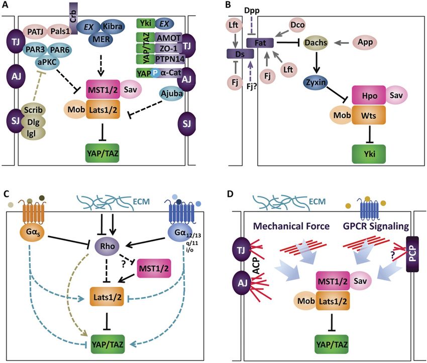
Downloaded from genesdev.cshlp.org on November 1, 2015 - Published by Cold Spring Harbor Laboratory Press

Yu and Guan

                                                                   2007; Lei et al. 2008; Oh and Irvine 2008), in which the
                                                                   interaction may be mediated by PPxY motifs on Lats1/2
                                                                   and WW domains on YAP/TAZ (Hao et al. 2008; Oka et al.
                                                                   2008). Lats1/2 are AGC family kinases and recognize
                                                                   the substrate consensus sequence HXRXXS (Zhao et al.
                                                                   2007). All five HXRXXS sites on YAP are directly phos-
                                                                   phorylated by Lats1/2 (Zhao et al. 2010b). The phosphor-
                                                                   ylated form of YAP is sequestered in the cytoplasm via a
                                                                   14-3-3 interaction, resulting in inhibition of target gene
                                                                   transcription (Zhao et al. 2007). Also, TAZ and Yki are
                                                                   phosphorylated by Lats1/2 or Wts, respectively, on mul-
                                                                   tiple HXRXXS sites (Kanai et al. 2000; Dong et al. 2007;
                                                                   Lei et al. 2008; Oh and Irvine 2008; Ren et al. 2010b).
                                                                   In contrast, when upstream kinases are inactive, Yki/
                                                                   YAP/TAZ will be hypophosphorylated and translocate
                                                                   into the nucleus to exert their functions on gene expres-
Figure 1. Implications of the Hippo pathway in cell biology.       sion (Kanai et al. 2000; Dong et al. 2007; Zhao et al. 2007;
The Hippo pathway modulates cell proliferation, differentiation,   Lei et al. 2008; Oh and Irvine 2008; Ren et al. 2010b). The
growth, and death. The coordination of different cellular pro-     phosphorylation status of YAP/TAZ also regulates their
cesses by the Hippo pathway may contribute to diverse physio-      protein stability. Phosphorylation of YAP (S381) and
logical and pathological conditions such as development, tissue
                                                                   TAZ (S311) by Lats1/2 primes subsequent phosphoryla-
homeostasis, and tumorigenesis.
                                                                   tion events by casein kinase 1 (CK1d/e); this sequential
                                                                   phosphorylation results in recruitment of b-transducin
                                                                   repeat-containing proteins (b-TRCP; a subunit of the SCF
kinase activity of MST1/2 is enhanced through interaction          ubiquitin E3 ligase) and consequently leads to degradation
with Sav1, which is mediated by SARAH (Sav/Rassf/Hpo)              of YAP/TAZ (CY Liu et al. 2010; Zhao et al. 2010b).
domains present in both MST1/2 and Sav1 (Callus et al.             Therefore, by affecting YAP/TAZ protein localization
2006). In addition, the thousand-and-one (TAO) amino               and stability, phosphorylation by upstream kinases rep-
acids kinase or TAOK1–3 has been shown to directly                 resents a central regulatory mechanism for YAP/TAZ
phosphorylate and activate Hpo or MST1/2 (Boggiano                 (Fig. 2).
et al. 2011; Poon et al. 2011). In Drosophila, RASSF                  YAP/TAZ do not contain intrinsic DNA-binding do-
competes with Sav for Hpo and recruits a PP2A com-                 mains but instead bind to the promoters of target genes
plex (dSTRIPAK) to dephosphorylate and inactivate Hpo
(Polesello et al. 2006; Ribeiro et al. 2010). However, mul-
tiple RASSF isoforms in mammals showed different roles
on the Hippo pathway (Praskova et al. 2004; Ikeda et al.
2009), suggesting a divergent role through evolution.
   MST1/2 directly phosphorylate Lats1/2 at the hydro-
phobic motif (Lats1 T1079 and Lats2 T1041), and this
phosphorylation is required for Lats1/2 activation (Chan
et al. 2005). Mob1, when phosphorylated by MST1/2,
binds to the autoinhibitory motif in Lats1/2, which in
turn leads to the phosphorylation of the Lats activation
loop (Lats1 S909 and Lats2 S872) and thereby an increase
of their kinase activity (Chan et al. 2005; Praskova et al.
2008). Sav1 may function as a bridge to bring MST1/2
and Lats1/2 together (Tapon et al. 2002; Callus et al. 2006)
and may enhance or inhibit the activity of Lats1/2 upon
phosphorylation by MST1/2 or salt-inducible kinases,
respectively (Callus et al. 2006; Wehr et al. 2012). The
requirement for MST1/2 to activate Lats1/2 might be cell
type-dependent. For instance, MST1/2 knockout in mouse
livers does not significantly affect Lats1/2 phosphoryla-
tion (Zhou et al. 2009), suggesting that additional ki-            Figure 2. The core Hippo pathway. MST1/2 phosphorylates Sav,
                                                                   Lats1/2, and Mob; Lats1/2 phosphorylates YAP/TAZ; and phos-
nases may regulate Lats1/2 activity. In addition to protein
                                                                   phorylated YAP/TAZ interacts with 14-3-3 and results in cyto-
phosphorylation, the protein levels of Lats1/2 kinases are         plasmic retention. Moreover, YAP/TAZ phosphorylation leads
controlled by Itch E3 ubiquitin ligase-mediated degrada-           to protein degradation. When dephosphorylated, YAP/TAZ enter
tion (Ho et al. 2011).                                             nuclei and induce gene transcription by interacting with tran-
   Lats1/2 directly interact with and phosphorylate YAP/           scription factors TEAD1–4. Drosophila orthologs for these core
TAZ (Huang et al. 2005; Dong et al. 2007; Zhao et al.              components are shown in brackets.

356     GENES & DEVELOPMENT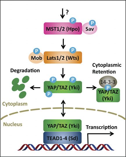
Downloaded from genesdev.cshlp.org on November 1, 2015 - Published by Cold Spring Harbor Laboratory Press

                                                                                                             Hippo pathway regulation

Table 1.   Hippo pathway components in mammals and Drosophila
Mammalian                                         Drosophila               Junctional localization         Cytoskeleton interaction
Core components
  MST1/2                                    Hpo (Hippo)                                                                U
  Sav1                                      Sav (Salvador)
  Lats1/2                                   Wts (Warts)                                                                U
  Mob1 (a and b)                            Mats
  YAP/TAZ                                   Yki (Yorki)                              U
  TEAD1–4                                   Sd (Scalloped)
Apical–basal polarity (TJs and AJs)
  Crb1–3                                    Crb (Crumbs)                             U
  Frmd6 (?)                                 Ex (Expanded)                            U                                 U
  NF2 (Mer)                                 Mer (Merlin)                             U                                 U
  Kibra                                     Kibra                                    U
  aPKC                                      aPKC                                     U
  PAR3                                      Baz (Bazooka)                            U
  PAR6                                      Par6                                     U
  PALS1                                     Sdt (Stardust)                           U
  Scrib                                     Scrib (Scribble)
  Dlg                                       Dlg
  Lgl                                       Lgl (Discs large)
  AMOT (angiomotin)                                     ?                            U                                 U
  PTPN14                                    Pez                                      U                                 U
  Ajuba/LIMD1/WTIP                          Jub                                      U                                 U
  a-Catenin                                 a-Catenin                                U                                 U
  b-Catenin                                 b-Catenin                                U
  ZO-1                                      ZO-1                                     U
  ZO-2                                      ZO-2
  E-cad (E-cadherin)                        E-cad                                    U
Planar cell polarity
  Fat1-4                                    Fat                                      U
  Dchs1/2                                   Ds (Dachsous)                            U                                 U?
  Fjx1                                      Fj (four-jointed)
  ?                                         Dachs
  Zyxin/Lpp/Trip6                           Zyx (zyxin)                                                                U
  Lix1, Lix1L                               Lft (lowfat)
  CK1d/e                                    Dco (Discs overgrowth)
  ZDHHCs                                    App (approximated)
Other components
  Taok1-3                                   Tao                                                                        U
  RASSF1-6                                  RASSF                                                                      U
  PP2A (STRIPAK)                            STRIPAK (PP2A)                                                             U?
  PP1                                       PP1                                                                        U
  Itch                                      Su(DX)
  bTRCP                                     Slimb
  14-3-3                                    14-3-3                                                                     U
Mammalian Hippo pathway components and their Drosophila orthologs are summarized. Check marks indicate localization to tight
junctions (TJs), adherens junctions (AJs), or actin cytoskeleton. Question marks indicate unsure information.

by interacting with DNA-binding transcription factors.               Apical–basal polarity: the polarized localization of hippo
YAP/TAZ mainly bind to the transcription factors                     components
TEAD1–4 to regulate genes involved in cell proliferation
and cell death (Vassilev et al. 2001; Goulev et al. 2008; Wu         Epithelial cells usually adhere to one another through
et al. 2008; Zhang et al. 2008; Zhao et al. 2008). Besides           cell–cell junctions such as adherens junctions (AJs), des-
TEADs, YAP/TAZ may also interact with other tran-                    mosomes, and tight junctions (TJs). TJs and AJs, with help
scription factors, such as Smad1 (Alarcon et al. 2009),              from different polarity complexes, divide the plasma
Smad2/3 (Varelas et al. 2008), Smad7 (Ferrigno et al.                membrane into an apical domain and a basolateral domain
2002), RUNX1/2 (Yagi et al. 1999), p63/p73 (Strano                   and thereby establish an apical–basal polarity in epithelial
et al. 2001), and ErbB4 (Komuro et al. 2003; Omerovic                cells (Martin-Belmonte and Perez-Moreno 2012). Interest-
et al. 2004); these interactions may mediate transcrip-              ingly, many upstream regulators identified for the Hippo
tion of diverse genes involved in proliferation, differ-             pathway are known components of TJs, AJs, or apical–
entiation, and development.                                          basal polarity protein complexes (Fig. 3A).

                                                                                                     GENES & DEVELOPMENT         357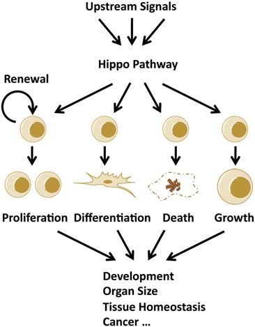
Downloaded from genesdev.cshlp.org on November 1, 2015 - Published by Cold Spring Harbor Laboratory Press

Yu and Guan

Figure 3. Regulatory mechanisms for the Hippo pathway. Regulation of the Hippo pathway by apical–basal polarity (A), PCP (B),
mechanical cues and GPCR signaling (C), and actin cytoskeleton (D). Arrowed or blunted ends indicate activation or inhibition,
respectively. Dashed lines indicate indirect or unknown mechanisms. Red lines in D represent actin filaments.

   Mer (Merlin; also known as NF2 for neurofibromatosis-          signal enhances MST1 kinase activity (Khokhlatchev et al.
2) and Ex (Expanded) are two proteins that belong to the          2002).
FERM (4.1, Ezrin, Radxin, and Moesin) domain-containing              Drosophila crumbs (Crb) has been identified as a cell
family of proteins. Both Mer and Ex have tumor suppressor         surface regulator for the Hippo pathway (Fig. 3A; CL
functions and work together to regulate cell proliferation        Chen et al. 2010; Grzeschik et al. 2010; Ling et al. 2010;
and differentiation (McCartney et al. 2000). In Drosophila,       Robinson et al. 2010). In Drosophila embryos, Crb is
genetic inactivation of both Mer and Ex revealed a dra-           localized at the subapical plasma membrane and plays an
matic overgrowth phenotype similar to that of the Hpo             important role in organizing apical–basal polarity (Tepass
mutants (Hamaratoglu et al. 2006). Later, Kibra (a WW and         et al. 1990). As a transmembrane protein, Crb has a large
C2 domain-containing protein) was identified to physi-            extracellular domain and a short intracellular domain.
cally interact with Mer and Ex, and these three proteins          The short intracellular domain contains a FERM-binding
activate Wts in a cooperative manner (Baumgartner et al.          motif (FBM) that can interact with Ex, and this interac-
2010; Genevet et al. 2010; Yu et al. 2010).                       tion modulates Ex localization and stability, which in
   Mer, Ex, and Kibra colocalize at the apical domain of          turn regulates the activity of Hippo pathway kinases and
polarized epithelial cells (Fig. 3A; Boedigheimer and             Yki (CL Chen et al. 2010; Ling et al. 2010; Robinson et al.
Laughon 1993; Boedigheimer et al. 1997; Yu et al. 2010).          2010). The connection of Crb with Ex and core compo-
Mer and Ex have been considered as a linker for the apical        nents of the Hippo pathway is also reflected by an over-
plasma membrane and actin cytoskeleton (Bretscher et al.          growth phenotype corresponding to Crb deficiency (Ling
2002). Kibra contains a C2 domain that interacts with             et al. 2010). Interestingly, overexpression of Crb leads to
phospholipids and may target interacting proteins to the          Ex mislocalization and inactivation of the Hippo path-
cell surface (Kremerskothen et al. 2003). Sav and Hpo             way, likely due to a dominant-negative effect of overex-
physically associate with Mer and Ex (Yu et al. 2010), and        pressed Crb (CL Chen et al. 2010; Grzeschik et al. 2010;
Kibra interacts with Wts (Genevet et al. 2010), suggesting        Robinson et al. 2010).
that the Mer/Ex/Kibra complex may recruit the Hippo                  Similar to the Crb complex, the Par apical complex also
pathway kinases to the apical plasma membrane for acti-           regulates the Hippo pathway (Fig. 3A). Overexpression of
vation. Indeed, it has been shown that Mats is activated          aPKC, a component of the Par complex, can induce Yki
at the plasma membrane (Ho et al. 2010), and targeting            activity and tissue overgrowth (Grzeschik et al. 2010;
MST1 to the plasma membrane by adding a myristoylation            Sun and Irvine 2011). The activity of the Par complex is

358     GENES & DEVELOPMENT

Downloaded from genesdev.cshlp.org on November 1, 2015 - Published by Cold Spring Harbor Laboratory Press

                                                                                                         Hippo pathway regulation

antagonized by the basal Scrib (Scribble) complex (Martin-           a-Catenin is a component of AJs that functions as
Belmonte and Perez-Moreno 2012). Indeed, depletion of             a linker for membrane cadherins and the actin cytoskel-
Scrib or Lgl also resulted in activation of Yki (Grzeschik        eton (Drees et al. 2005). An inhibitory role of a-catenin
et al. 2010; Menendez et al. 2010; Sun and Irvine 2011).          on YAP activity has also been reported, and this in-
   The regulation of apical–basal polarity on the Hippo           hibition of YAP may contribute to the tumor suppressor
pathway is largely conserved in mammals. NF2 is an                function of a-catenin (Fig. 3A; Schlegelmilch et al. 2011;
extensively studied tumor suppressor, and mutations in            Silvis et al. 2011). In keratinocytes, YAP strongly in-
the NF2 gene cause the development of nonmalignant                teracts with a-catenin, and this interaction is mediated
brain tumors, a syndrome called neurofibromatosis type 2.         by 14-3-3. Unlike AMOT, the phosphorylation of YAP at
Mice with conditional NF2 knockout in the liver develop           S127 is required for interaction with a-catenin because
hepatocellular carcinoma, cholangiocarcinoma, and bile            14-3-3 only binds to phosphorylated YAP (Schlegelmilch
duct hamartomas (Benhamouche et al. 2010; Zhang et al.            et al. 2011). The trimeric complex of a-catenin, 14-3-3,
2010). NF2 patients usually develop cataracts; mice with          and YAP sequesters YAP at AJs and prevents YAP de-
conditional NF2 knockout in lens epithelium also develop          phosphorylation/activation. In mammary epithelial Eph4
cataracts (Zhang et al. 2010). Interestingly, the phenotypes      cells, knockdown of a-catenin also induces YAP/TAZ
of NF2 knockout in the liver and eye were largely blocked         nuclear localization (Varelas et al. 2010), suggesting that
by heterozygous deletion of Yap (Zhang et al. 2010). In           the regulation of YAP/TAZ by a-catenin is present in
addition, overexpression of NF2 in mammalian cells re-            a variety of cell types.
sults in Lats activation and YAP inhibition (Zhao et al.             Another AJ component, protein tyrosine phosphatase
2007; Zhang et al. 2010). These results suggest that Mer is       14 (PTPN14), has also been shown to be a regulator of the
also an upstream component of the mammalian Hippo                 Hippo pathway (JM Huang et al. 2012; Liu et al. 2012;
pathway.                                                          Wang et al. 2012). PTPN14 can directly interact with
   Disruption of TJs or AJs in cultured mammalian cells           YAP, and this interaction is mediated by PPxY motifs of
(by depletion of extracellular calcium or knockdown of            PTPN14 and WW domains of YAP (JM Huang et al. 2012;
Crb3 or PALS1) causes induction of YAP/TAZ nuclear                Liu et al. 2012; Wang et al. 2012). PTPN14 also contains
localization and target gene expression (Varelas et al.           an N-terminal FERM domain, and the overall domain
2010). Moreover, Scrib also positively regulates the Hippo        organization is similar to that of Ex in Drosophila.
pathway kinases, and down-regulation of Scrib leads to            Moreover, Pez (the Drosophila ortholog of PTPN14) has
YAP/TAZ activation (Cordenonsi et al. 2011; Chen et al.           been shown to interact with Kibra and inactivate Yki
2012). In addition, many cell junction proteins, such as          (Poernbacher et al. 2012). PTPN14–YAP interaction re-
LIN7C, PATJ, MPDZ, PTPN14, angiomotin (AMOT), and                 sults in cytoplasmic localization of YAP and decreased
a-catenin, have also been identified as interacting part-         YAP activity; however, there are contradictory data on
ners of core Hippo pathway components (Fig. 3A; Varelas           the role of the tyrosine phosphatase activity of PTPN14
et al. 2010; Wang et al. 2011; KL Guan, unpubl.).                 on YAP (JM Huang et al. 2012; Liu et al. 2012; Wang et al.
   AMOT proteins, a family of proteins including AMOT,            2012).
AMOTL1, and AMOTL2, interact extensively with mul-                   Several other proteins important in establishing or
tiple TJ components and are important for maintaining TJ          maintaining apical–basal polarity have been shown to
integrity and epithelial cell polarity (Wells et al. 2006).       modulate the Hippo pathway. In mammalian cells, cell
Recently, an interaction between AMOT and YAP/TAZ                 adhesion mediated by homophilic binding of E-cadherin
has been identified (Chan et al. 2011; Wang et al. 2011;          led to YAP inactivation (Kim et al. 2011). Ajuba can
Zhao et al. 2011). The AMOT–YAP/TAZ interaction is                interact with Sav and Lats kinases in mammalian cells
not dependent on the YAP/TAZ phosphorylation status               and Drosophila and exhibits an inhibitory effect on
and is instead mediated by AMOT PPxY motifs and YAP/              YAP/Yki (Das Thakur et al. 2010). LKB1 (liver kinase
TAZ WW domains (Chan et al. 2011; Wang et al. 2011;               B1) is able to induce YAP phosphorylation (Nguyen
Zhao et al. 2011). AMOT proteins recruit YAP/TAZ to               et al. 2012). NPHP4 (nephronophthisis 4) can interact
TJs or the actin cytoskeleton, which consequently results         with and inhibit Lats1 (Habbig et al. 2011). ZO-2 (zona
in reduced YAP/TAZ nuclear localization and activity              occludens-2) can induce YAP nuclear localization (Oka
(Zhao et al. 2011). In addition, AMOT proteins also induce        et al. 2010), whereas ZO-1 has been shown to repress
YAP/TAZ phosphorylation at Lats target sites (Zhao et al.         TAZ activation (Remue et al. 2010). These results
2011); this might be due to a scaffolding function of AMOT        indicate that cell–cell contact, integrity of cell junc-
on Hippo pathway components such as MST2, Lats2, and              tions, and apical–basal polarity are important in regu-
YAP (Paramasivam et al. 2011). AMOT proteins can                  lation of the Hippo pathway. Apical–basal polarity can
therefore inhibit YAP/TAZ activity by both phosphoryla-           regulate the Hippo pathway by either recruiting the
tion-dependent and phosphorylation-independent mecha-             Hippo pathway kinases to the apical domain for activa-
nisms. Interestingly, AMOT has been shown to interact             tion or sequestering Yki/YAP/TAZ at cell junctions
with NF2 and is required for tumorigenesis caused by NF2          (Fig. 3A), both resulting in inactivation of YKi/YAP/
deficiency (Yi et al. 2011). An ortholog of AMOT in               TAZ. However, it is worth noting that the cellular
Drosophila has not been identified, suggesting that regu-         localization of YAP is mainly in the cytoplasm and
lation of AMOT on the Hippo pathway may be different              nucleus (Zhao et al. 2011); the interaction between cell
between Drosophila and mammals.                                   junctional proteins and YAP/TAZ may not result in

                                                                                               GENES & DEVELOPMENT           359
Downloaded from genesdev.cshlp.org on November 1, 2015 - Published by Cold Spring Harbor Laboratory Press
Yu and Guan
a predominant localization of YAP/TAZ at the cellular and Fat4 knockout mice with abnormal PCP (Mao et al.
apical domain. 2011). In zebrafish, Fat1 depletion was shown to activate
YAP in a Scrib-dependent manner (Skouloudaki et al.
2009). In mammals, an obvious Dachs ortholog is lacking,
Planar cell polarity (PCP): coordinates for the Hippo
suggesting that the connection between the Ft/Ds system
pathway
and the Hippo pathway may not be conserved in mam-
Epithelial cells are also polarized along an axis perpen- mals. The effect of the Fzi/Fmi PCP system on the Hippo
dicular to the apical–basal axis, in which clustered cells pathway is less well understood. Recently, it has been
within an epithelial plate are coordinated, aligned, and shown that overexpression of Frizzled 4 can activate YAP
orientated to the same direction, and this cell polarity is and that wnt signaling can activate TAZ in mammalian
termed PCP (Simons and Mlodzik 2008). In addition to cells (Azzolin et al. 2012; W Huang et al. 2012; Yu et al.
epithelial cells, PCP is also present in many other cell 2012b), suggesting that the Fzi/Fmi system may also
types, such as mesenchymal cells, and is important in cell regulate the Hippo pathway.
migration and cell interchalation (Simons and Mlodzik
2008). Two molecular networks are critical in establish-
G-protein-coupled receptor (GPCR) signaling: sensing
ing PCP: One is the Frizzled/Flamingo (Fzi/Fmi) system,
diffusible signals
and the other is the Fat/Dachsous (Ft/Ds) system (Simons
and Mlodzik 2008). The Ft/Ds PCP system has been shown A large number of growth factors regulate cell prolifera-
to regulate the Hippo pathway in Drosophila (Fig. 3B). tion by activating membrane receptors and intracellular
Ft is a tumor suppressor and affects tissue growth signaling pathways. It is reasonable to speculate that the
(Mahoney et al. 1991). Loss of Ft results in activation of YAP/TAZ oncoproteins are regulated by growth factors.
Yki by inactivating either Ex or Wts (Bennett and Harvey However, several well-known growth factors, such as in-
2006; Cho et al. 2006; Silva et al. 2006; Willecke et al. sulin and EGF, have no significant effect on YAP phos-
2006; Feng and Irvine 2007; Tyler and Baker 2007). Ft phorylation (Zhao et al. 2007; Yu et al. 2012b). In mam-
and Ds are both atypical cadherins, which form intercel- mals, the potential identity of extracellular ligands and
lular heterodimers (Cho and Irvine 2004; Matakatsu and their cognate receptors that regulate the Hippo pathway
Blair 2004), and this dimerization is regulated by Fj (four- remained elusive until recently. Two independent groups
jointed)-mediated phosphorylation (Ishikawa et al. 2008). have reported that serum could rapidly activate YAP/TAZ
Ds and Fj show gradient expression with opposite direc- in cultured cells. By extensive biochemical analysis, LPA
tions in many tissues, and this expression pattern might and S1P were identified as the major components in
be critical for Ft activity (Rogulja et al. 2008; Willecke et al. serum responsible for YAP/TAZ activation (Miller et al.
2008; Zecca and Struhl 2010). A sharp Ds gradient may 2012; Yu et al. 2012b). Both reports showed that LPA or
inhibit Ft activity, which leads to localization of atypical S1P bound to their corresponding membrane GPCRs and
myosin Dachs to subapical regions (Cho et al. 2006; Feng act through Rho GTPases to activate YAP/TAZ. Consis-
and Irvine 2007). Polarized Dachs promotes interaction tently, another report showed that thrombin, which acti-
between Zyxin and Wts, which in turn leads to Wts deg- vates protease-activated receptors (PARs; a GPCR), also
radation (Fig. 3B; Rauskolb et al. 2011). Several proteins stimulated YAP/TAZ activity via Rho GTPases (Mo et al.
have been reported to modulate the inhibitory effect of Ft 2012). These results suggest that YAP/TAZ can be regu-
on Yki. Dco (discs overgrown), a CK1 homolog, is able to lated by diffusible extracellular signals and cell surface
phosphorylate the intracellular domain of Ft and induce Ft receptors (Fig. 3C).
activity (Sopko et al. 2009). App (approximated), a palmi- Yu et al. (2012b) further showed that YAP/TAZ is
toyltransferase, can relieve the Ft inhibition on Dachs and robustly regulated by many GPCRs and their cognate
promote its apical localization (Matakatsu and Blair 2008). ligands and established a general function of GPCR in
Lft (lowfat) can bind to Ft and Ds and thereby increases YAP/TAZ regulation. GPCRs usually activate downstream
their protein stability (Mao et al. 2009). signaling through heterotrimeric G proteins. Ga12/13-,
The effect of the Ft/Ds PCP system on the Hippo path- Gaq/11-, or Gai/o-coupled signals induce YAP/TAZ activ-
way may be modulated by different morphogens. Dpp ity, whereas Gas-coupled signals repress YAP/TAZ activ-
(decapentaplegic; a BMP homolog) and Wingless (a Wnt ity (Fig. 3C). In the latter scenario, glucagon, epinephrine,
homolog) were shown to regulate the expression of Ds and a dopamine receptor agonist induce YAP/TAZ phos-
and Fj (Rogulja et al. 2008; Zecca and Struhl 2010), sug- phorylation. Interestingly, in a screen for YAP inhibitors,
gesting that these morphogens may help in establishing dobutamine has been shown to inhibit YAP (Bao et al.
the gradient of Ds and Fj. In addition, Fj is secreted and 2011). Dobutamine is an agonist for the b1 adrenergic
may function as a morphogen to regulate Ft/Ds phosphor- receptor, which likely inhibits YAP by activating Gas.
ylation (Ishikawa et al. 2008; Tagliabracci et al. 2012). These results suggest that the activity of YAP/TAZ can be
Our understanding of the Ft/Ds PCP system in the either up-regulated or down-regulated by GPCR signaling,
mammalian Hippo pathway is limited. There are two Ds depending on which Ga protein is activated. How up-
orthologs (Hchs1–2) and four Ft orthologs (Fat1–4) in stream G-protein signals are transmitted to the Hippo
mammals. Among the four Fat genes in vertebrates, Fat4 pathway is not fully understood, it is likely that these
has the highest homology with Drosophila Ft. However, signals regulate YAP/TAZ by modulating the actin cyto-
defects in YAP and Lats1 have not been observed in Dchs1 skeleton (also see below).
360 GENES & DEVELOPMENTDownloaded from genesdev.cshlp.org on November 1, 2015 - Published by Cold Spring Harbor Laboratory Press
Hippo pathway regulation
GPCR represents the largest family of plasma mem- (2012) also revealed that RhoA strongly enhances YAP/
brane receptors that can be activated or inactivated by TAZ activity. Moreover, Rac and Cdc42 might also
a wide range of physiological ligands or pharmaceutical regulate YAP/TAZ activity, although less potently when
drugs (Lappano and Maggiolini 2011). Therefore, YAP/ compared with RhoA (Zhao et al. 2012). These results are
TAZ activity might be fine-tuned by multiple GPCR consistent with a general role of Rho GTPases in regulat-
signals in a given cellular environment (Fig. 3C). Since ing dynamics of the actin cytoskeleton and promoting
most extracellular signals identified in these studies are cell proliferation (Jaffe and Hall 2005). In addition to actin
hormonal factors, it is possible that cells adjacent to cytoskeleton, microtubules have been shown to regulate
blood vessels will be preferentially regulated. Interest- YAP/TAZ phosphorylation (Zhao et al. 2012), indicating
ingly, YAP activation has been observed at perivascular that the Hippo pathway might be regulated by different
regions (Fernandez et al. 2009). YAP/TAZ could mediate types of cellular cytoskeleton.
the physiological functions of LPA or thrombin in induc- The regulation of the Hippo pathway by GPCR signal-
ing gene expression, cell migration, and/or cell prolifera- ing and mechanical cues strongly suggests that the actin
tion, and YAP/TAZ are activated in breast cancer induced cytoskeleton may function as a mediator and integrator
by transgenic expression of LPA receptor in mice (Mo of various upstream signals to the Hippo pathway. In-
et al. 2012; Yu et al. 2012b). As a downstream branch of deed, two studies in Drosophila have revealed a link
GPCR signaling, the Hippo pathway may mediate many between the actin cytoskeleton and the Hippo pathway
biological functions of GPCRs, particularly those related (Fernandez et al. 2011; Sansores-Garcia et al. 2011). De-
to cell proliferation, cell survival, and tissue growth (Yu pletion of actin-capping protein, which inhibits actin
et al. 2012a; also see below). polymerization, resulted in Yki activation and tissue out-
growth. Similar phenotypes were observed when Capulet
(an actin-binding protein that inhibits polymerization) was
Extracellular matrix (ECM) and cytoskeleton: sensing
inactivated or a constitutively active Diaphanous (Dia;
mechanical cues
induces actin polymerization) was overexpressed. All of
In vivo, cells are experiencing extensive mechanical these manipulations led to an increase in F-actin, suggest-
signals from neighboring cells, the ECM, and surrounding ing that F-actin can induce Yki activity. Overexpression of
biological fluids. In addition, cell shape and geometry also Wts, but not Ex or Hpo, significantly reversed the pheno-
generate mechanical forces. Cells are able to sense and type of constitutively active Dia, indicating that the effect
adapt to these mechanical signals and may undergo pro- of F-actin on Yki is mediated by Wts. In mice, knockout of
liferation, differentiation, apoptosis, and migration (Vogel destrin, which is an actin-depolymerizing factor, resulted
and Sheetz 2006). The cellular cytoskeleton can respond in an abnormal actin cytoskeleton and accelerated pro-
to and integrate extracellular mechanical signals. For in- liferation of corneal epithelial cells (Ikeda et al. 2003),
stance, actin filaments (F-actin) have been considered as suggesting that the role of F-actin in regulating YAP/TAZ
important regulators of cell proliferation (Provenzano and activity might be conserved in mammals.
Keely 2011), and the dynamics of microtubules are essen- Dupont et al. (2011) also showed that YAP/TAZ could
tial for cell division (Sorger et al. 1997). Mechanotransduc- sense cellular tension. Actomyosin (composed of myosin
tion is involved in not only normal physiology, but also and actin) is present in bundles in nonmuscle cells, where
tumorigenesis and cancer metastasis (Huang and Ingber it can generate contraction and tension following RhoA
2005). Both microtubules and F-actin are critical for cancer activation (Clark et al. 2007). When cells were treated
development and are targets of cancer therapies (Jordan with the nonmuscle myosin inhibitor blebbistatin, Rho
and Wilson 1998). kinase inhibitor Y27632, or myosin light chain kinase
Three recent studies have shown that YAP/TAZ is reg- inhibitor ML-7, nuclear YAP/TAZ localization was re-
ulated by mechanical cues in mammalian cells (Fig. 3C; duced (Dupont et al. 2011; Wada et al. 2011). Y27632 had
Dupont et al. 2011; Wada et al. 2011; Zhao et al. 2012). also been shown to block the effect of S1P on YAP/TAZ
Dupont et al. (2011) and Wada et al. (2011) showed that (Miller et al. 2012). However, in other reports, these drugs
YAP/TAZ was regulated by cell geometry, with active were unable to block the effect of LPA-induced, S1P-
YAP/TAZ present in cells that have undergone cell spread- induced, thrombin-induced, and cell attachment-induced
ing, and inactive YAP/TAZ found in round and compact YAP activity (Mo et al. 2012; Yu et al. 2012b; Zhao et al.
cells. Dupont et al. (2011) also showed that YAP/TAZ 2012). These discrepancies might be due to different
could respond to the stiffness of the ECM, with active YAP experimental settings, such as cell lines and conditions
(YAP predominantly in the nucleus) in cells seeded on stiff of treatment. However, it is also possible that additional
surfaces, and inactive YAP/TAZ in cells seeded on soft signaling pathways evoked by these mechanical or dif-
surfaces. Zhao et al. (2012) demonstrated that cell de- fusible signals are involved in the regulation of YAP/TAZ
tachment and attachment could either repress or induce activity (Fig. 3C). Therefore, further investigation is re-
YAP/TAZ activity, respectively, and inhibition of YAP/ quired to clarify whether cellular tension is responsible
TAZ was involved in cell detachment-induced anoikis (a for YAP/TAZ regulation.
specific type of apoptosis). All three studies revealed that It is still unclear how the actin cytoskeleton transmits
the rearrangement of actin cytoskeleton in response to upstream signals to the Hippo pathway. Both MST1/2 and
different mechanical cues is associated with changes of Lats1 have been shown to bind or colocalize with F-actin
YAP/TAZ activity. Dupont et al. (2011) and Zhao et al. (Densham et al. 2009; Visser-Grieve et al. 2011), suggesting
GENES & DEVELOPMENT 361Downloaded from genesdev.cshlp.org on November 1, 2015 - Published by Cold Spring Harbor Laboratory Press
Yu and Guan
a model in which F-actin may directly regulate the activity 2012); in addition, these adaptor proteins may regulate
of Hippo pathway kinases. In mammalian cells, the kinase activity of cdc42 and PAK1-Rac, thereby modulating the
activity of Lats1 and the phosphorylation status at the apical actin dynamics (Kissil et al. 2003; Perez-Moreno
hydrophobic motif and activation loop of Lats1/2 are and Fuchs 2006; Wells et al. 2006). Therefore, it is pos-
clearly sensitive to GPCR signaling and cell detachment; sible that the phenotypes of NF2, AMOT, a-catenin, and
however, the phosphorylation and in vitro kinase activity b-catenin deficiency on Yki/YAP/TAZ activity and cell
of MST1/2 are not significantly regulated by these up- proliferation are mediated by local actin rearrange-
stream signals (Mo et al. 2012; Yu et al. 2012b; Zhao et al. ments. In keratinocytes and mammary epithelial cells,
2012), indicating that MST1/2 are not direct targets of calcium depletion led to disruption of cell junctions
these upstream signals. On the other hand, Lats kinases and YAP/TAZ nuclear enrichment (Varelas et al. 2010;
were required for YAP/TAZ regulation by GPCR signaling, Schlegelmilch et al. 2011). Calcium-chelating agents
cell attachment, and cell geometry (Wada et al. 2011; Mo may regulate intracellular actin dynamics, as disruption of
et al. 2012; Yu et al. 2012b; Zhao et al. 2012). Meanwhile, cell junctions by calcium depletion resulted in an increase
actin cytoskeleton may also regulate YAP/TAZ phosphor- of actomyosin contraction in bovine corneal endothelial
ylation via a Lats-independent mechanism (Dupont et al. cells (Ramachandran and Srinivas 2010). However, it is
2011; Miller et al. 2012). In addition, the role of F-actin in also possible that the effect of actin rearrangements on the
Hippo pathway regulation might be indirect. F-actin may Hippo pathway is mediated by other cellular processes; for
function as a platform and facilitate signal transmission instance, the actin cytoskeleton is required for the estab-
between upstream regulators and core Hippo pathway lishment of cell polarity (Li and Gundersen 2008).
components. Protein kinases or phosphatases down- Actin cytoskeleton may also mediate the effect of PCP
stream from G proteins and Rho GTPases may regulate on the Hippo pathway. Dachs in the Fat/Ds system is
Hippo pathway components in an actin cytoskeleton- a myosin-like protein, which may directly or indirectly
dependent manner (Fig. 3C). Clearly, the mechanism affect actin dynamics (Rauskolb et al. 2011). In addition,
linking F-actin to the Hippo pathway is one of the most RhoA is a well-known downstream player of the non-
important questions yet to be answered in the field. canonical wnt pathway (Habas et al. 2001); thus Fzi/Fmi
As mentioned above, the Hippo pathway is regulated may regulate YAP/TAZ activity via RhoA and actin organi-
by apical–basal polarity in epithelial cells, PCP, mechan- zation. Collectively, actin dynamics emerges as a central
ical cues, and GPCR signaling. One common feature of mediator for YAP/TAZ regulation by a wide range of
these regulatory mechanisms is the involvement of actin stimuli (Fig. 3D).
cytoskeleton (Fig. 3D). The integration of multiple up-
stream signals to the Hippo pathway by F-actin may
Regulation of the Hippo pathway: implications
explain the YAP/TAZ activity regulation under diverse
in physiological and pathological conditions
conditions. Actin dynamics is tightly regulated by Rho
GTPases when cells are experiencing mechanical forces Recently, the Hippo pathway has been shown to regulate
or stimulation by extracellular ligands (Sah et al. 2000; the functions of stem cells. YAP and TAZ are required
Vogel and Sheetz 2006). It is well known that some GPCR for the maintenance of mouse and human embryonic
ligands, such as LPA, S1P, and thrombin, also induce con- stem cell pluripotency, respectively (Varelas et al. 2008;
tractive actin bundles in cells (Miller et al. 2012; Mo et al. Alarcon et al. 2009; Lian et al. 2010; Qin et al. 2012). In
2012; Yu et al. 2012b). On the other hand, some ligands for transgenic or knockout mice, elevated YAP/TAZ activity
Gas-coupled receptors, such as vasopressin, dopamine, and leads to an expansion of tissue-specific stem cells and the
parathyroid hormone, have been shown to counteract the blockage of cell differentiation (Camargo et al. 2007; Cao
formation of actin bundles (Ding et al. 1991; Egan et al. et al. 2008; Lee et al. 2008, 2010; Zhou et al. 2009, 2011;
1991b; Roma et al. 1998; Nguyen et al. 1999; Zhang et al. Benhamouche et al. 2010; Lu et al. 2010; Song et al. 2010;
2006). Mechanical cues such as cell spreading, cell geom- Schlegelmilch et al. 2011; Zhang et al. 2011). YAP/TAZ
etry, and matrix stiffness all result in rearrangement of the are important for mesenchymal stem cell (MSC) differ-
actin cytoskeleton (Dupont et al. 2011; Wada et al. 2011; entiation; knockdown of YAP/TAZ in MSCs decreases
Zhao et al. 2012). In cultured osteoblasts, the cross-linked osteogenesis and increases adipogenesis (Hong et al. 2005;
actin and myosin are reduced when cells are plated at high Dupont et al. 2011). In addition, YAP/TAZ have been
density (Egan et al. 1991a), indicating that cellular F-actin shown to be important in myogenesis (Jeong et al. 2010;
level is cell density-dependent, which may provide a mech- Watt et al. 2010; Judson et al. 2012). These results suggest
anism for cell density-dependent YAP phosphorylation that the Hippo pathway plays important roles in cell
(Zhao et al. 2007). differentiation (Fig. 4A). Both mechanical forces and GPCR
In epithelial cells, the actin cytoskeleton is more com- ligands contribute to the cell microenvironment. Stiff
plex due to polarization and cell–cell junctions. There are ECM and LPA have been shown to promote osteogenesis
apical or basal actin networks, and also connected actin and inhibit adipogenesis (McBeath et al. 2004; YB Liu et al.
cables that span multiple pairs of cells (Baum and Georgiou 2010). On the other hand, soft ECM and cAMP signaling
2011). Some actin bundles are linked to apical junctions via (e.g., IBMX treatment) could induce adipogenesis and re-
adaptor proteins such as catenins, NF2, and AMOT, and press osteogenesis (McBeath et al. 2004; Yang et al. 2008).
these physical connections will create continuity between Cell density and serum concentration are also critical dur-
actin cytoskeletons of neighboring cells (Gjorevski et al. ing cell differentiation; for instance, myogenesis requires
362 GENES & DEVELOPMENTDownloaded from genesdev.cshlp.org on November 1, 2015 - Published by Cold Spring Harbor Laboratory Press
Hippo pathway regulation
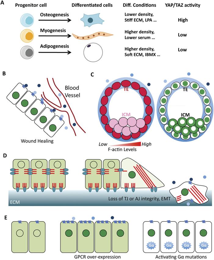
Figure 4. YAP/TAZ activity under different
physiological and pathological conditions. (A)
YAP/TAZ activity in cell differentiation. YAP/
TAZ activity is regulated by cell density, ECM,
and GPCR signaling. (B) Cells close to a wound
may experience different mechanical forces and
a higher concentration of GPCR ligands from
blood vessels. This may induce YAP/TAZ ac-
tivity and promote wound healing or tissue
regeneration. (C) In a blastocyst, cells in the
trophectoderm (TE) and inner cell mass (ICM)
have distinct YAP localization and F-actin/
tension distribution. (D) Disruption of cellular
junctions may induce mechanical stress and
result in changes in cell morphology, which can
enrich YAP/TAZ in nuclei under these condi-
tions. Activated YAP/TAZ may promote epi-
thelial–mesenchymal transition (EMT) and cell
migration. Cells that have escaped from epithe-
lium may also encounter more GPCR ligands.
Red lines represent actin filaments. (E) Aberrant
GPCR signaling may activate YAP/TAZ. Ele-
vated GPCR expression or activating Ga muta-
tions may induce YAP/TAZ nuclei localization
and activation and result in hyperproliferation
that may contribute to cancer development.
YAP/TAZ localization is represented by green
in B–E.
higher cell density and lower serum concentration— activity may facilitate wound healing processes (Fig. 4B).
conditions that favor YAP/TAZ inactivation. Altogether, For instance, thrombin is increased during blood clot-
a variety of microenvironmental signals may determine ting, a process directly associated with wounds, and
cell differentiation by regulating YAP/TAZ activity. may promote wound healing by activating YAP/TAZ.
The Hippo pathway is also involved in tissue regener- Moreover, LPA has been shown to facilitate wound closure
ation. In the intestines of mice, YAP protein levels were in a mouse model (Balazs et al. 2001). Therefore, Hippo
dramatically increased following dextran sulfate sodium pathway regulation is important for wound healing and
(DSS)-induced injury, and the damaged intestinal epi- regeneration.
thelium underwent regeneration; however, inactivation YAP plays important roles in early embryonic develop-
of YAP severely impaired regeneration (Cai et al. 2010). ment. Systematic knockout of YAP in mice is lethal, and
Furthermore, Yki is required for tissue regeneration in the embryo stops developing at embryonic day 8.5 (E8.5)
Drosophila midgut and wing discs (Karpowicz et al. 2010; with defects in the yolk sac, vasculogenesis, chorioallan-
Ren et al. 2010a; Shaw et al. 2010; Staley and Irvine 2010; tonic fusion, and body axis elongation (Morin-Kensicki
Grusche et al. 2011; Sun and Irvine 2011). Interestingly, et al. 2006). In a normal blastocyst, YAP shows nuclear
TEAD1 Y406H (tyrosine-to-histidine) mutation abolishes localization in the trophectoderm (TE) and cytoplasmic
YAP–TEAD interaction (Zhao et al. 2008; L Chen et al. localization in the inner cell mass (ICM), and this distinct
2010; Li et al. 2010), and this mutation is associated with distribution of YAP is important for lineage specification
Sveinsson chorioretinal atrophy (a human degeneration in the preimplantation mouse embryo (Fig. 4C; Nishioka
disease with absence of retinal pigment epithelium and et al. 2009). However, the underlying mechanism for this
additional retinal structures) (Jonasson et al. 2007), sug- patterned YAP localization is not clear. In blastocysts, the
gesting the importance of YAP/TEAD transcriptional ac- F-actin and myosin staining is strong in TE but not de-
tivity in tissue growth and homeostasis. Moreover, tissue tectable in the ICM, suggesting that contractive actin
damage may result in changes of mechanical or biochem- bundles are abundant in the TE (Slager et al. 1992). The
ical environments, and a local increase of YAP/TAZ distribution of F-actin is in nice correlation with YAP
GENES & DEVELOPMENT 363Downloaded from genesdev.cshlp.org on November 1, 2015 - Published by Cold Spring Harbor Laboratory Press Yu and Guan localization in blastocysts (Fig. 4C; Nishioka et al. 2007; Zhang et al. 2011; von Gise et al. 2012). Similarly, 2009). It is possible that the actin cytoskeleton is the inactivation of upstream core components of the Hippo determinant of YAP localization in this context. The pathway leads to tumor development in mice (St John distribution of F-actin in blastocysts might be regulated et al. 1999; Lee et al. 2008, 2010; Zhou et al. 2009, 2011; by multiple means: (1) Cells in the ICM are not polarized, Lu et al. 2010; Song et al. 2010; Nishio et al. 2012). whereas cells in the TE are polarized and may have more Moreover, genetic inactivation of NF2, a well-known F-actin and tension. (2) Like in cells growing in high den- tumor suppressor that acts upstream of the Hippo path- sity, cells in the ICM contact one another in all directions way (Rouleau et al. 1993; Ruttledge et al. 1994), resulted and may have less F-actin and tension. (3) The outside in tissue overgrowth in Drosophila and cancers in mice of the TE is exposed, and GPCRs that are expressed on (Hamaratoglu et al. 2006; Benhamouche et al. 2010; the surface might be influenced by maternal hormones, Zhang et al. 2010), and the phenotype was blocked by whereas the access of these signals to ICM cells is down-regulation of YAP (Zhang et al. 2010). YAP and TAZ prevented by the epithelium (TE). It would be interesting induced an epithelial–mesenchymal transition (EMT), a to further investigate the relationship between F-actin phenomenon crucial for the initiation of cancer metasta- and YAP localization and the upstream cues during early sis (Overholtzer et al. 2006; Lei et al. 2008; Thiery et al. embryogenesis. 2009). The role of YAP in promoting cancer metastasis The Hippo pathway is widely recognized as a signaling has also recently been demonstrated in mice (Chen et al. pathway that regulates organ size. In Drosophila, loss- 2012; Lamar et al. 2012). In addition, TAZ has been shown of-function mutants of Hpo, Sav, Wts, and Mob all lead to sustain self-renewal and induce tumor initiation of to significant tissue outgrowth, as indicated by enlarged breast cancer stem cells (Cordenonsi et al. 2011). These eyes, wings, or other appendages (Justice et al. 1995; Xu data suggest an important function of the Hippo pathway et al. 1995; Kango-Singh et al. 2002; Tapon et al. 2002; in cancer development. Harvey et al. 2003; Jia et al. 2003; Pantalacci et al. 2003; The connection between the Hippo pathway, mechan- Udan et al. 2003; Wu et al. 2003; Lai et al. 2005). Over- ical forces, and GPCR signaling also provides new in- expression of Yki also revealed similar tissue outgrowth sights in cancer development. Following disruption of phenotypes (Huang et al. 2005). The function of the Hippo cell–cell junctions and loss of cell polarity, cells may form pathway in organ size control is conserved in mammals. extensive focal adhesions with the ECM and thereby For instance, tissue-specific overexpression of YAP in the generate cellular tension, and these cells may further- mouse liver or heart resulted in a dramatic increase of more encounter stimulation by GPCR ligands (Fig. 4D). liver or heart size (Camargo et al. 2007; Dong et al. 2007; Activation of YAP/TAZ under these conditions may von Gise et al. 2012). Consistently, knockout of MST1/2 facilitate cell proliferation and cell migration or induce or Sav in the liver or heart also induced organ size (Zhou EMT. GPCRs are crucial players in cancer development, et al. 2009; Lee et al. 2010; Lu et al. 2010; Song et al. 2010; and dysregulated GPCR signaling has been identified in Heallen et al. 2011). Recently, a connection between the many types of human cancers (Dorsam and Gutkind Hippo pathway and the PI3K–TOR pathway has been 2007). GPCR signaling can contribute to cancer in a vari- demonstrated (Strassburger et al. 2012; Tumaneng et al. ety of ways (Fig. 4E). Increased production of some GPCR 2012b; Ye et al. 2012). This is significant because TOR ligands may promote cancer development; for instance, signaling is important in cell growth; thus, the Hippo overexpression of autotaxin (an enzyme critical for LPA pathway may control organ size in part by modulating synthesis) in mouse mammary glands has been shown to cell growth (Fig. 1; Tumaneng et al. 2012a). It is possible induce breast cancer (Liu et al. 2009). Up-regulated ex- that multiple upstream signals work together to modu- pression of GPCRs may activate intracellular signaling late the Hippo pathway to determine the final organ size. automatically, as indicated by high PAR1 expression in The mechanical forces may correlate with organ size and high-grade breast cancer patients (Hernandez et al. 2009). convey the organ size information to the Hippo pathway. Activating mutations of GPCRs have been identified in Similarly, tissue-specific GPCRs may also play a role. different type of cancers, such as the metabotropic gluta- For example, acetylcholine (a ligand for Gaq/11-coupled mate receptor mutations in melanoma and thyroid- receptor) signaling was required for salivary organogen- stimulating hormone receptor in thyroid carcinomas esis (Knox et al. 2010). Knockout of luteinizing hormone (Paschke and Ludgate 1997; Prickett et al. 2011). Activat- receptor resulted in dramatically smaller testis (Zhang ing mutations of Ga are also present in different types of et al. 2001), indicating a possible role of GPCR signaling cancers: Gaq/11-activating mutations have been iden- in tissue growth and organ size control. tified in >80% of uveal melanomas (Van Raamsdonk Hippo pathway members have been identified in the et al. 2009, 2010); Gai2a and Gas mutations have been search for tumor suppressor genes in Drosophila. Kinases identified in ovarian and endocrine tumors (Lania et al. in the Hippo pathway are generally tumor suppressors, 2001). In addition, aberrant activity of Rho GTPases whereas Yki/YAP/TAZ have oncoprotein-like functions. and GPCR regulatory molecules, such as GPCR-related Elevated YAP or TAZ expression and nuclear localization kinases, may also contribute to cancer by regulating are frequently observed in human cancers (Zhao et al. YAP/TAZ activity (Sahai and Marshall 2002; Metaye 2007; Chan et al. 2008; Steinhardt et al. 2008; Fernandez et al. 2005). Altogether, activation of YAP/TAZ by dysreg- et al. 2009; Xu et al. 2009). YAP transgenic mice display ulated GPCR signaling may play an important role in the hyperplasia and tumors (Camargo et al. 2007; Dong et al. development of human cancers. 364 GENES & DEVELOPMENT
Downloaded from genesdev.cshlp.org on November 1, 2015 - Published by Cold Spring Harbor Laboratory Press

                                                                                                              Hippo pathway regulation

Concluding remarks                                                   Boedigheimer MJ, Nguyen KP, Bryant PJ. 1997. Expanded func-
                                                                        tions in the apical cell domain to regulate the growth rate of
The Hippo pathway plays critical roles in normal phys-                  imaginal discs. Dev Genet 20: 103–110.
iology and pathogenesis, and pharmacological interven-               Boggiano JC, Vanderzalm PJ, Fehon RG. 2011. Tao-1 phosphor-
tions of this pathway have diverse clinical implications;               ylates Hippo/MST kinases to regulate the Hippo–Salvador–
therefore, it is very important to understand how this                  Warts tumor suppressor pathway. Dev Cell 21: 888–895.
pathway is regulated. The dynamics of the actin cyto-                Bretscher A, Edwards K, Fehon RG. 2002. ERM proteins and
skeleton appears to act as a focal point of different signals           merlin: Integrators at the cell cortex. Nat Rev Mol Cell Biol
pathways to modulate YAP/TAZ activity in either a Lats-                 3: 586–599.
                                                                     Cai J, Zhang N, Zheng Y, de Wilde RF, Maitra A, Pan D. 2010.
dependent or Lats-independent mechanism (Fig. 3D).
                                                                        The Hippo signaling pathway restricts the oncogenic poten-
However, several critical questions regarding the regu-                 tial of an intestinal regeneration program. Genes Dev 24:
lation of the Hippo pathway by these upstream regula-                   2383–2388.
tors still wait to be answered. How are these regulators,            Callus BA, Verhagen AM, Vaux DL. 2006. Association of
especially F-actin or cellular tension, sensed by the core              mammalian sterile twenty kinases, Mst1 and Mst2, with
components of the Hippo pathway? To what extent does                    hSalvador via C-terminal coiled-coil domains, leads to its
the Hippo pathway contribute to the physiological and                   stabilization and phosphorylation. FEBS J 273: 4264–4276.
pathological functions of these upstream regulators? Are             Camargo FD, Gokhale S, Johnnidis JB, Fu D, Bell GW, Jaenisch
one or more of these regulators the determining factors                 R, Brummelkamp TR. 2007. YAP1 increases organ size and
for organ size control? Answers to these challenging ques-              expands undifferentiated progenitor cells. Curr Biol 17: 2054–
                                                                        2060.
tions will advance our understanding of the regulation and
                                                                     Cao X, Pfaff SL, Gage FH. 2008. YAP regulates neural progenitor
function of the Hippo pathway.                                          cell number via the TEA domain transcription factor. Genes
                                                                        Dev 22: 3320–3334.
Acknowledgments                                                      Chan EH, Nousiainen M, Chalamalasetty RB, Schafer A, Nigg
We apologize for those primary works that are not cited due to          EA, Sillje HH. 2005. The Ste20-like kinase Mst2 activates
the scope of this review and space constraints. We thank Drs.           the human large tumor suppressor kinase Lats1. Oncogene
Jenna L. Jewell and Carsten G. Hansen for critical reading of this      24: 2076–2086.
manuscript. Research in the laboratory of K.L.G. is supported by     Chan SW, Lim CJ, Guo K, Ng CP, Lee I, Hunziker W, Zeng Q,
grants from the Nationa Institutes of Health and California             Hong W. 2008. A role for TAZ in migration, invasion, and
Institute for Regenerative Medicine.                                    tumorigenesis of breast cancer cells. Cancer Res 68: 2592–
                                                                        2598.
                                                                     Chan SW, Lim CJ, Chong YF, Pobbati AV, Huang C, Hong W.
References
                                                                        2011. Hippo pathway-independent restriction of TAZ and
Alarcon C, Zaromytidou AI, Xi Q, Gao S, Yu J, Fujisawa S, Barlas        YAP by angiomotin. J Biol Chem 286: 7018–7026.
   A, Miller AN, Manova-Todorova K, Macias MJ, et al. 2009.          Chen CL, Gajewski KM, Hamaratoglu F, Bossuyt W, Sansores-
   Nuclear CDKs drive Smad transcriptional activation and               Garcia L, Tao C, Halder G. 2010. The apical–basal cell
   turnover in BMP and TGF-b pathways. Cell 139: 757–769.               polarity determinant Crumbs regulates Hippo signaling in
Azzolin L, Zanconato F, Bresolin S, Forcato M, Basso G, Bicciato        Drosophila. Proc Natl Acad Sci 107: 15810–15815.
   S, Cordenonsi M, Piccolo S. 2012. Role of TAZ as mediator of      Chen L, Chan SW, Zhang X, Walsh M, Lim CJ, Hong W, Song H.
   Wnt signaling. Cell 151: 1443–1456.                                  2010. Structural basis of YAP recognition by TEAD4 in the
Balazs L, Okolicany J, Ferrebee M, Tolley B, Tigyi G. 2001.             hippo pathway. Genes Dev 24: 290–300.
   Topical application of the phospholipid growth factor lyso-       Chen D, Sun Y, Wei Y, Zhang P, Rezaeian AH, Teruya-Feldstein
   phosphatidic acid promotes wound healing in vivo. Am J               J, Gupta S, Liang H, Lin HK, Hung MC, et al. 2012. LIFR is
   Physiol Regul Integr Comp Physiol 280: R466–R472.                    a breast cancer metastasis suppressor upstream of the
Bao Y, Nakagawa K, Yang Z, Ikeda M, Withanage K, Ishigami-              Hippo–YAP pathway and a prognostic marker. Nat Med 18:
   Yuasa M, Okuno Y, Hata S, Nishina H, Hata Y. 2011. A cell-           1511–1517.
   based assay to screen stimulators of the Hippo pathway reveals    Cho E, Irvine KD. 2004. Action of fat, four-jointed, dachsous and
   the inhibitory effect of dobutamine on the YAP-dependent             dachs in distal-to-proximal wing signaling. Development
   gene transcription. J Biochem 150: 199–208.                          131: 4489–4500.
Baum B, Georgiou M. 2011. Dynamics of adherens junctions in          Cho E, Feng Y, Rauskolb C, Maitra S, Fehon R, Irvine KD. 2006.
   epithelial establishment, maintenance, and remodeling. J Cell        Delineation of a Fat tumor suppressor pathway. Nat Genet
   Biol 192: 907–917.                                                   38: 1142–1150.
Baumgartner R, Poernbacher I, Buser N, Hafen E, Stocker H.           Clark K, Langeslag M, Figdor CG, van Leeuwen FN. 2007.
   2010. The WW domain protein Kibra acts upstream of Hippo             Myosin II and mechanotransduction: A balancing act. Trends
   in Drosophila. Dev Cell 18: 309–316.                                 Cell Biol 17: 178–186.
Benhamouche S, Curto M, Saotome I, Gladden AB, Liu CH,               Cordenonsi M, Zanconato F, Azzolin L, Forcato M, Rosato A,
   Giovannini M, McClatchey AI. 2010. Nf2/Merlin controls               Frasson C, Inui M, Montagner M, Parenti AR, Poletti A, et al.
   progenitor homeostasis and tumorigenesis in the liver. Genes         2011. The Hippo transducer TAZ confers cancer stem cell-
   Dev 24: 1718–1730.                                                   related traits on breast cancer cells. Cell 147: 759–772.
Bennett FC, Harvey KF. 2006. Fat cadherin modulates organ size       Das Thakur M, Feng Y, Jagannathan R, Seppa MJ, Skeath JB,
   in Drosophila via the Salvador/Warts/Hippo signaling path-           Longmore GD. 2010. Ajuba LIM proteins are negative regula-
   way. Curr Biol 16: 2101–2110.                                        tors of the Hippo signaling pathway. Curr Biol 20: 657–662.
Boedigheimer M, Laughon A. 1993. Expanded: A gene involved           Densham RM, O’Neill E, Munro J, Konig I, Anderson K,
   in the control of cell proliferation in imaginal discs. De-          Kolch W, Olson MF. 2009. MST kinases monitor actin
   velopment 118: 1291–1301.                                            cytoskeletal integrity and signal via c-Jun N-terminal kinase

                                                                                                    GENES & DEVELOPMENT           365
You can also read