Dynamics in Fip1 regulate eukaryotic mRNA 3 end processing
←
→
Page content transcription
If your browser does not render page correctly, please read the page content below
Downloaded from genesdev.cshlp.org on November 4, 2021 - Published by Cold Spring Harbor Laboratory Press

Dynamics in Fip1 regulate eukaryotic
mRNA 3′ end processing
Ananthanarayanan Kumar,1,2 Conny W.H. Yu,2 Juan B. Rodríguez-Molina, Xiao-Han Li,
Stefan M.V. Freund, and Lori A Passmore
MRC Laboratory of Molecular Biology, Cambridge CB2 0QH, United Kingdom

Cleavage and polyadenylation factor (CPF/CPSF) is a multiprotein complex essential for mRNA 3′ end processing in
eukaryotes. It contains an endonuclease that cleaves pre-mRNAs, and a polymerase that adds a poly(A) tail onto the
cleaved 3′ end. Several CPF subunits, including Fip1, contain intrinsically disordered regions (IDRs). IDRs within
multiprotein complexes can be flexible, or can become ordered upon interaction with binding partners. Here, we
show that yeast Fip1 anchors the poly(A) polymerase Pap1 onto CPF via an interaction with zinc finger 4 of another
CPF subunit, Yth1. We also reconstitute a fully recombinant 850-kDa CPF. By incorporating selectively labeled Fip1
into recombinant CPF, we could study the dynamics of Fip1 within the megadalton complex using nuclear magnetic
resonance (NMR) spectroscopy. This reveals that a Fip1 IDR that connects the Yth1- and Pap1-binding sites remains
highly dynamic within CPF. Together, our data suggest that Fip1 dynamics within the 3′ end processing machinery
are required to coordinate cleavage and polyadenylation.
[Keywords: CPF; CPSF; RNA-binding protein; dynamics; mRNA processing; polyadenylation]
Supplemental material is available for this article.
Received May 16, 2021; revised version accepted September 9, 2021.

Protein-coding genes in eukaryotes are transcribed by RNA                   (Kumar et al. 2019). In yeast, CPF is comprised of three
polymerase II (Pol II) into precursor messenger RNAs (pre-                  enzymatic modules: a five-subunit polymerase module
mRNAs). Pre-mRNAs are modified by the addition of a 7-                      containing the poly(A) polymerase Pap1, a three-subunit
methylguanosine cap at the 5′ end, splicing, and 3′ end pro-                nuclease module containing the endonuclease Ysh1, and
cessing (Hocine et al. 2010). The 3′ end of an mRNA is                      a six-subunit phosphatase module that includes two pro-
formed by a two-step reaction involving endonucleolytic                     tein phosphatases (Glc7 and Swd2) that regulate transcrip-
cleavage at a specific site and the addition of a stretch of                tion (Casañal et al. 2017). Most CPF subunits are
polyadenosines [a poly(A) tail] to the new free 3′ hydroxyl                 conserved across all eukaryotes.
(Zhao et al. 1999). Poly(A) tails are essential for export of                  Insights into the molecular basis of polyadenylation
mature mRNAs into the cytoplasm, for their subsequent                       have been obtained through structural and biochemical
translation into proteins, and in determining mRNA half-                    studies. For example, a crystal structure of Pap1 in com-
life. Defects in 3′ end processing are associated with human                plex with ATP, poly(A) RNA, and Mg2+ confirmed that a
diseases including cancer, β-thalassemia, and spinal mus-                   two-metal ion-dependent nucleotidyl transfer mecha-
cular atrophy (Curinha et al. 2014). Understanding the                      nism is used in poly(A) tail synthesis (Balbo and Bohm
mechanistic basis of 3′ end processing and how cleavage                     2007). Together with kinetic studies, this structure pro-
and polyadenylation are coordinated with other mRNA                         vided a molecular basis for nucleotide specificity. Pap1
processing steps is therefore of great importance.                          is assembled into the polymerase module along with
   Eukaryotic 3′ end processing is carried out by a set of                  Cft1, Pfs2, the zinc finger-containing protein Yth1, and
conserved multiprotein complexes that includes the                          the low-sequence-complexity protein Fip1 (Casañal
cleavage and polyadenylation factor (CPF in yeast or                        et al. 2017). A similar mammalian polymerase module
CPSF in humans) and accessory cleavage factors (CF IA                       (mPSF) is sufficient for specific and efficient mRNA poly-
and CF IB in yeast or CF Im, CF IIm, and CstF in humans)                    adenylation in vitro (Schönemann et al. 2014). Cryo-elec-
                                                                            tron microscopy (cryoEM) structures of the polymerase
                                                                            modules from yeast and humans revealed an extensive
1
 Present address: Department of Molecular, Cellular, and Developmental      network of interactions between Pfs2 and Cft1 (WDR33
Biology, Yale University, New Haven, CT 06511, USA.
2
                                                                            and CPSF160 in humans), which function as a scaffold
 These authors contributed equally to this work.
Corresponding authors: passmore@mrc-lmb.cam.ac.uk, smvf@mrc-lmb.
                                                                            for assembly of the other subunits (Casañal et al. 2017;
cam.ac.uk
Article published online ahead of print. Article and publication date are
online at http://www.genesdev.org/cgi/doi/10.1101/gad.348671.121. Free-     © 2021 Kumar et al. This article, published in Genes & Development, is
ly available online through the Genes & Development Open Access             available under a Creative Commons License (Attribution 4.0 Internation-
option.                                                                     al), as described at http://creativecommons.org/licenses/by/4.0/.

                  GENES & DEVELOPMENT 35:1–17 Published by Cold Spring Harbor Laboratory Press; ISSN 0890-9369/21; www.genesdev.org               1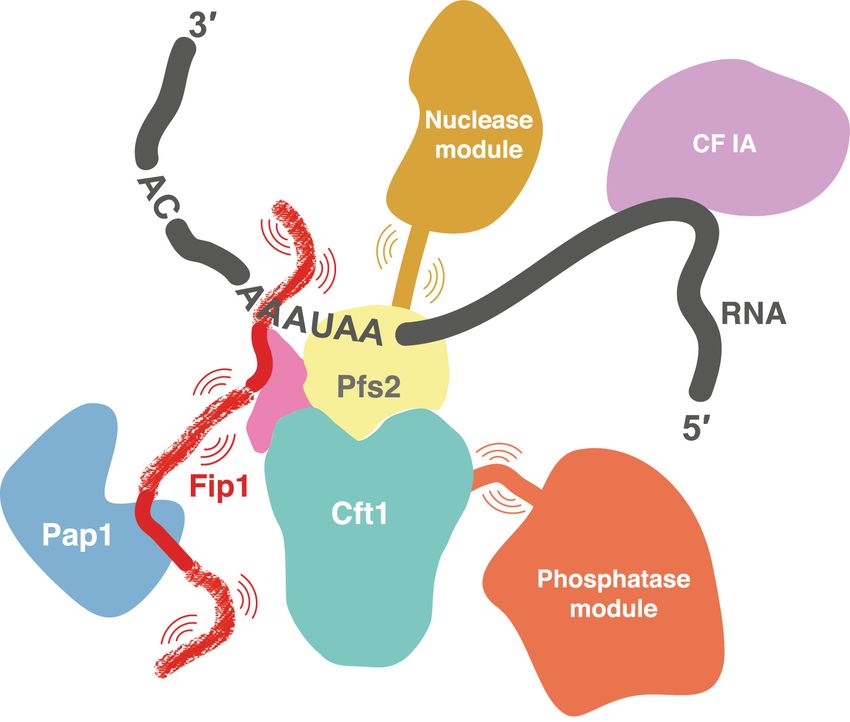
Downloaded from genesdev.cshlp.org on November 4, 2021 - Published by Cold Spring Harbor Laboratory Press
Kumar et al.
Clerici et al. 2017; Sun et al. 2018). The structures also Results
provided a rationale for how WDR33 and CPSF30 (Yth1
Yth1 binds Fip1, which in turn tethers Pap1 to the
in yeast) bind specific sequences in RNA (Clerici et al.
polymerase module
2018; Sun et al. 2018).
Fip1 and Pap1 interact directly, and a crystal structure To investigate how Fip1 and Pap1 interact with other CPF
of yeast Pap1 bound to residues 80–105 of Fip1 provided subunits, we first studied the five-subunit polymerase
the molecular details of their interaction (Meinke et al. module purified from a baculovirus-mediated insect cell
2008). Fip1 has also been reported to interact with other overexpression system as previously described (Casañal
CPF and CF IA components such as Pta1, Yth1, and et al. 2017). We used cryoEM to image this five-subunit
Rna14 (Preker et al. 1995; Barabino et al. 2000; Ohnacker complex (Supplemental Fig. S1). Selected 2D class averag-
et al. 2000; Tacahashi et al. 2003; Ghazy et al. 2009; es show a central structure that resembles the Cft1-Pfs2-
Casañal et al. 2017) but neither Fip1 nor Pap1 was visible Yth1 scaffold identified previously in the four-subunit
in cryoEM studies. Fip1 has an N-terminal acidic stretch complex lacking Pap1 (Fig. 1A; Casañal et al. 2017). In ad-
and a C-terminal Pro-rich region (Preker et al. 1995; Kauf- dition, a horseshoe-shaped structure was present in the
mann et al. 2004). These overall properties of Fip1 are con- 2D class averages at several different positions relative
served, but human FIP1 (hFIP1) is longer and additionally to Cft1-Pfs2-Yth1. This extra density resembles a 2D pro-
contains C-terminal Arg-rich and Arg/Asp-rich domains jection of the crystal structure of Pap1 (Bard et al. 2000),
compared with the yeast ortholog (Kaufmann et al. suggesting that Pap1 is positioned flexibly with respect
2004). Biochemical and genetic experiments led to the hy- to the Cft1-Pfs2-Yth1 scaffold. The lack of a defined
pothesis that Fip1 is an unstructured protein that acts as a position for Pap1 precluded high-resolution structure
flexible linker between Pap1 and CPF (Meinke et al. 2008; determination.
Ezeokonkwo et al. 2011). Very recently, a crystal struc- Next, to gain further insight into the architecture of the
ture of human CPSF30 bound to hFIP1 was reported polymerase module, we investigated subunit interactions
(Hamilton and Tong 2020). This showed that CPSF30 using pull-down assays. Using a StrepII tag on Pfs2, all five
binds two copies of hFIP1 with its zinc fingers 4 and subunits were copurified (Fig. 1B). When we removed Pap1
5. However, Fip1 structure has only been studied in isola- from the complex, the four remaining subunits were still
tion or in complex with Pap1 or Yth1; whether Fip1 re- associated. However, removal of Fip1 resulted in concom-
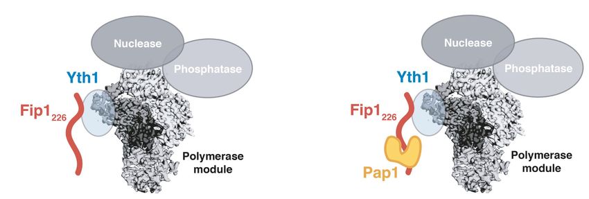
mains dynamic in the context of the entire 14-subunit itant loss of Pap1 from the complex (Fig. 1B). Thus, Fip1 is
CPF remains unclear. essential for Pap1 association with the polymerase mod-
The nuclease module subunits are flexibly positioned ule and, if it is flexible, it may contribute to the variable
with respect to the polymerase module (Hill et al. 2019; positioning of Pap1 in EM images (Fig. 1A).
Zhang et al. 2020), but they are hypothesized to become Fip1 is hypothesized to bind Yth1, which contains five
fixed upon CPF activation (Sun et al. 2020). It is likely zinc fingers. The N-terminal half of Yth1, including zinc
that the enzymes of CPF are regulated at several levels: fingers 1 and 2, interacts with Cft1 and Pfs2 (Casañal
First the nuclease must be activated. Second, the nuclease et al. 2017), zinc fingers 2 and 3 interact with RNA (Clerici
must be inactivated after cleavage has occurred. Finally, et al. 2018; Sun et al. 2018), and, in humans, zinc fingers 4
the RNA must be transferred to Pap1’s active site to allow and 5 interact with hFIP1 (Hamilton and Tong 2020). The
the poly(A) tail to be synthesized to the correct length. C-terminal half of Yth1 is not visible in cryoEM maps,
Conformational changes associated with regulation and suggesting that it may be flexible. To test whether the
function of multiprotein complexes frequently involve C-terminal region is required for interaction with other
dynamic IDRs (Fuxreiter et al. 2014; van der Lee et al. polymerase module subunits, we carried out pull-down
2014); however, it remains unknown whether CPF has dif- assays with versions of Yth1 containing C-terminal dele-
ferent conformational states and whether flexible IDRs tions. Deletion of the C-terminal half of Yth1, spanning
contribute to CPF function. zinc finger 4, zinc finger 5, and a C-terminal helical region,
Here, we aimed to understand the function and dynam- resulted in a complete loss of Fip1 and Pap1 association
ics of Fip1 using biochemical reconstitution, biophysical with the complex. Deletion of zinc finger 5 and the C-ter-
experiments, and NMR spectroscopy. We found that iso- minal helical region reduced, but did not abolish, the asso-
lated Fip1 is an intrinsically disordered protein in solu- ciation of Fip1 and Pap1 from the Pfs2 pull-downs.
tion, with defined binding sites for Yth1 and Pap1 that Deletion of zinc finger 4 resulted in loss of Pap1, but a
are connected by a low-complexity sequence. To fully weak band corresponding to a protein the size of Fip1
characterize Fip1 as an essential component of the CPF, was still present (Fig. 1B). There are a number of additional
we reconstituted a recombinant 850-kDa CPF. This al- bands present in this pull-down that may represent Pfs2
lowed us to incorporate an isotopically labeled Fip1 into degradation products. This suggests that deletion of
CPF for NMR studies, in which we show that, with the ex- Yth1 zinc finger 4 compromises the stability of the com-
ception of the Yth1- and Pap1-binding sites, Fip1 remains plex. Given that Pap1 is absent when zinc finger 4 is delet-
dynamic and largely disordered within CPF. Moreover, ed, the 50-kDa band may be a degradation product of Pfs2.
deletion of a highly flexible region in Fip1 impairs CPF nu- Based on these data, we hypothesize that Yth1 zinc finger
clease activity. Together, our data reveal that Fip1 dynam- 4 is the major binding site for Fip1 and is required for Fip1
ics are important in regulating eukaryotic mRNA 3′ end (and Pap1) incorporation into the fully recombinant poly-
processing. merase module, in agreement with a previously proposed
2 GENES & DEVELOPMENT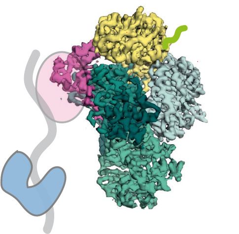
Downloaded from genesdev.cshlp.org on November 4, 2021 - Published by Cold Spring Harbor Laboratory Press

                                                                                                          Fip1 is dynamic within intact CPF

      A                                                    B

      C

Figure 1. Fip1 binds Yth1 and tethers Pap1 to the polymerase module. (A) Cartoon representation (top left) and selected 2D class averages
from cryoEM (right) of the polymerase module. The 2D averages show a central structure corresponding to the Cft1-Pfs2-Yth1 subunits,
and an additional horseshoe-shaped density (blue arrowheads). (Bottom left) Similarity to a 2D projection of the crystal structure of Pap1
(PDB 3C66) suggests that the horseshoe-shaped density is Pap1. (SII) StrepII. (B) SDS-PAGE of pull-down assays using StrepII-tagged (SII)
Pfs2 reveals interactions within the polymerase module. (Right) Domain diagrams of Yth1 indicate the constructs used. Fip1 is required for
Pap1 interaction. When the interaction between Yth1 and Fip1 is compromised, Fip1 is not pulled down and therefore Pap1 is also absent.
Yth1 subunits in the WT and “no Pap1” complexes contain a His tag, whereas Yth1 in the “no Fip1” and Yth1 truncation complexes do
not contain a His tag. With Yth1ΔZF4, protein degradation (potentially from Pfs2) is evident. (C) Histogram showing chemical shift per-
turbations (CSPs) in Yth1ZF45C (residues 118–208) spectra upon Fip1226 binding. Yth1ZF45C and Fip1226 were mixed in an equimolar ratio
to a final concentration of 75 µM. Gray circles indicate peaks that showed exchange broadening upon Fip1 binding. Most of the peaks that
are perturbed are in zinc finger 4. Homologous residues contributing >50 Å2 buried surface area in the human FIP1-CPSF30 structure
(Hamilton and Tong 2020) are highlighted with blue or purple circles. In that structure, two hFIP1 molecules (hFIP1-A and hFIP1-B)
are bound to one CPSF30.

role for this zinc finger in Fip1 binding (Tacahashi et al.             nM ± 40 nM (Supplemental Fig. S2G). Fip1226 binds more
2003; Hamilton and Tong 2020). Zinc finger 5 may con-                   tightly to the Yth1ZF45C construct, in agreement with
tain a low-affinity binding site for Fip1.                              zinc finger 5 also making contributions to the binding
   To characterize the Yth1-Fip1 interaction, we per-                   site (Supplemental Fig. S2H,I). We found that zinc finger
formed NMR analysis of two Yth1 constructs: the entire                  4 in Yth1ZF45C loses its structure upon incubation with
C-terminal half (Yth1ZF45C; residues 118–208) or zinc fin-              EDTA, suggesting that the zinc ions are exposed and sus-
ger 4 on its own (Yth1ZF4; residues 118–161) (Supplemen-                ceptible to metal chelation in the free protein (Supple-
tal Fig. S2A–C). We assigned backbone resonances of                     mental Fig. S2J). Interestingly, addition of EDTA results
Yth1ZF45C and mapped chemical shift perturbations                       in minimal changes in the Yth1ZF45C spectra in the pres-
upon addition of a Fip1 construct containing residues                   ence Fip1226 (Supplemental Fig. S2K). Therefore, Fip1 ren-
1–226 (Fip1226) (Fig. 1C; Supplemental Fig. S2D). (Details              ders the Yth1ZF45C zinc fingers resistant to loss of metal
on the choice of Fip1226 construct are described in the                 coordination. Taken together, Fip1 directly interacts
next section.) Addition of Fip1226 resulted in substantial              with and stabilizes Yth1.
chemical shift perturbations and line broadening. The ma-
jority of changes (19 out of 25 residues experiencing chem-
                                                                        Fip1 is largely disordered in solution
ical shift perturbation or line broadening) are located
within residues in zinc finger 4, with a similar pattern in             Next, we examined the structure of Fip1. We first per-
both Yth1ZF45C and Yth1ZF4 spectra. The remaining                       formed bioinformatics analyses on the sequence features
changes are located in zinc finger 5. Thus, Yth1 zinc finger            of the full-length, 327-amino-acid protein (Fig. 2A). Over-
4 is the primary interaction site for Fip1.                             all, >75% of Fip1 (∼250 residues) is predicted to be highly
   There are apparent domain boundaries on either side of               disordered, including an N-terminal serine-rich acidic re-
Yth1 zinc finger 4, but this region contains very little sec-           gion (residues 1–60), a central low-complexity region
ondary structure (Supplemental Fig. S2E,F). We confirmed                (LCR) rich in serine and threonine (residues 110–180),
a direct interaction of Yth1ZF4 and Fip1226 using isother-              and a C-terminal region with no net charge that is enriched
mal calorimetry, with a measured binding affinity of 240                in asparagine, proline, and phenylalanine (residues

                                                                                                         GENES & DEVELOPMENT             3
Downloaded from genesdev.cshlp.org on November 4, 2021 - Published by Cold Spring Harbor Laboratory Press

Kumar et al.

       A

       B                                     D

       C

Figure 2. Fip1 residues 1–226 are sufficient for reconstitution of the polymerase module. (A) Domain diagram of Fip1 showing low-com-
plexity regions (LCRs), Pap1-binding site, and Yth1-binding site (top), and bioinformatic analysis of the Fip1 sequence (bottom). In the plot,
the purple line indicates IUPRED2 disorder prediction score. Residues with an IUPred value >0.5 (gray dotted line) are classified as having
high propensity for being intrinsically disordered. Gray bars correspond to fraction of charged residues (FCR) over a sliding window of 10
residues. Red and blue bars represent net negative and positive charge per residue (NCPR), respectively, over the same window size. The
colored circles highlight the distribution of phenylalanine (black), asparagine (red), and proline (blue) residues. (B) Schematic showing con-
struct design of Fip1226 (residues 1–226; top) and SDS-PAGE of pull-down assays of a polymerase module using StrepII-tagged (SII) Pfs2
(bottom). (C ) In vitro polyadenylation assay with a recombinant polymerase module containing Fip1226. A 42-nucleotide CYC1 3′
UTR with a 5′ -FAM label was used as a substrate in the polyadenylation assay, and the reaction products were visualized on a denaturing
urea polyacrylamide gel. This gel is representative of experiments performed twice. (D) 1H,15N 2D HSQC of 75 µM Fip1226 shown with the
assignment of backbone resonances. The inset highlights a crowded region of the spectra. The bottom panels show examples of peaks. I171
and G194 show line broadening. BEST 1H,15N-TROSY spectra were acquired with four scans and a recycle delay of 400 msec, giving a final
spectral resolution of 0.9 Hz per points in the indirect dimension and an experimental time of 20 min.

243–327). This is consistent with previous evidence that                  ing the previously described Pap1-binding site (residues
isolated Fip1 is largely unfolded (Meinke et al. 2008; Ezeo-              80–105) is not highly conserved (Supplemental Fig. S3A).
konkwo et al. 2011). Interestingly, the only region that is               This is consistent with previous reports that the molecular
predicted to have low disorder propensity (residues                       details of the interaction between Pap1 and Fip1 are not
193–215) overlaps with sequences previously identified                    important for Pap1’s activity, but physical tethering is im-
as important for Yth1 binding (residues 206–220) (Helm-                   portant (Ezeokonkwo et al. 2011).
ling et al. 2001) and is highly conserved across eukaryotes                 The C-terminal region of Fip1 is poorly conserved com-
(Supplemental Fig. S3A). The N-terminal region contain-                   pared with the N-terminal region, both in length and in

4      GENES & DEVELOPMENT
Downloaded from genesdev.cshlp.org on November 4, 2021 - Published by Cold Spring Harbor Laboratory Press
Fip1 is dynamic within intact CPF
amino acid composition (Supplemental Fig. S3B), suggest- (Fig. 3B; Supplemental Fig. S4A). First, upon incubation
ing that the C-terminal region may not be crucial to the with Yth1ZF4, major chemical shift perturbations and sub-
function of Fip1. A previous study showed that deletion stantial line broadening were observed for resonances cor-
of residues 220–327 had negligible effect on the viability responding to Fip1 residues 170–220, indicating a potential
of yeast cells and no substantial effect on mRNA polyade- interaction between this region and Yth1ZF4 (Fig. 3B, top).
nylation in vitro (Helmling et al. 2001). Since overexpres- This region had also exhibited intrinsic line broadening in
sion of isolated full-length Fip1 resulted in insoluble the absence of Yth1ZF4 (Supplemental Fig. S3D,E). There-
protein aggregates, we removed residues 227–327, which fore, we used 13C-detect CON studies using deuterated
contain the aggregation-prone Asn-rich region, to create Fip1226. Although the signal intensities were reduced in
a Fip1226 construct that is stable for in vitro characteriza- the 13C-detect experiments due to the lower gyromagnetic
tion. As a helical stretch is predicted close to the trunca- ratio of 13C, line broadening was reduced by avoiding pro-
tion point, we chose proline 226 as a natural helix ton detection. These experiments provide an additional
breaker for the new C terminus. advantage of improved chemical shift dispersion for intrin-
We coexpressed Fip1226 with the other polymerase mod- sically disordered proteins. We could then unambiguously
ule subunits in Sf9 insect cells and performed pull-down define additional signals for C-terminal residues, provid-
assays using the StrepII tag on Pfs2 (Fig. 2B). This showed ing an independent confirmation that residues 180–220
that Fip1226 is incorporated into the recombinant polymer- are involved in Yth1 binding (Supplemental Fig. S4B,C).
ase module. This complex was active in in vitro polyade- Together, these data revealed that the major Yth1-binding
nylation assays (Fig. 2C). Together, this shows that site on Fip1 is within residues 180–220.
residues 1–226 are sufficient for Fip1 incorporation into Next, upon incubation with unlabeled 66-kDa Pap1,
the polymerase module and for polyadenylation activity, chemical shift perturbations were observed for signals
confirming our prediction that residues beyond 226 on within Fip1226 residues 58–80, and signal attenuation as
Fip1 are not essential for cleavage and polyadenylation. a result of line broadening was observed for resonances
Next, we analyzed Fip1226 using NMR. Several regions within residues 66–110 (Fig. 3B, middle). In addition,
contain substantial signal attenuation due to line broad- some resonances from the N-terminal acidic region were
ening, including residues between 170 and 220 (Materials slightly perturbed upon Pap1 binding, which may be the
and Methods; Supplemental Fig. S3C,D). We assigned 187 result of charge–charge interactions. Together, our obser-
out of 226 (83%) backbone resonances (Fig. 2D). Although vations are in agreement with binding of Fip1 residues 80–
several regions have some propensity to form secondary 105 to Pap1, as observed in the crystal structure (Meinke
structure (Supplemental Fig. S3E), Fip1226 generally has et al. 2008), but also suggest that additional Fip1 residues
a narrow 1H dispersion in a 1H,15N 2D HSQC spectrum (58–110) participate in the interaction. Interestingly, pre-
(Fig. 2D), consistent with Fip1 being a largely disordered vious studies had shown that Pap1 has higher affinity for
protein in solution. full-length Fip1 than for residues 80–105 (Meinke et al.
2008), consistent with involvement of additional residues
in this interaction.
Fip1 contains independent binding sites for Yth1 and
Finally, when both Yth1ZF4 and Pap1 were added to la-
Pap1
beled Fip1226, the binding patterns of each individual pro-
IDRs can either become ordered or remain dynamic upon tein were retained (Fig. 3B, bottom). This suggests that
binding to other subunits in a multiprotein complex (Fux- Fip1 contains two independent binding sites: one for
reiter et al. 2014; van der Lee et al. 2014). To understand Yth1 and one for Pap1. Interestingly, the central LCR
the conformational state of Fip1 and its contribution to was not involved in either of these interactions. Most of
CPF function, we investigated the dynamics of Fip1 the central LCR remains unperturbed, suggesting that
when bound to other subunits. We first sought to identify this region is still largely disordered and highly dynamic,
the interaction sites for polymerase module subunits even when Fip1 is bound to Pap1 and Yth1.
within Fip1 by making deletions in Fip1 and performing
pull-down assays using the StrepII tag on Pfs2 (Fig. 3A).
Reconstitution of a fully recombinant CPF
Residues 80–105 had previously been shown to mediate
Fip1 interaction with Pap1 (Meinke et al. 2008) and there- Residues 110–170 within the central LCR of Fip1 remain
fore we did not assess that region in pull-down assays. We dynamic in the presence of Yth1ZF4 and Pap1, raising the
found that Fip1 residues 190–220 are required for Fip1 (and possibility that they could also be dynamic in the context
Pap1) interaction with the polymerase module. In con- of the full CPF complex. To investigate this and dissect
trast, deletion of the N-terminal acidic region (residues the structural nature of Fip1 in CPF, we established a strat-
1–60) or the central LCR (residues 110–180) had minimal egy to purify a fully recombinant CPF complex containing
effect on Fip1 interactions. These experiments therefore all 14 subunits with selectively labeled Fip1226 that could
suggest that Fip1 residues 190–220 interact with Yth1 to be used for NMR analysis. Previous biochemical studies
promote association with the polymerase module. used native CPF purified from yeast (Casañal et al.
Next, we used NMR to gain residue-level insight into 2017). The recombinant system greatly simplifies genetic
the molecular interactions of both Yth1 and Pap1 with manipulation of CPF.
Fip1. We incubated Fip1226 with Yth1ZF4, Pap1, or Yth1ZF4 We used a modified version of the biGBac system
and Pap1 together, and mapped the changes in the spectra (Weissmann et al. 2016; Hill et al. 2019) to produce two
GENES & DEVELOPMENT 5Downloaded from genesdev.cshlp.org on November 4, 2021 - Published by Cold Spring Harbor Laboratory Press
Kumar et al.
A
B
Figure 3. Fip1226 contains bipartite binding sites for Yth1 and Pap1. (A) SDS-PAGE of pull-down assays using StrepII-tagged (SII) Pfs2.
Residues 190–220 on Fip1 are essential for reconstitution of the polymerase module. Yth1 is 6His-tagged for wild-type (WT) and “no
Pap1” samples, but untagged in all other samples. Diagrams of full-length and truncated Fip1 are shown at the right. The first three lanes
(WT, no Pap1 and no Fip1) are reproduced from Figure 1B. (B) Chemical shift perturbations of Fip1226 upon binding of Yth1ZF4 (blue, top),
Pap1 (orange, middle), and Yth1ZF4 and Pap1 together (green, bottom). Chemical shift perturbations are shown as histograms. The dotted
line indicates the relative peak intensities compared with free Fip1226 in the 1H,15N 2D HSQC spectra. Experiments were performed at 150
mM NaCl to minimize potential nonspecific binding.
bacmids: One bacmid contained genes encoding the eight tering (SEC-MALS) revealed a molecular weight of 879
subunits of the nuclease and polymerase modules, and a kDa ± 28 kDa, which is in agreement with the theoretical
second bacmid contained the six genes encoding the phos- molecular weight of CPF (860 kDa) with all subunits in
phatase module (Supplemental Fig. S5A). These two bac- uniform stoichiometry (Supplemental Fig. S5D). We also
mids were used for coinfection of Sf9 cells and the tested in vitro cleavage and polyadenylation activities to
complex was purified using a StrepII tag on the Ref2 sub- determine whether the complex is functionally active. Re-
unit. The isolated complex was then subjected to anion combinant CPF specifically cleaves the 3′ UTR of a model
exchange chromatography followed by size exclusion pre-mRNA and polyadenylates the cleaved RNA product
chromatography (Supplemental Fig. S5B). SDS-PAGE (Fig. 4B). Thus, we were able to purify a fully recombinant,
analysis of the purified CPF showed the presence of all active CPF.
14 subunits (Fig. 4A; Supplemental Fig. S5C). Size exclu- Next, we produced a variant of CPF lacking Fip1 (re-
sion chromatography coupled with multiangle light scat- ferred to as CPFΔFip1) using the same method
6 GENES & DEVELOPMENTDownloaded from genesdev.cshlp.org on November 4, 2021 - Published by Cold Spring Harbor Laboratory Press
Fip1 is dynamic within intact CPF
A B
C
Figure 4. Purification of a recombinant, active CPF. (A) SDS-PAGE showing all 14 subunits present in purified recombinant CPF. (SII)
StrepII affinity tag. (B) Schematic diagram (left) and denaturing urea polyacrylamide gel (right) of cleavage and polyadenylation assay of
recombinant CPF. A 259-nt CYC1 3′ UTR was used as a model pre-mRNA substrate. This gel is representative of experiments performed
twice. (C ) Workflow for preparing CPF with selectively labeled Fip1226. Recombinant CPFΔFip1 was purified from baculovirus expression,
while isotopically labeled Fip1226 was purified after overexpression in E. coli. Purified Fip1226 was combined with excess CPFΔFip1 (1:1.1)
and the resulting complex was used for NMR analysis. Free CPFΔFip1 is silent in NMR experiments, as it is unlabeled. Excess Pap1 was
added to study its interaction with Fip1 on CPF.
(Supplemental Fig. S5E). Notably, Pap1 was also absent, generate NMR samples of ∼10 µM selectively labeled
showing that, like in the polymerase module, Fip1 is essen- Fip1226-CPF.
tial for Pap1 incorporation into intact CPF. We separately
expressed and purified isotopically labeled Fip1226 from
Fip1 is largely dynamic in the intact CPF complex
E. coli. To make a selectively labeled CPF-Fip1226 chimeric
complex for NMR analysis, we mixed unlabeled CPFΔFip1 To investigate the dynamics of Fip1 when it is incorporat-
in 1.1-fold excess with isotopically labeled Fip1226 (Fig. ed into CPF, we used NMR to study the recombinant com-
4C). The small excess of CPFΔFip1 is not visible by plex. As this complex is ∼1 MDa in size, we used a
NMR and therefore does not contribute to signals ob- combination of 2H,13C,15N selectively labeled samples
served in the NMR experiments. Finally, we also added ex- and BEST 1H,15N-TROSY experiments to enhance sensi-
cess Pap1 to study the interaction between Pap1 and CPF- tivity. Spectra of CPF-Fip1226 were compared with spectra
Fip1226. of free Fip1226 at the same concentration of 11 µM (Fig.
To monitor the integrity and stoichiometry of these 5A). Strikingly, even in the spectra of the 850-kDa com-
complexes, we used mass photometry. By measuring the plex, Fip1226 signals can be clearly observed, indicating
light scattered by single molecules, mass photometry that a large proportion of Fip1 remains highly dynamic
can be used to determine molecular mass with minimal when bound to CPF. To ensure the signals in the CPF-
amounts of protein (10 µL of 100 nM samples) (Young Fip1226 spectra corresponded to CPF-bound Fip1226 and
et al. 2018). Mass photometry reported a molecular mass not to free Fip1226, 15N-edited 1H diffusion experiments
in the range of ∼850 kDa for recombinant CPF (Supple- were used to determine the diffusion coefficients of the
15
mental Fig. S5F), which is in agreement with the expected N-labeled species in the samples (Supplemental Fig.
mass and the SEC-MALS measurement (Supplemental S6). The measured diffusion coefficient of Fip1226 alone
Fig. S5D). Additionally, the reported molecular masses (4.6 × 10⁻¹¹ m2sec−1) is consistent with a highly mobile,
for CPFΔFip1, CPF-Fip1226, and CPF-Fip1226-Pap1 are all free protein, whereas the diffusion coefficient of CPF-
in good agreement with their expected molecular masses Fip1226 (2.8 × 10⁻¹¹ m2sec−1) is consistent with a larger,
(Supplemental Fig. S5F), confirming stable reconstitution more slowly diffusing CPF-bound species. Along with
of these complexes. These data show that we were able to the pull-down and mass photometry data (Supplemental
GENES & DEVELOPMENT 7Downloaded from genesdev.cshlp.org on November 4, 2021 - Published by Cold Spring Harbor Laboratory Press
Kumar et al.
A
B
C
Figure 5. The LCRs of Fip1226 are highly dynamic within the CPF complex. (A) 1H,15N 2D HSQC of Fip1226, alone (red, left) and bound to
CPFΔFip1 (blue, middle) or CPFΔFip1 and Pap1 (orange, right). Schematic diagrams show the proteins included in each experiment. All
spectra were collected at 950 MHz with 11 µM 13C,15N,2H Fip1226 in 150 mM NaCl buffer. Peaks analyzed in B are indicated in the spectra.
BEST 1H,15N-TROSY spectra were acquired with 64 scans and a recycle delay of 400 msec, giving a final spectral resolution of 2.2 Hz per
point in the indirect dimension and an experimental time of 142 min. (B) Selected Fip1226 peaks for all three samples. Perturbation or line
broadening of peaks specific to the defined regions for Yth1 and Pap1 binding was observed upon interaction with CPFΔFip1 and Pap1.
Peaks are mapped onto a diagram of the Fip1226 protein. Colors as in A. (C) T2 relaxation data for the first 170 residues of Fip1226 alone
(red) and incorporated into CPFΔFip1 (blue).
8 GENES & DEVELOPMENTDownloaded from genesdev.cshlp.org on November 4, 2021 - Published by Cold Spring Harbor Laboratory Press
Fip1 is dynamic within intact CPF
Fig. S5D–F), this shows that the CPF-Fip1226 complex is would be consistent with the flexible position of Pap1 in
stable and intact during NMR data acquisition. cryoEM analysis of the polymerase module (Fig. 1A). To
In general, the narrow dispersion of proton chemical identify whether the central LCR is functionally impor-
shifts in spectra of CPF-Fip1226 and free Fip1226, and the tant for the cleavage and polyadenylation activities of
relatively narrow line widths suggest regions of high local CPF, we deleted Fip1 residues 110–180 and purified a
flexibility. This is consistent with Fip1226 being largely CPF(Fip1Δ110–180) complex. SDS-PAGE analysis of puri-
disordered, both when it is free in solution and when it fied CPF(Fip1Δ110–180) showed that it contained all 14
is incorporated into CPF without Pap1. The exception to subunits in similar stoichiometry to wild-type CPF (Fig.
this is resonances from residues 170–226, which include 6A). Thus, the central LCR of Fip1 is not required for as-
the Yth1-binding region of Fip1: These residues show sembly of CPF.
selective line broadening in CPF-Fip1226 and therefore Next, we performed in vitro coupled cleavage and poly-
likely become ordered in the complex (Supplemental adenylation assays with both wild-type and mutant com-
Fig. S7A). For example, within this region, the peaks for plexes. A synthetic 56-nt CYC1 3′ UTR RNA was used as
G176 become much weaker and G204 undergoes chemi- a model pre-mRNA substrate. 5′ -FAM and 3′ -Alexa647
cal shift perturbation when Fip1226 is incorporated into fluorescent labels allowed visualization of the two cleav-
CPF (Fig. 5B). These data indicate that Fip1226 interacts age products and the polyadenylated RNA. Recombinant
with Yth1 via residues 170–226 to form a stable complex wild-type CPF cleaves the substrate RNA efficiently and
with CPFΔFip1. In agreement with this, Fip1 residues adds a poly(A) tail to the 5′ cleavage product (Fig. 6B,
S198, D199, Y200, N202, Y203, and W210 are implicated left). In contrast, more substrate RNA remains unpro-
in Yth1 binding based on the recently published crystal cessed at all time points in the assay for CPF(Fip1Δ110–
structure of hFIP1 bound to CPSF30 (Hamilton and 180) compared with wild-type CPF (Fig. 6B, right). These
Tong 2020). Other regions of Fip1 are relatively unaffected results could be explained by slower endonucleolytic
by incorporation into CPF and remain flexible. cleavage of the pre-mRNA or slower activation of the nu-
When excess Pap1 was added to CPF-Fip1226, selective clease in CPF lacking Fip1 central LCR.
line broadening and chemical shift perturbations were To assess whether the polyadenylation activity is also
also observed for resonances in the Pap1-binding region defective, we performed an uncoupled polyadenylation as-
(residues 60–110) (Supplemental Fig. S7A). For example, say. Using a 42-nt synthetic CYC1 RNA ending at the
resonances for G88, T92, and T107 disappear or undergo cleavage site (pcCYC1), we found that CPF(Fip1Δ110–
chemical shift perturbation upon addition of Pap1 (Fig. 180) has similar polyadenylation activity to wild-type
5B). This confirms that, similar to the isolated proteins CPF (Fig. 6C). Interestingly, polyadenylation defects
(Fig. 3B), interaction between Pap1 and Fip1 within CPF were observed in a previous study where residues 106–
likely extends beyond the region observed in the previous- 190 were replaced with another IDR sequence (Ezeo-
ly reported crystal structure (Meinke et al. 2008). konkwo et al. 2011). However, this replacement removed
Outside the Yth1- and Pap1-binding sites, sharp reso- some of the highly conserved residues within Fip1 (Sup-
nances are present in the CPF-Fip1226-Pap1 spectra. These plemental Fig. S3A) and therefore may have disrupted
likely represent highly flexible residues in Fip1 and sug- Yth1 positioning within the complex. Thus, we replaced
gest that Fip1226 does not have any additional major inter- residues 110–170 of the Fip1 central LCR with either a
actions with CPF. To investigate its dynamics, we scrambled version of the same sequence (Fip1scramble) or
determined the 15N transverse relaxation times (T2) for with an IDR of the same length from another protein,
free Fip1226, Fip1226-Yth1ZF4 and CPF-Fip1226 (Fig. 5C). Puf3 (Fip1Puf3) (Supplemental Fig. S8). These Fip1 variants
Resonances from the residues in the Yth1-binding region were incorporated into CPF and used to assess whether
show substantial line broadening consistent with being the flexibility, the amino acid composition, or the exact
ordered, and were therefore not included in this analysis. amino acid sequence of the central LCR is important. In-
We found that the interaction of Fip1 with CPF or free terestingly, CPF with either IDR replacement had cleav-
Yth1ZF4 does not substantially alter the flexibility of the age and polyadenylation activities that were essentially
first 170 residues of Fip1226 (Fig. 5C; Supplemental Fig. indistinguishable from wild-type (Supplemental Fig. S8).
S7B). We also analyzed the flexibility of Fip1226 in the Together, our in vitro assays show that the Yth1- and
presence of Pap1 (Supplemental Fig. S7B). This showed Pap1-binding sites on Fip1 must be separated by a flexible
that the Pap1-binding site becomes more rigid upon linker for efficient pre-mRNA cleavage.
Pap1 binding, but the residues outside the Pap1-binding
site remain highly dynamic. In conclusion, outside the
Yth1- and Pap1-binding sites, Fip1 is dynamic in the con- Discussion
text of CPF.
mRNA 3′ end processing by CPF is essential for the pro-
duction of mature mRNA. Here, using in vitro reconstitu-
The central low-complexity region of Fip1 plays a role in
tion and structural studies, we gained new insight into the
cleavage and polyadenylation
architecture of CPF. We show that the essential Fip1 sub-
The dynamic central LCR of Fip1 (residues 110–180) be- unit contains IDRs that are dynamic, even when Fip1 is
tween the Pap1- and Yth1-binding sites is of particular in- incorporated into the full CPF complex. Thus, a large
terest because it may flexibly tether Pap1 to CPF. This part of Fip1 does not become ordered in apo CPF and
GENES & DEVELOPMENT 9Downloaded from genesdev.cshlp.org on November 4, 2021 - Published by Cold Spring Harbor Laboratory Press
Kumar et al.
A B
C
D
Figure 6. The central LCR of Fip1226 is important for CPF function. (A) SDS-PAGE of purified CPF alongside CPF(Fip1Δ110–180). Dele-
tion of the central LCR in Fip1 does not affect the assembly and purification of CPF. The composition and stoichiometries of CPF subunits
are similar in both samples. (B) Denaturing gel electrophoresis of coupled cleavage and polyadenylation assays of a CYC1 3′ UTR contain-
ing a 5′ -FAM and a 3′ -A647 label. Each reaction contained 50 nM CPF or CPF(Fip1Δ110–180), 100 nM CYC1 3′ -UTR, and 300 nM CF IA
and IB. The cleavage activity of CPF is compromised in the absence of the central LCR in Fip1. This gel is representative of experiments
performed twice. (C ) Polyadenylation activity of wild-type CPF compared with CPF(Fip1Δ110–180). The RNA contains a 5′ -FAM label.
This gel is representative of experiments performed twice. (D) Model for CPF. The central IDR of Fip1 flexibly tethers Pap1 to the complex.
Additionally, IDRs within other subunits in each of the modules may allow flexibility to permit conformational remodeling.
may be required to impart flexibility between the RNA- within Fip1 using NMR but there were no major changes
binding sites in CPF and the Pap1 enzyme. in Fip1226 spectra outside these sequences after Fip1 incor-
poration into intact CPF. Thus, it appears that Fip1 does
not interact with other CPF subunits in this context.
Fip1 binds Yth1 zinc finger 4 and Pap1
Fip1 may acquire new interaction partners (e.g., the
Fip1 interacts directly with zinc finger 4 of Yth1 (Fig. 1A, Rna14 subunit of CF IA) during CPF activation.
B; Helmling et al. 2001; Tacahashi et al. 2003; Hamilton Our previous analysis of native yeast CPF showed that
and Tong 2020) and is proposed to bind additional 3′ end up to two Fip1 and two Pap1 molecules can associate
processing factors such as Pfs2 and Rna14 (Ohnacker with the complex (Casañal et al. 2017), and purified native
et al. 2000). We identified the Yth1- and Pap1-binding sites CPF contains a mixture of no, one, or two copies of Fip1
10 GENES & DEVELOPMENTDownloaded from genesdev.cshlp.org on November 4, 2021 - Published by Cold Spring Harbor Laboratory Press
Fip1 is dynamic within intact CPF
(and Pap1). In a recent crystal structure, two hFIP1 mole- natively, the Fip1 central LCR may be required for confor-
cules are bound to one copy of CPSF30 zinc fingers 4 mational rearrangements that occur upon pre-mRNA
and 5, with the same region of each hFIP1 bound to each binding and nuclease activation. Since the sequence of
zinc finger (Hamilton and Tong 2020). Notably, the bind- the central IDR can be changed with no substantial effect
ing affinity of hFIP1 for CPSF30 zinc finger 4 is ∼300-fold on cleavage activity (Supplemental Fig. S8; Ezeokonkwo
stronger than for zinc finger 5 (Hamilton and Tong 2020). et al. 2011), it seems unlikely that it would provide specif-
In agreement, yeast Fip1 also shows a preferential binding ic binding sites for other proteins; e.g., the accessory cleav-
toward Yth1 zinc finger 4 and weak interactions with zinc age factors that are necessary for efficient nuclease
finger 5 (Fig. 1C). Many of the CPSF30 residues involved in activity.
binding hFIP1 in the crystal structure undergo changes in
our NMR experiments with the yeast proteins, suggesting
IDRs are important for CPF function
a similar mode of binding (Fig. 1C). Although we estimate
one copy of Fip1 in recombinant CPF based on SEC- Unstructured proteins containing IDRs often bind other
MALS, mass photometry, and NMR diffusion experi- proteins to form higher-order complexes and mediate
ments, we cannot exclude the possibility that a second cellular processes (Wright and Dyson 1999; Dyson and
Fip1-binding site on Yth1 exists with a much weaker affin- Wright 2005). Fip1 is not the only subunit in CPF that
ity (Supplemental Fig. S5F). It is also possible that Fip1 contains IDRs. For example, Mpe1, Ref2, Yth1, and
binding to Yth1 zinc finger 5 is not recapitulated in our re- Pfs2 also contain regions that are predicted to be disor-
combinant systems. Like most aspects of 3′ end process- dered (Nedea et al. 2003, 2008; Casañal et al. 2017; Hill
ing, the interaction and stoichiometry of Fip1 and Yth1 et al. 2019). The unstructured N terminus of Yth1 binds
is likely conserved with that of the human proteins. to Pfs2 (Casañal et al. 2017), but roles for the other IDRs
remain unknown.
One possible function for IDRs within CPF is to medi-
Fip1 flexibly tethers Pap1 to CPF and is important for
ate flexibility to allow coordination of the four enzymes
nuclease activation
in CPF, promoting endonuclease activation, endonuclease
The poly(A) polymerase Pap1 was previously known to in- inactivation, polyadenylation, and transcription termina-
teract with Fip1 (Preker et al. 1995; Meinke et al. 2008). tion. Mpe1, a core subunit of the nuclease module, binds
However, some data had suggested that Pap1 may also directly to Ysh1 and is involved in nuclease activation
contact additional CPF subunits (Murthy and Manley (Hill et al. 2019; Rodriguez-Molina et al. 2021). Mpe1 con-
1995; Ezeokonkwo et al. 2011; Casañal et al. 2017), and tains ordered domains as well as low-complexity regions
it was unclear whether this would be required for stable and is dynamic within a 500-kDa, eight-subunit subcom-
Pap1 incorporation into CPF. Here, we show that Fip1 is plex of CPF (Hill et al. 2019). Ref2 is an intrinsically disor-
essential for Pap1 association with recombinant CPF, dered protein that is important for activation of CPF
but we did not find any evidence for Pap1 contacting other phosphatase activity and transcription termination
subunits, at least in the apo complex in the absence of (Nedea et al. 2008; Choy et al. 2012; Schreieck et al.
RNA and cleavage factors. 2014). IDRs in CPF may allow dynamics and remodeling
A primary functional role of Fip1 may be to flexibly of CPF.
tether Pap1 to CPF, acting as a nonrigid scaffold for the as- In this work, we established a recombinant CPF system
sembly of a fully functional complex. The binding of Yth1 for the first time. This provides us with the potential to
or Pap1 seems to have minimal effect on the dynamics of generate variants of CPF components to test additional
the rest of Fip1, including the central LCR. Poly(A) tails hypotheses regarding the activation and regulation of
are synthesized to a length of ∼60 nt in yeast and 150– the complex. Recombinant CPF was essential for studying
200 nt in mammals. We hypothesized that a flexible teth- the dynamics of Fip1 within CPF, and will allow further
er would allow Pap1 to follow the growing 3′ end of the studies of the dynamics of single proteins within this
poly(A) tail until all adenosines have been added, while al- large, multiprotein complex. Dynamics within large mul-
lowing CPF to remain bound to the polyadenylation signal tiprotein complexes, and specifically IDRs, are difficult to
in the 3′ UTR. However, deletion of the central LCR in study, especially because characterization of mobile re-
Fip1 did not substantially affect polyadenylation by CPF. gions is often elusive in cryoEM and X-ray crystallography
Instead, and surprisingly, it compromised pre-mRNA studies. On the other hand, NMR has an advantage in
cleavage (Fig. 6). studying dynamic proteins, but for multiprotein complex-
Activation of the CPF endonuclease must be highly reg- es, the large molecular masses and overlapping signals
ulated to prevent spurious cleavage. It is likely that upon from various components pose a major challenge. Our
RNA binding, conformational changes occur to activate strategy of using a fully recombinant megadalton CPF
the nuclease and allow RNA to access its active site. with selectively labeled Fip1 ensures the rest of the com-
Our data suggest that a flexible central LCR is required plex remains NMR-silent, and therefore allows a clean
for efficient nuclease activity, but the LCR sequence is and detailed analysis of a single protein within a large
not important. One possibility is that a dynamic Fip1 cen- complex. Flexible regions in large complexes can thus pro-
tral LCR is required so that the position of Pap1 within the duce sharp resonances in NMR spectra, opening up new
complex can remain dynamic, preventing steric occlusion possibilities to dissect the dynamics and functional roles
of other components (e.g., Ysh1, CF IA, and CF IB). Alter- of IDRs within biological assemblies.
GENES & DEVELOPMENT 11Downloaded from genesdev.cshlp.org on November 4, 2021 - Published by Cold Spring Harbor Laboratory Press
Kumar et al.
Materials and methods ed gene cassettes were introduced into linearized pBIG2ab. Cor-
rect insertion of the six phosphatase module genes into
Bioinformatics analysis pBIG2ab was verified by SwaI and PacI restriction digestion.
The combined nuclease and phosphatase module (“CPFcore”)
Disorder prediction was performed using IUPRED2A in long dis-
was assembled without any affinity tags in this work. First, the
order form (Meszaros et al. 2018). Net charge per residue and frac-
five-subunit polymerase module (Cft1, Pap1, Pfs2, Fip1, and
tion of charged residues were calculated using the localCIDER
Yth1) was cloned into pBIG1a and a three-subunit nuclease mod-
package with a window size of 10 residues (Holehouse et al.
ule (Cft2, Ysh1, and Mpe1) was cloned into pBIG1b with Gibson
2017). Sequences of homologs of yeast Fip1 were collected from
assembly. The multigene cassettes from pBIG1a and pBIG1b were
the UniProt database and aligned using ClustalOmega with de-
cut by PmeI restriction digestion and cloned into pBIG2ab. For
fault parameters (Madeira et al. 2019). The homologs were then
CPFΔFip1, Fip1 was omitted from the CPFcore construct. For
divided into N-terminal domain (NTD) and C-terminal domain
Fip1Δ110–180, the Fip1 variant was used.
(CTD) based on the alignment. Residues in the alignment corre-
CF IA subunits Rna14, Rna15, Pcf11, and Clp1 were cloned into
sponding to yeast Fip1 residues 1–226 were classified as NTD
the pBIG1c vector using Gibson assembly as described above for
and the rest as CTD. Amino acids were grouped by negatively
the polymerase module subunits.
charged residues (DE), positively charged residues (RK), amines
The sequence corresponding to Fip1 residues 1–226 was cloned
(NQ), small hydrophilic residues (ST), aromatic residues (FYW),
into pET28a+ using PCR with primers listed in Supplemental Table
aliphatic residues (LVIM), and other small residues (PGA). The
S1 and NdeI and HindIII restriction sites. The sequence correspond-
frequency of occurrence for each group is defined as the total
ing to Yth1 zinc finger 4 (residues 108–161) or Yth1 zinc fingers 4
number of residues in a specific group normalized by the length
amd 5 and the rest of the C-terminal region (residues 118–208)
of each sequence. The occurrence of histidines and cystines are
was cloned into pGEX6P-2 using PCR with primers listed in Sup-
minimal and hence omitted from the plot. Visualizations of
plemental Table S1 and BamHI and EcoRI restriction sites.
data were performed either using custom-written Python or R
scripts. Sequence logos were generated using WebLogo (Crooks
et al. 2004). Baculovirus-mediated protein overexpression
Plasmids encoding the protein or protein complex of interest were
transformed into E. coli DH10 EmBacY cells. Colonies that had
DNA constructs
successfully integrated the plasmid into the baculovirus genome
Cloning involving pACEBac1, pBIG1, or pBIG2 was performed in were picked using blue/white selection methods. A 5-mL over-
DH5α or TOP10 E. coli cells. DH10 EmBacY E. coli cells were night culture of the selected colony was set up in 2× TY media.
used to generate and purify all bacmids used in this study. CPF For pACEBac1 and pBIG1 vectors, 10 µg/mL gentamycin was
subunit genes were synthesized by GeneArt with their sequences used. For pBIG2 vectors, both 10 µg/mL gentamycin and 35 µg/
optimized for expression in E. coli. mL chloramphenicol were used. Bacmids were purified from these
The five-subunit wild-type polymerase module was cloned into cultures using protocols described earlier (Stowell et al. 2016).
the MultiBac protein complex production platform as previously A total of 10 µg of bacmid DNA was transfected into six wells of
described (Casañal et al. 2017). In brief, Cft1, Pfs2-3C-SII, and 2 × 10⁶ adherent Sf9 cells (at 5 × 10⁵ cells/mL) using the transfec-
8His-3C-Yth1 were cloned into the pACEBac1 plasmid. The tion reagent Fugene HD (Promega). Forty-two hours to 72 h after
Pfs2 subunit had a C-terminal 3C protease site and a twin StrepII transfection, the viral supernatant was isolated, diluted twofold
tag (SII). The genes for Pap1 and Fip1 were cloned into the pIDC with sterile FBS (Labtech), and filtered through a 0.45-µm sterile
vector. The five-subunit wild-type polymerase module was gener- filter (Millipore). This primary virus could be stored in the dark
ated by Cre-Lox recombination as described (Stowell et al. 2016; for up to 1 yr at 4°C. We then used 0.5 mL of the primary virus
Casañal et al. 2017). to infect 50 mL of Sf9 cells in suspension at ∼2 × 10⁶ cells/mL.
Polymerase module truncations and deletions were cloned us- The infection was monitored every 24 h by taking note of the
ing a modified version of the biGBac system (Weissmann et al. cell viability, cell count, and YFP fluorescence. Forty-eight hours
2016) as described previously (Hill et al. 2019). Yth1ΔZF45C, to 72 h after infection, the cells usually undergo growth arrest.
Yth1ΔZF5C, Yth1ΔZF4, Fip1Δ1–60, and Fip1226 were amplified by During this time, robust fluorescence indicating high levels of pro-
PCR using primers listed in Supplemental Table S1. For deletion tein expression can be observed. The supernatant from this sus-
of Yth1 zinc finger 4 and Fip1Δ110–180, overlap extension splicing pension culture was harvested by centrifugation at 2000g for 10
PCR was used. Variants were cloned into pACEBac1 by using min. The resulting supernatant or “secondary virus” was filtered
BamHI and XbaI restriction sites. The five subunits of the poly- using 0.45-µm pore size sterile filter (Millipore) and was used im-
merase module were then PCR-amplified from their parent plas- mediately to infect large-scale expression cultures. For large-scale
mid (pACEBac1 for Cft1, Pfs2-3C-SII, and Yth1, and pIDC protein expression, 5 mL of secondary virus was used to infect 500-
plasmid for Pap1 and Fip1) using biGBac primers as described mL suspension Sf9 cells (at 2 × 10⁶ cells/mL and viability >90%)
(Weissmann et al. 2016). Each of the five amplified PCR products grown in a 2-L roller bottle flask. All Sf9 cells were grown in in-
therefore contains the individual gene with its own promoter and sect-Express (Lonza) media, incubated at 140 rpm and 27°C. No
terminator sequences. The five PCR products were cloned into additional supplements were provided to the media.
pBIG1a using Gibson assembly to generate a polymerase module For the production of a recombinant 14-subunit CPF, the fol-
containing a Yth1 or Fip1 variant. The final plasmids were veri- lowing modifications were made. Five milliliters of primary virus
fied by SwaI digestion to ensure that the clone contained all was used to infect 100 mL of Sf9 cells (at 2 × 10⁶ cells/mL) grown in
five genes in uniform stoichiometry. For the ΔFip1 construct, suspension in a 500-mL Erlenmeyer flask. Forty-eight hours to 72
the Fip1 PCR product was omitted. h after infection, when the cell count was ∼3 × 10⁶ cells/mL (>90%
For the phosphatase module, Pta1 was cloned into pBIG1a, and viability) and ∼80% of the cells exhibited YFP fluorescence, the
Ssu72, Pti1, Glc7, Ref2-3C-SII, and Swd2 were cloned into supernatant or the secondary virus was harvested. For large-scale
pBIG1b by Gibson assembly. The Pta1 gene cassette from pBIG1a overexpression, 5 mL of the phosphatase module secondary virus
and the multigene cassette from pBIG1b were released by PmeI along with 5 mL of the CPFcore (combined nuclease and polymer-
digestion. Using a second Gibson assembly step, the PmeI-digest- ase modules) secondary virus were used to infect 500 mL of Sf9
12 GENES & DEVELOPMENTDownloaded from genesdev.cshlp.org on November 4, 2021 - Published by Cold Spring Harbor Laboratory Press
Fip1 is dynamic within intact CPF
cells (at 2 × 10⁶ cells/mL) grown in suspension in a 2-L roller bottle mL/min. The peak fractions from the size exclusion step were an-
flask. The cells were harvested either at 48 or 72 h after infection. alyzed by SDS-PAGE. The fractions were concentrated, flash-fro-
The exact time of harvest was decided by performing small-scale zen in liquid nitrogen, and stored at −80°C. For biochemical
protein pull-downs as described in the next section. Upon harvest- assays, pure CPF-containing fractions were used immediately af-
ing, the cell pellets were washed once with prechilled PBS, flash- ter the anion exchange purification step.
frozen in liquid nitrogen, and stored at −80°C. Recombinant CPF containing Fip1 central LCR variants
(Fip1scramble or Fip1Puf3) were prepared by coinfecting Sf9 cells in
suspension at ∼2 × 10⁶ cells/mL with the secondary viruses of
Small-scale pull-down assays the Fip1 variants, and a secondary virus of the Cft1, Pfs2-SII, and
Yth1 complex in a 1:1 ratio (by volume). The resulting four-protein
Small-scale pull-down assays were used to assess protein expres-
polymerase module was purified using the same protocol as wild-
sion. We used 0.5 mL secondary virus to infect 50 mL of Sf9 cells
type polymerase module. The enzyme Pap1 was purified from E.
(2 × 10⁶ cells/mL, viability > 90%) in a 200-mL Erlenmeyer flask.
coli (see details below). The nine-subunit complex of the com-
For 96 h after infection, ∼107 cells were harvested at 24-h time
bined nuclease and phosphatase modules was expressed by coin-
points by centrifugation at 2000g for 10 min. Cells were flash-frozen
fecting Sf9 cells in suspension at ∼2 × 10⁶ cells/mL with a
in liquid nitrogen and stored at −20°C. All subsequent steps were
secondary virus of the nuclease module, and a secondary virus
performed at 4°C unless otherwise stated. First, the pellets were
of the phosphatase module. The complex of the combined nucle-
lysed in 1 mL of pull-down lysis buffer and lysed using vortexing
ase and phosphatase modules was purified following the same pro-
for 2 min with glass beads in a 1.5-mL tube. The lysate was clarified
cedure described for recombinant CPF complexes. Finally, full
by centrifugation for 30 min in a tabletop centrifuge at maximum
CPF containing each of the Fip1 variants was assembled by mixing
speed. The supernatant was incubated for 2 h with 20 µL of Strep-
the three purified protein complexes (Pap1, Cft1-Pfs2-SII-Yth1-
tactin resin (GE) that had been washed and pre-equilibrated in
Fip1, and the nuclease-phosphatase module) and performing size
pull-down lysis buffer. Protein binding was carried out with mixing
exclusion chromatography using a Superose 6 Increase 3.2/300
for 2 h. Unbound proteins were separated from the resin by centri-
column (GE) with CPF wash buffer. Fractions containing all sub-
fugation at 600g for 10 min. The resin was then washed twice with 1
units of CPF were pooled and concentrated using a 100-kDa Ami-
mL of pull-down wash buffer. The bound proteins were eluted with
con centrifugal filter (Sigma) at 10,000 rpm in a tabletop
20 µL of pull-down elution buffer for 5 min. The elution fraction
centrifuge. In Fip1scramble, Fip1 residues 110–170 were replaced
was recovered by centrifugation at 600g for 10 min. Twelve micro-
by NTTDALSGAIGNPIMRTAVSTTVVDESTGLADGEVTKES
liters of eluted proteins was mixed with 4 µL of 4× NuPAGE LDS
DDKDIVIGTQKSTVEAKSKENT. In Fip1Puf3, Fip1 residues 110–
sample buffer (Thermo Fisher) and analyzed by SDS-PAGE (4%–
170 were replaced by S. pombe Puf3 residues 3–63 TAVNSNPNAS
12% Bis-Tris gradient gel [Thermo Fisher] with MOPS running
ESISGNSAFNFPSAPVSSLDTNNYGQRRPSLLSGTSPTSSFFNS
buffer, run at 180 V for 60 min at room temperature).
SMISSNY.
Purification of recombinant CPF complexes
Purification of cleavage factors
The wild-type five-subunit polymerase module was expressed
CF IB was purified as described previously (Hill et al. 2019). Puri-
and purified as previously described (Casañal et al. 2017). Buffers
fication of CF IA was carried out essentially as described for re-
for CPF purifications are listed in Supplemental Table S2. All
combinant CPF with the corresponding buffers listed in
steps were performed at 4°C unless otherwise stated and the fol-
Supplemental Table S2, and with the following modifications.
lowing amounts are given for a preparation from 2 L of cells. Fro-
Pooled eluate fractions from StrepTactin Sepharose HP resin
zen Sf9 cells pellets were resuspended in 120 mL of CPF lysis
was applied to a 5-mL HiTrap Heparin HP (GE) column equili-
buffer. The cells were lysed by sonication using a 10-mm tip on
brated in CF IA wash buffer, and subsequently eluted using a lin-
a VC 750 ultrasonic processor (Sonics). Sonication was performed
ear 0.25–1 M NaCl gradient over 100 mL. Following SDS-PAGE
at 70% amplitude with 5 sec on and 10 sec off. The lysate was
analysis and concentration of pooled fractions, CF IA was further
clarified by ultracentrifugation at 18,000 rpm in a JA 25.50 rotor
purified by gel filtration using a HiLoad 26/60 Superdex 200-pg
for 30 min. The clarified lysate was incubated with 2 mL of bed
column in CF IA wash buffer. The peak fractions were assessed
volume StrepTactin Sepharose HP resin (GE) that was pre-equili-
by SDS-PAGE for sample purity. During concentration of the
brated with CPF lysis buffer. Protein was allowed to bind for 2 h in
pooled fractions showing correct complex stoichiometry, care
an end-over-end rotor. The unbound proteins were separated from
was taken not to overconcentrate the sample (maximum 7 mg/
the resin by centrifugation at 600g for 10 min. The resin was then
mL). The concentrated purified protein complex was flash-frozen
washed in a gravity column with 200 mL of CPF wash buffer. Elu-
in liquid nitrogen and stored at −80°C until further use.
tion was performed at room temperature with 10 fractions, each
with 3 mL of ice-chilled CPF elution buffer incubated for 5–10
min on the gravity column. The eluted fractions were pooled
Protein expression and purification in E. coli
and loaded on to a 1-mL resource Q anion exchange column
(GE) that was equilibrated with CPF wash buffer. CPF was eluted Buffers for purifications are listed in Supplemental Table S2. Yth1
from the resource Q column using a gradient from 0.15 to 1 M KCl proteins were expressed in BL21 Star with an N-terminal GST tag.
over 100 mL. The eluted fractions were assessed by SDS-PAGE. Isotopically labeled proteins were overexpressed in M9 media (6
Such a shallow gradient elution across 100 mL aided in the com- g/L Na2HPO4, 3 g/L KH2PO4, 0.5 g/L NaCl) supplemented with
plete separation of the 14-subunit CPF complex from subcom- 1.7 g/L yeast nitrogen base without NH4Cl and amino acids
plexes. Next, CPF-containing fractions with stoichiometric (Sigma Y1251). We supplemented 1 g/L 15NH4Cl and 4 g/L 13C-
subunit amounts were pooled and concentrated in a 50-kDa Ami- glucose for 15N and 13C labelling, respectively. Expression was in-
con centrifugal filter (Sigma) at 4000 rpm in a tabletop centrifuge. duced with 1 mM IPTG for 16 h at 22°C. Harvested cells were
Fifty microliters of concentrated CPF sample was polished fur- lysed by sonication in buffer A supplemented with 2 μg/mL
ther by gel filtration chromatography using a Superose 6 Increase DNase I, 2 μg/mL RNase A, and protease inhibitor mixture
3.2/300 column (GE) with CPF wash buffer at a flow rate of 0.06 (Roche). Proteins were bound to GST resin (GE Healthcare) and
GENES & DEVELOPMENT 13You can also read