Chemotherapy induces senescence-like resilient cells capable of initiating AML recurrence
←
→
Page content transcription
If your browser does not render page correctly, please read the page content below
Author Manuscript Published OnlineFirst on January 26, 2021; DOI: 10.1158/2159-8290.CD-20-1375
      Author manuscripts have been peer reviewed and accepted for publication but have not yet been edited.

     Chemotherapy induces senescence-like resilient cells capable of
                                    initiating AML recurrence

Cihangir Duy1,2,3, Meng Li3, Matt Teater3, Cem Meydan4, Francine E. Garrett-Bakelman3,5,6, Tak C. Lee3,
     Christopher R. Chin4, Ceyda Durmaz4, Kimihito C. Kawabata3, Eugen Dhimolea7,8,9, Constantine S.
 Mitsiades7,8,9, Hartmut Doehner10, Richard J. D'Andrea11, Michael W. Becker12, Elisabeth M. Paietta13,
                           Christopher E. Mason4, Martin Carroll14, Ari M. Melnick3

 1
 Cancer Signaling and Epigenetics Program, Institute for Cancer Research, Fox Chase Cancer Center,
   Philadelphia, Pennsylvania; 2Cancer Epigenetics Institute, Fox Chase Cancer Center, Philadelphia,
 Pennsylvania; 3Department of Medicine, Division of Hematology and Oncology, Weill Cornell Medicine,
  New York, NY, USA; 4Department of Physiology and Biophysics, Weill Cornell Medical College, New
   York, NY, USA; 5Department of Medicine, University of Virginia School of Medicine; 6Department of
   Biochemistry and Molecular Genetics, University of Virginia School of Medicine, Charlottesville, VA;
 7
   Department of Medical Oncology, Dana-Farber Cancer Institute, Boston, MA, USA; 8Harvard Medical
 School, Boston, MA, USA; 9Broad Institute of MIT and Harvard, Cambridge, MA; 10Universitätsklinikum
    Ulm, Ulm, Germany; 11University of South Australia, Adelaide, Australia; 12University of Rochester
       Medical Center, Rochester, New York, USA; 13Oncology, Montefiore Medical Center, Bronx,
                        NY;14University of Pennsylvania, Philadelphia, PA, USA

For correspondence:
Ari Melnick,
Department of Medicine
Weill Cornell Medicine
413 E 69th Street
New York, NY 10021
E-mail: amm2014@med.cornell.edu

Cihangir Duy,
Fox Chase Cancer Center
333 Cottman Ave
Philadelphia, PA 19111
E-mail: cihangir.duy@fccc.edu

Running title: Senescence-like cells contribute to relapse of AML

Competing financial interests: A.M.M. receives research funding from Janssen and
Daiichi Sankyo. A.M.M has consulted for, Kdac, Epizyme and Constellation. M.C.
receives research funding from Incyte Pharmaceuticals and has consulted for Janssen
Pharmaceuticals. C.S.M. discloses employment of a relative with Takeda;
consultant/honoraria from Fate Therapeutics, Ionis Pharmaceuticals, and research
funding from Janssen/Johnson & Johnson, TEVA, EMD Serono, Abbvie, Karyopharm,
Arch, Sanofi and Nurix.

                                                                                                               1

Downloaded from cancerdiscovery.aacrjournals.org on February 3, 2021. © 2021 American Association for Cancer
                                                 Research.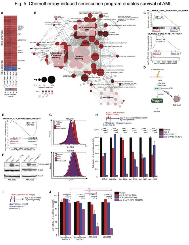
Author Manuscript Published OnlineFirst on January 26, 2021; DOI: 10.1158/2159-8290.CD-20-1375
    Author manuscripts have been peer reviewed and accepted for publication but have not yet been edited.

ABSTRACT

Acute myeloid leukemia (AML) patients frequently relapse after chemotherapy, yet the
mechanism by which AML reemerges is not fully understood. Herein, we show that
primary AML cells enter a senescence-like phenotype following chemotherapy in vitro and
in vivo. This is accompanied by induction of senescence/inflammatory and embryonic
diapause transcriptional programs, with downregulation of MYC and leukemia stem cell
genes. Single-cell RNA-seq suggested depletion of leukemia stem cells in vitro and in
vivo, and enrichment for subpopulations with distinct senescence-like cells. This
senescence effect was transient and conferred superior colony forming and engraftment
potential. Entry into this senescence-like phenotype was dependent on ATR, and
persistence of AML cells was severely impaired by ATR inhibitors.                           Altogether, we
propose that AML relapse is facilitated by a senescence-like resilience phenotype that
occurs regardless of their stem cell status. Upon recovery, these post-senescence AML
cells give rise to relapsed AMLs with increased stem cell potential.

SIGNIFICANCE

Despite entering complete remission after chemotherapy, relapse occurs in many AML
patients. Thus, there is an urgent need to understand the relapse mechanism in AML and
the development of targeted treatments to improve outcome. Here, we identified a
senescence-like resilience phenotype through which AML cells can survive and
repopulate leukemia.

                                                                                                               2

Downloaded from cancerdiscovery.aacrjournals.org on February 3, 2021. © 2021 American Association for Cancer
                                                 Research.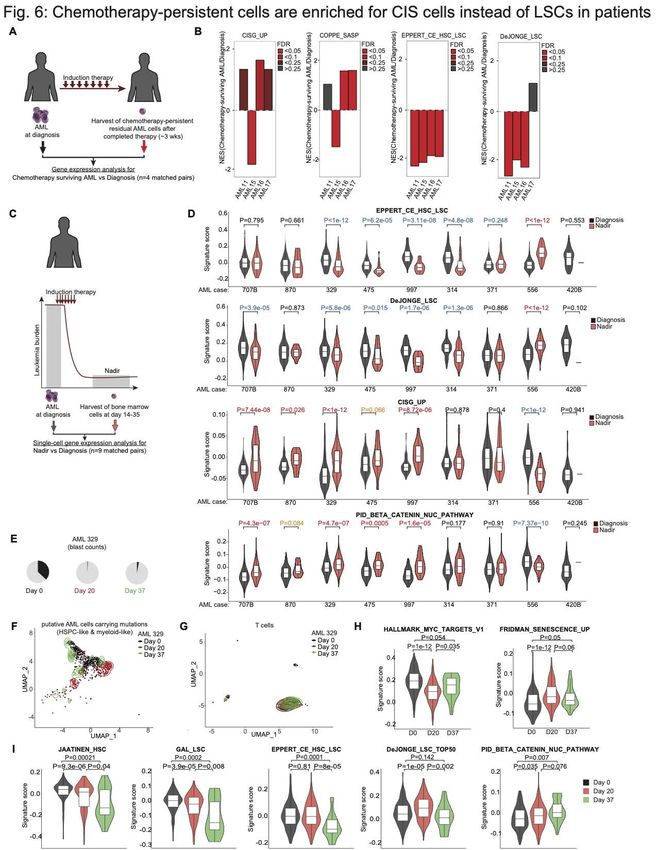
Author Manuscript Published OnlineFirst on January 26, 2021; DOI: 10.1158/2159-8290.CD-20-1375
    Author manuscripts have been peer reviewed and accepted for publication but have not yet been edited.

INTRODUCTION

AML is the most lethal of all blood cancers, killing 3 out of 4 patients within 5-years
(https://seer.cancer.gov/statfacts/). Even though 70–80% of AML patients (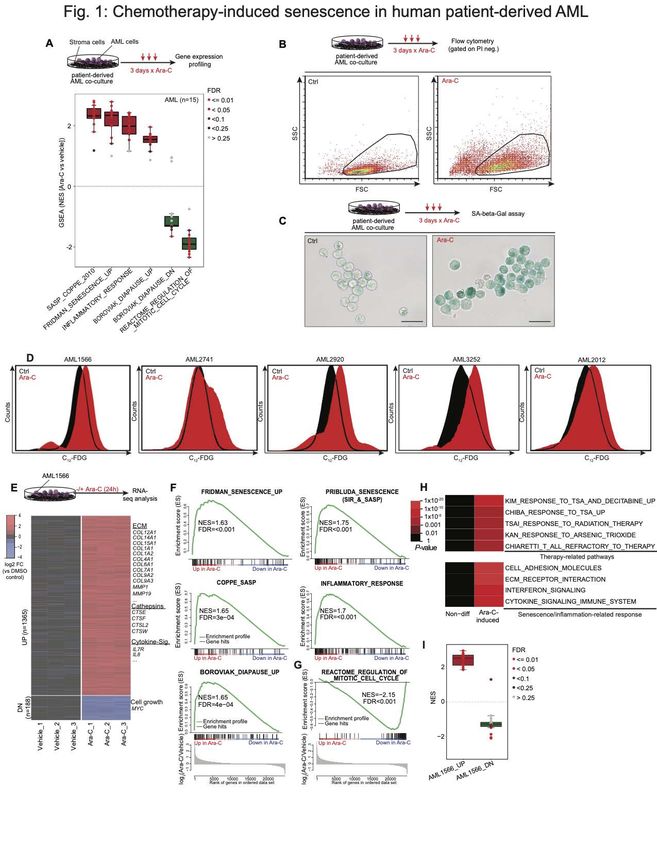
Author Manuscript Published OnlineFirst on January 26, 2021; DOI: 10.1158/2159-8290.CD-20-1375
    Author manuscripts have been peer reviewed and accepted for publication but have not yet been edited.

The initial studies on cellular senescence were performed on non-malignant cells
describing a natural barrier to unlimited proliferation, termed as “replicative senescence”
or the so-called Hayflick limit (23). Yet, cells that have not reached their replicative
lifespan can also undergo cell arrest in response to sublethal stress, often referred to as
(stress-induced) premature senescence (24). A landmark study in the late 90’s
demonstrated that expression of oncogenes in primary cells induced premature
senescence, defined as oncogene-induced senescence (OIS) that led to the concept that
senescence might function as a tumor-suppressive mechanism (25). However, more
recent studies have shown that expression of oncogenes in even fully transformed cancer
cell lines activates OIS, and that both malignant and non-malignant cells can escape OIS
after a prolonged period in the senescence arrested state (26,27). Thus, the premature
senescence phenotype is not as static and irreversible as initially thought, particularly in
malignant cells (27-30).

Overall, cellular senescence is a complex phenomenon, initiated by a wide range of
stimuli including DNA damage, oncogene activation, telomere erosion, protein misfolding,
oxidative damage, extracellular signaling by mitogens or cytokines, and high-fat diet (8).
One interpretation that potentially reflects the common effect of these various senescence
inducers could be that senescence acts as a conserved stress-response mechanism.
Given the frequency with which cells encounter environmental stress, it is not
unreasonable to expect cells to possess evolutionary conserved mechanisms to tolerate
harmful conditions. For example, the embryonic diapause is an evolutionary strategy in
many mammals that describes a reversible growth arrest (up to many months) of
blastocysts (31,32), presumably enabling survival during stressful environmental
conditions. We hypothesized that AML relapse is dependent on these cells undergoing a
senescence-like resilient state, potentially mimicking a diapause state, to survive
genotoxic stress caused by chemotherapy and eventually repopulate the disease.

RESULTS

                                                                                                               4

Downloaded from cancerdiscovery.aacrjournals.org on February 3, 2021. © 2021 American Association for Cancer
                                                 Research.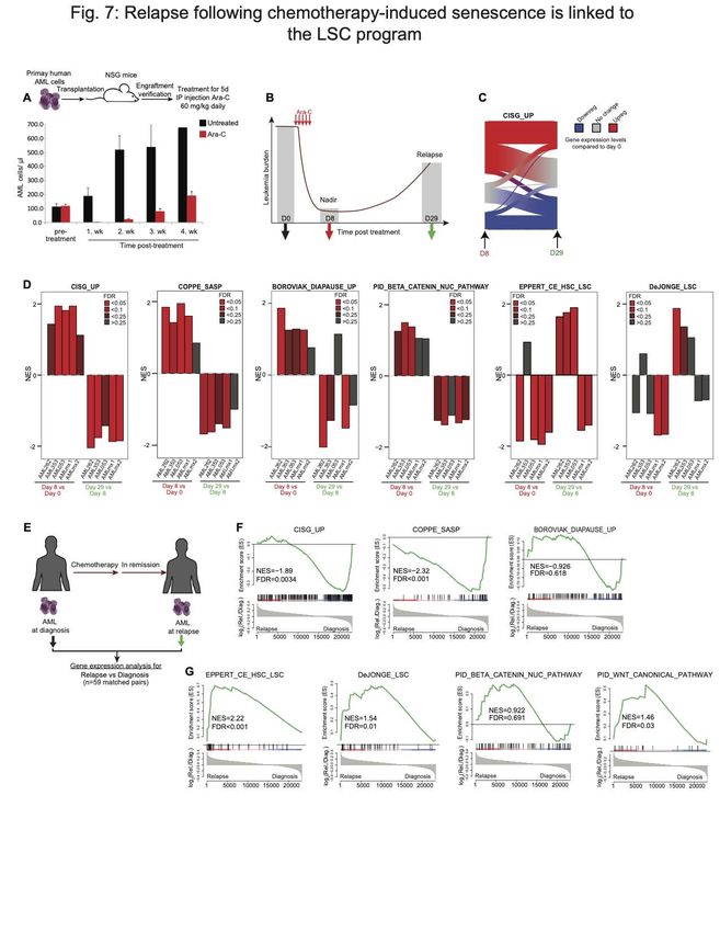
Author Manuscript Published OnlineFirst on January 26, 2021; DOI: 10.1158/2159-8290.CD-20-1375
    Author manuscripts have been peer reviewed and accepted for publication but have not yet been edited.

Chemotherapy induces cellular senescence in patient-derived AMLs.
To determine if chemotherapy induces a senescence-like state, we analyzed published
gene expression microarray profiles of 15 primary patient-derived AML specimens
exposed to Ara-C (a standard component of AML therapy) for three days in a co-culture
system (33) (Fig. 1A). Gene set enrichment analysis (GSEA) (34) showed significant
enrichment of senescence and SASP/inflammatory signatures in >90% of primary
patient-derived cases (Fig. 1A). We also observed significant enrichment for diapause-
associated genes (Table S1) (35) after chemotherapy (Fig. 1A).                            Reciprocally we
observed significant negative enrichment for genes that are downregulated during
diapause, as well as genes associated with cell division, consistent with the growth arrest
linked to senescence.

To validate whether chemotherapy induces a senescence-like phenotype in myeloid
leukemia cells, we examined induction of X-gal-based β-galactosidase staining as a
canonical biomarker for this state (9).           Using an ex vivo co-culture system that allows
reliable expansion of primary AML cells collected from patients (36), we observed key
features of senescence including increased cell size, granularity and SA-β-gal activity
after three-days exposure to Ara-C or anthracyclines, the two chemotherapy drugs used
for standard treatment of this disease, among cell lines and patient specimens (Fig. 1B,
C and Fig. S1A) (6). The X-gal based assay has two main limitations: the requirement for
chemical fixation, precluding viable cells for further analysis, and limited ability to quantify
populations of cells through microscopic assessment. In order to quickly and
quantitatively assess SA-β-Gal activity within viable cells, we used the fluorogenic
substrate 5-dodecanoylaminofluorescein di-β-D-galactopyranoside (C12-FDG) (37) (Fig.
S1B-C). Using this assay, we confirmed induction of this senescence marker as well as
increased granularity in additional patient-derived AML specimens (Fig. 1D, Fig. S1D).
Because of the inherent fluorescence property of anthracyclines (38) that would interfere
with the C12-FDG signal in our subsequent analyses, we focused largely on chemotherapy
treatment with Ara-C. Chemotherapy-induced senescence (CIS) occurred in myeloid
leukemia cells regardless of p16 or p53 mutation or expression (Fig. S2A, B), consistent
with previous reports (12,39,40). Senescence was not simply a marker of differentiation,

                                                                                                               5

Downloaded from cancerdiscovery.aacrjournals.org on February 3, 2021. © 2021 American Association for Cancer
                                                 Research.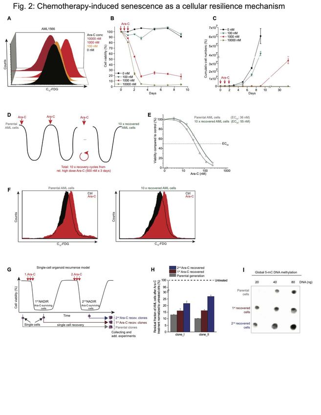
Author Manuscript Published OnlineFirst on January 26, 2021; DOI: 10.1158/2159-8290.CD-20-1375
    Author manuscripts have been peer reviewed and accepted for publication but have not yet been edited.

since a LSD1 inhibitor (36,41,42) induced strong expression of CD11b without an
increase of beta-Gal activity (Fig. S2C, D).

To explore the gene signatures induced by chemotherapy exposure at an earlier timepoint
that might further indicate the role of senescence as a primary effect on these cells, we
performed RNA sequencing on patient-derived AML cells exposed to Ara-C and
harvested 24 hours later. We identified 1553 differentially expressed genes (DEG; at least
2-FC and adjusted p value < 0.01) with approximately 88% of genes being upregulated
(Fig. 1E, Table S2). Among the upregulated genes were many that encoded typical
SASP factors including several members of the cathepsin family and cytokines like IL-8.
GSEA demonstrated significant enrichment for gene signatures associated with
senescence, inflammation, and diapause (Fig. 1F) (12,13,35,43,44). Reciprocally to the
senescence phenotype, cell cycle genes were significantly downregulated (Fig. 1G).
Among the downregulated genes was MYC (Table S2), depletion of which was shown to
induce a dormant state mimicking diapause (45). Additional pathway analysis of genes
upregulated by Ara-C exposure showed enrichment for gene signatures associated with
ECM modulation and transcriptional response to genotoxic stress (Fig. 1H).                                  We
observed significant enrichment of our early upregulated signature in 14/15 of the patient
specimens profiled at day 3 post-chemotherapy exposure (33), suggesting a conserved
response mechanism in AML (Fig. 1I).

Senescence-like AML cells can repopulate AMLs after a latent state.
Relapse of AML frequently occurs following a variable nadir phase. To mimic this, we
first seeded primary human AML cells in our co-culture organoid system and exposed
them to increasing Ara-C concentrations in the range of clinically-relevant concentrations
(ranging between the induction vs close to consolidation doses of this drug used in the
clinic) (46). We determined the association between the degree of genotoxic stress and
senescence induction by SA-β-Gal activity assessed by flow cytometry after 3 days of
Ara-C treatment, and observed little induction of senescence at 100 nM of drug, but much
stronger induction upon exposure to 1000 nM (Fig. 2A). In contrast, senescent cells were
markedly diminished after exposure to 10,000 nM Ara-C (Fig. 2A).

                                                                                                               6

Downloaded from cancerdiscovery.aacrjournals.org on February 3, 2021. © 2021 American Association for Cancer
                                                 Research.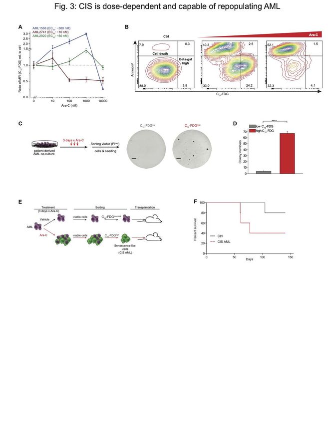
Author Manuscript Published OnlineFirst on January 26, 2021; DOI: 10.1158/2159-8290.CD-20-1375
       Author manuscripts have been peer reviewed and accepted for publication but have not yet been edited.

To model the nadir phase ex vivo and how leukemia regenerates after exposure to
chemotherapy, we determined cell viability by flow cytometry (dye exclusion) over the
course of 10 days after treating the cells with increasing doses of Ara-C for 3 days (Fig.
2B).      There was very little effect on viability with the 100 nM dose, but close to 100%
cell death with the 10,000 nM dose. In contrast the intermediate dose yielded a significant
fraction of cells (~20%) that persisted in a viable state (Fig. 2B). To determine if these
cells were end-stage or could recover and regenerate the leukemic population we
performed serial viable cell counts over an extended time period by flow cytometry up to
30 days post Ara-C treatment (Fig. 2C). Cells treated with 100 nM Ara-C manifested only
a slight delay in their expansion, whereas the few cells remaining after the highest dose
of Ara-C remained in a quiescent phase throughout the experiment. In contrast, those
AMLs treated with 1000 nM Ara-C remained in a latent state until day 9 and after which
they were able to re-expand (Fig. 2C).                      These results suggest that induction of
senescence is dose-dependent and reversible, allowing cells to potentially recover and
restore the disease.

Chemotherapy-induced senescence as a cellular resilience mechanism.

To explore whether the senescence-like phenotype is a reversible resilience response as
opposed to a process of selecting pre-existing chemotherapy-resistant C12-FDGhigh
subpopulations, we first sorted C12-FDGhigh vs C12-FDGlow AML cells in three different
patient-derived cases (Fig. S3A). Exposure to increasing doses of Ara-C or Daunorubicin
did not reveal evidence of differential sensitivity in these C12-FDGhigh cells (Fig. S3B). To
address this question in a more rigorous fashion, we next performed sequential cycles of
Ara-C treatment and recovery in primary patient-derived AML cells, using a dose (500
nM) that kills >90% of cells from this patient (Fig. 2D). These cells required approximately
1-2 months to recover after each Ara-C treatment and these cycles were repeated a total
of ten times (Fig. 2D). Notably, after the tenth cycle the AML cells manifested only a
small increase in their EC50 (Fig. 2E). Moreover, Ara-C induced a similar degree of
senescence in AML cells after the 10th recovery as compared to the initial parental cells
(Fig. 2F).

                                                                                                               7

Downloaded from cancerdiscovery.aacrjournals.org on February 3, 2021. © 2021 American Association for Cancer
                                                 Research.
Author Manuscript Published OnlineFirst on January 26, 2021; DOI: 10.1158/2159-8290.CD-20-1375
Author manuscripts have been peer reviewed and accepted for publication but have not yet been edited.
In addition, we created an ex vivo AML clonal recurrence model in which single primary
human cells were placed in culture before, or after serial Ara-C treatment from the residual
fraction of chemotherapy-persistent cells at nadir (Fig. 2G). Single cells from each time
point formed a monoclonal population thereby excluding genetic heterogeneity within the
recovered population. This procedure revealed a modest and gradual increase in the
fraction of surviving cells at nadir after each Ara-C recovery cycle (Fig. 2H), indicating
that the single cell-derived clones are slightly more resilient to chemotherapy, consistent
with progressive stress adaptation (47,48) rather than chemotherapy resistance, as there
was no major shift in the EC50 of previously exposed cells to Ara-C (Fig. 2H). Of note,
consistent with previous data linking DNA methylation with population fitness in AML
patients (49), we observed increased abundance of methylcytosine in recovered cell
populations (Fig. 2I). Collectively, these findings support that the senescence-like
phenotype is a reversible resilience state instead of a manifestation of an inherently
chemotherapy-resistant subpopulation that would have selected Ara-C-resistant clones
(Fig. 2F).
Induction of senescence-like phenotype is dose-dependent based on
chemotherapy sensitivity.
Patient-derived AML cells manifest significant variation in sensitivity (EC50) to Ara-C (Fig.
S4A). To explore the link between chemotherapy sensitivity and senescence, we next
examined the senescence response at four different doses of Ara-C in three patient
specimens with variable degree of sensitivity. Notably, the patient that was most
insensitive to Ara-C manifested the greatest induction of senescence that peaked at 1000
nM dose of drug (Fig. 3A), but did not display senescence at the highest dose (where
these cells were virtually all killed). The most sensitive case (AML 2741) peaked at 10
nM, after which there was loss of senescent populations. The intermediate case
manifested a weaker degree of senescence induction at lower doses than the resistant
patient (Fig. 3B). Hence in general we observed that higher concentrations of Ara-C
induced increased SA-β-Gal activity in AML cells until reaching a critical toxic dose that
killed these cells (Fig. 3A, B).
8
Downloaded from cancerdiscovery.aacrjournals.org on February 3, 2021. © 2021 American Association for Cancer
Research.Author Manuscript Published OnlineFirst on January 26, 2021; DOI: 10.1158/2159-8290.CD-20-1375
Author manuscripts have been peer reviewed and accepted for publication but have not yet been edited.
Our co-culture experiments suggested that acquisition of the senescence-like phenotype
was associated with persistence and retention of leukemia repopulating potential. To
further explore if this was the case, we treated patient-derived AML cells in co-culture with
1000 nM Ara-C for 3 days, after which the specimens were sorted into C12-FDGhigh and
C12-FDGlow viable (dye exclusion flow cytometry) fractions and plated on methylcellulose
for colony forming cell (CFC) assays. Two of our cases were able to form colonies, and
in both cases, the senescence-like cells had significantly higher clonogenic potential
(PAuthor Manuscript Published OnlineFirst on January 26, 2021; DOI: 10.1158/2159-8290.CD-20-1375
Author manuscripts have been peer reviewed and accepted for publication but have not yet been edited.
As expected, projecting the bulk RNA-seq gene expression signature derived from Ara-
C-treated cells in Fig. 1E showed predominant enrichment among the sorted senescent
cells (Fig. S5A). Genes linked to SASP upregulated upon Ara-C treatment (e.g.
cathepsins, IL-8) were confirmed to be highly expressed in the senescent population (Fig.
S5B, Table S3). On the other hand, MYC and genes associated with ribosome
biosynthesis (e.g. RPL38) were among the most downregulated genes (Fig. S5C). MYC
target genes were depleted in AML cells that highly expressed senescence- and diapause
gene signatures (Fig. S5D). Although there was some degree of heterogeneity among
AML cells, we found significant inverse correlation of expression between MYC target
genes and chemotherapy-induced stress genes (CISG) that we identified in this study
(Fig. S5E, Table S1).
To further delineate subpopulations among these cells we performed a k-nearest-
neighbor-based analysis, which yielded seven clusters (Fig. 4D and Table S4), that were
largely mutually exclusive between control and senescent cells, with the exception of
senescence cluster 4, which consisted of ~17% untreated control cells (Fig. 4E and Fig.
S4F). We next projected gene sets linked to senescence, diapause or LSC/HSCs on the
UMAP plots (Fig. 4F-G). As expected, genes associated with senescence and SASP
were highly expressed within Ara-C-treated cells that compose clusters: 0, 3, 4, and 6
(Fig. 4F, H). Diapause genes were most highly expressed in cells composing clusters 4
and 6. Among untreated cells, only the small fraction contained in cluster 4 manifested
strong association with senescence signatures, consistent with the presence of small
numbers of senescent cells under basal conditions (Fig. 4E, H). Analysis of stem cell
signatures showed the highest expression among control cell clusters 1, 2 and 5 (Fig.
4G, H). In contrast there were reduced numbers of cells and lower expression of these
signatures within senescent cell populations, although a sizable fraction of cells in clusters
3 and 0 still retained moderate expression of these gene sets (Fig. 4G, H). scRNA-seq
analysis of the senescence-like AML cells showed no enrichment of LSC markers that
could explain the association with increased AML engraftment (Fig. 3F, Fig. S5G).
WNT/beta-catenin pathways were reported to become de novo activated during
senescence-associated reprogramming in cancer cells (28) and were likewise induced
10
Downloaded from cancerdiscovery.aacrjournals.org on February 3, 2021. © 2021 American Association for Cancer
Research.Author Manuscript Published OnlineFirst on January 26, 2021; DOI: 10.1158/2159-8290.CD-20-1375
Author manuscripts have been peer reviewed and accepted for publication but have not yet been edited.
among cluster 6 post-treatment subpopulations compared to control cells (PAuthor Manuscript Published OnlineFirst on January 26, 2021; DOI: 10.1158/2159-8290.CD-20-1375
Author manuscripts have been peer reviewed and accepted for publication but have not yet been edited.
Chemotherapy-induced senescence gene signatures suggest potential therapeutic
vulnerabilities.
To gain deeper insight into biological functions affected by the AML chemotherapy
senescence response, we merged the RNA-seq profiles obtained in the two different
primary patient-derived AML specimens shown in Figs 1 and 3, and identified the subset
of genes most robustly differentially expressed after Ara-C exposure. This procedure
yielded a gene expression signature consisting of 262 upregulated and 26 downregulated
genes (2-FC, qAuthor Manuscript Published OnlineFirst on January 26, 2021; DOI: 10.1158/2159-8290.CD-20-1375
Author manuscripts have been peer reviewed and accepted for publication but have not yet been edited.
experiments confirmed induction of CHK1 phosphorylation vs. total CHK1 in AML
specimens after exposure to Ara-C. This effect was severely impaired when cells were
exposed to the specific ATR inhibitor VE821 (56) just prior to treatment with Ara-C for 24
hours (Fig. 5F). Since genetic deletion of ATR protein is lethal to dividing cells (57,58),
we therefore examined the effect of catalytic inhibition with VE821, a selective ATR
inhibitor chemically related to VX-970 (M6620), which is currently in clinical trials (59,60).
AML cells were pretreated for 2 hours with VE821 (2 µM) or vehicle and then exposed to
Ara-C or vehicle for 3 days, followed by C12-FDG flow cytometry. Strikingly, the VE821
severely impaired C12-FDG induction, suggesting that ATR activation plays a critical role
in inducing the senescent/SASP phenotype in AML context (Fig. 5G). If the senescent
phenotype has a protective role then it would be expected that VE821 would prevent
survival of AML cells after exposure to Ara-C. For this we exposed a panel of five primary
AML specimens as well as the KG-1 AML cell line to Ara-C, VE821, or the combination
of both for 3 days to assess cell viability one week after treatment (Fig. 5H). In half of
the cases Ara-C and VE821 killed roughly 50% of the AML cells, whereas the combination
killed 95% of cells (Fig. 5H). However, in the other three cases killing was supra-additive
since these cells were relatively resistant to either Ara-C or VE821, yet still manifested
>95% killing in the presence of the combination. These results were confirmed using a
second, more potent ATR inhibitor BAY-1895344, which is also in clinical trials (61) (Fig.
5I-J). In contrast these drugs did not induce a marked killing effect in normal donor
hematopoietic stem/progenitor cells (HSPCs) (Fig. 5J). Fordham et al. recently
demonstrated that in vivo treatment with ATR inhibitor and chemotherapy also yields
dramatically enhanced activity against patient-derived AML in mice (62). Altogether, these
results suggest that ATR targeted therapy strongly potentiates the efficacy of
chemotherapy in AML by preventing these cells from being rescued through induction of
the senescence resilience phenotype.
13
Downloaded from cancerdiscovery.aacrjournals.org on February 3, 2021. © 2021 American Association for Cancer
Research.Author Manuscript Published OnlineFirst on January 26, 2021; DOI: 10.1158/2159-8290.CD-20-1375
Author manuscripts have been peer reviewed and accepted for publication but have not yet been edited.
Senescence-like AML cells are enriched at post induction chemotherapy nadir in
AML patients.
If it is true that AMLs enter a senescence-like state that enables them to persist after
exposure to cytotoxic drugs, then we would expect that such cells would be enriched in
the bone marrow of AML patients after induction chemotherapy. To determine if this was
the case, we investigated gene expression profiles of residual AML cells that persist in
the bone marrow of four human subjects approximately three weeks following induction
therapy, as compared to their matched diagnostic specimens (Fig. 6A) (4). This analysis
revealed enrichment of our CISG_UP senescence and SASP signatures in the residual
AML cells in 3 of these patients (Fig. 6B). In contrast there was depletion of leukemia
stem cell signatures in the patients.
We next determined the distribution of the senescence signature across the population
of residual AML cells persisting 14 to 35 days post induction chemotherapy using single-
cell RNA-seq data collected from nine AML patients, as compared to their matched
diagnostic specimens (Fig. 6C) (63). Analyzing the gene expression profiles of myeloid,
myeloid-like, HSPCs, and HSPCs-like cells collected from the bone marrow of AML
patients, we found generally depletion or no enrichment of LSCs in 7 out of 9 AML patients
at nadir compared to diagnosis (Fig. 6D and Fig. S7). On the other hand, we found
around half of the patients (n=4 with PAuthor Manuscript Published OnlineFirst on January 26, 2021; DOI: 10.1158/2159-8290.CD-20-1375
Author manuscripts have been peer reviewed and accepted for publication but have not yet been edited.
(3% blasts) (Fig. 6E). This allowed us to exclude potential normal cells from our analysis
(63). AML cells at day 37 revealed closer clustering to diagnosis samples as compared
to nadir cells (Fig. 6F), whereas T cells did not manifest detectable transcriptional
difference among the three timepoints (Fig. 6G, controlling for variability between
specimens). We observed strong MYC target gene signature expression in AML cells at
diagnosis and significantly lower levels at nadir (PAuthor Manuscript Published OnlineFirst on January 26, 2021; DOI: 10.1158/2159-8290.CD-20-1375
Author manuscripts have been peer reviewed and accepted for publication but have not yet been edited.
mice, treated them with the same Ara-C regimen, and performed gene expression
analysis on AML cells purified cells pre-treatment, at nadir and upon relapse (Fig. 7D).
Using GSEA on these gene expression profiles revealed enrichment for the CISG_UP,
SASP senescence and diapause signatures as well as partial enrichment for beta-catenin
at nadir (day 8) vs pre-treatment (day 0) (Fig. 7D). In contrast there was no enrichment
for LSC signatures at day 8, which were instead often significantly depleted. However,
when comparing gene expression profiles at day 29 (relapse) vs day 8 nadir, we observed
the opposite effect: depletion of CISG_UP, SASP, diapause and beta-catenin signatures,
and enrichment for LSC profiles in at least three of the five cases. These data suggest
senescent programs resolve as AML cells recover from chemotherapy-induced
senescence-like state, and as they emerge from this state they feature enrichment for
more stem-like transcriptional programming. These findings are consistent with a recent
report showing that senescence is accompanied by epigenetic plasticity and reactivation
of stem cell functionalities (28). To further examine this point, we performed RNA-seq
analysis in 59 matched AML specimens collected at diagnosis vs relapse (Fig. 7E).
GSEA analysis demonstrated that AML cells exhibit a depletion for senescence gene
signatures and an enrichment for LSC-signatures at relapse (Fig. 7F, G). Relapsed AML
compared to diagnosis samples demonstrated partial enrichment of WNT/beta-catenin
programs (Fig. 7G). Overall, these data suggest that survival after exposure to
chemotherapy is most likely stochastic, involves a diapause/SASP-like resilience
response, and that subsequent enrichment for stem-like features at relapse could reflect
the consequence, rather than the cause of leukemic survival after exposure to
chemotherapy.
DISCUSSION
AML relapse occurs in a majority of patients after chemotherapy and portends incurable
disease. While selection of clones containing drug resistance mutations (e.g. TP53)
following chemotherapy are relatively rarely observed in relapsed AML, a number of
studies suggest that relapse is explained by the presence of inherently more resistant
leukemia stem cells as the origin of relapse (65,66). Yet, most of these studies analyzed
samples at diagnosis and relapse but not at nadir. Although LSC gene expression
16
Downloaded from cancerdiscovery.aacrjournals.org on February 3, 2021. © 2021 American Association for Cancer
Research.Author Manuscript Published OnlineFirst on January 26, 2021; DOI: 10.1158/2159-8290.CD-20-1375
Author manuscripts have been peer reviewed and accepted for publication but have not yet been edited.
signatures are predictive of relapse (67,68), it does not necessarily mean that these cells
are enriched during exposure to chemotherapy. Indeed, while we also observed an
enrichment of LSC gene signatures at relapse, we did not find LSC gene signatures in
the residual fraction of chemotherapy-persistent leukemia cells at nadir. This is consistent
with other recent studies reporting no enrichment of LSCs at nadir (4,5). Instead, we found
that AML cells surviving initial chemotherapy manifest a senescence-like resilient
phenotype, with potential to repopulate leukemia. Previous studies analyzing the transient
state of chemotherapy-persistent AML at nadir indicate the expression of genes related
to SASP/inflammation and senescence-like cells (4,5). Upon recovery these cells
manifested positive enrichment of LSC-gene signatures.
We suggest that LSC programming in many AML cases occurs during the senescence-
like resilient phenotype, explaining how these stem cells are enriched at relapse. This
hypothesis is supported by a recent study showing that doxorubicin-induced senescence
reprograms murine leukemia and lymphoma cells to cancer stemness (28). We do not
exclude that in certain AMLs with initially abundant LSCs, these cells could survive
chemotherapy without major harm and repopulate the disease. Given the heterogeneity
between AML patients, it is also possible that other resilience mechanisms could exist
that were not captured here. Yet, our data suggest that many AMLs enter a stress-
adaptive and senescence-like resilient phenotype regardless of whether they are LSCs
or not, and that this process plays a critical role in relapse, possibly through an acquired
stemness reprogramming process mediated by the WNT/β-catenin pathway as described
previously (28). Alternatively, adaptation may reflect either initial epigenetic heterogeneity
or epigenetic plasticity as possible mechanisms of resilience. Fundamental to
understanding this process is the inherent plasticity of cells to reprogram from one state
to another. Importantly in this regard, cancer stem cell functionality is not a fixed property
but rather can be induced under stress conditions (28,69). Another layer of complexity in
this context is that different AMLs require different doses of chemotherapy to induce a
comparable senescence phenotype in which some refractory AMLs will only manifest
overt senescence phenotypes when exposed to higher drug concentrations.
Furthermore, AML is a heterogenous disease and we cannot exclude that certain AML
subtypes induce the senescence phenotype to a different degree than other subtypes.
17
Downloaded from cancerdiscovery.aacrjournals.org on February 3, 2021. © 2021 American Association for Cancer
Research.Author Manuscript Published OnlineFirst on January 26, 2021; DOI: 10.1158/2159-8290.CD-20-1375
Author manuscripts have been peer reviewed and accepted for publication but have not yet been edited.
Our findings are consistent with a report that investigated drug-resistance in solid tumors
and revealed that the drug-persistent state shares a diapause-like dormancy signature
(70). The diapause principle in theory represents a long-term dormancy state. Almost half
of senescence-associated genes are also expressed in quiescent cells, potentially
indicating a mechanistic overlap between senescence and dormancy (71). The concept
of a transient stress-induced endurance state is a common phenomenon described in
developmental biology, whereby early embryos survive adverse conditions via transition
into diapause (32). MYC controls cellular growth and depletion of MYC can induce a
diapause-like state (45,72). Notably, translocations leading to constitutive expression of
MYC are associated with better prognosis in certain cancers (e.g. Burkitt lymphoma)(73),
presumably in part because of the aggressive cell proliferation that incorporates more
chemotherapy. Thus, suppression of MYC may represent a survival strategy of cells to
endure stress. We have used the phrase, “senescence-like phenotype” to describe our
results. As note, AML cells treated with chemotherapy induce a cell cycle arrest and
growth pause similar to replicative senescence but unlike replicative senescence the
growth pause is not permanent. The observed senescence transcriptional profiles are
most similar to SASP, a specific phenotype of the senescent state initially described in
response to genotoxic stress (13). SASP is characterized by an inflammatory response
that may contribute functionally to leukemic recovery, consistent with notions on
regenerative properties of AML nadir cells described by Boyd et al. (4). Among SASP
genes, we observed induction of IL-8, which has been described to protect AML cells
from the effects of chemotherapy (74)
Our data indicate that ATR plays a critical role in the chemotherapy-induced senescence
resilient phenotype. This is consistent with previous reports showing that ATR can induce
SASP through induction of NF-κB in a p53-independent manner (53,54). Indeed, we
show for the first time that induction of senescence as well as tumor survival and
persistence is dependent on ATR catalytic activity, through our experiments using
chemotherapy treatment in primary human AML cells. Moreover, others have shown that
treatment of primary AML cells with ATR inhibitor and chemotherapy is also remarkably
effective in vivo (62). ATR inhibitors may thus represent a novel approach for targeting
senescence resilience by preventing cells from harnessing this survival mechanism. It is
18
Downloaded from cancerdiscovery.aacrjournals.org on February 3, 2021. © 2021 American Association for Cancer
Research.Author Manuscript Published OnlineFirst on January 26, 2021; DOI: 10.1158/2159-8290.CD-20-1375
Author manuscripts have been peer reviewed and accepted for publication but have not yet been edited.
intriguing to consider the potential for ATR inhibitor to potentiate the effect of drugs that
directly target senescent cells such BCL2 inhibitors, which have been used to enhance
the efficacy of chemotherapy and other drugs in AML patients (75,76). ATR inhibitors
are currently in clinical trials both as single agent as well as in combination with various
chemotherapy drugs for solid tumors. Collectively, these and our data strongly point to
the rationale for initiating clinical trials of ATR inhibitor in AML patients. In summary, our
findings suggest that AML relapse can be explained by entry of these cells into an ATR-
dependent senescence-like resilient state of variable duration that endows them with
superior fitness and enhanced ability to repopulate the disease. Although this effect
appears to be independent of their initial stemness profiles, it may make these cells highly
vulnerable to drugs such as ATR inhibitors, with potential to better eradicate these tumors.
19
Downloaded from cancerdiscovery.aacrjournals.org on February 3, 2021. © 2021 American Association for Cancer
Research.Author Manuscript Published OnlineFirst on January 26, 2021; DOI: 10.1158/2159-8290.CD-20-1375
Author manuscripts have been peer reviewed and accepted for publication but have not yet been edited.
Acknowledgements: We acknowledge the ECOG-ACRIN Cancer Research Group and
the Weill Cornell Medicine Epigenomics Core for technical services. We thank Yaseswini
Neelamraju for assistance with data submission to dbGap. This work is supported by the
National Cancer Institute of the National Institutes of Health under the following award
numbers: NIH/NCI R01CA198089 (AM and MC), NIH/NCI UG1 CA233332 (AM). This
work was supported by grants NIH R01 CA050947 (C.S.M.), CA196664 (C.S.M.),
U01CA225730 (C.S.M); Leukemia and Lymphoma Society (LLS) Scholar Award
(C.S.M.); and Ludwig Center at Harvard (C.S.M). The content is solely the responsibility
of the authors and does not necessarily represent the official views of the National
Institutes of Health. This work was further supported by LLS SCOR 7013-17 (AM) and
U10CA180820 (ECOG). CD was supported by an LLS fellow award (LLS 5486) in
partnership with The Jake Wetchler Foundation, and is a Forbeck scholar.
Contributions: C.D. conceived the study and wrote the paper. C.D. and A.M. designed
experiments and interpreted data. C.D. and M.L. performed bench experiments. C.D.,
M.T., C.D., and C.M. performed computational analysis and interpreted data. A.M., M.T.
and M.C. edited the manuscript. M.T., F.G.B., C.M., M.L., T.L., C.C., E.D., C.S.M., H.D.,
R.A., M.B., E.M.P., C.M., and M.C. provided reagents, data and interpreted data.
20
Downloaded from cancerdiscovery.aacrjournals.org on February 3, 2021. © 2021 American Association for Cancer
Research.Author Manuscript Published OnlineFirst on January 26, 2021; DOI: 10.1158/2159-8290.CD-20-1375
Author manuscripts have been peer reviewed and accepted for publication but have not yet been edited.
MATERIAL & METHODS:
Primary AML samples. Primary human AML specimens were collected after written
informed consent and obtained from peripheral blood or bone marrow aspiration
according to the declaration of Helsinki for collection and use of sample materials. The
studies were approved by the institutional review board at Weill Cornell Medicine (IRB
protocol # 0805009783). Information on the primary AML cases used in this study are
described in Table S7 and in more detail elsewhere (36,49).
Culturing of primary patient-derived AML. Primary AML were cultured ex vivo as
previously described (36). In short, primary patient-derived AMLs were expanded on
stromal feeder layers with cytokine support.
AML cell lines. Human myeloid leukemia cell lines were purchased from ATCC or DSMZ
and maintained in IMDM media containing 10-20% fetal bovine serum, 100 IU ml-1
penicillin and 100 μg ml-1 streptomycin. Cell lines were validated via short tandem repeat
DNA profiling and monitored regularly for mycoplasma contamination.
Isolation of HSPCs. Mononuclear cells (MNCs) were isolated from fresh human umbilical
cord blood samples (New York Blood Center, NYBC) using Ficoll (Atlanta Biologicals,
GA) density gradient centrifugation. After lysis of red blood cells, hematopoietic stem and
progenitor cells (HSPCs) were selected via immunomagnetic enrichment of CD34+
MNCs using CD34 MicroBead Kit and Automacs from Miltenyi Biotec (Auburn, CA).
HSPCs were propagated in our ex vivo co-culture model as previously described (36).
Drug preparation and treatment. Stock solutions of inhibitors against ATR (VE-821,
BAY-1895344) derived from Selleckchem (Houston, TX) were prepared in DMSO and
stored at -80°C. Cells were exposed to 2 μmol l-1 (VE-821) or 20 nmol l-1 (BAY-1895344)
in a dilution factor that did not pass 0.3% DMSO content in growth medium during whole
treatment time. Cytarabine (Sigma-Aldrich, St. Louis, MO), Daunorubicin (EMD Millipore,
Billerica, MA), and Adriamycin/Doxorubicin (Selleckchem) was prepared in water and
kept protected from light at 4°C.
21
Downloaded from cancerdiscovery.aacrjournals.org on February 3, 2021. © 2021 American Association for Cancer
Research.Author Manuscript Published OnlineFirst on January 26, 2021; DOI: 10.1158/2159-8290.CD-20-1375
Author manuscripts have been peer reviewed and accepted for publication but have not yet been edited.
Classical SA-β-Gal activity assay using X-Gal. SA-β-Gal staining kit from Cell
Signaling was used for SA-β-Gal analyses and performed according to the manufacturer’s
protocol (BD Biosciences, San Jose, CA).
Fluorogenic SA-β-Gal activity assay using C12-FDG. Cells cultured for prolonged
period were maintained in well plates that had PBS or plain media in the outer wells to
reduce edge effects. Otherwise, cells cultured at outer wells exhibited stronger
evaporation effects that increased intracellular stress and SA-β-Gal activity. C12-FDG
procedure was performed as previously described (37) using 20 μM C12-FDG (Thermo
Fisher Scientific, Waltham, MA). C12-FDG was prepared in pre-warmed media and added
to cells with gently mixing of the cell suspension. Cells were protected from light and
incubated for 2 hours at 37°C and 5% CO2 before flow cytometry measurement.
Flow cytometry. Cell viability was assessed by the propidium iodide (PI)-negative cell
proportion, while cell growth was measured by absolute numbers of viable cells (PIneg)
including forward scatter (FSC) and sideward scatter (SSC) size discrimination. Annexin
V co-stained with propidium iodide (PI) was used for apoptosis analyses and performed
according to the manufacturer’s protocol (BD).
In vivo PDX relapse model. Mononuclear cells extracted from patients were
transplanted into NOD/SCID/IL2Rγnull (NSG) mice, which were obtained from The
Jackson Laboratory. Mice were randomly assigned to treatment groups. The investigators
were not blinded to allocation during experiments and outcome assessment. No power
analysis was used to predetermine sample size. Cytosine arabinoside (Ara-C)
administered using a physiologically relevant dose and schedule (60 mg/kg/day x 5 days)
to AML engrafted NSG mice induced a drop in peripheral blood leukemic cells and total
body leukemic burden by one week after initiation of therapy. Human AML cells were
collected from 4 bones and spleen after therapy. Cells from weeks 1 and 4 were purified
using human anti-CD45 antibody and analyzed for gene expression using RNA-seq or
Affymetrix Human Gene 1.0 ST arrays. All studies were conducted in accordance with
the guidelines of and approved by the Institutional Animal Care and Use Committee at
the institution where the work was performed.
In vivo leukemia repopulation of senescence-like cells. Untreated and treated AML
cells with Ara-C for 3 consecutive days were stained with C12-FDG on the following day.
22
Downloaded from cancerdiscovery.aacrjournals.org on February 3, 2021. © 2021 American Association for Cancer
Research.Author Manuscript Published OnlineFirst on January 26, 2021; DOI: 10.1158/2159-8290.CD-20-1375
Author manuscripts have been peer reviewed and accepted for publication but have not yet been edited.
Cells were washed and stained with sterile PI for cell viability discrimination. Cells were
sorted with a FACSAria II system (BD) into FBS and centrifuged after collection was
finished. Cell pellet was washed and resuspended in plain IMDM media. Cells were
transplanted into female NSG mice via tail vein injection of 500,000 cells per condition
per mouse.
Colony forming assay. After sorting, 5,000-25,000 cells/well of primary patient-derived
AML were seeded in MethoCult (H4534) medium (StemCell Technologies, Vancouver,
BC) supplemented with cytokines (36). The MethoCult medium-cell mixture was plated
into six-well plates with water supply in the inter-well chamber. Additionally, the six-well
plate was placed inside a tub with extra water dishes to prevent evaporation. After three
weeks of incubation at 37°C in 5% CO2, pictures were acquired by using ChemiDoc Touch
Imaging System (Bio-Rad, Hercules, CA) or STEMvision (Stemcell Technologies) and
colonies were counted using ImageJ software.
Immunoblot. Lysates from leukemia cells were lysed using PBS containing 1% NP-40
and 1 mM DTT supplemented with 1% protease/phosphatase inhibitor cocktail
(cOmplete, Roche, Indianapolis, IN; Phosphatase Inhibitor Cocktail A, Santa Cruz
Biotechnology, Dallas, TX) and were sonicated for 15 seconds using a tip sonicator
(Branson Sonifier 450). Protein lysates were resolved by SDS-PAGE, transferred to
PVDF membrane, and probed with the indicated primary antibodies: CHK1 (clone:
2G1D5, Thermo Fisher Scientific), phospho-CHK1 Ser345 (clone: 133D3, Cell signaling,
Danvers, MA), p53 (SC-126, Santa Cruz Biotechnology), p16 (ab108349, Abcam,
Cambridge, MA) or beta-actin (Sigma). Membranes were then incubated with a
peroxidase-conjugated correspondent secondary antibody and detected using Pierce
ECL chemiluminescent substrate (Thermo Fisher Scientific).
Depletion of apoptotic cells. When cell viability decreased below 50%, a depletion step
was introduced for all relevant assays except for flow cytometry. Dead cells and cellular
debris were removed via magnetic column depletion using the cell death removal kit
(Miltenyi Biotec, Auburn, CA) according to the company’s instructions.
Single-cell organoid AML recurrence: Individual clones were established before and
after killing the bulk leukemia following chemotherapy exposure. Briefly, parental and
nadir collected AML cells were serially diluted to a final concentration of 1 cell per two
23
Downloaded from cancerdiscovery.aacrjournals.org on February 3, 2021. © 2021 American Association for Cancer
Research.Author Manuscript Published OnlineFirst on January 26, 2021; DOI: 10.1158/2159-8290.CD-20-1375
Author manuscripts have been peer reviewed and accepted for publication but have not yet been edited.
wells in a 96- or 384-well plate. Each well was checked and monitored that contained a
single cell. Single cells were cultured using the ex vivo platform as previously described
(36). Recovered clones were picked from the wells and sub-cultured in larger wells.
5-mC immuno dot blot. Genomic DNA was extracted using the Purelink Genomic DNA
kit (Thermo Fisher Scientific) and quantified via NanoDrop (Thermo Fisher Scientific).
Dilution of DNA samples were denatured with 0.1 mol l-1 NaOH at 95°C for 5 min.
Subsequently, samples were rapidly chilled for 5 min on ice, spun down and neutralized
with 0.1 volume of 6.6 mol l-1 ammonium acetate. DNA samples were spotted onto a
positively charged nylon membrane (Immobilon-NY+, EMD Millipore, Bedford, MA), air
dried for 15 min and UV-crosslinked for 5 min. Membranes were blocked in PBS
containing 0.05% Tween-20 and 5% non-fat milk powder (PBS-TM) for 1h at room
temperature (RT). After blocking, membranes were washed in PBS-T for 3×5 min at RT
and incubated with the 5-mC antibody (NA81, EMD Millipore) in a 1:1000 dilution in PBS-
TM for 1 h. Subsequently, membranes were washed with PBS-T for 3×5 min at RT and
incubated with a secondary antibody for 1 h. After final washing, membranes were
incubated in Pierce ECL chemiluminescent substrate (Thermo Fisher Scientific).
Single-cell RNA-seq library preparation. AML cells were sorted with a FACSAria II
system (BD) into FBS and washed with PBS after collection was finished. Sorted viable
untreated AML cells and Ara-C-induced senescent AML cells were processed together
on the same day on a 10× Chromium instrument (10× Genomics) to generate barcoded
single-cell GEMs according to manufacturer’s protocol. Single-cell RNA-Seq libraries
were prepared using Chromium Single Cell 3’ v3 Reagent Kit (10× Genomics) following
the directions of the manufacturer. Briefly, the initial step consisted in performing an
emulsion where individual cells were isolated into droplets together with gel beads coated
with unique primers bearing 10× cell barcodes, unique molecular identifiers (UMI), and
poly(dT) sequences. GEM-Reverse Transcription (53 °C for 45 min, 85 °C for 5 min; held
at 4 °C) was performed in a C1000 Touch Thermal cycler with 96-Deep Well Reaction
Module (Bio-Rad, Hercules). After RT reaction, GEMs were broken and the single-strand
cDNA was cleaned up with DynaBeads MyOne Silane Beads (Thermo Fisher Scientific,
Waltham, MA). The cDNA was amplified for 12 cycles (98 °C for 3 min; 98 °C for 15 s,
63°C for 20 s, 72 °C for 1). Quality of the cDNA was assessed using an Agilent
24
Downloaded from cancerdiscovery.aacrjournals.org on February 3, 2021. © 2021 American Association for Cancer
Research.Author Manuscript Published OnlineFirst on January 26, 2021; DOI: 10.1158/2159-8290.CD-20-1375
Author manuscripts have been peer reviewed and accepted for publication but have not yet been edited.
Bioanalyzer 2100 (Santa Clara, CA), obtaining a product of about 1400bp. This cDNA
was enzymatically fragmented, end repaired, A-tailed, subjected to a double-sided size
selection with SPRIselect beads (Beckman Coulter, Indianapolis, IN) and ligated to
adaptors provided in the kit. A unique sample index for each library was introduced
through 12 cycles of PCR amplification using the indexes provided in the kit (98 °C for 45
s; 98 °C for 20 s, 54 °C for 30 s, and 72 °C for 20 s x 14 cycles; 72 °C for 1 min; held at
4 °C). Indexed libraries were subjected a second double-sided size selection, and libraries
were then quantified using Qubit fluorometric quantification (Thermo Fisher Scientific,
Waltham, MA). The quality was assessed on an Agilent Bioanalyzer 2100, obtaining an
average library size of 455bp. Libraries were pooled and normalized to 2 nM and
clustered on a HiSeq4000 high output mode on a pair end read flow cell and sequenced
for 28 cycles on R1 (10x barcode and the UMIs), followed by 8 cycles of I7 Index (sample
Index), and 98 bases on R2 (transcript), with a coverage around 130M reads per sample.
Primary processing of sequencing images was done using Illumina’s Real Time Analysis
software.
Single-cell RNA-seq data analysis. Single-cell expression was analyzed using the Cell
Ranger Single Cell Software Suite (v3.0.2) to perform quality control, sample de-
multiplexing, barcode processing, and single-cell 3’ gene counting (77). Sequencing
reads were aligned to the refdata-cellranger-hg19-3.0.0 transcriptome using the Cell
Ranger suite with default parameters. Single cells from control and Ara-C-treated
conditions, a total of 6278 single cells with a mean of ~3200 genes per cell were imported
into Seurat 3 package (78) in R statistical software. Only genes detected in at least two
cells were considered for further analysis. Apoptotic cells were filtered out by any cell
containing >15% of mitochondrial UMI counts. To detect changes in gene signatures
more robustly, any cell with 40000 UMIs or >6000 genes per cell were filtered out. Overall, a
total of 4627 cells were kept for downstream analyses. After merging control and Ara-C
treated cells using the merge function, cell expression levels were normalized and scaled
using default settings in Seurat. Principal component analysis (PCA) for dimensionality
reduction was then run on the normalized gene-barcode matrix. UMAP (51) embedding
was applied to visualize the cells in the two-dimensional space after selecting the first 20
25
Downloaded from cancerdiscovery.aacrjournals.org on February 3, 2021. © 2021 American Association for Cancer
Research.Author Manuscript Published OnlineFirst on January 26, 2021; DOI: 10.1158/2159-8290.CD-20-1375
Author manuscripts have been peer reviewed and accepted for publication but have not yet been edited.
principal components for the clustering analysis with a resolution parameter of 0.4. This
graph-based clustering method relies on a clustering algorithm based on shared nearest
neighbor (SNN) modularity optimization, which first calculates k-nearest neighbors to
construct the SNN graph. Cluster-specific genes were identified by running the Seurat
“FindMarkers/FindAllMarkers” function with the Wilcoxon Rank Sum test. Signature
scores were computed using the Seurat function “AddModuleScore” and the
corresponding gene set. A positive score suggests that the module of genes in a particular
cell is higher expressed than randomly expected, given the average expression of this
module across the population. To identify more robustly transcriptional programs for
certain phenotypes (JAATINEN HSC; GAL LSC) that exhibited a large fraction (>1/3) of
DEG both up- and downregulated, we collapsed the two sets into one score by taking the
difference of the signature scores for up- and down-regulated gene signatures. Gene
signature sets were compiled from the Molecular Signatures Database (MSigDB) or
elsewhere (described in GSEA section) and summarized in Table S1. UMAP, dot and
violin/box plots for single cell analysis were plotted using the Seurat package and R.
Single-cell RNA-seq analysis of bone marrow samples of AML patients. Processed
scRNA-seq data of bone marrow aspirates before and shortly after induction therapy (day
14 – 35, defined as nadir) including the annotation files were downloaded as tab-delimited
text files from the GEO series GSE116256 (63) (Table S8). The annotation file denoted
the cell types for each cell and the expression matrix contained the number of UMIs for
each gene (rows) and cell (columns). The files were converted into a Seurat object using
the functions “CreateSeuratObject” and “AddMetaData” in the Seurat package. After
normalization and scaling, gene enrichment scores were calculated using designated
gene sets with the “AddModuleScore” function. For downstream analysis, we first focused
on single cells composed of the following cell types in van Galen et al. (63): HSC, GMP,
Prog, Prog-like, ProMono, Mono, Mono-like, ProMono-like, HSC-like, or GMP-like. To
further focus on putative AML cells in the bone marrow, we used hematopoietic cells that
carried AML-associated mutant genes identified by van Galen et al. (63) and defined as:
Prog-like, Mono-like, ProMono-like, HSC-like, or GMP-like cells.
Gene expression of persistent AML cells in patients. Gene expression data were
downloaded from the GEO series GSE75086 (Table S8) (4). AML samples were obtained
26
Downloaded from cancerdiscovery.aacrjournals.org on February 3, 2021. © 2021 American Association for Cancer
Research.Author Manuscript Published OnlineFirst on January 26, 2021; DOI: 10.1158/2159-8290.CD-20-1375
Author manuscripts have been peer reviewed and accepted for publication but have not yet been edited.
from patients belonging to intermediate- and high-risk prognostic groups. Bone marrow
aspirates of persistent disease were collected approximately 3 weeks following the
completion of standard induction chemotherapy. Whenever leukemic blasts did not
compose the majority of the mononuclear cells, Boyd et al. (4) sorted leukemic blasts
from both pre- and post-treatment cells based on CD45 intensity and side scatter gating.
Bulk RNA-seq. High quality RNA of treated samples was isolated using guanidinium
thiocyanate-phenol-chloroform extraction (Trizol; Thermo Fisher Scientific) with
subsequent purification on silica-membrane columns using Qiagen RNeasy kit. Quality of
was RNA was validated using the 2100 Bioanalyzer system (Agilent, Santa Clara, CA).
Samples were multiplexed and single read 50 bp sequenced on HiSeq 2500 sequencing
system (Illumina, San Diego, CA). Reads were aligned to hg19 using STAR aligner (79).
Bulk RNA-seq analysis of ex vivo cultured AML cells. Hg19 annotated RefSeq
transcripts were counted with FeatureCounts (80) in the Rsubread package using the
union-exon based approach. Mapped counts were normalized by the trimmed mean of
M-values (TMM) method and DEGs were calculated using a generalized linear model in
the edgeR (81) package in R.
Bulk RNA-seq of clinical AML samples at diagnosis and relapse. Collecting
specimens and processing them for RNA-seq analysis are described in detail elsewhere
(49). Information of patients that were previously published and deposited at GEO under
GSE83533 and at dbGaP under accession number phs001027.v1.p1. Additional cases
of previously unpublished AMLs used in this study were added the same dbGaP number.
Gene set enrichment analysis (GSEA). Gene set enrichment analysis was performed
using GSEA (34) software (http://www.broadinstitute.org/gsea/) and R software.
Senescence-, inflammation-, diapause, mitotic cell cycle, H/LSC-associated gene sets
were obtained or compiled from previous publication (12,15,35,55,82) and the MSigDB
(http://software.broadinstitute.org/gsea/msigdb/search.jsp). The diapause signature was
defined by selecting differentially expressed genes (2-fold log change and adjusted p-
valueYou can also read