Systematic Review of Platelet-Rich Plasma Use in Androgenetic Alopecia Compared with Minoxidil, Finasteride, and Adult Stem Cell-Based Therapy - MDPI
←
→
Page content transcription
If your browser does not render page correctly, please read the page content below
International Journal of
Molecular Sciences
Review
Systematic Review of Platelet-Rich Plasma Use in
Androgenetic Alopecia Compared with Minoxidil®,
Finasteride®, and Adult Stem Cell-Based Therapy
Pietro Gentile 1, * and Simone Garcovich 2
1 Department of Surgical Science, Plastic and Reconstructive Surgery, “Tor Vergata” University,
00133 Rome, Italy
2 Institute of Dermatology, F. Policlinico Gemelli IRCSS, Università Cattolica del Sacro Cuore, 00168 Rome,
Italy; simgarko@yahoo.it
* Correspondence: pietrogentile2004@libero.it; Tel.: +39-3388-5154-79
Received: 19 March 2020; Accepted: 11 April 2020; Published: 13 April 2020
Abstract: The number of articles evaluating platelet-rich plasma (PRP) efficacy in androgenic alopecia
(AGA) have exponentially increased during the last decade. A systematic review on this field was
performed by assessing in the selected studies the local injections of PRP compared to any control
for AGA. The protocol was developed in accordance with the Preferred Reporting for Items for
Systematic Reviews and Meta-Analyses-Protocols (PRISMA-P) guidelines. A multistep search of
the PubMed, MEDLINE, Embase, PreMEDLINE, Ebase, CINAHL, PsycINFO, Clinicaltrials.gov,
Scopus database, and Cochrane databases was performed to identify studies on hair loss treatment
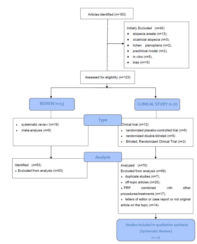
with platelet-rich plasma. Of the 163 articles initially identified, 123 articles focusing on AGA were
selected and, consequently, only 12 clinical trials were analyzed. The studies included had to match
predetermined criteria according to the PICOS (patients, intervention, comparator, outcomes, and
study design) approach. In total, 84% of the studies reported a positive effect of PRP for AGA
treatment. Among them, 50% of the studies demonstrated a statistically significant improvement
using objective measures and 34% of the studies showed hair density and hair thickness improvement,
although no p values or statistical analysis was described. In total, 17% of the studies reported
greater improvement in lower-grade AGA, while 8% noted increased improvement in higher-grade
AGA. Only 17% of the studies reported that PRP was not effective in treating AGA. The information
analyzed highlights the positive effects of PRP on AGA, without major side effects and thus it be may
considered as a safe and effective alternative procedure to treat hair loss compared with Minoxidil®
and Finasteride® .
Keywords: regenerative plastic surgery; regenerative medicine; androgenetic alopecia; AGA; hair
growth; platelet-rich plasma; PRP; hair loss
1. Introduction
The number of articles evaluating autologous platelet-rich plasma (PRP) efficacy in androgenic
alopecia (AGA) have exponentially increased during the last decade (2009–2019).
Autologous activated PRP (AA-PRP) and autologous not-activated PRP (A-PRP) are considered
standard routine for dermatologists and plastic surgeon experts in hair growth (HG). Preparation
procedures are not standardized yet and regenerative mechanisms are still the object of study due to
their various growth factors (GFs). The effects of the GFs contained in AA-PRP and A-PRP in HG have
been reported [1–4].
Int. J. Mol. Sci. 2020, 21, 2702; doi:10.3390/ijms21082702 www.mdpi.com/journal/ijmsInt. J. Mol. Sci. 2020, 21, 2702 2 of 26
In particular, the anti-apoptotic effect of A-PRP and AA-PRP has been suggested as one of the most
important factors stimulating HG via the activation of the Bcl-2 protein (anti-apoptotic regulator) and
Akt signaling, improving the survival of dermal papilla cells (DPCs) during the hair cycle (H-C) [1–4].
Additionally, the upregulation of fibroblast growth factor-7 (FGF-7)/b-catenin signaling pathways
with A-PRP treatment has been suggested to stimulate HG by inducing hair follicle stem cell (HFSC)
differentiation as well as prolonging the anagen phase of the HG cycle [1–4]. It also seems to stimulate
the perifollicular vascular plexus via the increase of vascular endothelial growth factor (VEGF) and
platelet-derived growth factor (PDGF) levels, which have angiogenic potential [1–4].
AGA is characterized by the miniaturization of follicles with a diminishment of the anagen phase,
accompanied by an increase in the percentage of resting hair follicles (HFs) and the telogen phase,
producing microscopic hairs [5]. Additionally, lymphocytes and mast cells were identified around the
miniaturized follicles [6], in the bulge area [7].
In a scalp suffering from hair loss (HLs), HFSC numbers remain unaltered, though the number of
more actively proliferating progenitor cells particularly diminishes [8]. This concept suggests that a
bald scalp either does not have an activator or has an inhibitor of hair follicle (HF) growth.
The total number of articles published on PRP in HLs is considerable (163 articles were identified
in PubMed). The results are very encouraging for the vast majority of the identified articles.
As previously described [1–4,7–9], there are several methods, kits, and procedures aimed at the
preparation of PRP. The differences depend on the centrifugation’s time and g-force or revolutions used
(revolutions per minute -RPM-), platelet’s amount, GFs, and chemokine release. These differences are
increased by wide biological and temporal variation [5]. Consequently, it seems to be very difficult to
select which methods, kits, and procedures for PRP are better [6] or which are more or less adequate
for treating different types of HLs. The efficacy of autologous PRP in patients who suffer AGA is clear
and it has also been reported several times by authors [1–4,7–9].
Autologous regenerative procedures are represented beyond the PRP, also from adult stem
cell-based therapy (ASCs-BT) to HFSC injections. Most recently, many articles [1,2,10,11] showed the
efficacy of autologous micro-grafts, containing unexpanded HFSCs, obtained by mechanical detachment
and slow centrifugation of scalp micro fragments (2 mm) according to minimal manipulation rules,
with promising results. HFSCs may be considered a cell population containing human hair follicle
mesenchymal stem cells (HF-MSCs) and human hair follicle epithelial stem cells (HF-ESCs) [10].
In addition, HF-MSCs, obtained by the intra- and an extra-dermal portion of the scalp, are named
human intra and extra dermal adipose tissue-derived hair follicle stem cells (HD-AFSCs) as previously
described [11].
In the present systematic literature review, the effectiveness of an autologous regenerative therapy
focused exclusively on PRP treatment for AGA was evaluated, with the conclusion that PRP was
effective in promoting HG in most articles.
2. Methods
2.1. Institutional Guidelines
The human autologous PRP is considered an emocomponent for non-trasfusional use, and thus
it is subject to regulations that define transfusion activities. In order to better understand the sense
of the current European Rules, it is necessary to differentiate between emocomponents for topical or
infiltrative use and those used in cell therapy, which involves complex techniques of bioprocessing of
therapeutic cells.
Reference is made to Regulation n.1394/2007 of the European Parliament (EC) and by the reflection
paper on the characterization of cutting edge treatment medicinal products draft concurred, 20 June
2014 European Medicines Agency (EMA)/Committee for Advanced therapies (CAT)/600280/2010 Rev 1,
in which the autologous applications in one-step surgery, minimal manipulations, and omofunctionalInt. J. Mol. Sci. 2020, 21, 2702 3 of 26
utilization are situations that do not require good manufacturing practices (GMPs) rules for processing,
good clinical practices (GCPs) for the clinical application, and the ethical committee’s endorsement.
It is necessary to highlight the differences between the productions of emocomponents suitable for
cell therapy compared with those directed to topical or infiltrative use. While the first require special
handling procedures and details of product derivation, the second, where the effect is extrinsic and an
amplification of the physiological function at the site of insertion, is much simpler, with the product
being easily derived by simple physical means, but nonetheless, it is important to include it within the
legislation while not limiting its use. All the rules have a common purpose: To guarantee the quality
and safety of the procedures and the products of transfusion medicine. The European rules related
to the use of PRP were represented both by Decree of 9 November 2007, n. 207, ‘Implementation of
Decree 2005/61/EC in means of traceability of blood components intended for transfusion and the
notification of adverse and severe reactions’, and by the Legislative Decree of 9 November 2007, n. 208,
‘Implementation of Directive 2005/62/EC relating to a quality system of blood’.
Currently, the PRP preparation must be performed respecting in Italy “Law-Decree of the Blood, 2
November 2015”, dispositions related to the quality and safety parameters of blood and emocomponents,
in which all patients receive detailed oral and written information about the study, including the risks,
benefits, and alternative therapies, and sign an informed consent form before any study procedures,
according to trasfusional service.
In this law-decree, it is established that each PRP procedure must take place in a structure
authorized by the reference blood transfusion service by means of a specific agreement. This Italian
law-decree established:
- Quantity of platelets to be obtained (1 × 10⁶ µL ± 20%);
- Exclusion criteria (platelets disorders, thrombocytopenia, anti-aggregating therapy, bone marrow
aplasia, uncompensated diabetes, sepsis, and cancer);
- Fields of application of the PRP only on the basis of available scientific evidence and guidelines
of the national blood center;
- Methods of preparation of the PRP (kits and procedure);
- How to use the PRP (only topical or infiltrative);
- Quality and sterility checks on the sample obtained;
- Blood volume to withdrew (within 55 cc for each patient);
- The volume of A-PRP and AA-PRP to be obtained (depending on the extension of the AGA area);
- Labeling of each sample of PRP;
- Informed consent;
- Adverse reaction form; and
- Data processing module.
All the PRP procedures must be performed in accordance with the European rules and institutional
guidelines, and they must be conducted following the principles outlined in the Declaration of Helsinki
and internationally consented ethics in clinical research [12], performing a quality assessment based on
the Strengthening the Reporting of Observational studies in Epidemiology (STROBE) checklist [13]
and respecting the national laws representing in Italy by “Law-Decree of the Blood, 2 November 2015”.
For all physicians that want to use PRP in this field, the authors suggest strictly respecting the
guidelines highlighted in the Italian decree, as it was reported to follow all the GCPs. This systematic
review was the object of a research contract (R. D. 1467/2017) between the first author and the University
of Rome “Tor Vergata”, Italy.
2.2. Search Strategy
Due to the growing interest in hair restoration, a number of investigations have been conducted
to assess the efficacy of A-PRP and AA-PRP as a treatment modality for hair loss. A systematic reviewInt. J. Mol. Sci. 2020, 21, 2702 4 of 26
protocol was developed in accordance with the Preferred Reporting for Items for Systematic Reviews
and Meta-Analyses-Protocols (PRISMA-P) guidelines. The search was conducted in accordance
with the PRISMA guidelines and the Cochrane handbook. A multistep search of the PubMed,
MEDLINE, Embase, PreMEDLINE, Ebase, CINAHL, PsycINFO, Clinicaltrials.gov, Scopus, and
Cochrane databases was performed to identify studies on HLs treatment with PRP searching without
a language or publishing-time restriction; 163 articles using the keyword “platelet-rich plasma
hair loss” and, 155 articles using the keyword “platelet-rich plasma androgenetic alopecia” were
identified. Of the 163 articles initially identified, 53 articles were reviews (including 19 systematic
reviews and 9 meta-analyses), 70 articles were clinical studies in AGA (including 12 clinical trials
-randomized placebo-controlled trial/randomized, double-blind, placebo- and active-controlled,
half-head study/double-blind, placebo-controlled pilot study/blinded, randomized clinical ttrial-), 13
article were related to alopecia areata, 3 articles to cicatricial alopecia, 2 articles to lichen planopliaris, 2
articles were pre-clinical models (mouse and rat), 5 articles were in vitro studies, and 15 articles were
identified as biased (not correct match with the key word used).
2.2.1. Study Assessment
The aim of the present systematic review was to assess the selected studies comparing local
injections of PRP compared to any control for AGA. Studies included in this article had to match
predetermined criteria according to the PICOS (patients, intervention, comparator, outcomes, and
study design) approach. Criteria for inclusion and exclusion are specified as following: P-Patients
(-inclusion criteria- age 18–76 years, males who showed AGA or male pattern hair loss (MPHL) in
stage I–V controlled by the Norwood–Hamilton classification scale and females with AGA or female
pattern hair loss (FPHL) in stage I–III controlled by the Ludwig classification scale; and exclusion
criteria: Other types of alopecia, alopecia areata, cicatricial alopecia, lichen planopliaris, patient with
platelets disorders, thrombocytopenia, anti-aggregating therapy, use of pharmacological therapeutics
targeting AGA as Finasteride® , similar drugs, and/or antiandrogens in the earlier year, bone marrow
aplasia, uncompensated diabetes, sepsis, cancer, an MPHL in stages over VI degree, a FPHL in stages
over III degree, use of topical medicines for AGA as lotions as Minoxidil® , prostaglandin analogs,
retinoid, or corticosteroids in the earlier year); I-Intervention (inclusion criteria: Local application
of autologous PRP; exclusion criteria: Combined use of PRP with other products); C-Comparator
(inclusion criteria: Any type of control, internal, external and different product; exclusion criteria:
Not applied); O-Outcomes (inclusion criteria: Hair count, hair density, hair thickness and hair
color improvement; hair loss reduction; exclusion criteria: Not applied); S-Study design (inclusion
criteria: Clinical trial, randomized placebo-controlled trial/randomized, double-blind, placebo- and
active-controlled, half-head study/double-blind, placebo-controlled pilot study/blinded, randomized
clinical trial; exclusion criteria: Reviews, expert opinion, comments, letter to editor, case report,
preclinical model (animal studies), in vitro studies, articles identified as bias (not correct match with
the key word used, group of study < 10 patients, shorter follow up than 3 months). No limitations
were applied on ethnicity or method of PRP processing.
This systemic review, performed on the PICOS approach in which only randomized
placebo-controlled trials/randomized, double-blind, placebo- and active-controlled, half-head
study/double-blind, placebo-controlled pilot study/blinded, randomized clinical trials, focused on
PRP in AGA were analyzed, is considered an EBM 1a level study according the Oxford Centre
for Evidence-Based Medicine (OCEBM), March 2009 (https://www.cebm.net/2009/06/oxford-centre-
evidence-based-medicine-levels-evidence-march-2009/).
2.2.2. Study Selection
Only AGA articles were considered and, for this reason, a total of 40 articles related to alopecia
areata (n = 13), cicatricial alopecia (n = 3), lichen planopliaris (n = 2), pre-clinical model (n = 2), in vitro
(n = 5), and bias (n = 15) were excluded initially.Int. J. Mol. Sci. 2020, 21, 2702 5 of 26
In total, 123 articles focused on AGA were identified and selected using Prisma Flow [14]
(www.prisma-statement.org) (Scheme 1). Consequently, it was decided to include only clinical trials
with male and female patients diagnosed with AGA, also referred to as MPHL or FPHL. In total, 53
Int. J. Mol. Sci. 2020, x, x FOR PEER REVIEW 5 of 32
articles were excluded as they were reviews, 7 articles were excluded as they were duplicate studies,
20 articles were excluded as they were off-topic, 17 articles were excluded as they assessed PRP in
combination with other procedures/treatments, and 14 articles were excluded as commentaries or
combination
letters of the editorwith or other
caseprocedures/treatments, and 14
reports or not original articles onarticles were
the topic. excluded
Twelve asstudies
original commentaries
were or
letters of the editor or case reports or not original articles on the topic. Twelve
included in this systemic review. These 12 studies were evaluated and summarized by their study original studies were
included
characteristicsin thisand systemic review. These
study outcomes (Table 12
1),studies were
treatment evaluated
protocols, andand summarized
mode by their study
of PRP preparation
characteristics
(Table 2). and study outcomes (Table 1), treatment protocols, and mode of PRP preparation
(Table 2).
Scheme
Scheme1.1.CONSORT
CONSORT(Consolidated Standards
(Consolidated of Reporting
Standards Trials)
of Reporting flow diagram.
Trials) flow diagram.
Table 1. The study design and results of the included studies. Abbreviations: M, male; F, female; wks,
weeks; mos, months. *p value not reported.Int. J. Mol. Sci. 2020, 21, 2702 6 of 26
Table 1. The study design and results of the included studies. Abbreviations: M, male; F, female; wks, weeks; mos, months. *p value not reported.
Characteristics of
Objective Assessment of Hair Subjective Assessment
Authors Study Type Enrolled Subjects Objective Measures Year Ref
Growth of Hair Growth
(Completed Study)
Randomized Controlled Blinded Half-head
1. Mean number of hairs (digital and
Patients reported less
dermoscopic imaging)
26 (26) depilation when
2. Mean HCS of hairs (digital and 1. Yes*
16 M, 10 F, aged 28–59, shampooing, greater
Takikawa et al. - Yes No No dermoscopic imaging) 2. Yes (p < 0.01) 2011 [15]
thin hair in the frontal bounce/resilience of hair,
3. Epidermal thickness, collagen and 3. Yes*
or parietal areas maintenance of healthy
blood vessel density around hair follicles
hairs
(4-mm punch biopsy)
64 (64)
1. Hair count and hair thickness
42 M, mean age 28,
using Jaeschke 15-point scale rating of 1. Yes (mean change in clinical
Schiavone et al. - No No No stage II–V; 22 F, mean N.a 2014 [16]
clinical change (macrophotographs rating of 3.2 and 3.9)*
age 32,
examined by 2 independent evaluators)
Stage I–II
Patient self-assessment
questionnaire: mean
22 (20) 1. Hair pull test 1. Yes* result rating of 7.1 on a
18 M, aged 24–72, stage 2. Hair density and quality 2. Yes, p < 0.001; overall 1–10 scale; 85% reported
Gkini et al. No No No No 2014 [17]
II-5a; 2 F, aged 58–72, (dermoscopic photomicrographs and improvement in hair density improvement in hair
Stage I macroscopic photographs) and quality per photographs quality and thickness;
65% reported increases in
hair density
1. Yes (81.81% achieved a
negative pull test at 12 wks.)
1. Hair pull test Patient satisfaction
11 (11) 2. Yes (average mean gain
2. Hair count (Trichoscan) questionnaire: mean
Khatu et al. No No No No 11 M, aged 20–40, stage of 22.09 follicular units/cm2 ) 2014 [18]
3. Hair loss (clinical examination, overall satisfaction rating
II–IV 3. Yes (moderate improvement
macroscopic photos) of 7 out of 10
in hair volume and coverage
with reduction in hair loss)
#1–4: Computerized phototrichogram
and global photography:
1. Hair count
1. Yes (p < 0.0001) at 3 mos
2. Hair density Physician and patient
10 (10) 2. Yes (p < 0.0001) at 3 mos
3. Terminal hair density global assessment
Cervelli et al. Yes Yes Yes Yes 10 M, aged 20–52 stage 3. Yes (p = 0.0003) at 3 mos 2014 [8]
4. Epidermal thickness and hair follicle scale—results not
IIa–IV 4. Yes (p < 0.05) at 3 mos
density (3-mm punch biopsy) reported
5. Yes (p < 0.05) at 14 wks.
5. Percentage of Ki67+ keratinocytes &
blood vessel density
(immunohistochemistry)Int. J. Mol. Sci. 2020, 21, 2702 7 of 26
Table 1. Cont.
Characteristics of
Objective Assessment of Hair Subjective Assessment
Authors Study Type Enrolled Subjects Objective Measures Year Ref
Growth of Hair Growth
(Completed Study)
Randomized Controlled Blinded Half-head
#1–3: Computerized phototrichogram
and global photography:
1. Yes (p < 0.0001)
1. Hair count and total hair density
2. Yes (p = 0.0003)
2. Terminal hair density Physician and patient
23 (20) 3. Yes (p < 0.05)
3. Epidermal thickness and hair follicle global assessment
Gentile et al. Yes Yes Yes Yes 20 M, aged 19–63 stage 4. Yes (p < 0.05) 2015 [3]
density (3-mm punch biopsy) scale)—results not
IIa–IV 5. Four patients reported
4. Keratinocyte proliferation and small reported
progressive hair loss at 12–16
blood vessel proliferation around hair
mos
follicles (immunohistochemistry)
5. Relapse of AGA
1. Yes, pulled hair count was
reduced by 65% (vs. 0% in
controls)*
20 (20) 1. Hair count (hair pull test)
2. Yes, hair growth noted in 6
Singhal et al. No Yes No No 16 M, aged 25–32 4 F 2. Hair growth, hair volume, hair N.a 2015 [19]
patients after 7 days but in 4
aged 32–35 quality, fullness (global photographs)
patients after 15 days; yet, all
patients (10) had good hair
growth after 3 mos*
#1–6: Phototrichogram and global PRP vs. placebo:
25 (24) photography 1. Anagen hair (%) 1–3, 5, 6. No (p > 0.05) 4. Yes, at
Alves and 11 M, aged 18–65, stage 2. Telogen hair (%) 3 and 6 mos (p < 0.05)
Yes Yes Yes Yes N.a 2016 [20]
Grimalt II–V; 11 F, aged 18–86, 3. Anagen: telogen ratio PRP vs. baseline:
Stage I–II 4. Hair density 1–5. Yes (p < 0.05)
5. Terminal hair density 6. Hair count 6. No (p > 0.05)
13.3% of treatment group
vs. 0% of control group
reported substantial
improvement in hair loss,
1. Hair count (photography) rate of hair loss, hair
26 (26) 1. No (p = 0.503) 2. No
Puig et al. Yes Yes Yes No 2. Hair mass index (Cohen HairCheck® thickness, and ease of 2016 [21]
26 F, stage II (p = 0.220)
system managing/styling hair;
26.7% of treatment group
vs. 18.3% of control
group reported feeling
coarser/heavier hair
19 (17) 1. Terminal hair count (magnifying
1. No (p = 0.25 at 6 mos) 2. No
Mapar et al. Yes Yes Yes Yes 17 M, aged 24–45, stage glass) n.a. 2016 [22]
(p = 0.23 at 6 mos)
IV–VI 2. Vellus hair count (magnifying glass)Int. J. Mol. Sci. 2020, 21, 2702 8 of 26
Table 1. Cont.
Characteristics of
Objective Assessment of Hair Subjective Assessment
Authors Study Type Enrolled Subjects Objective Measures Year Ref
Growth of Hair Growth
(Completed Study)
Randomized Controlled Blinded Half-head
Patient self-assessment
questionnaire: treatment
group reported 30 ±
1. Yes (39.7 ± 16.5% increase 13.1% mean improvement
1. Hair density (CapilliCare trichoscan) compared to baseline)* (range 10–70%); 93.3%
30 (30)
2. Hair diameter (CapilliCare trichoscan) 2. Yes (39.8 ± 17.2% increase reported complete
Gupta et al. - No No No 30 M, aged 25– 35 stage 2017 [23]
3. Independent observer clinical compared to baseline)* cessation of hair fall by 2
III–VII
evaluation (global macrophotographs) 3. Average improvement = 30.2 mos; 66.7% reported
± 12.2% increase in hair growth;
36.7% reported
improvement in hair
texture
#1–4 Computerized phototrichogram 1.
Hair density Patient self-satisfaction
2. Hair diameter score following a Likert
3. Terminal/vellus-like hair ratio scale: 7 = very satisfied,
4. Thin/regular/thick hair shafts among 6 = satisfied, 5 =
terminal follicles indifferent, 1 =
1. Yes (p < 0.05)
5. Independent observer clinical unsatisfied, and
19 (19) 2. Yes (p < 0.05)
evaluation (mean improvement score 0 = very unsatisfied; most
12 M, aged 27–60, stage 3. Yes (p < 0.05)
using global macro-photographs) patients (15/19) declared
Anitua et al. No No Yes No III–VI; 4. Yes (p < 0.05) 2017 [24]
6. Epidermal thickness perivascular noticeable hair loss
2 F, aged 32–60, stage 5. Yes; 0.75/1*
inflammatory infiltrate, rete ride number, decrease, 13/19 declared
II-frontal 6. Yes (p < 0.05 for most) 7. Yes
terminal/miniaturized hair ratio, and noticeable improvement
(p < 0.05 for most)
collagen, reticular fiber and elastic fiber in hair quality and
mesh quantity (3 mm punch biopsies) appearance, and 11/19
7. Proliferative epidermal/follicular cells, stated they would
newly formed blood vessels, and continue with PRGF
presence of bulge stem cell niches treatment
(immunohistochemistry)Int. J. Mol. Sci. 2020, 21, 2702 9 of 26
Table 2. Treatment protocols for the included trials. Abbreviations: n◦ treat, number of treatments; Int, interval; Centrif. Time, centrifugation time.
PRP n◦ Centrif. Blood
Authors Int Max F-up Type of PRP Injections Protocol Activators RPM orG PRP Volume
Treat Time Volume
Subcutaneous injection (3 mL) into
Takikawa selected 1 × 1 cm areas measured from a. 1700 rpm a. 15 min
5 2–3 wks 12 wks Manual Double Spi - 15 mL 3 mL
et al. [15] the nasal tip and upper part of the b. 3000 rpm b. 5 min
auricular base
After local anesthesia (xylocaine 1%,
with adrenaline 1:100,000) was a. 6–8 mL PRP + 3–4
administered, cutaneous inflammation GPS III Platelet Separation No (Scalp roller mL of plasmatic
Schiavone was induced via application of gentle System used to favor– a. 60 mL b. protein concentrate =
2 3 mos 6 mos - -
et al. [16] pressure using 1.0-mm-deep Scalp-roller a. Single spin at baseline b. platelet 40 mL 9–12 mL; 0.2–0.3 mL
to favor activation of injected platelets; Double spin at 3 months activation per injection
then, superficial injections were b. Same as above
administered 1 cm apart
Injections (0.05–0.1 mL/cm2 ) were Calcium
21 days
performed using nappage technique in gluconate
Gkini 3 (+1 (booster 6 RegenA-PRPCentri (Regenlab) 6 mL (0.05–0.1
1 year affected areas to a depth of 1.5–2.5 mm; a (0.1 mL per 0.9 1500× g 5 min 16 mL
et al. [17] booster) mos after Single spin method mL/cm2 )
specific area was checked at all times by mL of PRP; 1:9
onset)
defining a “V” (Kang’s point) ratio)
Nappage technique injections (2–3 mL)
into a prefixed 1 × 1 cm squared area
Calcium
Khatu over the right parietal area; anesthetic a. 1500 rpm a. 6 min b.
4 2 wks 12 wks Manual Double Spin chloride (1:9 20 mL 2–3 mL
et al. [18] cream was applied before each treatment b. 2500 rpm 15 min
ratio)
after cleaning the skin with cetavlon,
spirit, and povidoneiodine
1 year (at
Intradermal injections (0.1 mL/cm2 ) into
baseline and
2 of the 4 selected halves (e.g., frontal or
14 wks, 6
Cervelli parietal) (placebo was injected into the
3 4 wks mos, and Cascade-Selphyl-Esforax Kit Ca2+ 1100× g 10 min 18 mL 9 mL
et al. [8] other 2 halves) after the scalp was
12 mos after
cleansed with 70% alcohol; local
initial
anesthesia was not used
treatment)
Interfollicular injections of PRP (0.1
2 years (at mL/cm2 ) within 2 of the 4 selected areas
a. Cascade-Selphyl- Esforax a. 1100× g
baseline and of the scalp (physiologic solution into a. 10 min
system a. Ca2+ b. 1200 rpm a. 18 mL a. 9 mL
Gentile 2, 6, 12, 16, the other 2 areas), after cleaning skin b. 10 min
3 4 wks b. PRL platelet-rich b. Nothing c 1200 rpm + b. 55 mL b. 20 mL
et al. [3] and 23 mos with 70% alcohol; target areas were c. 10 min
lipotransfert system c. C-punt c. Nothing double spin c. 55 mL c. 20 mL
after initial marked with semi-permanent tattoos for +10 min
system 1900 rpm
treatment) subsequent treatment and evaluation;
local anesthesia was not used
Injections using nappage technique
3 mos (at (multiple small injections in linear Calcium
Singhal a. 1500 rpm a. 6 min
4 2–3 wks 1-wk pattern 1 cm apart) after area was Double spin method chloride (9:1 20 mL 8–12 mL
et al. [19] b. 2500 rpm b. 15 min
intervals) cleansed with spirit and ratio)
povidone-iodineInt. J. Mol. Sci. 2020, 21, 2702 10 of 26
Table 2. Cont.
PRP n◦ Centrif. Blood
Authors Int Max F-up Type of PRP Injections Protocol Activators RPM orG PRP Volume
Treat Time Volume
Injections (0.15 mL/cm2 ) within four 1 ×
1 cm selected circular areas of the frontal
and occipital scalp (marked with a dot
Alves and 6 mos (at Calcium
tattoo) depending on the
Grimalt 3 4 wks 3-mo Single spin method chloride (10%, 460× g 8 min 18 mL 3 mL
treatment-designated side of the scalp
[20] intervals) 0.15 mL)
(vs. control side of the scalp received
placebo (normal saline); no local
anesthesia was used
Single subcutaneous injection within the
26 wks (at 4 cm2 area in the central scalp (termed
Puig et al.
1 N.a 4-wk the “hair check data box”), after Angel PRP system (Cytomedix) Nothing - - 60 mL 10 mL
[21]
intervals) anesthesia (2% lidocaine and 0.5%
bupivacaine) was administered
Injections (1.5 mL of PRP) within one of
two 2.5 × 2.5 cm square regions, at least
3 cm apart, in the scalp randomly
6 mos (at 1, assigned to be a case square (control Calcium
Double spin method using
Mapar 3, and 6 mos square received 1.5 mL of normal saline); gluconate a. 3000 rpm a. 6 min
2 4 wks Tubex PRP tube (Moohan 9 mL 1.5 mL
et al. [22] after initial randomization of case and control (0.1 mL per mL b. 3300 rpm b. 3 min
Enterprise)
treatment) squares was performed using a random of PRP)
number table; iron oxide- and titanium
dioxide-containing substances were
used to tattoo the corners of the squares
Scalp was activated by micro-needling;
Gupta
6 2 wks 6 mos then, PRP was massaged into the vertex Double spin method - - - - -
et al. [23]
of the scalp (10 cm from the glabella)
1 mo for first
PRGF activator
4 sessions;
Anitua Intradermal injections of PRGF into (BTI
5 final session 1 year Single spin method 580 rpm 8 min 18 mL 3–4 mL
et al. [24] hair-depleted areas Biotechnology
7 mos after
Institute)
start pointInt. J. Mol. Sci. 2020, 21, 2702 11 of 26
2.2.3. Data Extraction
Data were independently collected by one investigator (PG) and checked by a second investigator
(SG) only from the retrieved articles. Any disagreement on the collected data was settled by a consensus
among PG and SG. No attempt was made to obtain specific or missing data from the authors. The
following data were extracted: First author, year of publication, study design, number of patients, type
of procedure, and primary and secondary outcomes.
The quality of the included studies was independently assessed using two investigators (PG
and SG) using the Cochrane Collaboration’s Risk of Bias Assessment tool for RCT15 while using the
Newcastle–Ottawa Scale to evaluate the individual non-randomized studies [25].
2.2.4. Outcome Measures
The primary outcome was the difference in hair density (HD), number of hairs per cm2 , and hair
count (HC), number of hairs per 0.65 cm2 . Secondary outcomes were hair thickness (HT) increase, hair
re-growth (HRG), and hair cross-size (HCS) percentage increase.
All results collected from the studies were reported with the same measurements retrieved from
the papers. From one paper, percentages were calculated from the patients’ individual data displayed
in the paper [15]. The patient’s contralateral scalp was used as a control in some of the included papers,
while in other studies, patients were respectively allocated into study groups when they underwent
PRP and to the control group when they underwent the placebo or other treatments. Missing data
were dealt with according to previously validated estimations [26,27].
2.3. Brief History Analysis of PRP Use in Androgenetic Alopecia
In total, 137 articles focusing on the use of PRP in AGA were published from 2015 to 2019 whereas
only 18 were published before (range 2011–2014). Selecting original articles alone and excluding other
types, Takikawa et al. [15] reported for the first time (2011) the effects of PRP-containing dalteparin and
protamine microparticles (D/P MPs) on HG; Kang et al. [28] reported, in 2014, the clinical efficacy of
interfollicular injection of a CD34+ cell-containing PRP preparation for patterned HLs; in the same year,
Cervelli and Gentile [8] reported for the first time in the literature the clinical and histomorphometric
evaluation of an AA-PRP injection in patients affected by AGA. In 2015, Gentile et al. [3] reported the
most important (for the journal’s impact factor, actually SCTM 5,9 IF) randomized placebo-controlled
clinical trial, including histological and trichospic analysis of the effect of AA-PRP and A-PRP in AGA.
Rodrigues et al. [29] reported recently (March 2019) the most important (for the journal’s impact
factor, actually J Am Acad Dermatol 7,01 IF) double-blind controlled study, including platelet number
and growth factor level analysis of the effect of PRP in AGA.
2.3.1. A-PRP and AA-PRP Devices for Hair Regrowth
Seven different devices were clearly identified and analyzed. Currently, each of them could have
a number of references that refer to commercial variants of a combination of fungible but that have a
common denominator that is the device described. Used devices sorted alphabetically by trade name
are as follows: Angel® (Arthrex, Inc. Corporate Naples, Florida, 1370), Cascade® (Musculoskeletal
Transplant Foundation, Edison, NJ 08837) and Selphyl® (Factor Medical, LLC Langhorne, PA 19047),
C-Punt® (Biomed Device, MO, Italy, 41126), i-Stem® Preparation System (i-Stem, Biostems, Co., LTD.,
Seoul, South Korea 138–843), MAG-18® (DTS MG Co., Ltd., Seul, Korea #B108-147), MyCells® (Kaylight
Technologies Ltd., Holon, Israel), and Regenlab® (En Budron b2, 1052 Le Mont-sur-Lausanne, Swiss).
The Angel® device allows the selction of the degree of the platelet concentration in a wide range
(3 to 18x ). It was used to prepare A-PRP (3 mL) from a large volume of peripheral blood (120 mL).
x
The A-PRP was then combined with 5 mL of platelet-poor plasma (PPP) to produce 8 mL of A-PRP
with a 5-fold increase in the platelet concentration over the whole blood. The A-PRP collected was
then triggered via the addition of 10% (v/v) calcium gluconate to obtain AA-PRP [4].Int. J. Mol. Sci. 2020, 21, 2702 12 of 26
Using the Cascade® or Selphyl® device, AA-PRP was prepared from a small volume of blood
(18 mL) collected in two different tubes (9 mL each one) from a peripheral vein using sodium citrate
(ACD) as an anticoagulant. The tubes were centrifuged at 1100× g for 10 min, with the final aim
of obtaining a platelet pellet; later the suspension contained in the tubes was activated through the
switch into two tubes containing CaCl2+ to induce platelet activation and exocytosis of the alpha
granules [3,8].
C-Punt® consists of a 60-mL syringe in which whole blood (55 mL) was collected from a peripheral
vein using sodium citrate as an anticoagulant. The syringe was centrifuged at 1200 rpm for 10 min;
later, the autologous platelet suspension PPP and PRP obtained, in an amount of 23 mL, was inserted
in a platelet selector device, and at the end of the procedure, 9 mL of A-PRP was harvested [3,4,9].
Using an hourglass system, the i-Stem® Preparation System, autologous blood (17.7 mL) was
harvested by adding ACD as an anticoagulant (2.2 mL). After the first spin (centrifugation at 3000 rpm
for 6 min), the PPP portion (1 mL) and RBCs (red blood cells) (2 mL) were removed and the suspension
was re-centrifuged for the second time (3000 rpm for 3 min). At the end of the procedure, 15 mL of
A-PRP were obtained [2].
Mag-18 PRP® is a hourglass system in which 18 mL of whole blood and 1 mL of ACD were
collected and centrifuged two times; the first time at 3000 rpm for 10 min and second time at 3400 rpm
for 6 min. Then, 1.5 mL of A-PRP were obtained in the middle portion of the hourglass, indicated as a
buffy-coat. It is very similar to the i-Stem® Preparation System and may be considered the evolution
protocol [2].
PRP Regen Blood Cell Therapy® tubes were used to obtain A-PRP (15 mL, 5 mL per BCT tube)
from whole blood (24 mL) taken from a peripheral vein using ACD. The top 2 mL of A-PRP from
each tube was then discarded, giving 9 mL of A-PRP. Alternatively, a Kit RegenLab® (Regen Lab SA,
Le Mont-sur-Lausanne, Switzerland) was used to process 40 mL of venous peripheral blood. Blood
was collected in five ATS (autologous thrombin serum) Regen® tubes (8 mL each). All tubes were
centrifuged at 1500× g for 15 min. After the centrifugation, PRP activated (AA-PRP) by autologous
thrombin consolidated in the tube [4].
2.3.2. PRP and Growth Factors Assessment
A-PRP and AA-PRP contain at least six major GFs, including basic fibroblast growth factor (b-FGF),
epidermal growth factor (EGF), transforming growth factor-β (TGF-β), insulin-like growth factor-1
(IGF-1), PDGF, and VEGF, which are released after platelet activation [2]. Each one of these major GFs
is involved in a specific bio-molecular activity during HRG. In this way, in fact, it is possible to identify
different types of PRP preparations depending on their cell content and fibrin architecture as reported:
- Leukocyte-poor PRP (LP-PRP) or pure platelet-rich plasma (P-PRP). PRP without leukocytes and
with a low-density fibrin network after activation;
- Leukocyte-PRP (L-PRP). PRP with leukocytes and a low-density fibrin network after activation
(most frequent);
- Leukocyte-poor platelet-rich fibrin (LP-PRF) or pure platelet-rich fibrin (P-PRF). PRF without
leukocytes and a high-density fibrin network.
- Leukocytes platelet-rich fibrin (L-PRF). PRF with leukocytes and a high-density fibrin network.
As highlighted, there are too many protocols for the preparation of A-PRP and/or AA-PRP
depending on the different time and rpm used, the number of platelets, and the availability of GFs and
chemokines. There is also wide biological (between patients) and temporal (day to day) variation [5].
So, it is difficult to assess which kit for PRP preparation is better and which is worse [6].
In each case, the GFs serve to promote angiogenesis, follicular cell proliferation, and initiation of
cell division, thus having a fundamental role in HRG [1–4,9].
The list of GFs present in PRP and their suspected mechanism in the treatment of AGA is reported
in Table 3.Int. J. Mol. Sci. 2020, 21, 2702 13 of 26
Table 3. List of GFs identified in PRP and their suggested bio-molecular pathway in the treatment
of AGA.
Growth Factors Bio-Molecular Pathway in Hair Re-Growth
Improves perifollicular angiogenesis;
Elevated expression in dermal papilla cells during anagen phase;
VEGF
Endothelial cell-specific mitogen;
Micro-vascular permeability and perifollicular vascularization;
Improves the activity and growth of follicle outer-root sheath cells by
activation of Wnt/β-catenin signaling;
EGF
Cell growth modulator during follicular differentiation;
Proliferation and migration of follicular outer root sheath cells;
Improves the advancement of hair follicles;
Anagen phase induction via B-catenin expression;
FGF
Angiogenesis;
Dermal fibroblast and hair follicle mitogen;
Up-regulate the genes associated with HF separation, induction, and
control of anagen;
PDGF Angiogenesis and vascularization;
Hair follicle dermal stem cell proliferation;
Mesenchymal stem cell mitogen;
Improves the migration, survival, and proliferation of HF cells;
IGF-1 Hair follicle proliferation during development;
Increase hair density and inhibit apoptosis;
Enhance the proliferation of follicular epithelial cells
HGF Hair follicle elongation;
Inhibits catagen phase induction;
Stimulates the signaling pathways that manage the Hair cycle;
Extracellular matrix synthesis;
TGF-ß
Fibroblast and mesenchymal stem cell proliferation;
Hair folliculogenesis and maturation;
IL-6 Involved in WIHN through STAT3 enactment
Manages the IGF-1 effect and its connection with extracellular matrix
IGFBP-1 to -6
proteins at the Hair follicle level
BMP Maintains the DPC phenotype (fundamental for stimulation of HFSCs)
BMPR1 Maintains the proper identity of the DPCs (basic for explicit DPC work)
M-CSF Involved in wound-induced hair growth
M-CSFR Involved in wound-induced hair growth
Wnt3a Involved in HF advancement through β-catenin signaling
PGE2 Stimulates anagen in HF
PGF2α Enhance change from telogen to anagen
BIO GSK-3 inhibitor
PGD2 Enhances follicle regeneration
Iron and l-lysine95 Still under examination
2.3.3. Protocol: Manual Versus Mechanical and Controlled Hair Injection of A-PRP and AA-PRP
In 7 studies (58%) extracted by 12 clinical trials, the scalp was separated into several regions
(half-head placebo-controlled study) in which a targeted and controlled area were identified.
The A-PRP and AA-PRP injections were performed manually more frequently (75%; 9/12) versus
the mechanical and controlled injection (17%; 2/12), as published in all the articles identified by
Gentile et al. [3,8], while massage of PRP on the scalp was performed in one article only (8%) [23].Int. J. Mol. Sci. 2020, 21, 2702 14 of 26
Currently, it is not possible to accept that the infiltration of PRP into the scalp is done in a totally
empirical way through the use of one’s hands. In fact, in this case, it is not possible to perform a
homogeneous and precise infiltration. The PRP injection, done by the hand of a plastic surgeon or a
dermatologist, it not able to compete with the mechanical and controlled infiltration achieved by a
mesotherapy gun equipped with software capable of scheduling the amount of PRP delivered for each
cm2 (0.2 mL), the depth (5 mm), and inclination of the needle.
Gentile et al. [1–4,9] realized an inter-follicular infiltration (0.2 mL/cm2 ) to AGA-affected regions
at a depth of 5 mm utilizing a mechanical and controlled injection via the Ultim Gun® (Anti-Aging
Medical Systems, Montrodat, France) outfitted with a 10-mL Luer lock syringe with 30-gauge needle,
in three sessions spaced 30 days apart.
Regarding the number of sessions performed, different protocols have been proposed.
Rodrigues et al. [29] reported four subcutaneous injections of PRP. Hausauer et al. [30] described,
in a prospective randomized single-blinded trial conducted on 40 moderate AGA patients, two different
protocols based on sub-dermal PRP injections: Protocol 1, in which three monthly sessions with a
booster 3 months later were performed or protocol 2 in which sessions every 3 months were done. At
6 months, both protocols produced statistically significant increases in hair count (p < 0.001). These
improvements occurred more rapidly and were greater for patients who underwent protocol 1 (mean
percent change: protocol 1, 29.6 ± 13.6 vs. protocol 2, 7.2 ± 10.4; p < 0.001).
Schiavone et al. [31] performed two injections, with a 3-month interval between the two
interventions. Jha et al. [32] performed autologous platelet-rich plasma injections with micro-needling
over a period of 3 months at 3-week intervals. Tawfik et al. [33] performed weekly for a maximum
total of four PRP sessions in females affected by AGA. Mapar et al. [22] performed PRP injections in
two sessions, 1 month apart.
Gentile et al. [1–4,9] reported very interesting results from performing three treatments that were
administered to each patient at 30-day intervals. At the end of the three treatment cycles, the patients
showed a mean HC increase of 33.6 hairs in 0.65 cm2 and a mean increase in total HD of 45.9 hairs per
cm2 compared with baseline values (p < 0.001).
Takikawa et al. [15] performed five treatments of 3 mL of PRP and D/P MPs at 2- to 3-week intervals.
All details are reported in Tables 1 and 2.
3. Results
3.1. Results Performing Literature Scans: PRP Studies with Hair Density and Hair Count Improvement
In 2011, Takikawa et al. [15] performed the first controlled clinical trial of PRP containing D/P MPs
in 26 patients affected by frontal or parietal HLs. Solutions of either PRP with D/P MPs, or PRP and
saline were infiltrated at sites of HLs (13 patients each), with controls being the opposing sides with
equal HD. Twelve weeks later after five treatments, an increased mean HC was seen in both PRP- and
PRP&D/P MP-treated regions relative to control sites. Additionally, significantly increased HCS was
observed in both PRP- and PRP&D/P MP-treated areas relative to control areas. The patients treated
reported, via their own subjective evaluation, HLs reduction, and greater hair texture. Microscopic
evaluation of punch biopsies displayed a thickened epidermis, proliferation of collagen fibers and
fibroblasts, and greater amount of blood vessels around HFs in sites treated with PRP. No infections,
hematomas, or other severe side effects were observed, though patients referred to temporary pain at
the infiltration areas.
Several observational works published in 2014 all concluded that PRP may be considered effective
for male and female patients affected by AGA.
In particular, Schiavone et al. [16] (2014) performed an observational work in which 64 patients
received two infiltrations of L-PRP mixed with plasmatic proteins 12 weeks apart. Six months later, HC
and HT were visibly improved; an average of 40.6% of the patients treated reached at least a moderate
level of improvement.Int. J. Mol. Sci. 2020, 21, 2702 15 of 26
Gkini et al. [17] led a prospective cohort study with 20 patients. Three injections later, they
displayed increased HD compared to baseline at 3, 6, and 12 months after PRP (p < 0.001), as well as
improvements in HD and HT. In this report, milder forms of HLs represented by patients classified
as II◦ –III◦ degree of the Norwood–Hamilton scale responded better to the PRP procedure than more
advanced cases, producing more interesting results on the vellus. The researchers also suggested that
the PRP treatment appeared to lead to increases in HT more than HC.
Khatu et al. [18] also performed a very small prospective cohort study to evaluate the effects of
PRP in 11 patients. Four PRP injections later, nine patients reverted to having a negative hair pull
test. HD and HC were improved and, in particular, HC was noted to be increased from 71 to 93.09 on
average. Both Gkini et al. [17] and Khatu et al. [18] assessed patients’ satisfaction, finding an average
score of 7.1 and 7.0 out of 10, respectively.
Gentile et al. [3] performed for the first time in the literature (2015) a randomized blinded half-head
study evaluating the effects of an inter-follicular injection of PRP (0.1 mL/cm2 ) with 30-gauge needles,
in selected scalp sites. In this work, 23 males suffering from AGA were treated by performing the
PRP infiltrations (without local anesthesia) three times at intervals of 30 days, in which the scalp of
each patient was divided into four sites: Frontal, parietal, vertex, and occipital. PRP injections were
performed on half of the scalp, while the other side received saline as a placebo (Table 1). The article
reported a statistically significant increase in mean HC, HD, and terminal HD after three months of the
last PRP injection compared to saline.
Cervelli et al. [8] performed a similar study, finding equally interesting results. All outcomes
displayed a statistically significant improvement as reported in Table 1. In both articles, the histological
evaluation indicated that the epidermal thickness and density of follicles were both increased compared
to baseline (p < 0.05) two weeks after the last PRP injection.
Additionally, immunohistochemistry revealed that the percentage of Ki67+ cells was also increased
in both basal keratinocytes of the epidermis and hair follicle bulge cells at 2 weeks after the PRP
procedure (p < 0.05 compared with baseline), suggesting an increase in keratinocyte proliferation. The
researchers also observed an increase in the small blood vessel amount around the HFs (p < 0.05 at 2
weeks after PRP compared with baseline), confirming the concept that PRP stimulates angiogenesis
via the discharge of vascular GFs.
Another randomized blinded half-head study was performed one year later (2016) by Alves and
Grimalt [20], testing the PRP injections in 22 patients divided into two groups: Group a, treated with 3
mL of PRP on the right side of the scalp and 3 mL of placebo on the left; and group b, treated with
the same two suspensions on opposite sides of the scalp. Three and six months later, interesting
improvements in mean anagen hairs, mean telogen hairs, HD, and terminal HD in PRP-treated sites
were reported when compared with baseline (p < 0.05). Mean total HD was the only measure found to
be significantly increased in PRP versus placebo-treated sites (p < 0.05).
Singhal et al. [19] (2015) performed a controlled clinical trial to compare PRP with medical
treatments in 20 patients, with 8 males and 2 females in each treatment group. HG was observed in 6
patients after just 7 days and in 4 patients after 15 days. Three months later, all evaluated parameters
(Table 1) displayed superior results in PRP-treated patients than in the control group, although no
statistical analysis was shown on the analyzed data. In comparison, the patients who underwent
medical treatments displayed no improvement in the hair pull test or HG.
In an open-labeled pilot study performed by Gupta et al. [23] (2017) involving 30 male patients, in
which each one received six PRP massage treatments after the scalp was first activated by micro-needling,
interesting outcomes were reported. In fact, six months later, a significant increase in HT and HD was
reported by a blinded evaluator, displaying an improvement of 30.2 ± 12.2% (Table 1). The procedure
response was more significant in those with lower-grade HLs in terms of HT (p = 0.0446) and HD
(p = 0.0196). Efficacy was more evident in patients who had a shorter duration of disease prior to
therapy and patients without family HLs history, with an improvement in HT (p = 0.0485 and p = 0.0272,
respectively) and HD (p = 0.0096 and p = 0.0114, respectively).Int. J. Mol. Sci. 2020, 21, 2702 16 of 26
Anitua et al. [24] (2017) reported the results obtained after five injections of plasma rich in growth
factors (PRGF) in 19 patients affected by AGA. One year after the treatment, the mean HD, HT, and
terminal/vellus hair ratio displayed a statistically significant improvement (p < 0.05). Histological
analysis displayed an improvement in the epidermal thickness, peri-follicular neo-angiogenesis, and
terminal/miniaturized hair ratio, as well as decreased perivascular inflammatory infiltrates.
3.2. Results Performing Literature Scans: PRP Studies without Hair Density and Hair Count Improvement
Only two articles did not display a statistically significant improvement in the results assessed.
These articles were published by Mapar et al. [22] and Puig et al. [21], respectively.
Puig et al. [21] performed a double-blind randomized placebo-controlled multicenter trial involving
26 patients with FPHL. Fifteen females were randomized to the PRP group (study group) and 11 to the
placebo group (control group). Researchers marked a 4-cm2 area in the central part of the scalp, where
hair was repeatedly evaluated during the work using the HairCheck® . Patients of the study group
received one infiltration of either PRP or normal saline within 4 cm from this area at week 0 (Table 2).
At week 26, no statistically significant difference was found between the study and control groups
in terms of HC (Table 1). Patients of the study group did, however, report a subjective reduction of
the HL rate, and an improvement of HT, and ease of hair styling, which none of the control group
participants noted. This work was the only study published in which the patients received only one
PRP or placebo treatment.
The second article was a prospective half-head comparative pilot study performed by
Mapar et al. [22] on 17 male patients affected by AGA. Researchers performed PRP or normal
saline infiltration (Table 2) during two sessions 1 month apart. Outcomes displayed a mean decrease
in the number of terminal and vellus hairs six months after the PRP, which was assessed using only
a magnifying glass. Consequently, the researchers found no statistically significant difference in the
results obtained between the treated area and baseline.
3.3. Critical Assessment of Study Design
Among the articles reviewed, it is evident that a standardized and widely shared protocol for the
use of PRP is lacking, as well as standardized evaluation procedures.
In particular, there is a lack of a widely shared consensus regarding the preparation procedure,
eventual addition of activators (calcium chloride -CaCl- Ca2+; thrombin, calcium gluconate, etc.),
g-force, RPM and timing of centrifuge to be used, platelet concentration that must be attained, volume
of blood to be collected, and amount of PRP to be used. Three works, for example, reported the use of
CaCl as an activator [18–20], while two works reported calcium gluconate [17,22], one work reported
PRGF activator [24], and two other works reported Ca2+ [3,8]. Protocols also varied in the number of
sessions, time interval between treatments, administration procedure, and follow-up period.
In terms of study design, they ranged from pilot studies to randomized blinded trials, and
this variation has contributed to the difficulty in interpreting the results across the included studies.
The studies were stratified considering not only the use of PRP alone or PRP combined with other
treatments, but also sex, degree of HLs, sample size, randomization, and control groups, further
obscuring PRP treatment results.
Of the articles reviewed, seven mentioned the use of a control group [3,8,15,19–22], and five
conducted the study without it [16–18,23,24]. Five articles mentioned the randomization of patients
into study or control groups [3,8,20–22], while three specifically mentioned that they did not randomize
patients [17–19], potentially introducing bias. Although a major part of the articles included both male
and female patients [15–17,19,20,24], others included only males [3,8,18,22,23] or only females [21]. Since
male and female patterns of HLs have different manifestations and may have different mechanisms, it
may be inappropriate to extrapolate the results to both sexes in studies examining only a single sex.
Moreover, most of the investigations were compromised due to the small sample sizes. Most studiesInt. J. Mol. Sci. 2020, 21, 2702 17 of 26
enrolled only 10–30 subjects [3,8,17–24], and the largest article examined 64 patients [34]. All articles
had inclusion and exclusion criteria.
3.4. Side Effects
No major side effects, such as scarring, progressive worsening, or infections, were reported in
the analyzed articles. Only mild headache, tolerable and temporary pain during the procedure, mild
itching and desquamation, and transient edema were reported by some subjects after PRP injection.
3.5. Considerations
In total, 84% of the studies (10/12) displayed a positive effect of PRP for AGA treatment. Among
them, 50% of the studies (6/12) displayed a statistically significant improvement in the objective measures
(e.g., HD, HT) following treatment with PRP [3,8,15,17,20,24] and 34% of the studies (4/12) reported
hair improvement with PRP, although no p values or statistical analysis were described [16,18,19,23].
In total, 17% of the studies (2/12) reported a greater improvement in lower-grade AGA [17,23], while
one reported increased improvement in higher-grade AGA [16]. In total, 9% of the studies (only one),
a study conducted by Mapar et al. [22], concluded that PRP was not effective in treating AGA via
analysis of the terminal and vellus HC. However, in this study, only two treatments were performed,
and outcomes were assessed using a magnifying glass only, which may not be the best method to use to
measure the results. Further, neither an objective team evaluation or a subjective patient self-assessment
were performed.
Another study, by Puig et al. [21], did not report a significant improvement in HC or the hair mass
index after PRP treatment. In this work, however, only one treatment was performed and the PRP
injected was not activated, thereby impeding its full therapeutic potential. Nevertheless, subjective
improvement was reported by the patients as lower HLs and improved HT [21]. In the articles in
which no statistical analysis was displayed [16,18,19,23], the investigators remarked positively on
the subjective parameters on HG, volume, coverage, and mean HD. Overall, all the investigations in
which a minimum of three PRP treatments were performed displayed an improvement in at least one
objective measure.
Among the articles reviewed, it appears to be evident that a standardized protocol for PRP
preparation (kits/procedures/methods) and PRP injection (mechanical and controlled vs. manual) is
lacking, as well as standardized evaluation methods (trichoscan/phototricograms/magnifying glass).
Without such standardized parameters, it appears to be more difficult to adequately assess the
effectiveness of PRP between the different studies in AGA patients, in terms of HG improvement.
Performing this analysis, as briefly introduced before, certain methodological differences were noted.
Regarding the procedure of PRP preparation, a lack of consensus regarding the choice of preparation
method, eventual addition of activators, timing of centrifuge, G-force and RPM used, platelet
concentration, volume of blood collected, and amount of PRP injected was noted (Tables 2 and 3). In
total, 25% of the articles (3/12), for example, used CaCl as an activator [18–20], while 17% of the articles
(2/12) used calcium gluconate [17,22], 9% of the articles (only one) used PRGF activator [24], and other
17% of the articles (2/12) used Ca2+ for PRP activation [3,8].
Treatment protocols also varied in the number of sessions, time interval between procedures,
administration procedure, and follow-up period (Tables 2 and 3). In terms of the study design, the
articles analyzed ranged from pilot studies to randomized blinded trials.
4. Discussion
4.1. PRP Compared with Minoxidil® and Finasteride®
AGA produces a yearly worldwide market income of US$4 billion and a growth rate of 1.8%,
demonstrating a developing consumer market [35].You can also read