MTOR Signaling at the Crossroad between Metazoan Regeneration and Human Diseases - MDPI
←
→
Page content transcription
If your browser does not render page correctly, please read the page content below

International Journal of
            Molecular Sciences

Review
mTOR Signaling at the Crossroad between Metazoan
Regeneration and Human Diseases
Yasmine Lund-Ricard, Patrick Cormier , Julia Morales                and Agnès Boutet *
 Centre National de la Recherche Scientifique (CNRS), Sorbonne Université, Integrative Biology of Marine
 Models (LBI2M), UMR 8227, Station Biologique de Roscoff (SBR), 29680 Roscoff, France;
 ylundricard@sb-roscoff.fr (Y.L.-R.); patrick.cormier@sb-roscoff.fr (P.C.); julia.morales@sb-roscoff.fr (J.M.)
 * Correspondence: agnes.boutet@sb-rosocoff.fr; Tel.: +33-298-29-2366

 Received: 27 February 2020; Accepted: 10 April 2020; Published: 14 April 2020

 Abstract: A major challenge in medical research resides in controlling the molecular processes of
 tissue regeneration, as organ and structure damage are central to several human diseases. A survey of
 the literature reveals that mTOR (mechanistic/mammalian target of rapamycin) is involved in a wide
 range of regeneration mechanisms in the animal kingdom. More particularly, cellular processes such
 as growth, proliferation, and differentiation are controlled by mTOR. In addition, autophagy, stem cell
 maintenance or the newly described intermediate quiescence state, Galert , imply upstream monitoring
 by the mTOR pathway. In this review, we report the role of mTOR signaling in reparative regenerations
 in different tissues and body parts (e.g., axon, skeletal muscle, liver, epithelia, appendages, kidney,
 and whole-body), and highlight how the mTOR kinase can be viewed as a therapeutic target to
 boost organ repair. Studies in this area have focused on modulating the mTOR pathway in various
 animal models to elucidate its contribution to regeneration. The diversity of metazoan species used to
 identify the implication of this pathway might then serve applied medicine (in better understanding
 what is required for efficient treatments in human diseases) but also evolutionary biology. Indeed,
 species-specific differences in mTOR modulation can contain the keys to appreciate why certain
 regeneration processes have been lost or conserved in the animal kingdom.

 Keywords: mTOR pathway; regeneration; whole-body; appendage; muscle; axon; epidermis; liver;
 kidney; autophagy; proliferation; stem cell; differentiation; human diseases

1. Introduction
      The mTOR signaling pathway is highly conserved in eukaryotic organisms including unicellular
photosynthetic eukaryotes [1], while the iconic developmental pathways (e.g., FGF, Hedgehog,
Wnt/β-catenin, TGF-β, Notch pathways) operating in tissue modeling both at the embryonic and
adult stage are only observed in metazoans. With the components of these developmental pathways
assembled early in metazoans [2], the mTOR signaling pathway might have played a more ancestral
role in the emergence of the eukaryotic cell and its metabolism. Protein and nucleotide synthesis are
important anabolic processes targeted by mTOR signaling together with cell growth, proliferation,
cell survival, and migration. The catabolic process of autophagy (Table 1) is another cellular function
regulated by mTOR activity. Nutrients, energy, oxygen, growth factors, glucose, cytokines rank
among signals within the cell microenvironment that are sensed and integrated by the mTOR pathway
to produce a suitable response (Figure 1). TOR is the catalytic subunit of two distinct protein
complexes TORC1 and TORC2 containing Raptor (regulatory-associated protein of mammalian target
of rapamycin) and Rictor (rapamycin-insensitive companion of mTOR) respectively. Rapamycin (or
sirolimus) is a potent inhibitory molecule that targets mTORC1. This small molecule was first identified
from a soil bacterium Streptomyces hygroscopicus in 1972 from Rapa Nui (Easter Island). mTORC2

Int. J. Mol. Sci. 2020, 21, 2718; doi:10.3390/ijms21082718                              www.mdpi.com/journal/ijms
Int. J. Mol. Sci. 2020, 21, x FOR PEER REVIEW                                                                          2 of 23
    Int. J. Mol. Sci. 2020, 21, 2718                                                                               2 of 23
was first identified from a soil bacterium Streptomyces hygroscopicus in 1972 from Rapa Nui (Easter
Island). mTORC2 is insensitive to acute rapamycin treatment but chronic exposure can disrupt its
     is insensitive to acute rapamycin treatment but chronic exposure can disrupt its structure. Raptor
structure. Raptor and Rictor protein scaffolds participate in assembling the different constituents of
     and Rictor protein scaffolds participate in assembling the different constituents of the complexes and
the complexes and binding substrates.
     binding substrates.
       Detailed   data about the structure, components and regulation of both TORC1 and TORC2
           Detailed data about the structure, components and regulation of both TORC1 and TORC2
complexes     are
     complexes areavailable  inin
                      available several
                                  severalrecent
                                          recentreviews
                                                  reviews [3–5].  S6K1/2(S6
                                                           [3–5]. S6K1/2  (S6kinase
                                                                                kinase1/2)
                                                                                         1/2) and
                                                                                            and    4E-BP
                                                                                                4E-BP  (4E(4E  binding
                                                                                                            binding
protein)    constitute
     protein)  constituteimportant   substratesphosphorylated
                          important substrates     phosphorylated      byserine/threonine
                                                                  by the   the serine/threonine       kinase
                                                                                             kinase TOR        TOR to
                                                                                                         to initiate
initiate  protein
     protein       translation
              translation        and
                           and are   are commonly
                                   commonly     used asused   as readout
                                                         readout  for TORC  foractivity.
                                                                                 TORC LST8activity. LST8
                                                                                               (lethal with(lethal
                                                                                                             SEC13with
SEC13protein 8) and deptor are present in both complexes. PRAS40 (Proline-rich Akt substrate 40 kD) is a 40
          protein  8) and  deptor  are  present   in both  complexes.    PRAS40     (Proline-rich    Akt  substrate
kD)specific
      is a specific inhibitor
              inhibitor        of mTORC1
                         of mTORC1           [6] Sin1
                                      [6] while  while   Sin1 (stress-activated
                                                      (stress-activated            map kinase-interacting
                                                                         map kinase-interacting      protein 1)protein
                                                                                                                is a
     regulatory  component    of mTORC2    [7] (Figure
1) is a regulatory component of mTORC2 [7] (Figure 1). 1).

          Figure
      Figure         A simplified
              1. A1.simplified     mTOR
                                mTOR       (mechanistic/mammalian target
                                         (mechanistic/mammalian      targetofofrapamycin)
                                                                                rapamycin)pathway
                                                                                            pathwaywith   upstream
                                                                                                       with upstream
          signals, which   activate or inhibit mTORC1    or  mTORC2    activities. mTORC1  activity
      signals, which activate or inhibit mTORC1 or mTORC2 activities. mTORC1 activity is sensitive  is sensitive to to
          growth   factors, energy  levels, oxygen, amino   acids, and stress while mTORC2   activity
      growth factors, energy levels, oxygen, amino acids, and stress while mTORC2 activity responds to responds  to
          growth factors only. Below, the main cellular processes, which are affected by mTOR activity. mTORC1
      growth factors only. Below, the main cellular processes, which are affected by mTOR activity.
          activity leads to cell growth, cell cycle progression with an increased phosphorylation of S6K1/2 (S6
      mTORC1 activity leads to cell growth, cell cycle progression with an increased phosphorylation of
          kinase 1/2) and 4E-BP (4E binding protein). mTORC1 activity inhibits autophagy. mTORC2 activity
      S6K1/2 (S6 kinase 1/2) and 4E-BP (4E binding protein). mTORC1 activity inhibits autophagy. mTORC2
          controls cell survival, proliferation, and migration.
      activity controls cell survival, proliferation, and migration.
           Regeneration ability is an endogenous tissue or organ-specific characteristic. In metazoans, a large
     Regeneration
    diversity           ability isis an
                of mechanisms            endogenous
                                     involved    to repairtissue   or organ-specific
                                                            or regrow                   characteristic.
                                                                       the missing structure,   organ or In  metazoans,
                                                                                                          body   part [8]: a
large
    Fordiversity
         example,of  themechanisms
                        use of residentisstem
                                           involved    tocommitted
                                               cells and   repair or progenitors,
                                                                       regrow the themissing   structure, organ
                                                                                        transdifferentiation        or body
                                                                                                              of existing
partcells
       [8]:thatFor
                are reprogrammed
                      example, the    anduse
                                           finally
                                                 oftheresident
                                                       cell cycle reactivation
                                                                   stem cellsof resident   differentiated
                                                                                  and committed           cells (Table 1). the
                                                                                                        progenitors,
    During organogenesis,
transdifferentiation             the mTOR
                          of existing          pathway
                                        cells that          is paramount inand
                                                     are reprogrammed          regulating  physiological
                                                                                  finally the              cell growth of
                                                                                                cell cycle reactivation
    and proliferation
resident    differentiated[3].cells
                               In adult   animal
                                     (Table        organisms,
                                              1). During         mTOR signaling
                                                              organogenesis,    the is involved
                                                                                    mTOR          in diverse
                                                                                              pathway         processes in
                                                                                                         is paramount
    of  tissue/body    regeneration.    Studies   that provide    a mechanistic  understanding
regulating physiological cell growth and proliferation [3]. In adult animal organisms, mTOR        of the regeneration
    processes contribute to the strategies that can stimulate regeneration at sites where regenerative failure
signaling    is involved in diverse processes of tissue/body regeneration. Studies that provide a
    is the norm. Regenerative failure has been shown to be linked to the microenvironment of the healing
mechanistic understanding of the regeneration processes contribute to the strategies that can
    wound and not to an inability of cells to respond in a pro-regenerative manner [9–15]. This review will
stimulate regeneration at sites where regenerative failure is the norm. Regenerative failure has been
    focus on the role of the mTOR pathway in regeneration. With selected examples from different tissue
shown to be linked to the microenvironment of the healing wound and not to an inability of cells to
respond in a pro-regenerative manner [9–15]. This review will focus on the role of the mTOR pathway
in regeneration. With selected examples from different tissue types, data will be presented on the
ways in which mTOR controls cellular behaviors to contribute to regeneration. This will highlight
Int. J. Mol. Sci. 2020, 21, 2718                                                                             3 of 23

types, data will be presented on the ways in which mTOR controls cellular behaviors to contribute to
regeneration. This will highlight how and where this pathway could be modulated to improve the
restoration of disease-induced damages.

                                   Table 1. Glossary of key terms used in this review.

                                                       Definition
 Stem cell differentiation is the process where a stem cell acquires a specific cell identity.
 Cellular transdifferentiation refers to the process whereby a differentiated cell changes into another
 differentiated cell identity.
 Cellular dedifferentiation is the process where a terminally differentiated cell reverts to a stem
 cell-like identity.
 Regenerative blastema is formed after tissue, body or appendage amputation and is composed of a mass of
 undifferentiated cells displaying the capacity to grow and regenerate the missing part.
 Autophagy is a lysosome-dependent degradation pathway that allows cells to recycle damaged or superfluous
 cytoplasmic content such as proteins, organelles, and lipids. Autophagy acts as an intracellular quality control,
 promotes the viability of cells deprived of nutrients/growth factors and participates in cell remodeling during
 differentiation.
 Asexual reproduction in metazoans is a type of reproduction by which a new individual arises directly from a
 single organism. It does not involve the fusion of gametes.

2. mTOR Signaling Involvement during Appendage Regeneration
      Many vertebrate species are capable of impressive feats of regeneration such as limb, tail or fin
regeneration. Adult salamanders and teleosts have a higher regenerative potential as they are able to
regenerate whole limbs and fins, which host a series of specialized tissues such as epidermis, nerves,
bone, endothelium, soft mesenchymal cells, and fibroblast-like cells. For limbs, amputation is followed
by the migration of epidermal cells to cover the wound and a blastema is formed, reminiscent of
the embryonic limb bud. Caudal fin regeneration in zebrafish also implies wound covering by skin
cells and blastema setup. Within this blastema, mesenchymal cells and osteoblasts proliferate a few
hours after the onset of amputation. In mammals, regeneration in which the structure of lost tissue is
recapitulated appears to be limited to the distal tip of the digit [9]. In mouse, the regeneration process
has shown the conservation of the same steps of wound closure, blastema setup, cell proliferation and
differentiation [9].
      A study using the suppression subtraction hybridization (SSH) technique combined with bioinformatics
analyses was performed on a freshwater teleost following fin amputation. Genes involved in TOR
signaling were identified as differentially expressed during the caudal fin regenerative process [16].
It, therefore, appears that the mTOR pathway is important in controlling the process of appendage
regeneration, which involves the proliferation and remodeling of numerous cell types.
      Following caudal fin amputation in zebrafish, TOR signaling, reflected by the levels of the
phosphorylated form of S6K (S6 Kinase), is active in wound epidermal cells, osteoblasts and proliferative
proximal region of the blastema [17] (Figure 2a). During regenerative outgrowth, mTORC1 inhibition
(with rapamycin treatment) suppresses blastema formation. mTORC1 is required for cell proliferation
involved in blastema formation and then for cell survival. Additionally, the mTORC1 function was
shown as an osteoblast proliferation and differentiation factor. Using the same model, pharmacological
inhibitions of Wnt/β catenin and IGF (Insulin-like Growth Factor) pathways block cell proliferation and
regeneration indicating that TOR activation is under their control [17] (Figure 2a). These results show
that growth factors like IGF acts as an mTOR activity-promoting signal and that other pathways like the
Wnt/β-catenin pathway, can also interact with the mTOR pathway and alter the regenerative response.
      In the regeneration examples detailed above, growth factors are efficient mTOR activity-promoting
agents. mTOR activity is necessary for cell proliferation, survival, differentiation, and growth during
appendage regeneration. Molecular crosstalk such as exists between the Wnt/β catenin and mTOR
pathways is an important element to consider in designing drugs that enhance regeneration. These
examples of appendage regeneration are on the more spectacular end of the regenerative spectrum of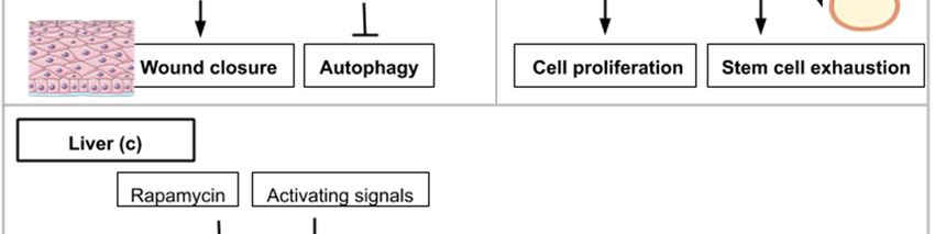
Int. J. Mol. Sci. 2020, 21, 2718                                                                    4 of 23

the animal kingdom. Studies with tissue-specific regeneration in animal models after injury have an
easier framework for which to extract data useful for treating human diseases. mTOR roles in specific
cell types will be investigated separately in the next paragraphs along with how mTOR modulations
can alter the microenvironment in a pro-regenerative fashion.

3. Adult Myofiber Growth and Formation Is Mediated by mTOR Signaling
      In muscle tissue, damages and atrophies are caused by mechanical shock or genetic diseases and
can seriously disrupt muscle functions. Chronic deterioration of muscular tissue is also observed
during the aging process. This makes the development of efficient therapies for muscle regeneration
of particular interest. Muscle regeneration is an event that is composed of two main steps: myofiber
neoformation and fiber growth [18]. In chordates, adult skeletal muscles possess their own stem cell
population, the myogenic satellite cells. These cells are positive for the myogenic Pax7 transcription
factor and are involved in muscle repair, growth, and regeneration. The existence of Pax3/7-expressing
muscle satellite cells has also been discovered in the crustacean Parhyale hawaiensis suggesting that this
stem population was present in the bilaterian common ancestor [19]. With the cellular basis for muscle
regeneration being evolutionarily conserved between arthropods and mammals, research benefits
from the use of animal models with regenerative capacities in deciphering the way in which the mTOR
pathway can serve or disserve regeneration. Examples of mTOR involvement in regeneration will be
illustrated below with mouse and axolotl (amphibian) models.
      mTOR activity is involved in homeostatic myogenesis and is associated with enhanced muscle
regeneration. The role of TOR signaling has been genetically demonstrated using a mouse model
harboring a conditional deletion of TOR in satellite cells [20]. Upon skeletal muscle injury, these mice
display necrotic fibers and fail to activate proliferation in satellite cells (Figure 2b). The myogenic
program is also affected by TOR deletion as shown by the reduced expression of Myf5, MyoD and
MyoG gene products in myoblasts [20]. Using transgenic mice in which Akt is constitutively active, Lai
et al., investigated changes in muscle mass [21]. Akt (also known as PKB) is a serine/threonine-specific
protein kinase that activates mTORC1. Akt participates in several processes such as glucose metabolism,
apoptosis, cell proliferation, transcription, and cell migration. Constitutive activation of Akt and by
extension mTORC1 in transgenic mice results in skeletal muscle hypertrophy [21].
      In contrast, in wild type adult mice, the addition of rapamycin inhibits muscle regeneration after
myotoxin exposure. This result shows mTORC1’s involvement in muscle regeneration. Investigating
the properties of adult pig satellite cells, Han et al. found that muscle growth (protein synthesis and
proliferation) in vitro is highly dependent on mTOR signaling activation after leucine and insulin-like
growth factor 1 (IGF-1) stimulation [22] (Figure 2b). Supplementation of amino acids like leucine [23]
or delivery of factors containing insulin-like growth factor 1 [24] have been successfully tested on rats
and mice as a means to ameliorate muscle regeneration. These studies show the necessity of mTOR
activity in adult satellite cells for proper stem cell activation and myofiber growth, which are essential
in muscle development and regeneration.
      Complementarily to myofiber growth, myofiber formation is an important process in muscle
regeneration. To dissect out the kinase activity of TOR in skeletal muscle repair, transgenic mice with an
inactive TOR kinase in skeletal muscles were designed [18]. This study revealed that myofiber growth
was impaired but not the formation of nascent myofibers. These data suggest that TOR-regulated
muscle regeneration displays kinase-dependent and -independent mechanisms [18] (see Figure 2b).
      More recently, a long non-coding RNA (lncRNA), highly expressed in mouse skeletal muscles, was
found to have the property to modulate TORC1 complex activity [25]. This lncRNA, called LINC00961,
is conserved in mice and humans [25] and has the particularity to host a small open reading frame that
is translated into a protein (SPAR, small regulatory polypeptide of amino acid response) displaying
co-localization with late endosome and lysosome markers in both HeLa and prostate cancer cell lines.
The TORC1 complex is a nutrient sensor, able to move from the cytoplasm to the lysosome after amino
acid stimulation. Investigating the effect of SPAR on amino acid-induced TORC1 activation, SPAR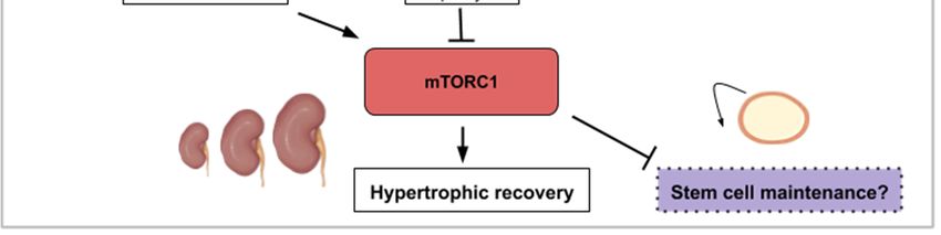
Int. J. Mol. Sci. 2020, 21, 2718                                                                                            5 of 23

   was discovered to impair mTORC1 correct localization to the lysosomes [25]. Spar-deficient mice
   (Spar−/− ) display a better record in tibialis anterior muscle regeneration compared to wild type mice
   (larger and more mature myofibers) and showed increased phosphorylation of S6K1/2 and S6 (see
   Figure 2b) [25]. This reflects a hyperactivation of mTORC1 complex when SPAR is knocked out in
   skeletal muscles. SPAR functions to negatively regulate mTORC1 recruitment, through interaction
   with v-ATPase. The modulation of regulatory elements of mTOR activity represents another possible
   method to enhance endogenous regeneration. A way to therapeutically boost myofiber proliferation
   and growth during muscle repair could then be to negatively target this atypical polypeptide hidden
Int.inJ. long  non-coding
         Mol. Sci.             RNA.
                   2020, 21, x FOR PEER REVIEW                                                        6 of 23

      Figure
        Figure2.2.This    figureillustrates
                   This figure     illustrates
                                            the the
                                                 waysways    in which
                                                       in which   activityactivity
                                                                           from the from
                                                                                      mTORthe      mTOR
                                                                                              signaling     signaling
                                                                                                         pathway         pathway
                                                                                                                    contributes
      contributes
        to appendageto appendage
                        (a) or muscle(a)   or muscle(b,c):
                                        regeneration    regeneration     (b,c):appendage
                                                             (a) Concerning      (a) Concerning     appendage
                                                                                            regeneration,          regeneration,
                                                                                                            the Wnt/β-catenin
        pathway
      the         and insulin-like
           Wnt/β-catenin      pathway growth
                                          andfactor (IGF-1) activate
                                               insulin-like    growth mTORC1.      This activity
                                                                         factor (IGF-1)          leadsmTORC1.
                                                                                            activate   to the wound    covering,
                                                                                                                    This  activity
        blastema
      leads  to theformation     and regenerative
                    wound covering,        blastemaoutgrowth
                                                       formationofand the regenerative
                                                                           appendage. (b)      During muscle
                                                                                           outgrowth             regeneration,(b)
                                                                                                         of the appendage.
        mTORC1
      During       activity
                muscle       is necessary mTORC1
                         regeneration,    for myofiber   growthisbut
                                                      activity        not myogenesis.
                                                                   necessary              mTORC1
                                                                                for myofiber         inhibition
                                                                                                 growth          by rapamycin
                                                                                                           but not   myogenesis.
        treatment   inhibits   regeneration   whereas    the  leucine  amino    acid, insulin-like
      mTORC1 inhibition by rapamycin treatment inhibits regeneration whereas the leucine amino       growth    factor 1 or Akt
                                                                                                                             acid,
        activity contribute   to mTORC1-mediated       muscle   regeneration.   SPAR   regulatory   protein
      insulin-like growth factor 1 or Akt activity contribute to mTORC1-mediated muscle regeneration.        (small  regulatory
        polypeptide of amino acid response) can inhibit mTORC1 activity and hinder muscle regeneration.
      SPAR    regulatory protein (small regulatory polypeptide of amino acid response) can inhibit mTORC1
        (c) mTORC1 signaling is necessary for muscle regeneration in the injured limb and to induce a Galert state
      activity and hinder muscle regeneration. (c) mTORC1 signaling is necessary for muscle regeneration
        in the contralateral limb. The Galert cells enter the cell cycle more rapidly and show an increase in size
      in the injured limb and to induce          a Galert state in the contralateral limb. The Galert cells enter the cell
        compared to quiescent satellite cells.
      cycle more rapidly and show an increase in size compared to quiescent satellite cells.
        Finally, in a study analyzing mouse satellite cells located in the contralateral muscle after injury
4. with
   The mTOR      Signaling
        myotoxins,    mTORC1  Roles
                                 wasduring    AxontoRegeneration
                                      discovered       control a novel state within the G0 quiescence phase
   newly termed Galert [26]. Two cell populations were compared; the muscle stem cells from the site of
4.1. The mTOR
   injury         Signaling
          and those          Roles in Vertebrate
                      of the contralateral limb. Retinal  Neuron
                                                   This study     Regrowth
                                                              found          after
                                                                    that these     Axotomy
                                                                                 cells enter the cell cycle more
   rapidly and  display  an increase in cell size compared  to quiescent satellite
      In general, axons after injury do not spontaneously regenerate in the adult  cells. These two parameters
                                                                                              mammalian     central
nervous system. Interestingly, the mTOR pathway is involved in the differentiation of human retinal
ganglion cells from induced pluripotent stem cells (iPS cells) [26]. This model of neurogenesis has
made evident that function and neuritogenesis of human retinal ganglion cells (RGCs) are dependent
on mTOR [28,29]. In addition, high levels of the phosphorylated form of S6 (pS6) are observed in the
embryonic chick retina as progenitors cells differentiate [30]. These studies show that mTOR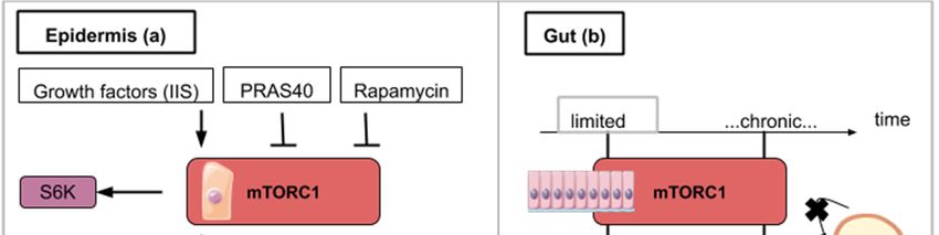
Int. J. Mol. Sci. 2020, 21, 2718                                                                      6 of 23

(cell size and cell cycle entry speed) are not as elevated as in satellite cells from the injured muscle but
in a kind of intermediate state (Galert ) (see Figure 2c). mTORC1 signaling is necessary and sufficient
to induce this “alert” response in satellite cells. The authors proposed that the quiescence status of
stem cells actually comprises several phases. This Galert state might allow stem cells to respond more
rapidly in case of injury and would then constitute a form of cellular memory as the one observed in
cell-mediated immune response [26].
      Interestingly, similar contralateral activation of mTOR was observed in the axolotl limb amputation
model [27]. The phosphorylation of S6 ribosomal protein, one of the main substrate of S6K1, is slightly
but significantly increased in the satellite cells of the contralateral intact limb compared to levels
observed in the limb of non-amputated animal [27]. The precise upstream signals, which activate
mTORC1 and enable cell-state transition, remain to be deciphered. Nonetheless, these remarkable
findings on an apparent systemic cellular activation response that occurs distant to the site of injury
could guide future research in regenerative medicine.
      Altogether, these data demonstrate that TORC1 activity is necessary to achieve the correct process
of muscle regeneration for cell growth and cell proliferation. The regeneration-competency of satellite
cells is also a factor that will influence the degree of recovery. The previously described Galert cell
state, which is under the control of mTOR activity, serves as an illustration for improved competency.
Any diseases displaying chronic muscle degradation might, therefore, consider inducible activators of
TORC1 as suitable therapeutic molecules such as growth factors, amino acids or modulators of the
regulatory elements of mTOR.

4. The mTOR Signaling Roles during Axon Regeneration

4.1. The mTOR Signaling Roles in Vertebrate Retinal Neuron Regrowth after Axotomy
     In general, axons after injury do not spontaneously regenerate in the adult mammalian central
nervous system. Interestingly, the mTOR pathway is involved in the differentiation of human retinal
ganglion cells from induced pluripotent stem cells (iPS cells) [26]. This model of neurogenesis has
made evident that function and neuritogenesis of human retinal ganglion cells (RGCs) are dependent
on mTOR [28,29]. In addition, high levels of the phosphorylated form of S6 (pS6) are observed in
the embryonic chick retina as progenitors cells differentiate [30]. These studies show that mTOR
signaling participates in the in vitro proliferation and differentiation of retinal stem cells. Upon retinal
injury in adult chicks, Müller glia cells (a type of retinal glial cell) can proliferate and give rise to a
population of progenitors (Müller glia-derived progenitor cells, MGPCs) that can be converted into
retinal neurons. This valuable property of Müller cells is observed in teleosts but strongly reduced in
amniotes. In adult chicks, retinal damage induced by NMDA (N-methyl-d-aspartate) is followed by
the activation of TOR signaling in Müller glia and this activation is necessary for Müller glia-derived
progenitor cells’ proliferation [30]. This suggests that regeneration mechanisms observed in adult
nerve cells are reminiscent of the machinery running during development. Targeting mTOR signaling
in adult cells could thus potentially stimulate this developmental reactivation.
     PTEN and TSC1/2 are two important molecular actors regulating the mTOR pathway. PTEN is a
phosphate and tensin homolog, which acts upstream of mTOR as an inhibitor. TSC1/TSC2 or Tuberous
sclerosis proteins 1 and 2 form a protein complex that inhibits mTOR activity. Retinal ganglion cells
(RGCs) in the adult mouse normally degenerate after section of the optic nerve. Hypothesizing that
manipulation of the intrinsic properties of retinal neurons can change their ability to regrow after
axotomy, mice carrying the deletion of the lipid phosphatase PTEN in adult retinal ganglion cells
were generated [31]. These mice were able to regrow their optic axon indicating that TOR activation
might be a crucial step in this process. According to molecular crosstalk, the mTOR pathway is
englobed by a larger PI3K/AKT/mTOR pathway. Thus, as a consequence of PTEN deletion, Akt is
strongly activated and the kinase GSK3β appears to be a core component of the Akt-induced axon
regeneration [32] (Figure 3a). Similarly, another study used small interfering RNA (siRNA) to invalidate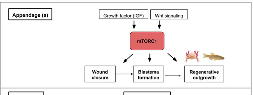
Int. J. Mol. Sci. 2020, 21, 2718                                                                   7 of 23

RTP801 (another inhibitor of TOR induced upon hypoxia and stress conditions) (Figure 3a). RTP801
(also known as Redd1) is a stress-induced protein that suppresses mTOR signaling by stabilizing
TSC1/TSC2 (inhibitors of mTORC1/2). Intravitreal injection of this mTOR activating siRNA was found
to promote the release of several neurotrophins and RGC neuroprotection [33]. A study combining
PTEN inhibition (PTEN KO mice) with ambroxol treatment demonstrated significant enhancement
of axonal outgrowth in dorsal root ganglion (DRG) neurons [34]. Ambroxol was shown to increase
the expression of a transcriptional network that coordinates central nervous system regeneration [34].
Altogether, these data show that a constitutive activation of the mTOR pathway promotes both retinal
ganglion cell survival and axon regrowth.
     In adult mice, inflammatory stimuli that activate resident retinal glial cells (astrocytes/Müller
cells) greatly favor axon regeneration of retinal ganglion cells [35]. Indeed intravitreal application
of Pam3 Cys, an agonist of toll-like receptor that constitutes an inflammatory stimulus, impairs the
decrease of TOR activity normally observed after optic nerve crush in wild type mouse (Figure 3a).
Surprisingly the inhibition of TOR (rapamycin treatment) does not modify the conversion of optic
neurons from a non-regenerative to a regenerative status upon inflammatory stimulation meaning that
additional signaling pathways can take over this intrinsic neuron property. Thus, mTOR activity is not
generally required for neuroprotection or switching mature neurons into an active regenerative state,
but it is important for the maintenance of the axonal growth state [35].
     Studies that focus on the mechanistic explanations behind optic nerve axon regeneration have
revealed which mTOR components are most likely involved. In the mouse optic nerve crush model,
mTORC1 downstream effectors (S6K1 and 4E-BP) are differentially involved. S6K1 activation after
crush clearly induced axon elongation and survival of optic neurons. In contrast, the deletion of
4E-BP (4E-BP1/2 double KO mice), which is normally phosphorylated and inhibited upon TOR activity,
had no effect on regrowth [36] suggesting that S6K1 or other TOR substrates are operating. It is also
worth mentioning that axon recovery upon optic nerve damage is still observed on PTEN deleted
neurons when they are treated with rapamycin suggesting the implication of mTORC1-independent
pathways [31]. Since PTEN deletion constitutively activates mTOR signaling but rapamycin inhibits
mTORC1 activity, this suggests an mTORC2-mediated recovery (Figure 3a). mTORC2 controls Akt,
which regulates cellular processes such as metabolism, survival, apoptosis, growth, and proliferation.
Rapamycin-resistant axon recovery and TOR-independent mechanisms of axogenesis are also observed
in peripheral sensory neurons [23,24].
     Finally, when considering retinal neuron axon regeneration, outgrowth following injury cannot
be the only investigated parameter in a human disease-context. Juvenile and adult mice with either
PTEN and SOCS3 (suppressor of cytokine signaling 3) co-deletion, or co-overexpression of osteopontin
(OPN)/insulin-like growth factor 1 (IGF1)/ciliary neurotrophic factor (CNTF), showed regrowth of
retinal axons and formation of functional synapses in the superior colliculus (SC), but not significant
recovery of visual function [37]. PTEN and SOCS3 deletions as well as the growth factors’ (OPN,
IGF1 and CNTF) overexpression activate mTOR signaling (Figure 3a). The regenerated axons failed to
conduct axon potentials from the eye to the brain. With functional recovery in mind, the administration
of voltage-gated potassium channel blockers was shown to restore conduction and results in increased
visual acuity [37]. When rebuilding circuits and evaluating regeneration after optic nerve injury, both
the axon regrowth and proper myelination seem to be required. In general, for each regeneration
challenge, attention must be paid in the specificities required for functional regeneration.
     The medical challenge resides in finding the exact ways to stimulate mTOR activity to maximize
the optic nerve’s regenerative properties. As exposed in this review, current leads include inhibitors of
mTOR inhibitors (PTEN, RTP801 or TSC1/TSC2), mTOR activators and growth factors. Conditions like
glaucoma where the optic nerve is damaged and which is the leading cause of blindness in people
over 60 could benefit from such treatments.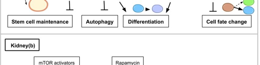
Int. J. Mol. Sci. 2020, 21, 2718                                                                                                 8 of 23

4.2. The mTOR Pathway Mediates Drosophila Neuron Regrowth and Remodeling during Metamorphosis
     The TOR pathway is also involved in axon regrowth in Drosophila. During metamorphosis, neurons
from the mushroom body are reshaped. This consists in parceling exiting axons followed by the
re-extension of new ones from the cellular body. This phenomenon mimics cellular events occurring after
axotomy in neuron and is different from initial axon growth from a newborn neuron. During mushroom
body remodeling, axon regrowth is mediated via the TSC-Rheb-TOR pathway, which is activated by the
nuclear receptor UNF [38]. In this system, TOR is also regulating neurite sprouting but its activation is
under the control of PI3K/PTEN [39].
Int. J. Mol. Sci. 2020, 21, x FOR PEER REVIEW                                                                                      9 of 23

                  Thisfigure
      Figure 3.3.This
     Figure             figureillustrates
                                illustrates how
                                             how activity
                                                   activity from
                                                             from the mTOR signaling
                                                                                  signaling pathway
                                                                                               pathwaycontributes
                                                                                                          contributestotoaxon  axon
      regeneration ininthe
     regeneration          selected
                         the        examples
                             selected           of retinal
                                         examples          nerve damage
                                                      of retinal            (a) and spinal
                                                                  nerve damage       (a) and cordspinal
                                                                                                  injurycord
                                                                                                         (b,c): injury
                                                                                                                 (a) Concerning
                                                                                                                          (b,c): (a)
      retinal nerveretinal
     Concerning      regeneration,   mTOR activity
                            nerve regeneration,         enhances
                                                     mTOR          axon
                                                              activity    elongation
                                                                        enhances    axonand   survival leading
                                                                                           elongation              to functional
                                                                                                         and survival      leading
      regeneration.     Signals   that  upregulate     mTOR     activity  like   PTEN     inhibition,
     to functional regeneration. Signals that upregulate mTOR activity like PTEN inhibition, siRTP801,  siRTP801,      Pam   3 Cys
      (pro-inflammatory       signal),   CNTF    (ciliary   neurotrophic      factor)  or   IGF-1
     Pam3Cys (pro-inflammatory signal), CNTF (ciliary neurotrophic factor) or IGF-1 lead to axon    lead   to  axon    recovery.
      TOR-independent
     recovery.              axon recovery
                  TOR-independent         axonexists   however
                                                 recovery         as rapamycin-treated
                                                              exists                           PTEN-deleted PTEN-deleted
                                                                       however as rapamycin-treated               neurons and
      Pam3 Cys
     neurons      untreated
                and  Pam3Cys  neurons
                                untreatedstillneurons
                                               showedstill
                                                         recovery.
                                                             showed  (b)recovery.
                                                                         For spinal (b)cord
                                                                                        For injuries,   mTORC1
                                                                                              spinal cord   injuries, activity
                                                                                                                        mTORC1  in
      astrocytes  hinders  neuronal    recovery  with   the formation    of glial scars  (gliosis). Growth
     activity in astrocytes hinders neuronal recovery with the formation of glial scars (gliosis). Growth     factors   like  EGF
      (epidermal
     factors  like growth   factor) can growth
                    EGF (epidermal       activate mTORC1.
                                                    factor) can(c)activate
                                                                   On the contrary,
                                                                              mTORC1.   mTORC1
                                                                                            (c) Onsignaling     in hemisection
                                                                                                     the contrary,      mTORC1
      spinal  cord injuries  promotes     growth   and  functional    regeneration.
     signaling in hemisection spinal cord injuries promotes growth and functional regeneration.
 4.3. The Cell-Specific mTOR Signaling Roles in Mammalian Spinal Cord Neurons
5. The mTOR Signaling Roles in Wound Healing during Epidermis Regeneration
       Spinal cord injuries (SCI) have been connected to mTOR signaling in different ways. The spinal
      The  Drosophila
 cord connects         monolayered
                  the brain             epidermis
                             to the peripheral      is alsosystem.
                                                  nervous   a good system  to study
                                                                    The mTOR          tissuehas
                                                                                 pathway     repair
                                                                                                beenafter  injury.
                                                                                                       shown   to
One   of the first events after   injury is the  formation   of an actomyosin   cable  that will tighten
 promote or inhibit axon regeneration in SCI animal models. Most frequently, two types of SCI animal      around
the wound.
 models        Then,spinal
          are used;  the wound      closure
                            cord injury  andis spinal
                                               ensured   byhemisection
                                                      cord  the association
                                                                        withofthe
                                                                                crawling
                                                                                  former cells
                                                                                          aimingto for
                                                                                                   lamellipodia-
                                                                                                       complete
carrying   cells surrounding    the  wound.   In the next  experiments, Drosophila  larvae   were
 anatomical and functional damage of the spinal cord while the latter is an incomplete injury with subjected  to a
laser beam that ablated cells of the epidermis. Wound closure clearly involves Insulin/IGF signaling
(IIS) as shown in flies carrying the deletion of three insulin ligands or the expression of a dominant-
negative version of the insulin receptor [44–46]. Wound healing in Drosophila is also delayed in the
presence of rapamycin [44] (Figure 4a). Similarly, targeted inhibition of mTORC1 in transgenic mice
overexpressing PRAS40 in basal keratinocytes resulted in delayed wound healing [45] (Figure 4a).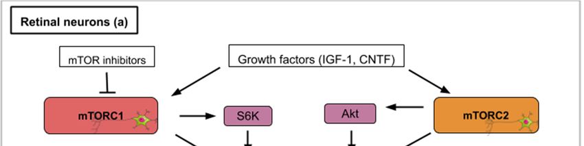
Int. J. Mol. Sci. 2020, 21, 2718                                                                      9 of 23

a certain remaining nerve connectivity. Glial scars, formed by the activation of inactive astrocytes,
involve the proliferation or hypertrophy of astrocytes, which create a physical and chemical barrier
that hinders neuronal recovery [40]. The conversion of astrocytes depends on epidermal growth
factors (EGF), which activates the Rheb/mTOR pathway [5]. Overexpressed PTEN attenuates gliosis at
three days after SCI and enhances motor functional recovery [5] (Figure 3b). In general, in astrocytes,
a hyperactive mTOR is a negative regulator for recovery following SCI [41]. Restravol is an example
of an mTOR inhibiting drug that has been successful in restoring nerve function following SCI [42].
However, in spinal cord hemisection, mTOR activation efficiently promotes regrowth and regeneration
of the corticospinal tract [5] (Figure 3c). In dorsal root ganglion neurons, mTOR activation increases
regrowth and recovery following peripheral nervous system injury [43]. Diseases like amyotrophic
lateral sclerosis (ALS), which specifically affects motoneurons, could gain from treatments which alter
mTOR activity in the spinal cord. To recapitulate, hyperactivity of mTOR in astrocytes is correlated to
an inflammatory response that is deleterious for neuron recovery while mTOR activation in spinal
cord neurons enhances axon regeneration. Importantly, functions of mTOR in varying neuronal injury
models can be different or even opposing, which might be attributed to the type of cell in which the
mTOR activity takes place.

5. The mTOR Signaling Roles in Wound Healing during Epidermis Regeneration
      The Drosophila monolayered epidermis is also a good system to study tissue repair after injury.
One of the first events after injury is the formation of an actomyosin cable that will tighten around the
wound. Then, the wound closure is ensured by the association of crawling cells to lamellipodia-carrying
cells surrounding the wound. In the next experiments, Drosophila larvae were subjected to a laser
beam that ablated cells of the epidermis. Wound closure clearly involves Insulin/IGF signaling (IIS) as
shown in flies carrying the deletion of three insulin ligands or the expression of a dominant-negative
version of the insulin receptor [44–46]. Wound healing in Drosophila is also delayed in the presence of
rapamycin [44] (Figure 4a). Similarly, targeted inhibition of mTORC1 in transgenic mice overexpressing
PRAS40 in basal keratinocytes resulted in delayed wound healing [45] (Figure 4a). These animal model
studies show that mTOR activity at the site of the wound is required for cell migration and wound
healing. The exploitation of the mTOR pathway as a source of novel pharmacological strategies for
wound healing is ongoing and is reviewed by Castilho et al. [46].

6. The mTOR Signaling Roles during Homeostatic Growth and Stem Cell Maintenance in the
Gut Epithelium
      Barrier epithelia such as the posterior midgut epithelium of Drosophila is another paradigm for
the study of tissue repair. One of the steps of Drosophila’s midgut regenerative mechanism is the
endoreplication of enterocytes (EC). Intestinal stem cells of the gut proliferate to form an enteroblast (EB)
population in which cells are able to differentiate into enterocytes (EC) or enteroendocrine cells. As they
differentiate, EC undergo endoreplication which contributes to an increase in cell size. The endocycle
is different from the mitotic cycle as DNA replication occurs without nuclear division resulting in
polyploid cells with increased cell size. Analyzing how endocycling-induced EC growth is used
during fly gut epithelium repair following stress, EC endoreplication was shown to be controlled by
EGFR/MAPK (epidermal growth factor receptor and mitogen-activated protein kinases respectively)
but not by TOR signaling [47]. The EGFR/MAPK pathway promotes cell growth and proliferation.
In normal conditions, however, the fly posterior midgut will use the Insulin/PI3K/TOR pathway for EC
endocycling and growth [47]. These data serve as an illustration for mTOR being implicated in the
physiological homeostatic growth of the gut but not in the regenerative process following epithelium
stress. In consequence, the therapeutic activation of mTOR aiming at promoting gut regeneration after
mechanical or chemical injury might induce a deleterious effect on the non-affected gut epithelium
portion undergoing physiological cell turn-over. This highlights the importance to favor the local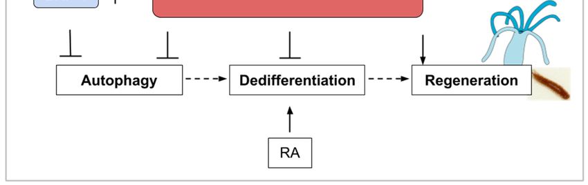
Int. J. Mol. Sci. 2020, 21, 2718 10 of 23
administration of drugs over a wide-spread action. This strategy might better target the damaged
tissue and preserve the intact epithelium.
The fruit fly intestine hosts intestinal stem cells (ISCs) that proliferate in response to regenerative
stimuli. Using enteropathogen infection to mimic regenerative stimuli, fly guts were then dissected
and labeled with anti-phospho-4E-BP antibodies. TOR activation was found to be necessary for the
quick activation of the proliferation of ISCs [48]. However, the pool of ISCs is significantly reduced
when TOR is continuously activated after several rounds of regenerative inputs in the posterior midgut.
This decline is explained by an accelerated differentiation of intestinal stem cells into enterocytes
and enteroendocrine cells [48]. Similar findings have been observed on stem cells (basal cells) of the
mouse tracheal
Int. J. Mol. Sci. 2020,epithelium
21, x FOR PEER upon SO2 exposure-induced injury: TOR is transiently activated
REVIEW 11 upon
of 23
chemical-induced damage but chronic activation of TOR induced a decline of basal cells population [48]
thirds 4b).
(Figure ablation,
These livers thatoutline
results have suffered
the dual90%rolehepatectomy are not
of TOR in stem cellable to regrow.ItUnsurprisingly,
homeostasis. is important to
TOR isproliferation
trigger not activatedimmediately
in the remnant liver
after after such
damage but massive resection
a low-grade [57].is necessary to ensure long
activity
stem cell Liver diseasesThis
longevity. stem from various
trade-off between etiologies and can be fatal.
mTORC1-mediated Therapies
inductions enhancing
of stem endogenous
cell proliferation and
regeneration following resection should inescapably focus on the activation of the mTOR
differentiation-induced loss seems to be widely conserved across cell types [26,29,48–52]. Pharmaceutical pathway to
create a more favorable microenvironment for hepatocyte reactivation. A recent review cites some of
alterations of the mTOR pathway have to be designed accordingly so as not to deplete adult stem
the chemical drugs that stimulate mTOR signaling to improve liver regeneration after partial
cell populations.
hepatectomy [5].
Figure
Figure 4. 4.
ThisThis figure
figure illustrates
illustrates the ways
the ways in whichin which
activityactivity
from thefrommTOR thesignaling
mTOR pathway
signalingcontributes
pathway
tocontributes to epidermis
epidermis (a), gut (b) and (a), gutregeneration
liver (b) and liver(c):regeneration
(a) mTORC1 (c):activity
(a) mTORC1 activityfor
is necessary is wound
necessary for
closure
wound
and can beclosure and by
enhanced caninsulin/insulin-like
be enhanced by insulin/insulin-like growth(IIS).
growth factor signaling factor signaling
S6K1/2 (IIS). target
is a TOR S6K1/2which
is a
TORwound
helps target which helps
closure. wound
mTOR closure. mTOR
inhibitory signalsinhibitory signals or
like rapamycin likean
rapamycin or an overexpressed
overexpressed PRAS40 delay
PRAS40 delay wound closure and promote autophagy. (b) For
wound closure and promote autophagy. (b) For gut regeneration, mTORC1 activity gut regeneration, mTORC1 activity for
is required is
required
initial cell for initial cell proliferation
proliferation but chronicleads
but chronic activation activation leads tostem
to intestinal intestinal stem cell exhaustion.
cell exhaustion. (c)
(c) For liver
For liver regeneration,
regeneration, mTORC1 activitymTORC1 activity
leads to cellleads
cycletoreentry
cell cycle
andreentry and recovery.
functional functionaleIF4E
recovery. eIF4E
(Eukaryotic
(Eukaryotic
Initiation Initiation
Factor Factor 4E)translation
4E) dependent dependentistranslation is activated
activated with mTORC1 withactivity
mTORC1 andactivity
leads to and leadsD
Cyclin
to Cyclin D expression. Cyclin D amplifies cell proliferation and leads
expression. Cyclin D amplifies cell proliferation and leads to functional liver recovery. to functional liver recovery.
8. The mTOR Signaling Roles during Bone Formation and Resorption
Bone regeneration is a physiological process of bone formation and is observed during normal
fracture healing and in continuous remodeling and growth throughout an organism’s lifespan. Chen
and Long describe the roles of mTOR signaling in skeletal development and disease in a recent review
[58]. Osteoblasts are specialized mesenchymal stem cells that differentiate into the major cellularInt. J. Mol. Sci. 2020, 21, 2718 11 of 23
7. The mTOR Signaling Role in Hepatocyte Proliferation during Liver Regeneration
Upon partial hepatectomy, the remaining and quiescent mammalian hepatocytes are able to
reenter the cell cycle synchronously and proliferate to restore liver volume. The liver does not rely
on a resident stem cell population but on the aptitude of its hepatocytes to reactivate proliferation
after local signals such as tissue resection. The use of mice mutants (S6K1−/− , S6K2−/− ) and rapamycin
intraperitoneal injection to abrogate TOR activity in a 70% partial hepatectomy (PH) mouse model
demonstrated the importance of the TOR pathway in the initial proliferation phase after resection [53,54].
More specifically decrease in levels of Cyclin D1, a cyclin known to be involved in the G1-S phase
transition, is observed in S6K1−/− , S6K2−/− hepatocytes accounting for the cell cycle progression delay
reported in these genetically modified hepatocytes. In addition, rescue experiments using Cyclin D1
overexpression in S6K1−/− , S6K2−/− hepatocytes showed that cell cycle entry timing can be rectified
after PH [53]. The link between mTOR and cell cycle entry can be explained by the fact that mTOR
inhibition induces phosphorylation of eIF4E (eukaryotic initiation factor 4E) [55] that participates in the
synthesis of the Cyclin D1 protein [56]. In a context of mTOR inhibition, eIF4E-dependant translation is
inhibited [55] which results in the decrease of Cyclin D1 protein levels and cell cycle progression delay
(Figure 4c). Nevertheless, liver regeneration can happen to a limited extent only. While TOR-mediated
proliferation of hepatocytes allows liver regeneration after two-thirds ablation, livers that have suffered
90% hepatectomy are not able to regrow. Unsurprisingly, TOR is not activated in the remnant liver
after such massive resection [57].
Liver diseases stem from various etiologies and can be fatal. Therapies enhancing endogenous
regeneration following resection should inescapably focus on the activation of the mTOR pathway to
create a more favorable microenvironment for hepatocyte reactivation. A recent review cites some
of the chemical drugs that stimulate mTOR signaling to improve liver regeneration after partial
hepatectomy [5].
8. The mTOR Signaling Roles during Bone Formation and Resorption
Bone regeneration is a physiological process of bone formation and is observed during normal
fracture healing and in continuous remodeling and growth throughout an organism’s lifespan. Chen
and Long describe the roles of mTOR signaling in skeletal development and disease in a recent
review [58]. Osteoblasts are specialized mesenchymal stem cells that differentiate into the major
cellular components of bone. Additionally, osteoclasts derive from hematopoietic progenitors from the
bone marrow and form the structural components of bone with osteoblasts. Stimulation of osteoblast
differentiation has been attributed to mTORC1 as a common effector mediating the bone anabolic effect
of insulin-like growth factor-1, Wnt and bone morphogenetic protein (BMP). mTORC1 activity drives
bone anabolism by stimulating osteoclast differentiation (Figure 5a). mTORC2 too has been found to
promote osteoblast differentiation and function [58] (Figure 5a). In ovariectomized rats, bone marrow
stromal cells (BMSC) lacking Rictor gene exhibited reduced osteogenic potential, but an increased
capacity to undergo adipogenic differentiation in vitro [59]. Thus, mTORC2 may serve as a potential
therapeutic target for treating age-related bone loss.
mTOR modulation has been linked to improve symptoms in certain bone diseases. Bone health
is determined by the correct balance of bone resorption and bone formation. Osteoporosis and
osteoarthritis are two chronic diseases, which show an imbalance between bone resorption and
formation. Osteoarthritis (OA) is a chronic degenerative joint disease characterized by gradual loss of
articular cartilage, synovial inflammation, and subchondral bone remodeling. The mTOR pathway
has been shown to be overexpressed in human OA chondrocytes [60]. Drugs inhibiting mTORC1
(local application of rapamycin or torin) have shown therapeutic promise [61–65]. One explanation
for the pathology is proposed to be linked to the overexpression of mTOR and its induced repression
of autophagy [60,64]. The expression of autophagy markers is suppressed in human OA cartilages
as well as animals models of OA and the inhibition of autophagy causes chondrocyte apoptosis and
OA-like pathogenesis in vitro and in vivo [66,67].Int. J. Mol. Sci. 2020, 21, 2718 12 of 23
Osteoporosis (OP) is a disease in which the density and quality of bone are reduced. A study in
rats showed that rapamycin treatment reduced senile osteoporosis by activating osteocyte autophagy
and preserving osteocytes and by a decrease in apoptosis of osteocytes and a decrease in number of
osteoclasts [68]. Another study with Everolimus (equivalent to rapamycin) in rats showed a decrease
of 60% in cancellous bone loss, the first type of bone structure, which is affected in osteoporosis.
Both studies showed that rapamycin or a rapalog decrease osteoporosis by blocking mTOR and
decreasing the activity of osteoclasts and the preservation of osteocytes by increasing autophagy.
In general, mTORC1 deficiency in osteoclast precursors may promote both osteoclast formation and
bone resorption [61].
Bone and dentin derive from stem cells from the apical papilla (SCAP) which are a subpopulation
of mesenchymal stem cells (MSCs). SCAP are identified as a population of postnatal mesenchymal stem
cells with the capacity for self-renewal and multipotent differentiation into osteoblasts/odontoblasts,
adipocytes, and neural cells. Looking specifically at the Phosphoinositide 3 kinase (PI3K)-Akt-mTOR
pathway, it was found that its suppression played a role in enhancing the in vivo and in vitro
osteogenic/dentinogenic differentiation of stem cells from the apical papilla [69]. A novel approach for
SCAP-based bone and dentin regeneration could then involve a suppressive regulation of PI3K-Akt-
mTOR signal pathway. This study showed the role of the PI3K-Akt-mTOR pathway in cell fate and
stem cell differentiation capacity (Figure 5a). Such data should be taken into account when elaborating
possible therapeutic treatments as the mTOR pathway can also play a role in cell fate.
9. The mTOR Signaling Roles during Kidney Repair, Disease and Stem Cell Maintenance
The functional unit of the kidney is the nephron and adult neonephrogenesis is unequally distributed
in the animal kingdom. The nephron is composed of a glomerulus and a tubule. The glomerulus contains
podocytes, which help carry out the initial blood filtration. The tubule is where diverse molecules are
secreted and reabsorbed. Adult mammals cannot form new nephrons contrary to cartilaginous fish,
bony fish or certain amphibians [70–73]. However, cellular mechanisms can contribute to the reparation
of mammalian renal structures. More specifically, this consists in proliferation of epithelial cells in
the tubules—which can originate from dedifferentiated cells (Table 1) or recruited resident progenitor
cells—leading to nephron hypertrophy. This repair depends on the ability of the remaining tubular cells
to proliferate and restore the injured tubular epithelium [74]. However, the increased intracapillary
pressures and flows associated with adaptive glomerular hypertrophy in humans can ultimately lead to
podocyte injury and proteinuria [75]. Proper glomerular filtration which regulates blood homeostasis
is paramount for functional kidney regeneration. Nephron hypertrophy is mTOR-dependent and
rapamycin treatment delays recovery of renal function after acute kidney injury in rats [75]. The effect
of rapamycin might be due to the dual effects of inhibition of proliferation and induction of apoptosis of
tubular cells.
Medical approaches for treating kidney diseases have been focusing on the stimulation of endogenous
reparative mechanisms or on stem cell therapies [76] and the use of organoids [77]. mTOR has emerged
as an important modulator of several forms of renal diseases [78]. Activation of mTOR within the kidney
occurs in animal models of diabetic nephropathy and other causes of progressive kidney disease. This
kinase has also been implicated in the development of glomerular disease, polycystic kidney disease and
kidney transplant rejection [78]. Rapamycin ameliorates several key mechanisms believed to mediate
changes associated with the progressive loss of glomerular filtration rate in chronic kidney disease [75].
These include glomerular hypertrophy, intrarenal inflammation, and interstitial fibrosis. mTOR also
plays an important role in mediating cyst formation and enlargement in autosomal dominant polycystic
kidney disease [75]. Inhibition of mTOR by rapamycin or one of its analogues represents a potentially
novel treatment for autosomal dominant polycystic kidney disease. mTOR modulators for renal recovery
are already ongoing clinical trials and were reviewed in 2018 [79].
During embryogenesis, nephrons are derived from a pool of self-renewing progenitors. Mammalian
nephron progenitors stop propagation and are terminally differentiated within a few days after birth [80]Int. J. Mol. Sci. 2020, 21, 2718 13 of 23
indicating that neonephrogenesis is not possible in the adult as mentioned above. In contrast, formation of
nephrons de novo has been reported in the adult little skate, Leucoraja erinacea, an elasmobranch cartilaginous
fish, after partial nephrectomy [70]. Similarly, in the adult zebrafish kidney and using transplantation
experiments, a small group of cells were discovered as able to form functional nephrons [71,73]. In the
skate, histological analysis revealed an enhancement of nephron growth in the nephrogenic zone, both in
the remnant tissue and in the contralateral kidney. In addition, stem cell-like mesenchymal cells were
identified in the nephrogenic zone [70]. This strongly suggests that a pool of nephron progenitors is
maintained in the renal tissue of adult cartilaginous fish giving them the possibility to form new nephrons.
What are the molecular properties that allow these renal progenitors to be maintained in the adult? One
hypothesis might be that these cells display low rates of protein synthesis. Interestingly, genes/pathways
that are differentially expressed in young versus old renal progenitors include pathways with known
effects on organism/stem cells aging (such as TOR) and the translational machinery [81] (Figure 5b).
More generally, recent reviews have highlighted that both embryonic and adult stem cells are dependent
on low rates of translation to maintain an undifferentiated state [4,82]. Pathways controlling protein
synthesis such as the mTOR pathway might then have a crucial role in maintaining renal progenitors in
adult cartilaginous fish. Retaining a low rate of protein synthesis and more specifically mTOR activity
might then be a way to keep renal stem cells in the mammalian adult kidney and to avoid the decline of
the progenitor pool around birth. As described in the examples in the bone subsection, mTOR activity
effects are characterized by a certain trade-off between stem cell maintenance and differentiation.
Int. J. Mol. Sci. 2020, 21, x FOR PEER REVIEW 14 of 23
Figure5.5. This
Figure Thisfigure
figureillustrates
illustratesthe
theways
waysininwhich
which the the mTOR
mTOR signaling
signaling pathway
pathway contributes
contributes to bone
to bone (a)
(a)kidney
or or kidney (b) regeneration.
(b) regeneration. (a) mTOR
(a) mTOR activity
activity (mTORC1
(mTORC1 and and mTORC2)
mTORC2) is required
is required for osteoblast
for osteoblast and
and osteoclast
osteoclast differentiation.
differentiation. mTORC1
mTORC1 is activated
is activated by boneby bone morphogenetic
morphogenetic protein
protein (BMP),
(BMP), insulin-
insulin-like
like growth factor 1 (IGF-1) and Wnt signaling. mTORC1 inhibition promotes
growth factor 1 (IGF-1) and Wnt signaling. mTORC1 inhibition promotes stem cell maintenance andstem cell maintenance
and autophagy
autophagy whilewhile
mTORC2 mTORC2 inhibition
inhibition leads toleads to cell
cell fate fate change.
change. (b)kidney,
(b) In the In thehypertrophic
kidney, hypertrophic
recovery
recoveryon
depends depends
mTORC1 on mTORC1 activity.
activity. mTOR mTORhas
activity activity
beenhas been correlated
correlated to stem
to stem cell cell depletion
depletion such as such
that
as that inhibition
mTOR mTOR inhibition
(purple)(purple)
might playmight play
a role inamaintaining
role in maintaining kidney progenitors.
kidney progenitors.
10. The mTOR Signaling Roles during Whole-Body Regeneration and Asexual Reproduction in
Cnidarians, Planarians, and Tunicates
The capability to regenerate the entire body is observed in a wide range of metazoans (mainlyYou can also read