Assessing the infl uence of FDM to the postoperative healing processes in distal fracture of the radius
←
→
Page content transcription
If your browser does not render page correctly, please read the page content below
Assessing the influence
 of FDM to the postoperative
       healing processes
in distal fracture of the radius

   Master Thesis to obtain the degree of
    Master of Science in Osteopathy
         at the Donau Universität Krems
Zentrum für chin. Medizin & Komplementärmedizin

                     presented
      at the Wiener Schule für Osteopathie
               by Tomasz Teszner

                Vienna, February 2011

                   Supported by

               Prof. Dr. Andrzej Zyluk
        Clinic of General and Hand Surgery
         Pomeranian Medical University
                         Head:
               Prof. Dr. Andrzej Zyluk

                      Translated by:
                    GET IT Sp. z o.o.
         ul. Krasińskiego 2a, 01-601 Warszawa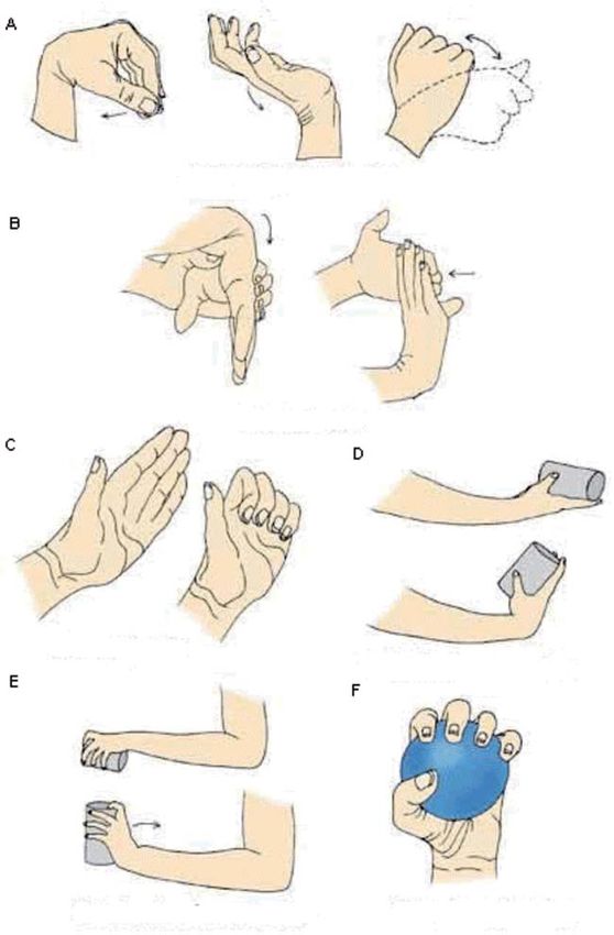
EIDESSTATTLICHE ERKLÄRUNG

  Hiermit versichere ich, die vorgelegte Masterthese selbständig verfasst zu haben.
  Alle Stellen, die wörtlich oder sinngemäß aus veröffentlichten oder nicht veröffentlichten
  Arbeiten anderer übernommen wurden, wurden als solche gekennzeichnet. Sämtliche
  Quellen und Hilfsmittel, die ich für die Arbeit genützt habe, sind angegeben. Die Arbeit
  hat mit gleichem Inhalt weder im In- noch Ausland noch keiner anderen Prüfungsbehörden
  vorgelegen.

  Diese Arbeit stimmt mit der von dem/der Gutachter/in beurteilte Arbeit überein.

DECLARATION

  Hereby I declare that I have written the present master thesis on my own.
  I have clearly marked as quotes all parts of the text that I have copied literally or
  rephrased from published or unpublished works of other authors.
  All sources and references I have used in writing this thesis are listed in the list of
  references. No thesis with the same content was submitted to any other examination
  board before.

  Datum / Date                               Unterschrift / Signature
ABSTRACT

    Introduction: Distal radius fractures are among the most common types of fractures. Irre-
spective of the choice of therapy (whether conservative or surgical), these fractures may entail
negative consequences in the form of limited range of motion and diminished muscle strength.
Such sequelae cause limited hand performance, which, considering the important function of
the hand, may negatively affect the quality of life and impair patient’s independence in perfor-
ming everyday activities Despite a considerable progress in medicine and physical therapy over
the last several years, distal radial fracture outcomes seem to be unsatisfactory. Conventional
mobilization methods do not increase the number of very good and good outcomes. Never-
theless, the effects of a therapist’s efforts concentrated on specific tissues of the musculoskeletal
system, such as fasciae, seem to be an effective treatment method rapidly restoring the normal
range of motion and muscle strength and consequently – full hand function.

  Aims: To present the Fascial Distortion Model (FDM) as a potentially effective treatment of
musculoskeletal dysfunctions after distal radius fractures.

    Methods: A total of 65 patients (12 men, 53 women, 22 to 81 yerars of age) suffering a distal
radial fracture were randomized into a study group (n = 33) and control group (n = 32). Apart
from the standard recommendations and exercise instructions, the study group underwent
three sessions with the use of FDM techniques. These therapeutic sessions were conducted once
a month. The therapy was adjusted to individual limitations and patient feedback related to pain.
The utilized therapeutic techniques included triggerbands, herniated triggerpoints, continuum
distortion, folding distortion, cylinder distortions, and tectonic fixation.
    An efficiacy analysis of the FDM techniques was done by pre- and posttherapeutic measu-
rements of grip strength, the range of motion (extension, flexion, adduction and abduction) at
the radiocarpal joint, of the ability to perform daily tasks (DASH 100 scale) and the level of pain
(100 mm VAS).

     Results: Single FDM therapy sessions conducted in the evaluation group resulted in imme-
diate improvement in the range of motion and grip strength and a significant improvement in
all studied parameters (p < 0.005). In comparison with the control group, patients treated with
the use of the FDM techniques achieved better results in grip strength and range-of-motion
assessment.The effects achieved after each session were maintained or improved in the period
before the next session. No negative effects of therapy, such as a decrease in strength or limited
range of motion, were observed in any patient.

   Conclusion: The results indicate very high efficacy of the FDM as a therapeutic technique
rapidly improving the muscle strength and the range of motion in the affected joint.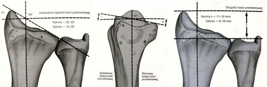
Acknowledgements (optional)
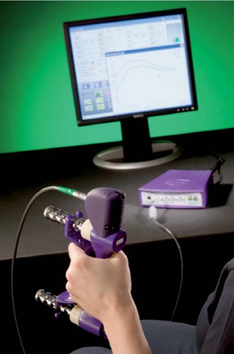
TABLE OF CONTENTS

Introduction and aim of the study

1. Background
1.1. Anatomy of the distal radius area
     1.1.1. Bones and joints
     1.1.2. Muscles
     1.1.3. Fasciae
1.2. Selected biomechanical aspects
     1.2.1. The radiocarpal joint
     1.2.2. The distal radioulnar joint
1.3. Distal radial fractures
     1.3.1. Mechanisms of injury and types of fractures
     1.3.2. Epidemiology of fractures
     1.3.3. Diagnostics
     1.3.4. Treatment of distal radial fractures
            1.3.4.1. Conservative management
            1.3.4.2. Surgical treatment
            1.3.4.3. Complications
            1.3.4.1. Physical therapy
1.4. The Fascial Distortion Model (FDM)
     1.4.1. Fasciae
     1.4.2. Fascial distortions
     1.4.3. Treatment techniques
     1.4.4. Contraindications to FDM

2. Material and methods
2.1. Material
2.2. Methods
     2.2.1. Grip strength assessment
     2.2.2. Range-of-motion assessment in the radiocarpal joint
     2.2.3. Assessment of patient’s functional performance
2.3. Statistical analysis methods

3.     Results
3.1.   Grip strength and the wrist range of motion
3.2.   Functional performance and the level of pain
3.3.   Efficacy of FDM sessions
3.4.   Analysis of correlations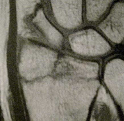
4.     Discussion
4.1.   Grip strength and the wrist range of motion
4.2.   Functional performance and the level of pain
4.3.   Study result correlations
4.4.   Efficacy of FDM

5.     Conclusions

Summary
References
List of figures and sources
List of tables
Annexes
      A study assessment form for the evaluation group
      A study assessment form for the control group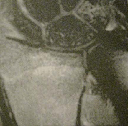
Introduction and aim of the study

    Distal radius fractures of the radius are among the most common types of fractures. In young
people, these are usually direct-mechanism fractures or high-energy injuries. In the elderly, di-
stal radial fractures are caused by a low-energy trauma such as a fall from the standing height
[57, 61]. Irrespective of the choice of therapy (whether conservative or surgical), these fractures
may entail negative consequences in the form of limited range of motion and diminished mu-
scle strength. Such sequelae cause limited hand performance, which, considering the important
function of the hand, may negatively affect the quality of life and impair patient’s independence
in performing everyday activities [12, 24, 26].

    Despite a considerable progress in medicine and physical therapy over the last several years,
distal radial fracture outcomes seem to be unsatisfactory. A number of patients, especially the
elderly, still complain of limited function and performance in the injured hand. Conventional
mobilization methods do not increase the number of very good and good outcomes [9, 30, 48].
Meanwhile, the number of publications on the use of novel therapies, and particularly the os-
teopathic methods, remains low. Nevertheless, the effects of a therapist’s efforts concentrated on
specific tissues of the musculoskeletal system, such as fasciae, seem to be an effective treatment
method rapidly restoring the normal range of motion and muscle strength and consequently –
full hand function [49, 75].

The aims of this study are:
•   To present the problem of distal radial fractures as function-limiting injuries of the hand,
•   To present the Fascial Distortion Model (FDM) as a potentially effective treatment of mus-
    culoskeletal dysfunctions,
•   To present the results of our studies on the efficacy of FDM techniques in the treatment of
    radial fracture patients,
•   To review the available literature concerning previous studies.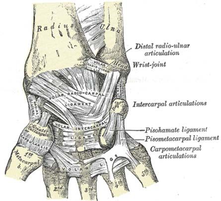
1. Background

1.1. Anatomy of the distal radius area

1.1.1. Bones and joints

   The articulations at the distal end of the radius include the radiocarpal joint and the distal
radioulnar joint (DRUJ).
   The DRUJ comprises the circumference of the head of the radius and the radial notch of the
ulna serving as its socket. The articular capsule is loose yet strong.
    The radiocarpal joint connects the radius with the proximal carpal bones, comprising the
following bones: the scaphoid, lunate, triquetral, and pisiform (however, the pisiform bone is
not part of the articular facet). The articular facet of the distal end of the radius constitutes 75%
of the joint’s socket and the remaining part of the socket is made up by the articular disc filling
the space between the head of the ulna and the carpal bones. The articular socket is slightly inc-
lined toward the ulna and tilted anteriorly, which results in an increased range of adduction and
flexion. The head of the joint, comprising three carpal bones (the scaphoid, lunate, and triquet-
ral), is ellipsoid in shape (Fig. 1) [5, 22].

                                                           S – scaphoid
                                                           L – lunate
                                                           P – pisiform
                                                           Tr – triquetral
                                                           H – hamate
                                                           C – capitate
                                                           T – trapezoid
                                                           Tz – trapezium
                                                           U – ulna
                                                           R – radius
                                                           RCJ – radiocarpal joint
                                                           DRUJ – distal radioulnar joint

Fig. 1. The bones forming the radiocarpal and the distal radioulnar joint.

                                                                                                  1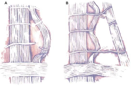
The proximal carpal bones are connected via arthrodial joints of limited mobility held firmly
together by ligaments, which facilitates synchronized movements of all three bones constituting
the radiocarpal joint with respect to the radius. The articular capsule is loose. The radiocarpal li-
gaments, which strengthen the articular capsule and control the movements in the joint include:
•   the radial collateral ligament – extends from the styloid process of the radius to the scaphoid
    bone, controls the adduction (ulnar abduction) of the hand and transfers the rotational mo-
    vements of the forearm onto the hand,
•   the ulnar collateral ligament – extends from the styloid process of the ulna to the triquetral
    bone and to the pisiform bone; it controls the abduction (radial abduction) of the hand and,
    together with the radial collateral ligament, transfers pronation and supination of the fore-
    arm onto the hand,
•   the palmar radiocarpal ligament – extends from the styloid process and the palmar margin
    of the radius to all four bones of the proximal carpal row; it controls the extension and su-
    pination of the hand,
•   the dorsal radiocarpal ligament – has its origin on the dorsal margin of the distal radius and
    its insertion on the dorsal surface of the proximal carpal bones, controls the palmar flexion
    and pronation, but is weaker than the one mentioned above,
•   the palmar arcuate ligament of the wrist – combines fibers of the palmar radiocarpal liga-
    ment and ulnar collateral ligament; it controls the extension in the joint,
•   the dorsal arcuate ligament of the wrist – connects only the scaphoid and triquetral bones; it
    controls the flexion and abduction (Fig. 2) [5, 22].

A                                                    B

Fig. 2. Ligaments of the radiocarpal joint; palmar view (A) and dorsal view (B).

                                                                                                  2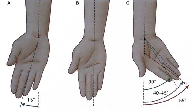
1.1.2. Muscles

The muscles mobilizing the joints at the distal end of the radius belong to a group of forearm
muscles. They can be divided into three groups:
•   the anterior (palmar) group – comprising eight muscles: the pronators teres and quadratus,
    the flexors carpi radialis and ulnaris, the flexors digitorum superficialis and profundus, the
    flexor pollicis longus, and the palmaris longus muscle; this group is responsible for the flexi-
    on of the radiocarpal joint and pronation of the forearm,
•   the posterior (dorsal) group – comprising seven muscles responsible for the extension of the
    radiocarpal joint: the extensors digitorum, indicis, digiti minimi, pollicis longus and exten-
    sor brevis, as well as the extensor carpi ulnaris and the abductor pollicis longus,
•   the lateral (radial) group – comprising four muscles: the brachioradialis (not involved in
    wrist movements), the extensors carpi radialis longus and brevis, and the supinator muscle.
    This group of muscles is responsible for the extension in the radiocarpal joint and supination
    in the radioulnar proximal and distal joints (Fig. 3) [5, 22].

Fig. 3. Muscles of the forearm; anterior views (A and B) and posterior views (C and D).

1.1.3. Fasciae

    The antebrachial fascia, which is a continuation of the brachial fascia, surrounds all the mus-
cles of the forearm. From the anatomical point of view, it can be divided into the proximal part,
the cubital fascia, surrounding the structures of the elbow joint and enclosing the cubital fossa,
and the distal part continuing into the fascia of the hand at the wrist level.

                                                                                                 3
Joined with the posterior margin of the ulna along its entire length, the antebrachial fascia
forms intermuscular septa separating individual groups of antebrachial muscles. Moreover, the
fascia forms multiple divisions separating individual muscles. Fibres of the antebrachial fascia
run circularly and are particularly thick and strong where the fascia continues into the fascia of
the hand.
The fascia of the hand is divided into four laminae. Two of them – the palmar deep fascia
and the dorsal interosseous fascia – are the deep layers. More superficially, on the dorsal side,
the superficial dorsal fascia of the hand can be found, beneath which lie the tendons of the ex-
tensors digitorum longus. On the palmar side, there is the superficial palmar fascia of the hand.
In its middle part, it thickens markedly and forms the palmar aponeurosis, whose palmar fibers
intertwine with the palmar longus muscle tendon, and the dorsal (deep) fibers interlace with the
extensor retinaculum [5, 22, 27, 68].
41.2. Selected biomechanical aspects
1.2.1. The radiocarpal joint
The radiocarpal joint is ellipsoid, with the distal part of the radius and the articular disc for-
ming the socket, and the proximal carpal bones forming the head. This is an articulation with
two degrees of freedom. The possible movements occur in a sagittal plane around a transverse
axis (flexion and extension) and in a frontal plane around a sagittal axis (adduction and abduc-
tion). These movements can be combined into circumduction around the long axis of the arm
[6, 29, 32, 34].
All of the above movements involve both the radiocarpal and the midcarpal joints (the lat-
ter connecting the bones of the proximal and distal carpal rows) as well as the arthrodial joints
between all the carpal bones. These articulations are conjoined, thus their combined mobility is
being considered.
The range of movements in a frontal plane is extensive, as it is 85º for active flexion and
active extension each. The passive range of these movements is even greater at 95º and 100º,
respectively (Fig. 4, 5) [29]. During flexion, the radiocarpal joint is responsible for 50º of mobi-
lity, and the midcarpal joint for 30º. During the extension, the greater role can be attributed to
the mobility in the midcarpal joint (45º), whereas the radiocarpal joint is responsible for only
approximately 35º of the range of extension [6].
Fig. 4. The range of active flexion and extension in the wrist.
5Fig. 5. The range of passive flexion and extension in the wrist.
Range of motion in a frontal plane is smaller at 15º of the active abduction (radial abduction)
and 40–45º of the active adduction (ulnar abduction) (Fig. 6) [6, 29, 32, 34].
Fig. 6. The range of abduction (A) and adduction (C) of the wrist starting from the intermediate
position (B).
1.2.2. The distal radioulnar joint
This articulation is a pivot joint with one degree of freedom. The movements of pronation
and supination of the forearm occur in a horizontal plane around the long axis of the forearm.
This articulation is functionally coupled with the proximal radioulnar joint, formed by the cir-
cumference of the head of the radius and the radial notch of the ulna. This coupling means that
movement in both of these joints is necessary in order to achieve rotation of the forearm. Move-
6ment in these joints is controlled by the pronator and supinator muscle flexion and, in extreme
positions, by the articular capsules. The interosseous membrane stabilizes the movements in
those joints controlling the mobility of the ulna and the radius, relative to each other in the long
axis of the forearm. The range of supination in the forearm is 90º and pronation – 85º (Fig. 7)
[6, 29, 32, 34]].
Fig. 7. The range of supination (A) and pronation (C) of the forearm from the intermediate positi-
on (B).
71.3. Distal radial fractures
Distal radial fractures are among the most common injuries reported in emergency depart-
ments and A&Es. Moreover, these are among the most common fractures in the elderly, alt-
hough they are not uncommon in the young adult population or children and adolescents where
they occur as a result of high-energy injuries [10, 20, 26].
1.3.1. Mechanisms of injury and fracture classifications
A distal radial fracture is most commonly a result of indirect force, i.e. fall on the hand.
Direct fractures caused by an impact of a heavy object are rare. Depending on the mechanism
of injury, fractures can be divided into fractures in extension mechanism (when the hand was
extended during the fall) and, less common fractures in flexion mechanism (when the hand was
flexed).
Usually, conventional names are used for the different types of distal radial fractures:
• Colles’ fracture – a distal metaphyseal fracture of the radius, where the fracture slit may
reach the articular surface; with angulation and radial shortening,
• Smith’s fracture – also known as a reverse Colles’ fracture, characterized by volar displace-
ment of distal fracture fragments, it may be extra-articular or may involve the radiocarpal
joint,
• Barton’s fracture – involves the dorsal or palmar margin of the radial articular surface and is
complicated by wrist subluxation,
• Chauffeur’s fracture – an intra-articular fracture of the radial styloid process,
• Die-Punch fracture – involves a depression fracture of the lunate fossa or a depression of a
central facet fragment.
There are also other classification systems intended to facilitate communication among the
medical personnel and help the clinician to select the most effective treatment. Among the ol-
dest, there is the Frykman’s classification which divides fractures into intra-articular and extra-
articular, and then according to the damage to the styloid process of the ulna; this classification
comprises eight types of fractures (Fig. 8) [18].
8Fig. 8. The Frykman’s classification of distal radial fractures.
The fracture classification most commonly found in literature is the AO classification, due to
its simplicity on one hand and precise division of fractures into types and subtypes on the other.
The AO classification divides fractures into three main categories:
A – extra-articular fractures,
B – partly intra-articular fractures,
C – fully intra-articular fractures.
Fracture subtypes can be classified based on the extent of injury to the joint and metaphyseal
fragmentation (Fig. 9). This classification is of practical benefit, considering that assigning a
given fracture to the right type has bearing on the selection of treatment approach.
9Fig. 9. The AO classification of distal radial fractures [Muller].
Similar principles are used in the so-called universal classification of fractures into four main
types, as well as in the Medoff ’s classification based on radiographic findings and the selected
treatment method. These classification systems, however, are less commonly applied.
Relatively frequently encountered is the Fernandez classification, which divides fractures
according to the mechanism of injury and co-existing damage as well as the recommended
treatment into:
Type I – bending fracture of the metaphysis,
Type II – shearing fracture of the joint surface,
Type III – compression fracture of the joint surface,
Type IV – avulsion fracture, radiocarpal fracture with dislocation,
Type V – complex fractures of types I-IV, high-energy fracture [Brown, Sanders].
10Distal radial fractures can be divided into indirect (more common) and direct (less com-
mon) mechanism injuries. Based on the kind of trauma, there may be high-energy or low energy
fractures. High-energy fractures occur usually in young people as a result of falls from a height,
a forceful impact or a traffic accident. Low-energy fractures are caused by falls from the standing
height and are typical for the elderly suffering from osteoporosis. As mentioned above, distal
radius fractures may result from a fall on an extended hand (Colles’ fracture), which is the most
common fracture type or on a flexed hand (Smith’s fracture) [15, 26, 79].
The following may be associated with distal metaphyseal fractures of the radius:
• fracture of the ulnar styloid process,
• fracture of the scaphoid bone and other carpal bones (particularly in children),
• periscaphoid dislocations,
• injury to the triangular fibrocartilage complex,
• ligament injuries (especially of the interosseous ligaments),
• injury to tendons, nerves, and other soft tissues surrounding the fracture.
Fracture-associated soft tissue injuries occur in about 70% fractures, which in the case of
misdiagnosis may lead to carpal instability [15, 20, 26, 35].
1.3.2. Fracture epidemiology
Distal metaphyseal fractures of the radius constitute 12–15% of all fractures. A vast majority
of distal radial fractures are osteoporotic. These are seven times more frequent in women over
60 than in men of the same age. Incidence of these fractures ranges from 0.5% to 2% annually,
and the number of people suffering from this injury grows rapidly in the age group of 60 to 69.
Risk factors for fractures in this population include mainly low bone mineral density (BMD)
and a fracture in the family. This is often the first sign of osteoporosis, particularly in regions
where early osteoporosis diagnostic tests are neglected. Poor mechanical strength of bones is
another factor predisposing to fragment displacement during treatment, late instability, and
deformities [21, 41, 57, 61, 62].
1.3.3. Diagnostics
Early assessment of the injury involves visual inspection and physical examination. The wrist
is often deformed, immobilized in one position, and any attempt at movement causes severe
pain. Before any further diagnostics or treatment is undertaken, the distal limb has to be as-
sessed for pulse and superficial sensibility.
Radiographic imaging is the evaluation of choice in suspected distal radial fracture. Routine
radiographic images are postero-anterior and lateral views showing the fracture line (Fig. 10).
11Additionally, a lateral view may be obtained with the wrist positioned in a neutral position and
elevated by 10º off the image plate. This projection more accurately shows the radiocarpal joint
surface.
A B
C D
Fig. 10. A radiographic image of a radial fracture in antero-posterior (A) and lateral (B)
views; C, D – distal radius fracture – type C3.
12Computed tomography (CT) and magnetic resonance imaging (MRI) are among the ad-
ditional examinations used in differential diagnosis assessing the associated injuries, such as
ligament and tendon tears, as well as in assessing the joint surface fit in trans-articular fractures
(Fig. 11) [26, 71].
A B
Fig. 11. An MRI scan of an intra-articular fracture of the radius: the fracture line in the T1-
weighted sequence (A) and a hyperintense area of bone marrow edema in the FST2-FSE sequence
(B).
1.3.4. Treatment of distal radial fractures
The treatment goals in fractures of the distal radius are reconstructing the anatomical angles
of the radiocarpal joint surface, as well as maintaining the proper radial height and the stability
of the distal radioulnar joint. The congruence of articular surfaces of the scaphoid and lunate
fossas is of major importance, as it allows forces to be properly distributed across the wrist and
ensures the execution of smooth movements in the radiocarpal joint. Important from the point
of view of restoring the hand function is reconstruing the normal biomechanical parameters of
the wrist, in particular:
• the radial inclination angle, normally between 22–23º, with an acceptable range of 13-10º,
• the palmar tilt of the distal radius (norm 11–12º, range 0–28º),
• the radial height in comparison with the ulna (norm 11–12 mm, acceptable range 8–18 mm)
(Fig. 12).
The expected long-term treatment effects are the return of full range of flexion, extension,
radial abduction and ulnar abduction of the wrist, as well as forearm rotation [12, 24, 26].
13A B C
Fig. 12. Normal positioning of the distal end of the radius: radial inclination angle (A), radial tilt
(B), and radial height (C).
1.3.4.1. Conservative management
Conservative management indications include:
- extra-articular fractures with or without displacement,
- intra-articular extension fractures without displacement,
- compression fractures with slight fragment displacement.
Moreover, conservative management is used in patients with contraindications to general
anesthesia.
In fractures with displacement, reduction of fragments is required prior to fragment immo-
bilization. This is most commonly done using closed methods under local anesthesia. During
the reduction procedure, the fragments are pulled apart with the help of another person or
using finger traction. Non-displaced and reduced fragments are stabilized with the use of an
individually moulded sugar-tong splint. This splint remains in place for 2–3 weeks with a week-
ly inspection for the axial positioning of fragments. After splint removal, the forearm is placed
in a cast for 3–5 weeks. After 6 weeks of immobilization, the cast is removed and passive wrist
movements are introduced. Further rehabilitation is similar to that following surgical treatment
and its purpose is to restore full joint mobility, muscle strength, and the ability to perform daily
activities.
If any fracture displacement occurs within 2 weeks after the fracture reduction and immobi-
lization, surgical stabilization should be considered [16, 24, 26, 40].
141.3.4.2. Surgical treatment
Surgical treatment indications are:
- intra-articular fractures with displacement,
- unstable fractures,
- fractures with significant initial fragmentation,
- fractures with a significant shortening of the radius, particularly compression fractures,
- fractures impossible to reduce using the closed methods.
The methods of choice in surgical treatment of distal radial fractures include: percutaneous
Kirschner-wire stabilization, external fixation, external fixation with K-wireing or internal fixa-
tion, and internal fixation with the use of plates and pins. Fracture stabilization is preceded by
closed, open or arthroscopic-assisted reduction [26, 40, 73].
Percutaneous wire stabilization is conducted after closed reduction of the fracture. Kirsch-
ner wires are introduced via small incisions in the skin through the styloid process of the radius
and into the cortical layer of the proximal fragment of the ulna (Fig. 13). In the Kapandji me-
thod, the wires are utilized as levers for the entire fracture and help reduce the fracture as well
as maintain the required shape of joint surfaces. After the wires are introduced, the wrist is im-
mobilized in a splint or plaster cast for 4–5 weeks. This method is not recommended for treating
fractures in the elderly; however, in younger patients, it leads to significantly better outcomes
than conservative management [4, 24, 26, 65, 73].
A B
Fig. 13. Post-surgery radiographic images of a fracture treated with Kirschner wires – antero-
posterior (A) and lateral (B) views.
15Good long-term effects are also achieved with external fracture stabilization. This is perfor-
med with the use of an external fixation device, comprising pins introduced into the bone and
external bars (Fig. 14). External stabilization is sometimes supported with the Kirschner-wire
fixation or internal stabilization, a bone graft or arthroscopic-assisted fragment reduction. The
use of external stabilization with the support of Kirschner wires is an effective means of frag-
ment immobilization and it decreases the risk of repeated surgery. However, it may increase the
incidence of infection [14, 21, 25, 26].
A B
Fig. 14. The use of external fixation in radial fracture management.
Arthroscopic reduction of fragments is particularly useful in injuries with extensive frag-
mentation of the epiphysis and appears to be a more effective method of assessing the articular
surface of the reduced fragments than fluoroscopy. Moreover, it facilitates the diagnosis of any
co-existing damage to ligaments and the triangular cartilage [24, 26].
In the case of fractures impossible to reduce, open reduction and internal fixation are used.
Internal fixation is also used in multiple-fragment and compression fractures, with a co-existing
ulnar fracture, and in people with osteoporosis. In multiple-fragment fractures, stabilization can
be achieved with the use of screws and Kirschner wires. With fewer fragments, a dorsal distrac-
tion plate or fixed-angle plates can be used dorsally or volarly. Distraction plates are utilised in
high-energy fractures with considerable fragmentation of the distal radial epiphysis. Stabiliza-
tion with the use of a dorsal plate is indicated in Barton’s fracture (shear type) and in fractures
with displacement of the dorsal margin of the articular surface. This technique, however, is less
and less common, as it may result in irritation or damage to the extensor digitorum tendons.
Stabilization with the use of palmar plate helps to restore the length of the radius and achieve ap-
propriate ulnar inclination (Fig. 15). Some authors report clinically asymptomatic joint instabi-
lity following this treatment. It is worth noticing that the use of internal stabilization contributes
to a quicker restoration of full range of motion, whereas its long-term effects seem to be similar
to those achieved with the use of external stabilization. Mechanical stabilization with a plate is
equally effective to that with external fixators, although clinical studies show inconsistent results
of comparative evaluation of these types of stabilization among different authors [8, 14, 17, 25,
26, 36, 52, 53, 55, 64, 72].
16A B
Fig. 15. A post-operative radiographic image of a fracture stabilized with a fixed-angle
palmar plate (A) and an image of a palmar plate (B).
In the case of significant bone loss, which is most often due to fragment impaction, grafts
of bone or bone replacement materials are implemented. Bone grafts are also utilized with the
placement of stabilizing plates in order to replace any larger bone defects [26, 64].
The outcomes achieved one year after the injury seem to be similar irrespective of the treat-
ment method, provided that it has been properly matched to the type of fracture and the patient’s
condition [14, 50].
1.3.4.3. Complications
Management of distal radial fractures is associated with a high rate of complications that
eventually contribute to a significant number of unsatisfactory outcomes. Complications of both
conservative and surgical management include:
- displacement resulting in incorrect bone union,
- delayed, or lack of, bone union,
- permanent nerve damage due to injury, surgical procedures or long-term pressure,
- inflammation of the joint and periarticular tissues (also as a result of infection), which
may cause bone nonunion,
- tendon rupture,
- inadequate mobility or instability of the radiocarpal or distal radioulnar joint,
17- Sudeck’s atrophy (reflex sympathetic dystrophy syndrome),
- Volkmann’s ischemic contracture,
- rarely: pressure sores caused by incorrectly applied plaster cast, carpal tunnel syndrome.
Incorrect union is one of the easily manageable sequelae causing most problems with resto-
ring the hand function. Bone axis correction is achieved by intra-articular or extra articular
osteotomy. An osteotomy procedure does not guarantee full joint function recovery, however, in
most cases the results are satisfactory [26, 38, 43, 63, 78].
In the case of lack of union or delayed bone union, attempts are undertaken at conservative
treatment with the use of physical therapy procedures (magnetotherapy with high-induction
fields) as well as surgical treatment (resection of the fractured bone ends, re-fixation, cortico
spongeous graft) or compression-distraction osteosynthesis (the Ilizarov technique), particu-
larly with a co existing radial shortening and axis warping. If the treatment is ineffective, carpal
arthrodesis can be performed, which improves the hand function with relatively few complica-
tions [39, 56, 60, 66].
Treatment of other complications is consistent with the generally accepted management pro-
cedures and will not be addressed in this article due to the detailed character of the subject.
1.3.4.4. Physical therapy
The objectives of physical therapy following the removal of an immobilization device or the
union of surgically fixed fragments are:
• restoring the full range of motion at the radiocarpal and distal radioulnar joints,
• achieving the full muscle strength, particularly grip strength,
• full recovery of the affected hand in terms of daily functioning.
The rehabilitation period can be divided into three phases:
• the early phase, lasting from fracture immobilization to approximately week 6,
• the intermediate phase, lasting from week 6 to week 8 post injury,
• the late phase, beginning approximately in week 8 post injury and lasting until the full
hand function is restored (approximately week 12).
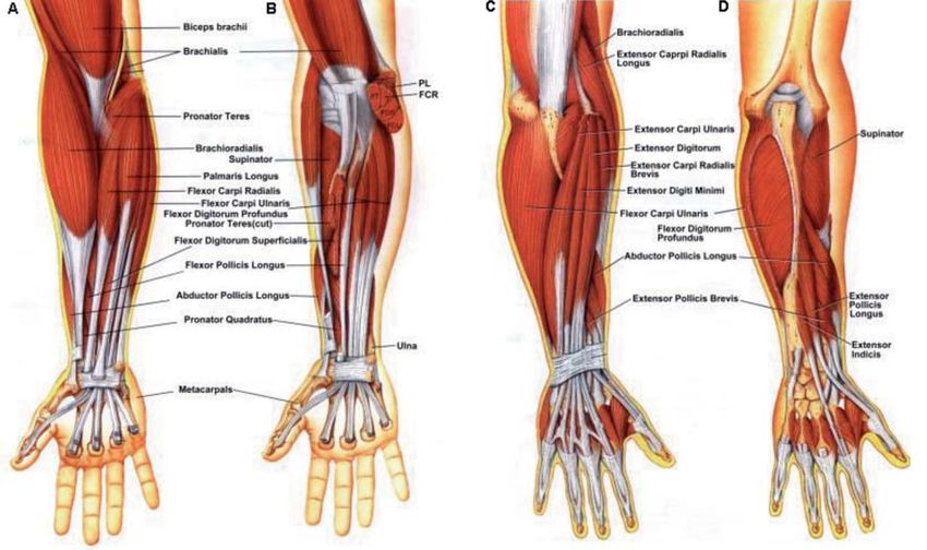
Early phase (weeks 0–6)
The main purpose of this phase is to decrease the rigidity and edema of the hand. Effective
means include hand elevation above the heart level, frequent movements of fingers, the use of
hand and wrist compression supports (or appropriate adhesive tapes). Active and passive finger
range-of-motion exercises are also recommended (Fig. 16). The patients should use the hand as
much as they can in performing light daily tasks, especially in the case of stable or successfully
surgically stabilized fractures. If there are no contraindications for forearm rotation, forearm su-
pination exercises should begin already in the early phase of rehabilitation, as this is one move-
18ment that can quickly become limited. Other wrist movements can be also performed, provided
there are no contraindications and wrist stability is maintained (e.g. with palmar plate fixation
or non-bridging external fixation). Post operative management requires the gently massaging
of the scar to prevent its hypertrophy. Additionally, active exercises of the elbow and shoulder
joints are recommended in order to prevent limitation of the range of motion in these joints.
Magnetotherapy, local cryotherapy and, in the case of conservative management, electrotherapy
are used to minimize pain, as well as to accelerate bone union and normal remodeling of the
existing union.
Fig. 16. Finger exercises for flexor tendon mobilization.
Intermediate phase (week 6–8)
After approximately 6 weeks, Kirschner-wire or external stabilization is removed. Also with
other treatment methods, the patients should be encouraged to gradually give up external im-
mobilization after 6 weeks. This phase focuses on increasing the limited range of motion of the
wrist (flexion and extension, abduction and adduction) and forearm (supination and pronati-
on). To this end, active-assistive and passive exercises are used, as well as supination splints or
other dynamic splints, if required. Any physical therapy initiated to that point should be conti-
nued in this phase.
Late phase (weeks 8–12)
After about 8 weeks following the injury, complete bone union is achieved, allowing the
patient to begin the strengthening exercises of the hand using soft balls of various types and
rubber hand trainers, as well as small weights, dumbbells or specially constructed devices for
strength training in various movements. Additionally, wrist, metacarpal, finger, and forearm
range-of-motion exercises are continued (Fig. 17). An important element of the late phase of
rehabilitation is restoring the normal hand function. This is achieved through exercises with the
use of various common objects – mugs, balls, cylinders, knobs, door handles, dials and other
elements used daily by the patient. If necessary, electrotherapy (to fight pain as well as in the
form of electric muscle stimulation) and local cryotherapy are used to prevent development of
post-exercise edema and pain.
19Fig. 17. Wrist exercises:
A – increasing the range of motion,
B – stretching the wrist into flexion and extension,
C – tendon mobilization exercises,
D – wrist flexion while holding a cylinder,
E – wrist extension while holding a cylinder,
F – increasing the grip strength.
20Typically, a large proportion of patients receive instructions on how they should exercise and
do the assigned exercises on their own at home. According to many authors, there is no signifi-
cant benefit in conducting the rehabilitation program at the clinic, and that benefits, if any, are
mainly due to greater patient satisfaction and decreased pain. Unfortunately, the role of physio-
therapy in the treatment of distal epiphyseal fractures of the radius and in quick restoration of
the hand function in these patients is still underestimated, which leads to not fully satisfactory
outcomes or significant delays in achieving the full limb function [9, 30, 43, 44, 48, 66, 69].
211.4. The Fascial Distortion Model (FDM)
The Fascial Distortion Model (FDM) is a manual therapy method created and developed in
the United States by Stephen Typaldos, an osteopath with many years’ experience. This tech-
nique is also known as TMT (Typaldos Manual Therapy).
The tenets of this technique are based on the knowledge and diagnosis of the types of fascial
structural, and consequently functional, abnormalities (distortions). Typaldos considers these
distortions to be more significant as the causative factor of pain as well as muscle motor and
function limitations than other injuries, such as sprains, luxations, fractures, mechanical muscle
injuries. Thus, management of fascial distortions directly affects the other elements of the mu-
sculoskeletal system by alleviating pain, reducing movement limitations or edema. This gives
the way to quicker and more effective treatment of injuries to other musculoskeletal system
structures [75].
1.4.1. Fasciae
Fasciae are fibrous structures composed of connective tissue and located in all parts of the
human body – they make up tendons, ligaments, superficial and deep fasciae, pericardium, and
other structures the function of which is to join, protect, separate, isolate, and envelop internal
organs, muscles and systems of the body.
As a result of their structure, fasciae have poor blood supply. A major portion of oxygen and
nutrients as well as metabolites are transported via diffusion between cells and fascial perfusion
fluid. This has important consequences in the case of fascial distortions described further in this
chapter.
Due to their diverse functions and locations, fasciae differ in structure and mechanical proper-
ties. Fascial structures can be divided into the following types:
• fascial bands – including tendons, ligaments, and the iliotibial tract,
• spiral bands – surrounding parts of limbs, trunk, blood vessels, and internal organs,
• folded fasciae – including joint capsules, interosseous membranes and fascial septa,
• smooth fascial bands – lining joints, lining the abdominal cavity beneath other types of fa-
sciae (except folded fasciae).
The function of all fascial types is the protection of various structures. Fascial bands pro-
tect joints, blood vessels, tissues, and some areas of the trunk and limbs against perpendicular
external forces. Spiral bands of fascia protect extra-articular tissues against harmful effects of
traction or compression forces. Bands of irregular, plicated structure are to protect the joints
against longitudinal forces, i.e. traction and compression. Finally, smooth fascial bands maintain
adequately low level of friction between the different structures, which allows them to easily
shift against each other.
22Apart from their protective function, fasciae also have a very important function as a struc-
ture able to receive mechanical signals. Parallel fibers of connective tissue forming fasciae are
excellent transducers of mechanical forces, received by mechanoreceptors located in both the
fasciae and adjoining tissues. Mechanoreceptors react both to stretching and compression that
affects the pressure in surrounding tissues and in the receptor cell itself. Stimuli received by
receptors are transferred to the central nervous system. It is vibration, sensed via single fascial
fibers and proportional to the level of external stimulation, that plays a significant role in the
reception of stimuli. Moreover, the vibration frequency of fascial fibers determines the cha-
racteristics of perceived discomfort: pulling, burning, numbness or pain. This fascial receptor
function is used extensively by the central nervous system in controlling the muscle contraction
and motion in the joint [75].
1.4.2. Fascial distortions
Fascial distortions can be divided into:
• triggerbands,
• herniated triggerpoints,
• continuum distortion,
• folding distortion,
• cylinder distortion,
• tectonic fixation.
The type of fascial functional abnormality is determined based on medical history. What
calls for particular attention is the manner in which the patient shows the painful area and
describes the nature of discomfort (burning, stabbing, pulling, etc.). In fascial distortions, it is
significant that an injury not only limits the range of motion, diminishes proprioception, and
impairs normal muscle function, but it also significantly disturbs fluid transport between fascial
laminae, and thus unsettles the chemical balance of the fascia and connecting tissues [75].
Triggerbands
Triggerbands are fascial bands that have been twisted, separated, torn or wrinkled (Fig. 18).
The patient reports burning or pulling sensation along the fascial band and shows the pain with
a wide movement of his/her hand along the affected fibers. The wider the movement the larger
fascial area has been damaged. The pressure of fingers against the skin will be grater with fascia
located deeper than with superficial fascial injuries.
The aim of treatment in this type of injury is to break the existing fascial adhesions, which
had formed after the injury and changed the band structure (in chronic conditions), and to res-
tore the normal arrangement of fibers. If fascial bands have been twisted, the first action will be
to rotate them back the other way. Secondly, the torn or separated fascial bands are approxima-
ted to allow for their heeling by restoring their normal anatomy.
23Fig. 18. Acute (A) and chronic (B) fascial band distortion.
Herniated triggerpoints
Herniation of fascial bands occurs when the underlying tissues protrude in an area of wea-
kened connective tissue. This type of injury may cause a number of discomforts such as: pain
in the cervical spine, shoulder, abdominal pain or the over-stretching of gluteal muscles. The
patient indicates the painful area with one or several fingers pressing the injured site. The range
of motion in neighboring joints is limited.
The treatment of herniated triggerpoints is to apply adequate perpendicular force to the in-
jured site in order to “press” the herniated tissues back in and restore their normal anatomical
relations.
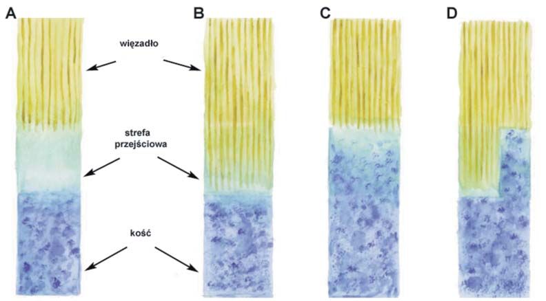
Continuum distortion
This type of distortion is characterized by structural imbalance in the transition zone bet-
ween the tendon, ligament or any other fascial structure and bone. As a result of this, the altered
transition zone structure becomes more vulnerable to external forces. The structure alteration
is mostly due to the growth of bone or tendon tissue that takes over the transition zone. This
results in a loss of the transition zone or its significant shift (Fig. 19). Such injuries are mostly
acute. These include tarsal joint sprains, over-stretching of neck muscles, and sacroiliac joint
pain. In conditions of this type, the patient always indicates the painful site with a single finger.
These injuries may be misdiagnosed as minor fascial band disturbances. Diagnosis should be
based on the efficacy of a particular treatment technique.
Treatment aims to “shift” the overgrowing tissue (whether tendon or bone) back into place
and to “expand” the transition zone to its normal size and position. A complementary treatment
of continuum distortion is ice massage, which reduces the general discomfort around the joint.
24Fig. 19. Transition zone alterations – neutral state (A), ligamentous state (B), bony state (C) and
“mixed-state” (continuum distortion) (D).
Folding distortion
Distortion in fascial folds is due to the traction or compression forces that exceed the me-
chanical resistance of periarticular fascia on which they are exerted. Based on their mechanism,
folding distortions can be divided into traction distortions and compression distortions (Fig.
20). The resulting joint pain can easily be relieved by applying the forces in the same direction
as those that caused the injury – traction is used in traction-related injuries, and compression is
effective in compression injuries. These actions help the overly stretched or compressed tissues
to return to their physiological state and the “organized” structure. Treatment also involves the
often co-existing structure abnormalities caused by joint rotation at the time of injury.
Fig. 20. Fascial distortion mechanism in a joint area following sudden traction and rotation.
25Cylinder distortion
This type of distortion affects the fascia cylindrically encircling the individual segments of
limbs (excluding the joints), the torso, and internal organs. As a result of compression or trac-
tion exceeding fascial resistance, the fibre arrangement shifts causing a disruption in the parallel,
organized fascial structure (Fig. 21). Patients characterize their pain as situated deeply, despite
the actual superficial location of its cause. More often than not, they are unable to determine the
exact location of discomfort. This discomfort may sometimes seem to be neurological due to its
character: tingling, numbness or reflex sympathetic dystrophy. While indicating the pain site,
the majority of patients repeatedly squeeze the affected soft tissues. The pain may spontaneously
relocate with time.
The treatment aims to restore the physiological arrangement of fascial fibers both with res-
pect to each other and to the long axis of the limb. This is achieved by simultaneously twisting
and pulling or compressing the damaged fascia. As with the already described treatment of pe-
riarticular fascial distortions, the direction of therapeutic force should be opposite to that which
had led to the given fascial injury.
Fig. 21. Normal (A) and distorted (B) structure of spiral fascial fibres of the forearm.
Tectonic fixation
Tectonic fixation (fascial adhesion) occurs as a result of reduction in the amount of fluid pro-
duced by smooth fasciae. Adhesions cause limitations of fascial mobility in relation to itself and
the surrounding tissues. There are also disturbances in the nourishment of cells incorporated in
the fascial structure. Adhesions of the fasciae surrounding the shoulder, hip, and intervertebral
joints are the most significant ones from the clinical point of view, as they produce the most
severe symptoms.
The treatment of fascial adhesions should first address the other co-existing problems and
then focus on increasing the tissue fluid perfusion. The final step of treatment is to restore the
fascial mobility in relation to the adjoining tissues by severing the existing adhesions [75].
261.4.3. Treatment techniques
The FDM treatment techniques combine precision and relatively high force that needs to be
applied to restore the fascial structure. These techniques can be divided into:
• manipulative techniques performed with the thumb – these include the treatment of
triggerbands, herniated triggerpoints, continuum distortions, and some of the techniques used
in cylinder distortions. Thumb techniques allow the application of significant force in a single
site at a certain angle, which increases the precision of these procedures, however, the area of
their application is limited,
• manipulative techniques performed with the whole hand – these are used in the treat-
ment of folding distortions, tectonic fixation, and some cylinder distortions. These techniques
are characterized by a smaller degree of precision, however, they allow the use of a greater force
applied over a larger affected area; there is also a possibility of applying a traction or compressi-
on force to the joint or to extra-articular soft tissues [75].
1.4.4. Contraindications to FDM
The main contraindications to the use of FDM techniques are:
• venous thromboembolism,
• conditions involving bleeding,
• confirmed aneurysm,
• phlebitis,
• other peripheral vascular conditions,
• history of stroke,
• severe oedema,
• open cuts and wounds in the treated area,
• acute bacterial, viral, and fungal infections,
• osteitis,
• septic arthritis,
• fractures,
• connective tissue disorders,
• neoplasm,
• pregnancy (in therapies involving abdominal, pelvic, and lumbosacral spine areas).
Very frequently, these techniques are painful, thus relative contraindications should include
low pain threshold or an existing psychiatric condition. In addition, caution should be exercised
when applying these techniques in children.
Following the therapy, there may develop erythema, bruising, and other reflex skin reactions
in the treated area. Sympathetic reactions such as nausea, vertigo, and vomiting are rare [75].
272. Materials and methods
2.1. Materials
A total of 65 patients (12 men, 53 women) at ages ranging from 22 to 81 were included in this
study. Members of this group were randomized into the study group (n = 33) and control group
(n = 32). Table 1 shows the detailed characteristics of groups.
Table. 1. Study and control groups broken down by gender and age.
Gender Age
Women Men Mean ± SD Range
Study group 25 8 62 ± 14 22 – 81
Control group 28 4 61 ± 13 30 – 80
All study participants suffered a distal radial fracture in the period from February to July
2009. The fractures were more common in the left limb (22 patients from the study group and
22 from control group) than in the right (11 and 10 patients, respectively). Table 2 shows the
types of fractures according to the AO classification. All patients underwent the treatment with
Kirschner-wire stabilization and a 6-week cast immobilization.
Table. 2. Types of fractures in the study and control groups according to the AO classification.
Fracture type A B C
Subtype A1 A2 A3 B1 B2 B3 C1 C2 C3
Number of fractures in the study group 3 2 2 0 1 1 7 13 4
Number of fractures in the control group 0 5 0 1 1 2 22 1 0
Apart from the standard recommendations and exercise instructions, the study group un-
derwent 3 sessions with the use of FDM techniques mentioned above. These therapeutic ses-
sions were conducted once a month. The therapy was adjusted to individual limitations and
patient feedback related to pain. The utilized therapeutic techniques included:
• triggerbands,
• herniated triggerpoints,
• continuum distortion,
• folding distortion,
• cylinder distortions,
• tectonic fixation [27, 68, 75].
28The selection of therapeutic techniques was based on detailed history and observation of
the patient during history-taking. Particular attention was being paid to pain location and the
patient’s body language when indicating the painful area.
Twenty-four patients underwent three sessions each, 3 patients underwent two sessions
each, and 6 patients one session each. The control group received only exercise instructions and
recommendations about managing their hand injury.
2.2. Methods
In order to conduct an efficacy analysis of the study therapy, the following were assessed:
- grip strength,
- range of motion at the radiocarpal joint: extension, flexion, adduction and abduction,
- ability to perform daily tasks,
- level of pain.
2.2.1. Grip strength assessment
Grip strength was assessed with the use
of the Biometrics Ltd. E-Link H500 dyna-
mometer. Grip strength was defined as a
mean of three consecutive measurements
and expressed in kilograms approximated
to one decimal place [3, 40] (Fig. 22).
Fig. 22. Muscle strength assessment with the
H500 dynamometer.
292.2.2. Range-of-motion assessment in the radiocarpal joint
Range of motion in the radiocarpal joint was measured with a manual goniometer according
to the established standards (Fig. 23, 24) [29, 67, 80].
A B
Fig. 23. Measurement of the range of flexion (A) and extension (B) in the radiocarpal joint.
A B
Fig. 24. Measurement of the range of abduction (A) and adduction (B) in the radiocarpal joint.
2.2.3. Assessment of patient’s functional performance
A subjective hand function assessment was conducted with the DASH (Disabilities of the
Arm, Shoulder and Hand) scale. This scale measures the patient’s limitations in performing 23
everyday activities, such as housework, strength tasks, personal hygiene, social life and work,
as well as 7 subjectively rated symptoms including pain, limb weakness, spasticity and the im-
pact of these discomforts on sleep. Figure 25 presents the full version of the DASH scale. Each
activity is scored from 1 (not at all difficult) to 5 points (unable to perform). The level of pain is
rated in a similar manner from 1 (none) to 5 points (unbearable). In order to get the final result,
the patient has to answer at least 27 out of 30 questions. The points from each answer are added
and divided by the number of answers. For the result to be comparable with those achieved in
other scales, the final score should be reduced by 1 and multiplied by 25. This way, the result falls
within the 0-100-point range and is called the DASH 100 score. A higher score means greater
limb disability [2, 33, 74].
30The Disabilities of the Arm, Shoulder and Hand (DASH) Score
Date of completion.................................................
Clinician‘s name (or ref)…………………………………………. Patient‘s name (or ref)…………………………………….......................
No difficulty Mild Moderate Severe Unable
difficulty difficulty difficulty
1. Open a tight or new jar 1 2 3 4 5
2. Write 1 2 3 4 5
3. Turn a key 1 2 3 4 5
4. Prepare a meal 1 2 3 4 5
5. Push open a heavy door 1 2 3 4 5
6. Place an object on a shelf above your head 1 2 3 4 5
7. Do heavy household chores (eg wash walls, wash floors) 1 2 3 4 5
8. Garden or do yard work 1 2 3 4 5
9. Make a bed 1 2 3 4 5
10. Carry a shopping bag or briefcase 1 2 3 4 5
11. Carry a heavy object (over 10 lbs) 1 2 3 4 5
12. Change a lightbulb overhead 1 2 3 4 5
13. Wash or blow dry your hair 1 2 3 4 5
14. Wash your back 1 2 3 4 5
15. Put on a pullover sweater 1 2 3 4 5
16. Use a knife to cut food 1 2 3 4 5
17. Recreational activities which require little effort
(eg cardplaying, knitting, etc) 1 2 3 4 5
18. Recreational activities in which you take some force or impact through
your arm, shoulder or hand (eg golf, hammering, tennis, etc) 1 2 3 4 5
19. Recreational activities in which you move your arm freely (eg playing
risbee, badminton, etc) 1 2 3 4 5
20. Manage transportation needs (getting from one place to another) 1 2 3 4 5
21. Sexual activities 1 2 3 4 5
22. During the past week, to what extent has your arm, shoulder or hand
problem interfered with your normal social activities with family,
friends, neighbours or groups? 1 2 3 4 5
23. During the past week, were you limited in your work or other regular
daily activities as a result of your arm, shoulder or hand problem? 1 2 3 4 5
Please rate the severity of the following symptoms in the last week No difficulty Mild Moderate Severe Unable
difficulty difficulty difficulty
24. Arm, shoulder or hand pain 1 2 3 4 5
25. Arm, shoulder or hand pain when you performed any specific activity 1 2 3 4 5
26. Tingling (pins and needles) in your arm, shoulder or hand 1 2 3 4 5
27. Weakness in your arm, shoulder or hand 1 2 3 4 5
28. Stiffness in your arm, shoulder or hand 1 2 3 4 5
29. During the past week, how much difficulty have you had sleeping
because of the pain in your arm, shoulder or hand? 1 2 3 4 5
30. I feel less capable, less confident or less useful because of my arm,
shoulder or hand problem 1 2 3 4 5
number of responses: DASH total DASH 100:
Fig. 25. The DASH scale.
31You can also read