Cancer - Disease of the Age - Recent revolution in cancer diagnosis, treatment, management, prevention and control
←
→
Page content transcription
If your browser does not render page correctly, please read the page content below

www.esciencecentral.org/ebooks

Cancer - Disease of the Age
Recent revolution in cancer diagnosis,
treatment, management, prevention
and control

                                         Edited by
                Dr. Ahmed M El-Sharkawy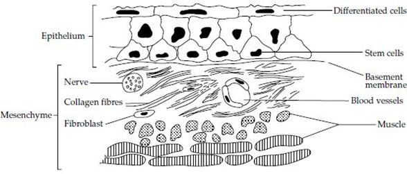
Copyright agreement
                  eBooks

Cancer: Disease of the Age Recent Revolution
in Cancer Diagnosis, Treatment, Management,
Prevention and Control
Chapter: Tumorigenesis
Edited by: Ahmed M El-Sharkawy
Published Date: July 2014

Published by OMICS Group eBooks

731 Gull Ave, Foster City. CA 94404, USA

Copyright © 2014 OMICS Group
This eBook is an Open Access distributed under the Creative Commons Attribution 3.0
license, which allows users to download, copy and build upon published articles even for
commercial purposes, as long as the author and publisher are properly credited, which
ensures maximum dissemination and a wider impact of our publications. However, users
who aim to disseminate and distribute copies of this book as a whole must not seek
monetary compensation for such service (excluded OMICS Group representatives and
agreed collaborations). After this work has been published by OMICS Group, authors have
the right to republish it, in whole or part, in any publication of which they are the author,
and to make other personal use of the work. Any republication, referencing or personal use
of the work must explicitly identify the original source.

Notice:
Statements and opinions expressed in the book are these of the individual contributors
and not necessarily those of the editors or publisher. No responsibility is accepted for the
accuracy of information contained in the published chapters. The publisher assumes no
responsibility for any damage or injury to persons or property arising out of the use of any
materials, instructions, methods or ideas contained in the book.

A free online edition of this book is available at www.esciencecentral.org/ebooks
Additional hard copies can be obtained from orders @ www.esciencecentral.org/ebooks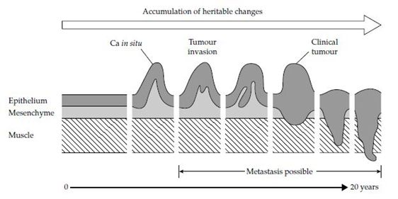
eBooks

       Tumorigenesis
                                  Ahmed M El-Sharkawy*
                                  Biochemistry department, Faculty of Science, Alexandria University, Alexandria, Egypt
                                  *Corresponding author: Ahmed M ElSharkawy, Biochemistry department, Faculty
                                  of Science, Alexandria University, Alexandria, Egypt, Tel: +201000512872; E-mail:
                                  ahmedm.elsharkawy83@gmail.com

Introduction
    Cancer has been known since human societies first recorded their activities. It was well
known to the ancient Egyptians and to succeeding civilizations but, as most cancers develops
in the latter decades of life, until the expectation of life began to increase from the middle of the
nineteenth century onwards, the number of people surviving to this age was relatively small.
Now that the infectious diseases, the major causes of death in the past, have been controlled
by improvements in public health and medical care, the proportion of the population at risk of
cancer has increased dramatically. Although diseases of the heart and blood vessels are still
the main cause of death in our ageing population, cancer is now a major problem. At least one
in three will develop cancer and one in four men and one in five women will die from it. For
this reason, cancer prevention and control are major health issues. However, cancer research
has wider significance. Cancer is not confined to man and the higher mammals but affects
almost all multicellular organisms, plants as well as animals. Since it involves disturbances in
cell proliferation, differentiation, and development, knowledge of the processes underlying this
disease help us to understand the very basic mechanisms of life. About 140 years ago a German
microscopist, Johannes Mueller, showed that cancers were made up of cells, a discovery which
began the search for changes which would help to pinpoint the specific differences between
normal and cancer cells. In the intervening period a huge amount of information has been
acquired about the cancer cell. In the past two decades in particular, rapid technological
progress has allowed us to begin to dissect the cancer genome, transcriptome, and proteome
in unprecedented detail and today there seems no limit to the amount of information that
can be obtained. However, this does not naturally answer all of the questions posed by
those early cancer biologists. Some fundamental questions remain unanswered, despite our
technical prowess and the availability of commercial ‘kits’ for most basic assays. Even the
most advanced technology is of no value if it is not applied appropriately and it is still too early
for the benefits of some recent technical advances to be clear. In the past, some of the major
questions for the cancer biologist concerned what types of experiments were possible and the
development of new techniques to extend these possibilities formed a major part of the work
done. Now that almost anything seems technically possible, the key issue for the twenty-first
century biologist is to identify the right questions to ask. This can make the difference between
a deluge of uninterpretable data and a real improvement in understanding. This book does
not aim to identify what these ‘right’ questions are but to provide an introduction to current
understanding of cancer, its causes, biology, and treatment. However, we do indicate areas in

Cancer: Disease of the Age Recent Revolution in Cancer Diagnosis, Treatment, Management, Prevention and Control   3
Edited by: Ahmed M El-Sharkawy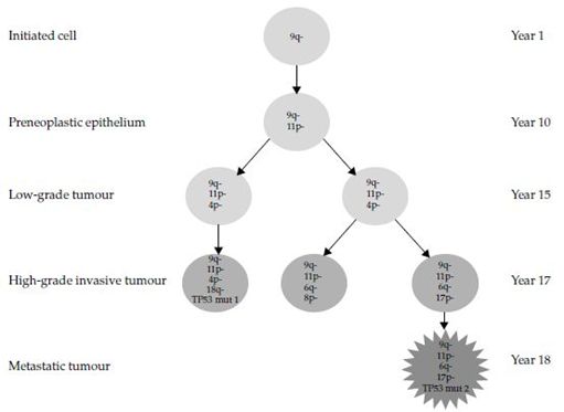
eBooks

which new and exciting discoveries are being made and those in which key questions remain
unanswered.
   Cancer is a disorder of cells and although it usually appears as a tumour (a swelling) made
up of a mass of cells, the visible tumour is the end result of a whole series of changes which
may have taken many years to develop. In this chapter, we discuss in general terms what is
known about the changes that take place during the process of tumour development, consider
tumour diagnosis and nomenclature, and provide some definitions. Succeeding chapters deal
with specific aspects in more detail.
Normal cells and tissues
    The tissues of the body can be divided into four main groups: the general supporting tissues
collectively known as mesenchyme; the tissue-specific cells-epithelium; the ‘defense’ cells-the
haematolymphoid system; and the nervous system. The mesenchyme consists of connective
tissue-fibroblasts which make collagen fibres and associated proteins, bone, cartilage,
muscle, blood vessels, and lymphatics. The epithelial cells are the specific, specialized cells
of the different organs, for example, skin, intestine, liver, glands, etc. The haemato-lymphoid
system consists of a wide group of cells, mostly derived from precursor cells in the bone
marrow which give rise to all the red and white blood cells. In addition, some of these cells
(lymphocytes and macrophages) are distributed throughout the body either as free cells or
as fixed constituents of other organs, for example, in the liver, or as separate organs such as
the spleen and lymph nodes. Lymph nodes are specialized nodules of lymphoid cells, which
are distributed throughout the body and act as filters to remove cells, bacteria, and other
foreign matter. The nervous system is made up of the central nervous system (the brain and
spinal cord and their coverings) and the peripheral nervous system, which is comprised of
nerves leading from these central structures. Thus, each tissue has its own specific cells,
usually several different types, which maintain the structure and function of the individual
tissue. Bone, for example, has one group of cells responsible for bone formation and a second
group responsible for bone resorption and remodeling when the need arises, as in the repair
of fractures. The intestinal tract has many different epithelial cell types responsible for the
different functions of the bowel, and so on. The specific cells are grouped in organs which have
a standard pattern (Figure 1). There is a layer of epithelium, the tissue-specific cells, separated
from the supporting mesenchyme by a semipermeable basement membrane. The supporting
tissues (or stroma) are made up of connective tissue (collagen fibers) and fibroblasts (which
make collagen), which may be supported on a layer of muscle and/or bone depending on the
organ. Blood vessels, lymphatic vessels, and nerves pass through the connective tissue and
provide nutrients and nervous control among other things for the specific tissue cells. In some
instances, for example, the skin and intestinal tract, the epithelium which may be one or more
cells thick depending on the tissue, covers surfaces. In others it may form a system of tubes
(e.g. in the lung or kidney), or solid cords (e.g. liver), but the basic pattern remains the same.
Different organs differ in structure only in the nature of the specific cells and the arrangement
and distribution of the supporting mesenchyme.

Cancer: Disease of the Age Recent Revolution in Cancer Diagnosis, Treatment, Management, Prevention and Control   4
Edited by: Ahmed M El-Sharkawy
eBooks
Figure 1: A typical tissue showing epithelial and mesenchymal components.
Control of growth in normal tissues
The mechanism of control of cell growth and proliferation is one of the most intensively
studied areas in biology. It is important to make the distinction between the terms ‘growth’
and ‘proliferation’. Growth is used here to refer to an increase in size of a cell, organ, tissue,
or tumour and proliferation to an increase in the number of cells by division. ‘Growth’ is
often used as a loose term for both of these processes but the distinction is particularly
important now that factors controlling both of these processes are becoming clear. In normal
development and growth there is a very precise mechanism that allows individual organs to
reach a fixed size, which for all practical purposes, is never exceeded. If a tissue is injured,
the surviving cells in most organs begin to divide to replace the damaged cells. When this has
been completed, the process stops, that is, the normal control mechanisms persist throughout
life. Although most cells in the embryo can proliferate, not all adult cells retain this ability. In
most organs there are special reserves or stem cells, which are capable of dividing in response
to a stimulus such as an injury to replace organ-specific cells. The more highly differentiated
a cell is, for example, muscle or nerve cells, the more likely it is to have lost its capacity to
divide. In some organs, particularly the brain, the most highly differentiated cells, the nerve
cells, can only proliferate in the embryo, although the special supporting cells in the brain
continue to be able to proliferate. A consequence of this, as we shall see later, is that tumours
of nerve cells are only found in the very young and tumours of the brain in adults are derived
from the supporting cells. In other tissues there is a rapid turnover of cells, particularly in
the small intestine and the blood and immune system. A great deal of work has been done
on the control of stem cell growth in the red and white cells (haemopoietic system), and the
relationship of the factors involved in this process to tumour development. For reasons that
are still unclear, rapid cell division itself is not necessarily associated with an increased risk of
tumour development, for example, tumours of the small intestine are very rare. In the embryo
there is a range of stem cells, some cells capable of reproducing almost any type of cell and
others with a limited potential for producing more specific cells, for example, liver or kidney.
In the adult, there is now unequivocal evidence for the existence of stem cells capable of
perpetuating themselves through self-renewal to generate specialized cells of particular tissues.
Striking parallels exist between the properties of stem cells and cancer cells. This, together
with the potential for the use of human stem cells in various types of regenerative medicine,
makes this a very active area of research [1]. Control of organ or tissue size is achieved via
a fine balance between stimulatory and inhibitory stimuli. When the balance is shifted, for
Cancer: Disease of the Age Recent Revolution in Cancer Diagnosis, Treatment, Management, Prevention and Control 5
Edited by: Ahmed M El-SharkawyeBooks
example, when the tissue is damaged and repair is needed, when a specific physiological
stimulus is applied, for example, hormonal stimulation or because extra work is required from
an organ, the component cells may respond in one of two ways to achieve these objectives.
This may be by hypertrophy, that is, an increase in size of individual components, usually of
cells which do not normally divide. An example is the increase in size of particular muscles
in athletes. The alternative is hyperplasia, that is, an increase in number of the cells. When
the stimulus is removed, commonly the situation returns to the status quo as exemplified by
the rapid loss of muscle mass in the lapsed athlete. Some of the stimuli that lead to these
compensatory responses are well-known growth factors and hormones that are discussed in
more detail in Chapter 5. Recent work on the insulin/IGF (insulin-like growth factor) system,
particularly in the fruit fly Drosophila, has demonstrated that this plays a pivotal role in
the control of organ and organism size [2]. It is of note that several molecules involved in
these processes are known to act as oncogenes or to be dysregulated in cancer. For example,
IGFs are commonly overexpressed and the phosphoinositide3-kinase (PI3K) pathway, which is
activated by insulin/IGF signaling, is functionally disrupted in various ways in cancer cells [3].
The Definition of Cancer
As humans we are comprised of many millions of cells. Some cells are specific to certain
tissues, for example epithelial cells are found throughout the gastrointestinal tract, bladder,
lungs, vagina, breast and skin. This group of cells accounts for approximately 70% of cancers
[4,5]. However, any cell has the potential to undergo malignant changes and lead to the
development of a carcinoma. Cancerous cells are not confined to localized ‘overgrowth’ and
infiltration of surrounding tissue, but can spread to other parts of the body via the lymphatic
system and bloodstream, creating secondary deposits known as ‘metastases’ [6-8]. This can
occur when ‘normal’ cell control mechanisms become disrupted or indeed fail [5]. Surgical
removal of the original tumour is not always a successful treatment in malignant disease, due
to microscopic spread. Malignant tumours are often irregular in shape, with ill-defined margins
[7,9]. The potential for microscopic spread occurs when the tissue surrounding the visible
tumour appears to the eye (macroscopic examination) to be unaffected by cancer. Microscopic
examination of the surgical resection margins can reveal the presence of malignant cells. If
left untreated, these cells will result in localized recurrence of the cancer and eventual spread
(metastasis). The spread of the malignant cells extends outward from the original tumour,
and has been described as resembling the appearance of a crab. This is the origin of the term
‘cancer’, which was derived from the Latin meaning ‘crab’ [7]. The earlier a cancer is detected,
the less likely it is to metastasize, and so the more favourable the prognosis for the individual
[10].
Metastatic spread
All cells replicate themselves. This usually happens about 50−60 times before the cell
eventually dies (see Chapter 4) [5,11]. However, as malignant cells replicate, they grow in
an irregular pattern, infiltrating surrounding tissue. This can result in infiltration of the
lymphatic’s and/or blood vessels. By gaining access to these vessels, malignant cells can be
carried to other sites within the patient’s body, where they will replicate and grow, rather like
rodents establishing colonies in various parts of a town by gaining access to sewer systems
[7,9]. In order to ensure that these malignant cells receive the nourishment they need to thrive,
angiogenesis occurs. This is the formation of new blood vessels [11].
Cancer: Disease of the Age Recent Revolution in Cancer Diagnosis, Treatment, Management, Prevention and Control 6
Edited by: Ahmed M El-SharkawyeBooks
Lymphatic spread
Malignant cells gain access to the lymphatic system and travel along the vessels to the
‘regional draining’ lymph nodes [7]. The malignant cells can then establish residency in these
regional nodes, where they replicate and eventually replace the lymph node with a malignant
tumour − that is, cancer. Malignant cells from this tumour can then travel, via the lymphatic
system, to the next group of lymph nodes, thereby spreading the malignancy throughout the
patient’s body [7]. Lymphomas and squamous cell carcinoma of the head and neck are two
examples of where cancer commonly spreads via the lymphatic system [11].
Blood spread: As the lymphatic spread, malignant cells can also infiltrate the vascular
system and travel along the vessels until they arrive at an area where they can become lodged
and subsequently replicate to form a secondary (metastatic) deposit. The malignant cells can
then migrate via the smaller blood vessels − that is, the capillaries [7]. However, there is
evidence that only a small percentage of cells entering the vascular system actually survive to
give rise to blood-borne metastatic spread [7]. Malignancies which are linked to blood-borne
spread include melanoma and small cell carcinoma of the lung [11].
Liver: The commonest site for blood-borne metastases is the liver. Malignancies originating
from the gastrointestine, including the pancreas, commonly metastasize to the liver. Other
malignancies which can result in secondary deposits in this organ include breast, melanoma,
lung and urological cancers [7,9].
Lung: The lung is the second most common site for metastatic spread. Tumours that are
associated with metastasizing here include the breast, teratomas, melanomas and sarcomas
[7,9].
Bone: Bone metastases are commonly associated with malignancies of the breast,
prostate, kidney, lung and thyroid. Patients with bone metastases can often present with
pain. Pathological fractures are not uncommon due to the damage caused to the bone by the
malignant cells − that is, the cancer cells replacing the healthy cells and thereby weakening
the bone, making it more prone to fracture [7,9].
Brain: Brain metastases are closely associated with primary malignancies of the lung, but
can also arise from other sites, including the breast, teratomas and malignant melanoma [7,9].
Adrenal glands: Breast and lung primary malignancies are more frequently associated with
secondary deposits in the adrenal glands, compared to cancers arising from other sites within
the body [7,9].
Transcoelomic spread: Transcoelomic spread is the term used to describe invasion of the
serosal lining of an organ by malignant cells. The malignant cells trigger an inflammatory
response, which results in a serous exudate. This is commonly seen in the peritoneal cavity,
where it is associated with ovarian and colonic malignancies [7,9].
The Process of Carcinogenesis
Carcinogenesis (the process of cancer development) is a multistage process (Figure 2).
In an animal, the application of a cancer-producing agent (carcinogen) does not lead to the
immediate production of a tumour. Cancers arise after a long latent period and multiple
carcinogen treatments are more effective than a single application. Experiments carried out
Cancer: Disease of the Age Recent Revolution in Cancer Diagnosis, Treatment, Management, Prevention and Control 7
Edited by: Ahmed M El-SharkawyeBooks
on mouse skin in the 1940s by Berenblum and Shubik [12] indicated that at least three major
stages are involved. The first was termed initiation and was found to involve mutagenic effects
of the carcinogen on skin stem cells. The second stage, which can be induced by a variety of
agents that are not directly carcinogenic in their own right, was termed promotion. Following
chronic treatment of carcinogen-initiated mouse skin with promoting agents, papillomas
(benign skin tumours) arise. The major effect of promoters seems to be their ability to promote
clonal expansion of initiated cells. Finally in the third stage, progression, some of these benign
tumours either spontaneously or following additional treatment with carcinogens, progress
to invasive tumours. The terms coined to describe this animal model are still commonly
applied to describe the process of carcinogenesis in man. The mouse skin model indicated that
carcinogenesis is a multistep process and clearly this is also the case for human cancer. For
example, most solid tumours of adults arise in the later decades of life, usually a long time
after exposure to a specific carcinogenic insult or after a long period of continuous exposure
and this can be explained in terms of the requirement for several distinct heritable changes.
The nature of some of these changes is now known in detail and is discussed at length in
several of the following chapters. These include genetic alterations to proto-oncogenes and
tumour suppressor genes (Chapters 2) and epigenetic alterations.
Histopathological observations also provide evidence for a long preneoplastic period,
sometimes with morphologically identifiable lesions such as benign tumours or in situ
dysplasia, which may persist for many years and within which a malignant tumour eventually
arises. The latent period between initiation and the appearance of tumours is great. In man,
after exposure to industrial carcinogens, it may take over 20 years before tumours develop.
Even in animals given massive doses of carcinogens, it may take up to a quarter or more of the
total lifespan before tumours appear. The requirement for acquisition of multiple events is the
likely explanation for this. In the tumour that finally emerges, most of the genetic and epigenetic
changes seen are clonal, that is they are present in the entire population of cells. It is likely
that a series of selective phases of clonal expansion takes place in the tumour such that after
each event, there is outgrowth of a clone of cells with a selective advantage. Evidence for this
has come from studies on many tissues and particularly where areas of surrounding tissue or
multiple related lesions can be sampled at surgery. In these circumstances, it is common to
find several shared clonal events in different lesions and occasionally in the apparently ‘normal’
surrounding epithelium and additional events in the most histopathologically advanced lesion.
Figure 2: Tumour development showing progression from normal to invasive tumour via accumulation of
heritable changes over a long period of time.
Cancer: Disease of the Age Recent Revolution in Cancer Diagnosis, Treatment, Management, Prevention and Control 8
Edited by: Ahmed
TheMrate
El-Sharkawy
of acquisition of these changes will be influenced by environmental exposures and host response.eBooks
Early Mutational Events in Carcinogenesis
Alterations of the genetic code
Analysis of the DNA of tumor cells reveals that a finite number of gene mutations are
responsible for the transmission of the phenotypic changes characteristic of the tumor from
one cell to the other during cell division. These mutations may have arisen sporadically in a
somatic cell through misrepair of endogenous DNA damage arising from oxidative stress and
DNA replication errors, or through mistakes in somatic recombination events. Alternatively,
they may be induced exogenously through the DNA-damaging action of environmental agents,
such as ionising radiation, UV, and mutagenic alkylating or intercalating agents. Failure of
the damage control processes to correct the damage before it is incorporated permanently into
the genome during replication is critical. Phenotype or may even leave the encoded amino acid
unchanged (silent mutations). Occasionally, the single base change may generate a premature
stop codon, truncating the protein, which frequently leads to rapid degradation of the abnormal
protein by the misfolded protein recognition system in the endoplasmic reticulum and the
proteasome. Insertions and deletions of a single base alter the reading frame of the gene. As
most genes have evolved with multiple stop codons protecting the two non-coding frames,
the frame-shifted sequence will most probably contain a stop codon close to the position of
the insertion/deletion. In some infrequent instances, the mutated single base may lie in a
critical structural element of the gene, such as the promoter site regulating gene activity, or
in a recognition site critical for RNA processing, for example splice site mutations resulting in
exon skipping deletions in the E-cadherin gene [13]. In addition to the intragenic mutations
described above, there is a range of additional mechanisms whereby the genome may become
perturbed during tumor development. Alterations in the copy number of cellular genes are
commonly described in human tumors. Both allelic gains and losses are encountered, and
their biological consequences are described elsewhere in this review. Amplification of genetic
regions may take the form of intrachromosomal duplications, leading to the in situ amplification
of a gene with oncogenic potential. Transcription of the amplified gene complex subsequently
leads to overexpression of the gene product. Alternatively, the amplification may occur
extrachromosomally, leading to the formation of multiple copies of chromosomal fragments
(double minutes) containing one or more transcriptionally active genes with an oncogenic
capacity. The spectrum of mutational events in tumor cells can also include chromosomal
translocation and inversion events leading to the structural rearrangement of parts of the
genome. This may result in a fusion of two unrelated gene fragments, creating a chimeric gene
instructing production of a protein with abnormal function. Alternatively, the rearrangement
may transpose an endogenously active promoter to coding sequences from a gene that is
normally either tightly regulated or transcriptionally silent in the tissue. This form of mutation
leads to the inappropriate expression of the protein, for example, in parathyroid tissue where the
CCND1 (Cyclin D1) gene is placed under the control of the highly active parathyroid hormone
gene promoter [14]. This is also seen in thyroid tissue where the transcriptionally inactive
glial-derived Neurotrophic Factor Receptor (RET) tyrosine kinase gene is placed under the
control of one of a number of different promoters active in thyroid tissue [15]. As a result of this
translocation event, the neuroendocrine tissue-restricted RET protein is produced in thyroid
cells and delivers cell proliferation signals in a ligand-independent. Functional translocations
are also frequent in the lymphoid and myeloid lineages, presumably due to the propensity
of these cells to undergo chromosomal rearrangements during immunoglobulin and T cell
receptor maturation. Failure to restrict the high level of chromosomal rearrangement activity
Cancer: Disease of the Age Recent Revolution in Cancer Diagnosis, Treatment, Management, Prevention and Control 9
Edited by: Ahmed M El-SharkawyeBooks
to the correct locus may explain the abundance of such alterations in immature stages of the
lineages. In solid tumors translocations are seen primarily in the endocrine tissues mentioned
above and in the pediatric tumors rhabdomyosarcoma and Ewing’s sarcoma, both of which
involve activation of genes regulating developmental pathways. Translocations are reported
less frequently in other solid tumors, and here their biological relevance remains uncertain.
Significantly, in none of the solid tumor types showing translocations is there any evidence
for endogenous chromosomal rearrangement processes that could explain the phenomena.
Two non-mutational events are also implicated in the changes in gene expression during
oncogenesis. In the first situation, transcriptional silencing of an essential tumor suppressor
gene is associated with non-mutational changes to the structure of the gene promoter region.
Changes in the methylation status of individual nucleotides of the DNA, as well as to the
methylation and acetylation status of the DNA-binding histone core proteins, are involved in
regulating local gene expression. A second non-mutational event is discussed below, where gene
silencing through endogenous RNA binding microRNA molecules has been suggested to be an
additional step in transcriptional control, leading to silencing in a post-transcriptional manner.
An altogether different mutational mechanism is seen almost exclusively in animal model
systems, where insertion of retroviral sequences or retroviral-like elements into the genome
results in the disruption of cellular genes. In humans, the role of insertional mutagenesis is
less clear. Retroviral insertion leading to proto-oncogene overexpression has been implicated
in the development of retroviral gene therapy-associated lymphoproliferative malignancies in
a small number of cases. Nevertheless, the general applicability of this mutational mechanism
for human cancer is unclear, and it is certainly uncommon. In addition to retroviral insertion,
viruses have evolved a range of strategies for productive infection of mammalian cells that
subvert defense and regulatory pathways. As a consequence of these actions, the viral proteins
elicit an oncogenic action through growth stimulation, suppression of apoptosis or inactivation
of endogenous tumor suppressor gene function.
Events accompanying progression
Mathematical and molecular studies on tumor tissues have each established that tumors
can arise and develop through a series of intermediate stages. The clonal expansion paradigm
suggests that discrete stages arise through evolutionary selection of appropriate phenotypes
that are themselves defined by mutational events. Histopathological studies deliver a partially
convergent concept, where morphologically distinct stages of tumor formation and development
are discernable in almost all tumor entities. The combination of the morphological models
of tumor development and analysis of molecular events suggests that tumor development
indeed follows a series of steps from pre-cancerous lesions (hyperplasia, atypical hyperplasia)
that lead either directly or indirectly to full neoplasia (infiltrative and metastatic growth).
During this progression, the normally differentiated phenotype may become either partially
or completely lost [16]. Estimates of the number of mutations and steps that are required to
create a full malignant phenotype vary wildly. In vitro studies suggest that mutation of as
few as three key genes is sufficient, whilst massive DNA resequencing studies of tumor cell
genomes have revealed hitherto undiscovered complexity in the magnitude and diversity of DNA
alterations; however, it remains unclear which of these, if any, are required for the acquisition
of a malignant phenotype [17]. Three conceptual models can help in partly reconciling these
differences. Kinzler and Vogelstein suggested, at least for the model of colon carcinogenesis,
that there is a linear evolution of the cells within the developing tumor, which follows a well-
circumscribed and sequential series of events [18,19]. Each step in their model is represented
Cancer: Disease of the Age Recent Revolution in Cancer Diagnosis, Treatment, Management, Prevention and Control 10
Edited by: Ahmed M El-SharkawyeBooks
by the mutation of a single key gene. However, the analysis of the gene alterations present
in different areas of some tumors shows that some clones lack the full complement of gene
mutations. This may indicate that a simple linear monoclonal evolution is not always followed
[20]. An alternate view to the Vogelstein model is that mutations are acquired in a cumulative
manner, with some clones in the tumor acquiring mutations that lead to them branching
off to an evolutionary dead end and others only being required at specific points in the
tumor development. Hanahan and Weinberg [21] have suggested that key cellular pathways
related to functional changes in tumor cell biology are individually targeted by mutational
events, explaining how the development of malignancy can result from a finite number of
mutations. Finally, systems theory and pathway analysis suggest that each functional activity
of the cell described by Hanahan and Weinberg requires multiple hits to remove backup and
alternative pathways. It is, however, worthy of note that tumor cells cannot tolerate wholesale
genomic alterations; consequently, there cannot be an unlimited number of mutations as
some functional pathways are essential for continued cell survival. A discrepancy of orders
of magnitude between the sporadic rate of mutational activity observed in cells and the level
of mutations found in tumors has prompted Loeb [22] to suggest that a key process in tumor
cell development must be the acquisition of a mutational activity (mutator phenotype, loss
of caretaker function). Although tumor suppressor and apoptosis genes could be considered
candidate mutator genes, no convincing evidence for a specific increase in mutation rate due
to loss of these genes has been presented. Genes involved in maintaining genomic integrity,
such as the DNA mismatch repair genes, whilst implicated in cancer susceptibility, provide no
clear evidence of mutator-gene driven genome changes.
Proliferation modifying genes
A major category of the genes influencing cell proliferation contains members of signaling
pathways involved in the regulation of cellular growth. At the cell surface this can be seen by
the uncontrolled production of stimulatory growth factors, the abnormal expression of growth
factor receptors or the production of a mutated form of the receptor that has acquired the
capacity to autonomously engage and activate the downstream intracellular signaling cascade.
A related functional set of tumor genes is that involved in the transmission of the growth-
regulating signal to the transcriptional apparatus, which includes signal-transducing kinases
and transcription factors. An additional group of proliferation genes plays a role in steering the
transit of cells into, through and out of the cell cycle. Inappropriate functioning of these genes
leads to uncontrolled cell cycle activity and the failure of proliferating cells to differentiate.
In the case of cell cycle checkpoint control genes, this can allow cells with non-repaired
DNA damage or chromosomal aberrations to continue through the cycle, yielding genetically
aberrant daughter cells. Failure to eliminate damaged cells is an additional feature of the
mutations influencing a further set of cancer genes, those involving the cellular pathways
regulating programmed cell death (apoptosis and anoikis, a form of apoptosis that is induced
in anchorage-dependent cells detaching from the surrounding cells and/or matrix). The failure
of tumor cells to initiate a normal apoptotic death response after stress and/or mutation
of DNA, or to initiate apoptosis after loss of cell–cell and cell–matrix contact, can involve
inactivation of the intrinsic (mitochondrial) pathway and extrinsic (ligand-receptor) apoptosis-
inducing pathways. This can be brought about by inappropriate overexpression of anti-
apoptotic proteins or by inactivation of pro-apoptotic proteins. More recently, the protective
sequestration of cells bearing oncogenic gene mutations into a pathway of Oncogene-Induced
Senescence (OIS) has been described. The regulation of this pathway is poorly understood,
Cancer: Disease of the Age Recent Revolution in Cancer Diagnosis, Treatment, Management, Prevention and Control 11
Edited by: Ahmed M El-SharkawyeBooks
but escape from growth restrictions imposed by the activation of the senescence programme
appears to be a critical step in oncogenesis and may involve overcoming cell cycle arrest by
removing expression of the p16 cyclin-dependent kinase inhibitor. It remains to be seen which
other protein activities regulate entry and exit from OIS and how mutations of these genes
influence tumorigenesis.
Acquisition of the invasive/metastatic phenotype
Although changes in proliferative regulation pathways are critically important, the
acquisition of an invasive/ metastatic phenotype is a major step in solid tumor formation. The
necessary changes in gene expression may occur through mutation or through changes in
more global programmes of cell regulation, such as the Epithelial to Mesenchymal Phenotypic
Transition (EMT). Tumor invasion into surrounding tissues requires distinct phenotypic
alterations. Loss of cell-specific adhesion allows tumor cells to detach from neighboring cells
and the underlying extracellular matrix. This may be accompanied by upregulation of an
alternative programme of adhesion, allowing the tumor cell to adhere to anomalous cells or
matrixes (e.g. a switch from epithelial specific E-cadherin to the mesenschymal-cell specific
cadherin’s in breast cancer) [23]. At the same time as acquiring an abnormal adhesive profile,
the tumor cells may also develop a programme allowing for the degradation of the surrounding
matrix proteins. Here, overexpression of specific proteases may facilitate local destruction of
matrix that allows the non-adherent tumor cell to exit the parental tissue and migrate [24].
Recent evidence suggests that the mobilization of tumor cells may be driven by local gradients
of cell- and tissue-specific chemokine molecules. Changes in the expression pattern of surface
chemokine receptors of tumor cells may permit them to respond to a different chemokine
milieu and has been suggested to be partly responsible for homing of tumor cells to specific
distant sites such as bone marrow [25]. Separation of the tumor cell from surrounding parental
tissue would normally be expected to initiate the anoikis programme of apoptosis, but as
described above, this pathway is inactivated as part of the loss of proliferative regulation. The
final stage in malignant growth, the acquisition of the capacity to generate new blood vessels
that infiltrate the tumor and oxygenate the expanding cell mass, angiogenesis, is discussed in
other chapter of this book.
Inherited susceptibility
Within a population there is a proportion of individuals who are predisposed to develop
cancer, either as an apparently sporadic disease or in response to an environmental challenge,
such as exposure to tobacco smoke or ionising radiation. The abnormally high frequency of
some tumor types within related members of large families provided evidence that cancer is, in
some circumstances, a heritable disease. Genetic linkage studies of these families has revealed
that a number of these cancer syndromes occur as simple Mendelian traits, usually with
a highly penetrant dominant pattern of inheritance. Many hereditary cancer susceptibility
genes, such as Breast Cancer 1 and 2 (BRCA1/2) and the group of DNA mismatch repair
genes, have a known function in the DNA repair. Incomplete functioning of DNA repair
appears to render somatic cells highly susceptible to carcinogenetic noxae and spontaneous
DNA mutations, leading to an accumulation of genetic damage and ultimately transformation.
Other susceptibility genes involving impaired DNA repair lead to cancer prone syndromes such
as xeroderma pigmentosa, Bloom’s disease and Hereditary Nonpolyposis Colorectal Cancer
(HNPCC), also known as Lynch syndrome. Yet, there are inherited susceptibility genes having
no direct function in DNA repair, but still showing an autosomal dominant familial pattern. Von-
Cancer: Disease of the Age Recent Revolution in Cancer Diagnosis, Treatment, Management, Prevention and Control 12
Edited by: Ahmed M El-SharkawyeBooks
Hippel-Lindau syndrome is a dominantly inherited hereditary cancer syndrome predisposing to
a variety of malignant and benign tumors of the eye, brain, spinal cord, kidney, pancreas and
adrenal glands. Other inherited cancer syndromes include ataxia telangiectasia, Li-Fraumeni
syndrome, retinoblastoma, Wilms’ tumor, familial adenomatous polyposis, multiple endocrine
neoplasia 1 and 2, just to mention a few. The hereditary mutations associated with cancer
syndromes only have a big impact on the risk of a population if they are common. Thus, whilst
mutations in the breast cancer susceptibility genes BRCA1 and BRCA2 are found in almost
10% of women with breast cancer, the PTCH1 gene mutation responsible for the Gorlin/
basal nevus syndrome occurs in less than 1 per 50,000 of the population. However, it must
be appreciated that the gene mutation frequencies vary considerably between populations,
especially if the populations are isolated for geographical, religious or other reasons. Good
examples in this context are BRCA2 mutations in Iceland and BRCA1/2 mutations among the
Ashkenazi Jewish population. Inaccuracies in population estimates may bias clinical judgement
and allocation of diagnostic resources [26]. Susceptibility to many diseases has been shown
to be polygenic, with a multitude of low-penetrance common polymorphisms contributing
to the risk of developing disease. These complex trait genes may contribute significantly to
risk estimations of certain cancers. Therefore, it is useful to quantify the relative importance
of known genes in the burden of disease by using the Population Attributable Fraction
(PAF) that states the contribution of the studied gene to disease aetiology, independent of
the environmental or other genetic factors that may interact with the gene in question [27].
New approaches, such as Genome-Wide Association Studies (GWAS) using Single Nucleotide
Polymorphism (SNP) arrays, have provided tools to map and potentially identify some of the
low penetrance hereditary cancer-susceptibility genes. Future developments here will require
large-scale multinational collaborations, similar to those conducted on breast cancer [28].
Genetic Instability, Clonal Selection and Tumour Evolution
Our recent ability to dissect the cancer genome at both the gross chromosomal and
nucleotide level has revealed extensive genetic change. Often this is complex, particularly in
advanced epithelial cancers and is commonly referred to as genetic or genomic instability.
Recent studies have revealed that genetic instability can take distinct forms and a debate
has arisen over whether these represent cause or effect. One type of instability is that which
results from inactivation of Mismatch Repair (MMR) genes such as MSH2 and MLH1. Defects
in MMR lead to numerous changes in short simple sequence repeats spread throughout the
genome (called microsatellites; MMR is also termed Microsatellite Instability, MIN). MIN is
characteristic of tumours found in patients with Hereditary Non-Polyposis Colorectal Cancer
(HNPCC) who inherit mutations in MSH2 or MLH1. Interestingly, MIN tumours usually have
a diploid karyotype which contrasts with non-MIN epithelial cancers which commonly show
complex karyotypic abnormalities, commonly termed Chromosomal Instability (CIN). The
causes of CIN have been less obvious. There are several possibilities including alterations in
mitotic checkpoint genes or genes involved in centrosome function or chromosomal segregation
as discussed above. Already some tumours have been found to contain this type of alteration.
It is also possible that once a cell has become aneuploid by chance, this in itself predisposes it
to become even more aneuploid. This might happen, for example, at mitosis where segregation
of aberrant or large numbers of chromosomes is more error-prone. A final mechanism is
the inherent CIN which is generated in cells at senescence when chromosomes have severe
telomere attrition. As indicated above, shortening of telomeres in advance of re-expression of
telomerase can lead to severe chromosomal rearrangement via end-to-end fusion followed by
Cancer: Disease of the Age Recent Revolution in Cancer Diagnosis, Treatment, Management, Prevention and Control 13
Edited by: Ahmed M El-SharkawyeBooks
breakage at segregation. There is evidence for all of these mechanisms and it is likely that one
or more may contribute to the development of any given tumour and that the mechanism that
is active will shape the genome in specific and recognizable ways that may well be tissue or
tumour type specific. More detailed analysis of tissue samples taken throughout the course of
tumour development should help to clarify these issues in the next few years.
While it is clear that tumours often have MIN or CIN, it is not yet clears whether this is an
early event in the process, nor whether it is necessary for tumour development. It has been
argued that the probability of tumours acquiring the necessary number of genetic alterations
is too low without some additional mutator effect. This type of calculation is difficult and to
date no clear answer is apparent, though there is no doubt that some tumour cells have this
phenotype while others, particularly early in their development, have little genomic alteration
that can be identified. It is probable however, that the level of generation of mutations is critical
and that too much instability is likely to impede tumour development rather than promote it.
Already it is known that the type of genetic instability present in the tumour cell has an effect
on the type of mutation found. Thus, for example, MIN colorectal tumours tend to inactivate
the two alleles of the APC gene via two point mutations, whereas CIN tumours tend to have
one point mutation and one allele lost by deletion. Two recent reviews explore these concepts
in depth using what is known about colorectal carcinogenesis, possibly the best-studied model
system, as an example [29,30].
Selection of altered clones
The process by which cancer cells develop and spread involves not only mutation but
also selection of altered clones. These processes are the drivers of tumour evolution. It is
thought that repeated rounds of mutation and selection occur during somatic evolution of a
tumour. As the lineage evolves, the tumour cells acquire increased autonomy and eventually
the capacity for metastasis. This is often compared to Darwinian evolution where in this case
the fittest cell survives and multiplies. The low rate of mutation, calculated as 2×10⁻⁷ per
gene per cell division for cultured human cells [31] precludes the acquisition by a single cell of
multiple mutations simultaneously. Even when large carcinogen doses are applied, the large
number of potentially lethal mutations sustained at the same time as any set of mutations
with potential advantage is likely to lead to cell death rather than instant tumorigenicity. Thus
the expectation is that events occur singly and in a particular sequence in each cancer. This is
frequently referred to as a genetic pathway or progression pathway and for several cancer types
attempts have been made to map the pathway in genetic terms. As indicated above, colorectal
cancer is arguably the best elucidated model in this regard [32]. In the colon, mutation of
the APC gene is the initiating event. The resulting ‘early’ adenoma then commonly acquires
mutations in KRAS, SMAD4, and TP53, respectively as it progresses histopathologically via
‘intermediate’ and ‘late’ adenoma to carcinoma. The frequency of each of these changes in
each of the lesion types suggests that there is a preferred order of events in this case but
this does not appear to be invariable. Results from other tumour types where samples can
be obtained from lesions at different stages in the process, or from cancers with different
malignant potential, also show shared lesions and temporal ordering of events in some cases.
There is also evidence that alternative pathways can lead to the same result, and in different
tissues specific mechanisms may dominate. For example, many tumours show inactivation
of TP53 via mutation while some others show amplification of the negative regulator of p53,
MDM2. In the Rb pathway, some tumours show direct mutation of RB while others show
inactivation of the pathway via inactivation of the negative regulator p16. The order of events
Cancer: Disease of the Age Recent Revolution in Cancer Diagnosis, Treatment, Management, Prevention and Control 14
Edited by: Ahmed M El-SharkawyeBooks may differ in different tissues. For example, mutations of TP53 are found frequently in lung cancer but patients with a germline mutation in TP53 (Li–Fraumeni syndrome) do not develop lung cancer as part of the syndrome. Possibly this reflects inability of loss of p53 function to act early in the pathway to lung cancer but its suitability as an early event in the other tumours that develop in these Li–Fraumeni patients. The ultimate result of clonal evolution is escape from the normal growth restraints imposed on the cell in its normal tissue milieu. It follows therefore that the way in which this is achieved will depend to a great extent on what those growth restraints are. Hence finding the tissue and cell type specific genetic alterations, different timing of alterations, etc. It is easy to envisage selection of mutations that increase proliferation or allow resistance to apoptosis or any of the other key features of cancer cells. However, mutations that increase mutation frequency such as those that generate CIN or MIN do not in themselves confer an immediate advantage to the cell. At present, it is not clear how such a phenotype is selected. One plausible explanation is that such mutations may occur rarely in the same cell as a second mutation that does confer an immediate advantage and thus are selected as ‘passenger’ or ‘bystander’ events. Many forms of treatment, for example, radiation and chemotherapy may provide additional mutagenic and selective stimuli and may precipitate the emergence of more aggressive variants. An obvious example is the destruction of X-ray-sensitive cells by X-ray treatment. If the tumour also contains X-ray-resistant cells, the cancer cells which are left after treatment will be X-ray resistant. Although progression is usually towards greater malignancy, this is not invariably so. There are a number of cases, unfortunately small, in which rapidly growing tumours have ceased to grow or even disappeared completely. Although we do not yet have a full explanation for this, some studies indicate that this may be related to the development of anti-tumour immunity in the host. Thus, a series of changes occur in a cell as carcinogenesis proceeds. As the tumour progresses, more and more normal characteristics are lost and it is common to observe what has been described as dedifferentiation within the tumour tissue. This refers to the loss of normal structure and cellular functions characteristic of the tissue. Specialized products of the cell, for example, secretions or structural components may no longer be produced as the cell begins to take on new characteristics. The loss of normal differentiated features is referred to by a pathologist as anaplasia and the degree of such changes identified in tissue sections is used by the pathologist to ‘grade’ tumours. In general, less well-differentiated tumours have a poorer prognosis than those that retain the differentiated characteristics of the normal tissue. As a rule, there is an approximate correlation between tumour grade and growth rate. The most differentiated tumours (low grade, i.e. Grade I) tend to be more slow-growing and the most anaplastic (high grade, i.e. III or IV) the more rapidly growing. Human breast cancers are graded in this way and it has been shown that about 80% of patients with well differentiated Grade I breast cancers will be alive and well at five years (and often much longer) but only 20% of patients with Grade IV tumours will survive for this time. It is of course equally obvious from these figures that although 80% of patients with Grade I cancers survive, 20% with the same structural type of tumour do not. Tumour growth and progression is influenced by factors other than tumour structure, and these may range from the rate of mutation and type of mutation they contain to the reaction of the patient’s own defense mechanisms. In recent years much effort has been made to identify additional tests that can be carried out in the pathology laboratory at the time of tumour diagnosis to add both diagnostic and prognostic (predictive) information and the search for molecular markers (proteins or DNA changes) that can supplement the repertoire of morphological tests is intense. In fact, there are many examples of success in identifying such markers for use in tumour classification, prediction of prognosis or response to therapy, disease monitoring, and markers that can be used as therapeutic targets. These Cancer: Disease of the Age Recent Revolution in Cancer Diagnosis, Treatment, Management, Prevention and Control 15 Edited by: Ahmed M El-Sharkawy
eBooks

are described in several of the other chapters of this book. Possibly, the identification of such
markers has been the earliest and most clinically applicable result of the intense effort of the
past two decades to characterize human tumours at the molecular level. More successes will
undoubtedly follow.
Tumour clonality
    We have alluded to clonal evolution during tumour development but what of the origin of
the tumour? Tumour clonality refers to the cellular origin of cancers. A monoclonal tumour
develops from a single progenitor cell and a polyclonal tumour develops from multiple cells.
In many tissues, a solitary primary tumour is the norm and this may or may not recur or
progress. In this circumstance the question of clonality concerns only this single tumour and
its direct descendants. However, in some tissues the situation is more complex and when the
structure of the organ is examined in detail widespread abnormal pathology may be found. In
such a tissue multicentric tumours are sometimes found. Could there be many cells involved
in the generation of a tumour or does each tumour arise from a single initiated cell? The
appearance of multiple preneoplastic lesions in a tissue has been described as a ‘field change’.
Figures 3 and 4 illustrate such a possible field effect. These tissues show a gradation from
benign to malignant (as in Figure 2) but here the ‘progression’ is in space rather than time. This
has been particularly described in tissues such as the bladder, colon, oesophagus, and oral
mucosa where there is a large epithelial surface available for study and in which essentially
all of the cells have received similar exposure to environmental agents. Here the question of
clonality can be addressed to each individual tumour that arises, that is, tumour clonality,
but of equal interest both to biologist and clinician, is the relationship between all the lesions
in a single patient. This can be referred to as the clonality of the disease. With the advent
of molecular genetic techniques there has been an explosion of information concerning the
genetic relationship of such synchronous lesions.
    There are several possibilities based on the clonality of each lesion and of the overall disease
in the patient:
     a. Each individual tumour consists of lineages derived from multiple normal parent cells.
    Such a tumour would be described as polyclonal. A tumour derived from a few parent cells
    would be termed oligoclonal.
     b. Each tumour has a single parental cell of origin and multiple tumours in the same
    organ arise via seeding or direct spread of cells. Each is therefore a monoclonal tumour and
    this is monoclonal disease.
     c. The disease is polyclonal, that is, more than one initiated cell progresses to generate
    multiple tumours each of which is derived from a single cell (monoclonal).
    There is in fact evidence for all three situations, though the majority of human cancers are
solitary tumours of monoclonal origin and there is ongoing debate over whether true polyclonal
tumours do exist [33]. The methods most commonly used to assess tumour clonality are
X-chromosome inactivation and Loss of Heterozygosity (LOH) analysis by microsatellite typing.
During the course of embryonic development in females, genes on one of the X chromosomes
are silenced by methylation of cytosine residues in the promoter. Such methylation is heritably
maintained and prevents transcriptional activation within the promoter region. This process
is random and in any tissue, 50% of cells have methylation of each copy of X. A monoclonal
tumour will therefore have inactivation of any gene on only one of its X chromosomes and

Cancer: Disease of the Age Recent Revolution in Cancer Diagnosis, Treatment, Management, Prevention and Control   16
Edited by: Ahmed M El-Sharkawy
eBooks
this can be detected at the molecular level. Polymorphisms in X-linked genes have been
used to identify individual parental alleles and when assessed in combination with the use of
methylation sensitive restriction endonucleases, which cut only non-methylated DNA, assays
for allele specific methylation can be developed. Several X-chromosome loci have been used
including Glycerophosphate Kinase (PGK), Hypoxanthine Phosphoribosyl Transferase (HPRT),
and the Androgen Receptor Gene (HUMARA) [34]. Such analyses are restricted to female tissues
and to those women who are heterozygous at the locus of choice, namely those that have
distinguishable maternal and paternal alleles. While there are some problems in interpretation
of X-inactivation assays for clonality, these assays have the significant advantage that the
feature studied is not itself part of the neoplastic process.(Figure:3,4)
Figure 3: Section of the edge of a squamous cell carcinoma of skin, with normal skin (a)
on the left and increasing dysplasia (b) and (c) leading into the main mass of the tumour (d)
below right. Stained with haematoxylin and eosin (×50).
Figure 4: Detail of the areas marked in Figure 3 at higher magnification.
Cancer: Disease of the Age Recent Revolution in Cancer Diagnosis, Treatment, Management, Prevention and Control 17
Edited by: Ahmed M El-SharkawyYou can also read