Pan-London Haemato-Oncology Clinical Guidelines - Acute Leukaemias and Myeloid Neoplasms Part 4: Myeloproliferative Neoplasms - King's ...
←
→
Page content transcription
If your browser does not render page correctly, please read the page content below
Pan-London
Haemato-Oncology
Clinical Guidelines
Acute Leukaemias and Myeloid Neoplasms
Part 4: Myeloproliferative Neoplasms

January 2020
CONTENTS

Contents

Pan-London Haemato-Oncology Clinical Guidelines ................................................................ 1
Acute Leukaemias and Myeloid Neoplasms Part 4: Myeloproliferative Neoplasms ................. 1
Contents .................................................................................................................................. 1
1     Introduction ....................................................................................................................... 4
2     Clinical Features ............................................................................................................... 5
      2.1        Essential thrombocythaemia (ET) .......................................................................... 5
      2.2        Polycythaemia vera (PV) ....................................................................................... 5
      2.3        Primary myelofibrosis (PMF) including Prefibrotic-MF (Pre-MF)............................. 5
3     Referral Pathways ............................................................................................................. 6
      3.1        Children ................................................................................................................. 7
      3.2        Teenagers and young adults ................................................................................. 7
4     Investigation and Diagnosis .............................................................................................. 8
      4.1        Essential thrombocythaemia (ET) .......................................................................... 8
      4.2        Polycythaemia vera (PV) ..................................................................................... 10
      4.3        Prefibrotic myelofibrosis (pre-MF) and primary myelofibrosis (PMF) .................... 14
      4.4        Pathology ............................................................................................................ 17
      4.5        Imaging................................................................................................................ 17
5     Risk Stratification ............................................................................................................ 18
      5.1        Primary myelofibrosis (PMF) ................................................................................ 18
6     Patient Information/Support............................................................................................. 21
7     Treatment ....................................................................................................................... 22
      7.1        Essential thrombocythaemia (ET) ........................................................................ 22
      7.2        Polycythaemia vera (PV) ..................................................................................... 27
      7.3        Primary (or secondary) myelofibrosis (PMF, PPV-MF, PET-MF).......................... 31
      7.4        MPN in accelerated or blast phase ...................................................................... 34
      7.5        Fertility ................................................................................................................. 35
8     Supportive Care .............................................................................................................. 36
      8.1        Anaemia .............................................................................................................. 36
      8.2        Haemostasis and thrombosis ............................................................................... 36
      8.3        Hyperviscosity syndrome ..................................................................................... 36
      8.4        Infection ............................................................................................................... 36
      8.5        Pain management ............................................................................................... 36

                                                                                                                                                  1
CONTENTS
8.6 Other symptom control ........................................................................................ 37
8.7 Breathlessness .................................................................................................... 37
8.8 Weight loss .......................................................................................................... 37
8.9 Cardiovascular risk assessment .......................................................................... 38
8.10 Complex symptom management ......................................................................... 38
9 Special circumstances .................................................................................................... 38
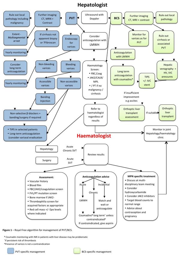
9.1 Splanchnic vein thrombosis ................................................................................. 38
9.2 Pregnancy ........................................................................................................... 38
9.3 Peri-operative management................................................................................. 39
9.4 Acute stroke presentation .................................................................................... 39
9.5 Treatment summary and care plan ...................................................................... 39
10 Follow-up Arrangements ............................................................................................. 40
11 End-of-life Care ........................................................................................................... 41
12 Data Requirements ..................................................................................................... 41
References ............................................................................................................................ 42
Annex 1: Nurse-led MPN Clinic SOP and Referral Guideline [an example] ............................ 45
Annex 2: Data Requirements ................................................................................................. 50
The Cancer Outcomes and Services Dataset (COSD) .................................................... 50
Systemic Anti-Cancer Therapy dataset (SACT) .............................................................. 50
Radiotherapy Dataset (RTDS) ........................................................................................ 50
Cancer Waiting Times dataset ........................................................................................ 51
Annex 3: Tables and Flowcharts ............................................................................................ 52
Annex 4: Response Criteria and Definition of Resistance ...................................................... 54
Annex 5: Guide on Dosing/Monitoring Cytoreductive Agents ................................................. 57
Hydroxycarbamide (HC) .................................................................................................. 57
IFNα (pegylated) (e.g. Pegasys) ..................................................................................... 57
Busulfan .......................................................................................................................... 57
Radioactive phosphorus (32P).......................................................................................... 57
Anagrelide ....................................................................................................................... 57
Annex 7: High-risk Pregnancy ................................................................................................ 60
2CONTENTS
Lead authors 2020:
Professor Claire Harrison, Guy’s and St Thomas’ NHS Foundation Trust
Dr Mallika Sekhar, Royal Free London NHS Foundation Trust and University College London Hospitals NHS
Foundation Trust
Dr Christopher Mitchell, North Middlesex University NHS Foundation Trust
Dr Donal McLornan, Guy’s and St Thomas’ NHS Foundation Trust and University College London Hospitals
NHS Foundation Trust
2018 Edition;
Professor Claire Harrison, Guy’s and St Thomas’ NHS Foundation Trust
Dr Mallika Sekhar, Royal Free London NHS Foundation Trust and University College London Hospitals NHS
Foundation Trust
Disclaimer
These guidelines should be read in conjunction with the latest NICE guidance, and all applicable
national/international guidance. The prescribing information in these guidelines is for health professionals
only. It is not intended to replace consultation with the Haematology Consultant at the patient’s specialist
centre. For information on cautions, contra-indications and side effects, refer to the up-to-date prescribing
information. While great care has been taken to see that the information in these guidelines is accurate, the
user is advised to check the doses and regimens carefully and if there is any uncertainty about the guidance
provided, you should discuss your queries with a Haematology Consultant or Senior Pharmacist. No set of
guidelines can cover all variations required for specific patient circumstances. It is the responsibility of the
healthcare practitioners using them to adapt them for safe use within their institutions and for the individual
needs of patients.
Contact us
The writing cycle for the guidelines will be from May-July each year. If you wish to be part of the writing
group, please contact us through the following link: Pan London Blood Cancer (or via
uclh.panlondonbloodcancer@nhs.net).
If you wish to report errors or omissions that require urgent attention please contact us via the same email
addresses.
© RM Partners, South East London Cancer Alliance, North Central and East London Cancer Alliance
2020
3INTRODUCTION
1 Introduction
Myeloproliferative neoplasms (MPN) include essential thrombocythaemia (ET), polycythaemia vera
(PV) and primary myelofibrosis (PMF). They are all closely related and have an intrinsic tendency
to evolve into acute myeloid leukaemia (AML), confirming their classification as haemato-
oncological disorders. They are characterised by an increased incidence of thrombosis in the
region 20 -30% over 15 years, and premature death for the majority of patients. MPNs are perhaps
the orphan diseases of haemato-oncology, but these patients, if managed judiciously, have
prolonged survival, with a median survival greater than 10–15 years for ET and PV. However,
available treatments have significant side-effect profiles and need to be chosen with care, particularly
in young patients. The last decade has seen the publication of a considerable body of clinical data
informing clinical decisions. Many therapeutic options, however, remain unlicensed. The following
sections contain current management protocols for ET, PV and MF (including MF in patients with
an antecedent history of ET and PV and pre-fibrotic MF).
Other entities within the MPN group – MPNU, chronic eosinophilia, chronic neutrophilic leukaemia
and mast cell disorders – are not covered in these guidelines. These conditions listed in the World
Health Organization (WHO) criteria for MPN 2016 (WHO Classification of Tumours of
Haematopoietic and Lymphoid Tissues. IARC, Lyon 2016) are:
Myeloproliferative neoplasms
Chronic myeloid leukemia (CML), BCR-ABL1+
Chronic neutrophilic leukemia (CNL)
Polycythemia vera (PV)
Primary myelofibrosis (PMF)
PMF, prefibrotic/early stage
PMF, overt fibrotic stage
Essential thrombocythemia (ET)
Chronic eosinophilic leukemia, not otherwise specified (NOS)
MPN, unclassifiable
Mastocytosis
Myeloid/lymphoid neoplasms with eosinophilia and rearrangement of
PDGFRA, PDGFRB, or FGFR1, or with PCM1-JAK2
Myeloid/lymphoid neoplasms with PDGFRA rearrangement
Myeloid/lymphoid neoplasms with PDGFRB rearrangement
Myeloid/lymphoid neoplasms with FGFR1 rearrangement
Provisional entity: Myeloid/lymphoid neoplasms with PCM1-JAK2
Myelodysplastic/myeloproliferative neoplasms (MDS/MPN)
Chronic myelomonocytic leukemia (CMML)
Atypical chronic myeloid leukemia (aCML), BCR-ABL1-
Juvenile myelomonocytic leukemia (JMML)
MDS/MPN with ring sideroblasts and thrombocytosis (MDS/MPN-RS-T)
MDS/MPN, unclassifiable
4CLINICAL FEATURES
2 Clinical Features
2.1 Essential thrombocythaemia (ET)
ET is characterised by a persistent thrombocytosis. The previously accepted platelet count
threshold >600 x 109/L has been revised to >450 x 109/L. Short-term complications of ET include
thrombosis and, less frequently, haemorrhage. In common with PV, long-term problems include a
risk of transformation to MF and acute leukaemia, although these are less frequent in ET.
Thrombotic events affect the arterial and venous macro and microvasculature, as well as the
placental circulation. Microvascular events predominate in ET typically causing erythromelalgia
(asymmetric erythema, congestion and burning pain in the hands and feet), which may progress to
ischaemia and gangrene, migraine-like headaches and transient ischaemic attacks (TIAs).
Approximately 30–50% of patients are symptomatic at presentation.
2.2 Polycythaemia vera (PV)
PV is characterised by an erythrocytosis (packed cell volume (PCV) >0.52 in men and >0.48 in
women, WHO 2008, BSH 2019 criteria; >49% in men and >48% in women, WHO 2016 criteria)
and sometimes thrombocytosis and neutrophilia according to the BCSH. Recent WHO and BSH
criteria are shown below. The median age at presentation is 55–60 years.
Vascular thromboses, especially arterial events and more rarely bleeding, are major events over
the patient’s lifetime. In the longer term (10–15 years), MF or ‘spent phase’ may occur and up to 5-
10% of patients may develop AML.
Aquagenic pruritus, gout and splenomegaly are also classical clinical features, but only occur in
a few patients.
2.3 Primary myelofibrosis (PMF) including Prefibrotic-MF (Pre-MF)
Chronic idiopathic myelofibrosis, or PMF may arise de novo or as a late phase of ET, and
particularly PV known as post-PV (PPV)-MF and post-ET (PET)-MF respectively. Fibrosis is
thought to arise from an interaction between clonal megakaryocytes, releasing mitogens such as
platelet-derived growth factor (PDGF) and transforming growth factor that directly increase
fibroblast proliferation.
PMF has a median age of presentation of 50–60 years. Symptoms relate to bone marrow failure
(anaemia, infection, bleeding) or progressive splenomegaly and a pro-inflammatory state (pain,
weight loss, sweating). Progression to acute leukaemia occurs in up to 25% of patients (more than
PV or ET) and may occasionally be associated with extramedullary collections of myeloid
progenitors (chloromas).
Pre-fibrotic MF (Pre-MF) is a relatively recently described entity. Akin to ET, pre-MF is
characterised by a chronic thrombocytosis, but it differs from ET in terms of bone marrow
morphology and prognosis (Thiele, et al., 2011). The bone marrow in pre-MF is markedly
hypercellular, with pronounced granulocytic hyperplasia, erythroid hypoplasia, and distinctive
megakaryocytic morphology, distinguishing it from ET. However, there is no significant increase in
reticulin fibres in pre-MF, distinguishing it from PMF. The life expectancy in pre-MF is probably
intermediate between ET and PMF, but the natural history of pre-MF is unknown and studies are
on-going. This is something we could usefully contribute to across London, i.e. defining natural
history and outcome.
5REFERRAL PATHWAYS
3 Referral Pathways
Patients with a high WBC, haemoglobin/haematocrit or platelet count and/or suspected MPN by
other features (e.g. splenomegaly, unprovoked and unusual site for a thrombotic episode) should
be referred to a haematologist for assessment.
All new patients should be referred to the MDT for confirmation of diagnosis, prognosis and
management plan, taking into account their performance status, needs and co-morbidities. A joint
approach with elderly care physicians and palliative care teams may be appropriate, depending on
the performance status of the patient and the phase of disease.
The following patients should be referred to the MDT:
All new patients with MPN in order to confirm the diagnosis and treatment plan
All patients where a new line of therapy needs to be considered
All patients with a restaging assessment
All patients in whom an allogeneic stem cell transplant is a consideration.
Information to be ideally captured and documented prior to, or during, the MDT includes:
Demographic information
Referring physician and/or GP
Performance status
An indicator of co-morbidities (e.g. co-morbidity score)
Any relevant history
Pertinent positive and negative findings on physical examination (splenomegaly, rashes, etc.)
Spleen size (by ultrasound)
FBC, haematinics, LFTs, U&E, LDH, urate, reticulocyte count, serum erythropoietin (for cases
of erythrocytosis), transfusion dependency
Bone marrow aspirate and trephine histology (where available)
Bone marrow aspirate immunophenotyping, if relevant
Cytogenetic status, if relevant
Mutational status (in most cases this will include the driver mutations JAK2/CALR (subtype
where available)/MPL but in patients with atypical features, so-called ‘triple negative’ MF and
patients where SCT is considered a wider mutational panel should be considered where
available as this will aid prognostication).
Specific diagnosis/category of MPN and prognostic risk score (we recommend recording which
diagnostic criteria were used, i.e. WHO or BCSH)
Other relevant imaging
Availability of a clinical trial/research study and whether the patient is eligible
Management and treatment plan
Key worker/clinical nurse specialist
Named consultant or team (as per local work patterns).
6REFERRAL PATHWAYS
Patients with PV, ET and MF may be managed at facilities with at least BSCH Level 1 designation.
Complex patients may be referred to centres with specific expertise or which have suitable and
available trials (examples of such patients include, though are not limited to, those displaying
therapy intolerance/ failure, unusual site thromboses such as splanchnic vein thromboses or for
complex or higher risk pregnancies). Patients who are being considered for an allogeneic stem cell
transplant should be referred to a JACIE-accredited centre. All patients with MF eligible for a
transplant option should be referred for a transplant opinion, ideally early in the pathway to
facilitate donor identification. This is dependent upon local practice.
3.1 Children
Children below the age of 16 years with a diagnosis of MPN must be referred to the paediatric
oncology team at the principal treatment centre (PTC) and must not be managed exclusively by
adult site-specific teams. However adult input in this specialty which is extremely rare in the
paediatric setting is critical.
The joint PTC for children aged below 16 years for South Thames is The Royal Marsden
(Sutton)/St George’s Hospital.
The PTC for North Thames (including North West London) is Great Ormond Street Hospital/
University College London Hospitals.
All patientsINVESTIGATION AND DIAGNOSIS
4 Investigation and Diagnosis
A thorough clinical history and examination should be performed, focusing upon exclusion of
secondary causes.
For patients with unprovoked blood clots (in particular of the splanchnic or cerebral venous
circulation, or other unusual sites), check JAK2 and CALR + MPL mutational status even if blood
counts are normal.
Diagnostic criteria: in this document we present both BCSH and WHO diagnostic criteria; we
recommend consistent use of one of these options.
4.1 Essential thrombocythaemia (ET)
There is no diagnostic hallmark for ET. The diagnosis is made by excluding other MPNs, and a
reactive or secondary thrombocytosis. Causes of a reactive thrombocytosis include iron deficiency
anaemia, chronic inflammation (e.g. rheumatoid arthritis, inflammatory bowel disease),
splenectomy, acute haemorrhage, and malignant disease. In an otherwise well patient, the
diagnosis is generally uncomplicated. However, where conditions co-exist which may cause a
reactive thrombocytosis, this may make the diagnosis more difficult.
Historically, the diagnostic criteria for ET were those of the polycythaemia vera study group. Forty
years on, continual development of the diagnostic criteria for MPNs set the stage for the World
Health Organization (WHO) Diagnostic Criteria 2001, modified in 2008 and again in 2016. The
revised WHO criteria (2016) require characteristic bone marrow morphology, a platelet threshold of
450 x 109/L and molecular analysis for the JAK2 V617F mutation and other clonal markers.
Modified BCSH (British Committee for Standards in Haematology, 2013) criteria or WHO criteria
may be used.
4.1.1 WHO ET criteria (2016)
Major criteria
1. Platelet count ≥450 x 109/L
2. BM biopsy showing proliferation mainly of the megakaryocyte lineage with increased numbers of
enlarged, mature megakaryocytes with hyperlobulated nuclei. No significant increase or left shift in
neutrophil granulopoiesis or erythropoiesis and very rarely minor (grade 1) increase in reticulin fibres
3. Not meeting WHO criteria for BCR-ABL11 CML, PV, PMF, myelodysplastic syndromes, or other
myeloid neoplasms
4. Presence of JAK2, CALR, or MPL mutation.
Minor criterion
Presence of a clonal marker or absence of evidence for reactive thrombocytosis.
Diagnosis of ET requires meeting all 4 major criteria or the first 3 major criteria and the minor
criterion.
8INVESTIGATION AND DIAGNOSIS
4.1.2 BCSH (2013) diagnostic criteria for ET
Diagnosis requires A1–A3 or A1 + A3–A5
9
A1 Sustained platelet count >450 x 10 /L
A2 Presence of an acquired pathogenetic mutation (e.g. in JAK2, CALR or MPL genes)
† ‡ §
A3 No other myeloid malignancy, especially PV*, PMF , CML or MDS
A4 No reactive cause for thrombocytosis and normal iron stores
A5 Bone marrow aspirate and trephine biopsy showing increased megakaryocyte numbers displaying a
spectrum of morphology with predominant large megakaryocytes with hyperlobated nuclei and abundant
cytoplasm. Reticulin is generally not increased (grades 0–2/4 or grade 0/3)
* Excluded by a normal haematocrit in an iron-replete patient.
† Indicated by presence of significant bone marrow fibrosis (greater or equal to 2/3 or 3/4 reticulin) AND
palpable splenomegaly, blood film abnormalities (circulating progenitors, tear-drop cells) or unexplained
anaemia (Barosi, et al., 1999; Mesa, et al., 2007).
‡ Chronic myeloid leukaemia; excluded by absence of BCR-ABL1 fusion from bone marrow or peripheral
blood.
§ Myelodysplastic syndrome; excluded by absence of dysplasia on examination of blood film and bone
marrow aspirate.
Investigations to be performed on all patients include:
FBC and blood film
Haematinics
Renal/liver profiles and CRP
ANA and RhF
Chest X-ray (most patients, all smokers).
Where there is a high index of suspicion on first appointment, otherwise at second visit:
JAK2 V617F, CALR (with subtype) and MPL W515L/K screen
Abdominal ultrasound scan
Bone marrow aspirate and trephine (BMAT), cytogenetics in all patients 1000 x 10 /L and haemorrhagic symptoms
The decision to proceed to formal cytogenetic analysis on any sample received is made at the diagnostic
multidisciplinary (MDT) meeting.
The recent WHO criteria (2016) for ET place greater emphasis upon bone marrow histology, in
particular megakaryocyte morphology, and also emphasis is placed on discriminating both PV and
prefibrotic MF from JAK2 positive ET. The other condition that must be excluded when diagnosing
ET is myelodysplastic syndrome. This is usually associated with a low rather than high platelet
9INVESTIGATION AND DIAGNOSIS
count and is characterised by dysplastic features morphologically and particular chromosomal
abnormalities. Note that some patients with refractory anaemia with ring sideroblasts (RARS) or
chromosome 5 abnormalities and MDS may also carry the JAK2 V617F mutation (see WHO
classification for MDS-RARS-T).
4.2 Polycythaemia vera (PV)
An erythrocytosis is defined as an HCT >0.52 in men and >0.48 in women (per BCSH, 2007,
2019), although the WHO (2016) have a lower HCT threshold (0.49 for men; 0.47 for women) this
has not yet been widely adopted. The JAK2 V617F mutation negative erythrocytosis cases may
still be a PV case without a genetic marker or with a JAK2 exon12 mutation; alternatives include a
pseudo/apparent, primary congenital, secondary congenital or acquired, or idiopathic
erythrocytosis, all of which require definition. Diagnostic criteria for JAK2 wild type PV are listed
below.
Table 1: WHO (2016) diagnosis of PV
Diagnosis needs both major and one minor criteria OR major criterion no.1 with two minor criteria
(after exclusion of secondary causes):
Major 1. Hb >16.5 g/dL in men; >16.0 g/dL in women or hematocrit >49% in men; >48% in women
or increased red cell mass (RCM)*
2. BM biopsy† showing hypercellularity for age with trilineage growth (panmyelosis) including
prominent erythroid, granulocytic, and megakaryocytic proliferation with pleomorphic,
mature megakaryocytes (differences in size)
3. Presence of JAK2V617F or JAK2 exon 12 mutation
Minor Subnormal serum erythropoietin level
Diagnosis of PV requires meeting either all 3 major criteria, or the first 2 major criteria and the minor
criterion†.
*More than 25% above mean normal predicted value.
†Criterion number 2 (BM biopsy) may not be required in cases with sustained absolute erythrocytosis: Hb
>18.5 g/dL in men (haematocrit> 55.5%) or >16.5 g/dL in women (haematocrit > 49.5%) if major criterion 3
and the minor criterion are present. However, initial myelofibrosis (present in up to 20% of patients) can only
be detected by performing a BM biopsy; this finding may predict a more rapid progression to overt
myelofibrosis (post-PV MF).
The current BSH 2018 erythrocytosis guideline (McMullin et al, 2019) suggests a three-stage
approach to investigation, this is practically helpful in the wider context of erythrocytosis. The
procedure outlined below is an adaptation of this guidance, based upon modified diagnostic criteria
suggested by Campbell and Green (2006), simplifying the diagnosis and the need for investigation
in JAK2-positive PV. Determination of a case of JAK2-negative PV or an alternative cause of
erythrocytosis will require further investigation.
10INVESTIGATION AND DIAGNOSIS
JAK2-positive PV (Diagnosis requires both to be present)
A1 PCV >0.52 men, >0.48 in women or a raised RCM (>25% above predicted)
A2 Mutation in JAK2
JAK2-negative PV (Diagnosis requires A1 + A2 + A3 + either another A or two B criteria)
A1 Raised Red Cell Mass (>25% above predicted) or a PCV >0.60 in men, >0.56 in women
A2 Absence of mutation in JAK2
A3 No cause of secondary erythrocytosis
A4 BM histology consistent with polycythaemia vera
A5 Palpable splenomegaly
A6 Presence of acquired genetic mutation (excluding BCR-ABL) in haemopoietic cells
9
B1 Thrombocytosis: platelet count >450 x 10 /L
B2 Neutrophil leucocytosis (N>10x109/l in non-smokers and > or equal to 12.5 x 10⁹/l in smokers)
B3 Radiological evidence of splenomegaly
B4 Low serum erythropoietin
The primary clinical assessment of an erythrocytosis case should include a thorough history and
examination seeking out possible secondary causes, followed by Stage 1 investigations to confirm
or refute a diagnosis of a JAK2 V617F-positive PV. The majority of patients (excluding borderline
erythrocytosis) and all ex- and current smokers will require a chest X-ray. Urinalysis is a simple
effective screen for renal disease, which should be performed in all patients at the initial visit.
Patients may present with co-morbidities; thus, regardless of a diagnosis of PV, a review of
potential secondary causes is pertinent. Additional investigation of possible secondary causes will
vary according to symptoms or signs present.
Erythrocytosis Stage 1 Investigations:
FBC and blood film
Renal and Liver Function tests
Serum Calcium levels
Serum ferritin levels
JAK2 V617F mutational analysis
Chest X-ray (smokers or otherwise indicated)
Urinalysis
Serum erythropoietin level
Pulse oximetry and venous carboxyhaemoglobin
If the initial screening tests are negative for a JAK2 mutation and there is no obvious secondary
cause, further investigations are indicated. A Red Cell Mass (RCM) may be required to define
whether a case is a pseudo/apparent or an absolute erythrocytosis at this point and should be
discussed with the Haematology consultant. Of particular note, a RCM is not interpretable if a
venesection has been performed. A PCV of >0.60 and >0.56 in a man or woman, respectively, can
be assumed to have an absolute erythrocytosis and a RCM would not be indicated. Cases
11INVESTIGATION AND DIAGNOSIS
confirmed as an absolute erythrocytosis require remaining Stage 2 tests, as appropriate, with
consultant guidance.
Erythrocytosis Stage 2/3: (see Flow chart 1)
Abdominal ultrasound (may be moved earlier)
RCM performed in nuclear medicine – discuss with consultant if appropriate. As per BSH guidelines
this may be helpful to confirm an absolute erythrocytosis versus an apparent erythrocytosis. Patients
with Hct >0·60 (males) or >0·56 (females) can be assumed to have an absolute erythrocytosis.
Access to this test is variable nationally as is accepted in the current BSH guidelines.
Haemoglobinopathy screen
Further testing is based upon the serum EPO level measured during stage 1 investigations
Normal or low serum EPO levels
JAK2 exon 12 mutational analysis
Bone Marrow Histology can aid diagnosis of PV from secondary erythrocytosis
High Serum EPO levels
Consider further investigation for secondary cause if clinically suspected (referral to respiratory/
renal or sleep studies as required)
Consider Lung function tests as required dependent on clinical phenotype
CT or MRI imaging head and neck (cerebellar haemangioblastoma/ meningioma/ parathyroid
adenoma or carcinoma)
For cases of both low/normal and high serum EPO - consider indications for further genetic testing
(see below) if no diagnosis is yet revealed:
Red cell directed NGS panels (including VHL, PHD and erythropoietin receptor mutations (EPOR)
and EGLN1) where available for detection of acquired genetic abnormalities
Of note this has some limitations as some mutations are non-specific
P50 testing is labour intensive and not readily available but may be considered where required (n.b
may not be required if red cell panel is performed which include HbA and HbB)
12INVESTIGATION AND DIAGNOSIS
The serum erythropoietin level, JAK2 exon12 mutation screen and abdominal ultrasound can aid
the diagnosis of JAK2 V617F-negative PV. Ultimately a bone marrow biopsy is critical ideally
before cytotoxics are commenced.
Hypoxaemia causing a secondary erythrocytosis can be screened for by assessing oxygen
saturation using pulse oximetry (92% is the arbitrary cut-off for significance) and the
carboxyhaemoglobin level available from biochemistry. It is important to subtract the
carboxyhaemoglobin level from the oxygen saturation to obtain the correct estimate of oxygen
saturation.
Abdominal ultrasound can also exclude secondary causes of erythrocytosis, particularly renal and
hepatic pathology, including hepatocellular carcinoma. The abdominal ultrasound combined with
urinalysis and GFR estimation enables a renal disease screen.
A BMAT should be performed in all cases of JAK2 V617F-negative absolute erythrocytosis where
no cause of secondary erythrocytosis is found. The presence of typical histology will support a
diagnosis of JAK2-negative PV, whereas its absence will suggest an alternative cause. A baseline
BMAT should also be considered in all JAK2 V617F-positive patients who are under 60 for future
reference regarding progression, although it is not essential to confirm a diagnosis. A BMA sample
should be sent for cytogenetics – the decision whether to formally proceed to assess these
samples is made in the diagnostic MDT meeting.
For those patients where the aetiology of absolute erythrocytosis is still undefined. Stage 3,
consisting of further specialised investigations, needs to be considered and discussed with a
consultant. Symptoms, including snoring, daytime somnolence and a body habitus suggestive of
sleep apnoea, may warrant referral for sleep studies. The Epworth score is a useful indicator for
referring for sleep studies. A red cell panel genetic screen including the erythropoietin receptor
mutation, HIF-1alpha and proline dehydroxylase mutations amongst others can be sent to the local
lab with facilities for red cell gene panel analysis e.g. the haematology lab at Viapath at King’s
College Hospital, and may lead to a diagnosis of a primary congenital erythrocytosis. A p50
estimation/beta chain sequencing (via red cell NGS) may demonstrate a high affinity haemoglobin
causing a secondary congenital erythrocytosis. As mentioned previously a p50 is increasingly
replaced by a red cell panel has been performed as this screens for haemoglobin variations.
13INVESTIGATION AND DIAGNOSIS
4.3 Prefibrotic myelofibrosis (pre-MF) and primary myelofibrosis (PMF)
For diagnosis of PMF, exclude other MPNs (PV, ET and CML) and disorders in which marrow
fibrosis can develop as a secondary feature (e.g. metastatic carcinoma, lymphoma, irradiation,
TB and leishmaniasis).
The following are generally necessary to confirm PMF:
Splenomegaly
Increased BM fibrosis. In later stages, new osteoid bone is formed (osteomyelofibrosis)
Leucoerythroblastic blood film
Absence of other MPN, including CML (perform FISH for bcr-abl)
Exclude secondary causes of myelofibrosis (see above)
It is useful to obtain an LDH level and cytogenetics (consider from peripheral blood) periodically
to monitor
In patients with atypical features, so called ‘triple negative’ MF and patients where SCT is
considered a wider mutational panel should be considered where available.
The following tests should be performed:
FBC and blood film, blast count
Haematinics
Renal, liver profile, LDH and urate level
JAK2 V617F, CALR (subtype) and MPL W515L/K screen
Chest X-ray
Abdominal ultrasound scan
BMAT with samples sent for cytogenetics and FISH for bcr-abl.
14INVESTIGATION AND DIAGNOSIS
Table 2: WHO diagnostic criteria of pre-MF: need to meet all three major and at least one minor
criterion
Major 1. Megakaryocytic proliferation and atypia, without reticulin fibrosis grade 1*,
accompanied by increased age-adjusted BM cellularity, granulocytic proliferation, and
often decreased erythropoiesis
2. Not meeting the WHO criteria for BCR-ABL11 CML, PV, ET, myelodysplastic
syndromes, or other myeloid neoplasms
3. Presence of JAK2, CALR, or MPL mutation or in the absence of these mutations,
† ‡
presence of another clonal marker, or absence of minor reactive BM reticulin fibrosis
Minor Presence of at least 1 of the following, confirmed in 2 consecutive determinations:
a. Anaemia not attributed to a comorbid condition
9
b. Leukocytosis ≥11 x 10 /L
c. Palpable splenomegaly
d. LDH increased to above upper normal limit of institutional reference range
Diagnosis of pre MF requires meeting all 3 major criteria, and at least 1 minor criterion.
*See Table 5.
†In the absence of any of the 3 major clonal mutations, the search for the most frequent accompanying
mutations (eg, ASXL1, EZH2, TET2, IDH1/IDH2, SRSF2, SF3B1) are of help in determining the clonal
nature of the disease.
‡Minor (grade 1) reticulin fibrosis secondary to infection, autoimmune disorder or other chronic inflammatory
conditions, hairy cell leukaemia or other lymphoid neoplasm, metastatic malignancy, or toxic (chronic)
myelopathies.
Table 3: WHO overt PMF criteria
Major 1. Presence of megakaryocytic proliferation and atypia, accompanied by either reticulin
and/or collagen fibrosis grades 2 or 3*
2. Not meeting WHO criteria for ET, PV, BCR-ABL11 CML, myelodysplastic syndromes,
or other myeloid neoplasms
3. Presence of JAK2, CALR, or MPL mutation or in the absence of these mutations,
presence of another clonal marker,† or absence of reactive myelofibrosis‡
Minor Presence of at least 1 of the following, confirmed in 2 consecutive determinations:
a. Anemia not attributed to a comorbid condition
9
b. Leukocytosis ≥11 x 10 /L
c. Palpable splenomegaly
d. LDH increased to above upper normal limit of institutional reference range
e. Leukoerythroblastosis
Diagnosis of overt PMF requires meeting all 3 major criteria, and at least 1 minor criterion
*See Table 5.
†In the absence of any of the 3 major clonal mutations, the search for the most frequent accompanying
mutations (eg, ASXL1, EZH2, TET2, IDH1/IDH2, SRSF2, SF3B1) are of help in determining the clonal
nature of the disease.
‡BM fibrosis secondary to infection, autoimmune disorder, or other chronic inflammatory conditions, hairy
cell leukemia or other lymphoid neoplasm, metastatic malignancy, or toxic (chronic) myelopathies.
15INVESTIGATION AND DIAGNOSIS
Table 4: BCSH diagnostic criteria
BCSH diagnostic criteria (2015) for PMF: requires A1 + A2 and any two B criteria
A1 Bone marrow fibrosis ≥ 3 (on 0–4 scale)
A2 Pathogenetic mutation (e.g. in JAK2, CALR or MPL), or absence of both BCR-ABL1 and reactive
causes of bone marrow fibrosis
B1 Palpable splenomegaly
B2 Unexplained anaemia
B3 Leucoerythroblastic blood film
B4 Tear-drop red cells
1
B5 Constitutional symptoms
B6 Histological evidence of extramedullary haematopoiesis.
BCSH diagnostic criteria for post-PV and post-ET MF: requires A1 + A2 and any two B
criteria
A1 Bone marrow fibrosis ≥ 3 (on 0–4 scale)
A2 Previous diagnosis of ET or PV
B1 New palpable splenomegaly or increase in spleen size of ≥ 5cm
B2 Unexplained anaemia with 2g/dL decrease from baseline haemoglobin
B3 Leucoerythroblastic blood film
B4 Tear-drop red cells
*
B5 Constitutional symptoms
B6 Histological evidence of extramedullary haematopoiesis.
0
* Drenching night sweats, weight loss >10% over 6 months, unexplained fever (>37.5 C) or diffuse bone
pains.
Table 5: Grading of myelofibrosis
Myelofibrosis grading
MF-0 Scattered linear reticulin with no intersections (crossovers) corresponding to normal BM
MF-1 Loose network of reticulin with many intersections, especially in perivascular areas
MF-2 Diffuse and dense increase in reticulin with extensive intersections, occasionally with focal
bundles of thick fibres mostly consistent with collagen, and/or focal osteosclerosis*
MF-3 Diffuse and dense increase in reticulin with extensive intersections and coarse bundles of thick
fibres consistent with collagen, usually associated with osteosclerosis*
Semiquantitative grading of BM fibrosis (MF) with minor modifications concerning collagen and
osteosclerosis. Fibre density should be assessed only in hematopoietic areas.
* In grades MF-2 or MF-3 an additional trichrome stain is recommended.
Source: Arber, et al. The 2016 revision to the World Health Organization classification of myeloid neoplasms
and acute leukemia. Blood. 2016;127:2391–2405
16INVESTIGATION AND DIAGNOSIS
4.4 Pathology
Careful attention must be paid to the labelling of forms and samples before sending to
the Specialist Integrated Haematological Malignancy Diagnostic Service (SIHMDS).
Samples are unlikely to be processed unless clearly and correctly labelled.
BMAT:
Slides for morphology to SIHMDS lab
2–5ml in EDTA for immunophenotyping with a slide
2–5ml in EDTA for molecular genetics
2–5ml in heparin (PFH or lithium heparin) for cytogenetics/FISH
Trephine for histopathology
4.5 Imaging
All patients should have an ultrasound of the abdomen performed at diagnosis to document spleen
(and liver) size, and thereafter when clinically appropriate.
17RISK STRATIFICATION 5 Risk Stratification We recommend that all patients be risk stratified at diagnosis and yearly thereafter. Risk stratification systems for ET and PV: please choose a system and stay with it. Table 6: Risk stratification systems in MPN for vascular/thrombotic events MPN Risk BCSH IPSET BSH group ET ET PV Low Age
RISK STRATIFICATION
Passamonti, 2010). A more recent scoring system is the DIPSS-Plus (Gangat, 2011), which takes
into account transfusion dependence and thrombocytopenia. Even more recently The MIPSS-
70/MIPSS-70 plus (http://www.mipss70score.it), the Grinfeld or personalised prognostic score
(https://jg738.shinyapps.io/mpn_app/_w_b33c5c1f/) and the MYSEC (for PPV and PET MF)
scores (http://www.mysec-pm.eu) have been introduced these are available via websites.
Only the MYSEC score has been validated for post-ET or post-PV MF (Passamonti, et al 2017)
Variable IPSS DIPSS
Age >65 years
Constitutional symptoms
Haemoglobin 25 x 10 /L
Circulating blasts ≥1%
1 point each 1 point each but Hb=2
DIPSS-plus add one to the DIPSS score for each of:
9
Platelet countRISK STRATIFICATION
Linking Scores to Prognosis
IPSS DIPSS DIPSS-Plus MIPSS70 MIPSS70-Plus GIPSS MPN
Personalised Risk
Calculator
Criteria - Age >65y (1) - Age >65y (1) - Age >65y (1) - Hb 25 x10 /L (2) (1) karyotype (3) - WCC
9
x10 /L (1) (1) (1) - PB blasts ≥ 2% - Constitutional - High-risk - Platelet count
- PB blasts ≥ 1% - PB blasts ≥ 1% - PB blasts ≥ 1% (1) Sx (1) karyotype (1) - Gender
(1) (1) (1) - Constitutional - Unfavourable - JAK2 (2) - Prior
- Constitutional - Constitutional - Constitutional Sx (1) karyotype (3) - MPL (2) Thrombosis
Sx (1) Sx (1) Sx (1) - Platelets < 100 - Absence of - CALR type 2 (2) - Splenomegaly
9
- Unfavourable x10 /L (2) CALR type 1/like - ASXL1/SRSF2 (1) - JAK2 V617F
a
Karyotype (1) - Absence of mutations (2) - Triple negative - MPL
- Transfusion CALR type 1/like - HMR category (2) - CALR
dependency (1) mutations (1) (1) - JAK2 Exon 12
- Platelets < 100 - HMR category - ≥2 HMR - Other
9
x10 /L (1) (1) mutations (2) mutations/cytoge
a
- ≥2 HMR netic anomalies
mutations (2)
- BM fibrosis
grade ≥2 (1)
Available http://www.siem https://qxmd.co http://www.mips http://www.mips https://cancer.sa
online web- atologia.it/LG/DIP m/calculate/calcu s70score.it/ s70score.it/ nger.ac.uk/mpn-
links SS/DIPSS.htm lator_315/dipss- multistage/
plus-score-for-
prognosis-in-
myelofibrosis
Risk groups,
scores (OS)
- Low 0 (11.3y) 0 0 (185m) 0-1 (27.7y) 0-2 (20y) 0 N/A as risk
- Int-1 1 (7.9y) 1-2 (14.2y) 1 (78m) 2-4 (7.1y) 3 (6.3y) 1-2 (9y) personalised and
- Int-2 2 (4y) 3-4 (4y) 2-3 (35m) 2-3 (5y) not grouped
- High ≥3 (2.3y) ≥5 (1.5y) ≥4 (16m) ≥5 (1.9y) 4-6 (3.9y) ≥5 (2.2y)
- Very High ≥7 (1.7y)
IPSS – International Prognostic Scoring System; DIPSS – Dynamic international prognostic scoring system; MIPSS –
Mutation enhanced international prognostic scoring system; GIPSS – Genetic inspired prognostic scoring system; y –
years; Hb – Haemoglobin; WCC – white cell count; PB – peripheral blood; Sx – symptoms; m - months; HMR - high
molecular risk
a - Other mutations and cytogenetic anomalies included - ASXL1, TET2, SRSF2, TP53, DNMT3A, EZH2, U2AF1,
SF3B1, CBL, NF1, IDH2, PPM1D, NFE2, ZRSR2, NRAS, GNAS, SH2B3, KRAS, PTPN11, CUX1, SETBP1, KIT, BCOR,
IDH1, RUNX1, GATA2, PHF6, FLT3, MLL3, GNB1, STAG2, MBD1
9pUPD, Tri 9, 1pUPD, 1q+, 4qUPD, 5q-, 7q-, Tri 8, 11q-, 12pUPD, 13qUPD, 14qUPD, 17p, 18qUPD, 19pUPD, 20q-
20PATIENT INFORMATION/SUPPORT
6 Patient Information/Support
All patients must have access to a key worker. This is usually (but not always) the clinical nurse
specialist.
The clinical nurse specialist/key worker should be present at diagnosis and at any significant
discussion where treatment changes and outcomes are discussed. In the absence of the clinical
nurse specialist, a senior nurse may deputise who must ensure that all conversations are
documented in the patient’s notes and on the electronic patient record. Where it is not possible for
the clinical nurse specialist or a deputy to be present, patients should be given the clinical nurse
specialist’s contact numbers. The clinician leading the consultation should advise the clinical nurse
specialist who should then arrange to make contact with the patient.
The clinical nurse specialist should ensure that all patients are offered a Holistic Needs
Assessment (HNA) at key pathway points, including within 31 days of diagnosis; at the end of each
treatment regime; and whenever a person requests one. Following each HNA, every patient should
be offered a written care plan. This plan should be developed with the patient and communicated
to all appropriate healthcare and allied healthcare professionals.
Written and verbal information is essential and the key worker/clinical nurse specialist plays a key
role in ensuring that patients have access to appropriate and relevant written information about
their condition.
Information booklets are available to download from the Bloodwise, Macmillan Cancer Support,
and MPN Voice websites:
www.bloodwise.org.uk/info-support/myeloproliferative-neoplasms
www.macmillan.org.uk/Cancerinformation/Cancerinformation.aspx
www.mpnvoice.org.uk
21TREATMENT
7 Treatment
Formal written consent should be obtained for all patients before commencing any
cytoreductive therapy (red cell-, white cell- or platelet-controlling drugs) including
hydroxyurea (hydroxycarbamide/HU), anagrelide, interferon-alpha, ruxolitinib, busulfan
or radioactive phosphorus.
MPN related symptoms can have a major impact upon the quality of life of MPN patients and
these should be documented formally and regularly reviewed using a validated tool such as the
MPN- symptom assessment form (MPN-SAF) or MPN10.
Cardiovascular risk factors should also be identified, discussed and regularly reviewed with all
MPN patients. The GP should be informed to undertake regular CV risk assessment.
7.1 Essential thrombocythaemia (ET)
7.1.1 Management and prognosis
Patients with ET, akin to those with PV, are predisposed to thrombosis, which is a major cause of
morbidity and mortality. Haemorrhage occurs less frequently and is particularly associated with
platelet counts in excess of 1500 x 109/L and acquired von Willebrand disease.
Initial management should address lifestyle issues and risk factors associated with vascular
events, including smoking, diabetes, hypertension and hyperlipidaemia. Most patients would
benefit from 75mg aspirin daily or alternative anti-platelet drugs. The exceptions are those with
active haemorrhage, aspirin intolerance, active or previous peptic ulcer disease, and, in aspirin,
should be used with caution in those patients with platelets >1000 x 109/L. There is some
provisional data that low-risk CALR positive patients may not benefit from aspirin, but at the
present time each case should be considered on its own merits.
An acquired von Willebrand disease should be considered in patients with a haemorrhagic
phenotype by testing vWF:Ag and ristocetin cofactor activity.
The likelihood of thrombosis and haemorrhage is significantly reduced by therapy to control the
platelet count to 1500 x 109/L. For patients under 40 with none
of these risk factors, aspirin alone is probably sufficient. For patients aged between 40 and 60 and
lacking any of the risk factors, the management strategy is far from clear. Best practice would be to
randomise such patients into an appropriate clinical trial, if available.
22TREATMENT
7.1.2 Aims of treatment
The aims of treatment are to reduce the incidence of thrombotic and haemorrhagic complications
and potentially reduce long-term risk of transformation to myelofibrosis.
7.1.3 Evidence
HC reduces the incidence of thrombotic episodes in high-risk patients according to a
randomised controlled trial (Cortelazzo, et al., 1995).
IFN usage is based upon retrospective case series (Elliott and Tefferi, 1997; Reilly, 1996;
Radin, 2003, Destero et al 2019).
ANA may be inferior to HC according to the results of the high-risk arm of the MRC-PT1 and
the EXEL study (Harrison, 2005, Birgegard, 2018)
Evidence on which to base a management strategy for patients aged 40-59 with no high-risk
features has recently been published (Godfrey, 2018).
There is evidence that HC reduces long-term risk of transformation to myelofibrosis in PV,
although there is no direct evidence for ET (Najean, et al., 1996).
Patients who are resistant or intolerant to HU have a worse prognosis (Hernandez-Boluda,
2015).
7.1.4 Treatment protocol
Identify and aggressively manage all reversible risk factors for arterial disease including
smoking, hypercholesterolaemia, hypertension and diabetes. Responsibility for this to be
defined with primary care.
Document MPN related symptoms.
Aspirin for all in absence of contraindications as above. Consider screen for acquired von
Willebrand disease in those with platelets >1000 x 109/L. Consider clopidogrel if intolerant of
aspirin.
Evidence grade level overall Ib-III
High-risk patients
First-line therapy is HC, pegylated IFN should be considered in younger patients.
Ensure counselling of all patients of reproductive age regarding teratogenicity.
Uncertain effects upon fertility in long-term use and reiterate necessity for
contraception (see above).
Evidence grade level Ib
Second-line therapy (in those patients who are refractory/intolerant to first-line therapy or
developing PMF or progressive splenomegaly):
Patients aged >70 years, consider busulfan or combination therapy with ANA and HC.
23TREATMENT
Patients aged 60* years
st
1 line: hydroxycarbamide or pegylated IFN in selected cases
nd
2 line: consider clinical trial or pegylated interferon**, anagrelide*** alone or in combination; if
32
>75 years busulfan or PTREATMENT
Aspirin
75mg per day for all patients without a clear contraindication (asthma, history of peptic ulceration,
haemorrhage, platelet count in excess of 1000 x 109/L). Clopidogrel 75 mg/day may be used if the
patient is intolerant of aspirin.
Hydroxyurea/hydroxycarbamide
Refer to local or regional protocol. HC is the only treatment with demonstrated benefit in a
randomised controlled trial; benefit for intermediate-risk patients was assessed in the MRC-PT1
study. Limiting side effects include teratogenicity and the development of a refractory state in 10–
15% of patients. There is no published evidence that HC monotherapy is leukaemogenic but HC
may enhance the leukaemogenic effect of other cytoreductive agents such as busulfan and
32
phosphorus (risk of AML 15–30% if used in combination with alternative leukaemogenic agents
(Murphy, et al., 1997; Sterkers, et al., 1998).
Prior to conception, a three-month wash-out period is required. Women should be
reviewed, stop HC and discuss alternative treatment if necessary with IFN. Similarly,
men wishing to father a child should discontinue HC three months prior to planned
conception and discuss treatment if necessary with IFN. Young male patients should
be offered sperm cryopreservation before starting this treatment.
Interferon
Use of pegylated interferon in specific clinical circumstances should be considered as standard of
care across London hospitals. IFN reduces the risk of complications in high-risk patients and
pegylated IFN is much better tolerated. is a suitable second-line agent and also the only treatment
currently used in pregnancy (Harrison, 2002). Pegylated-IFN is better tolerated and is the
formulation of choice for these patients.
The relative benefit of HC versus IFN first-line is yet to be determined.
For all interferons, it is important to screen regularly for liver and thyroid disease, as well as active
surveillance for depression.
Anagrelide
Refer to local or regional protocol. ANA is currently licensed for high-risk patients as a second-line
agent for patients refractory or intolerant of first-line therapy. Limitations include cardiac toxicity
and teratogenicity. Preliminary data from the MRC-PT1 study suggest ANA is not as effective as
HU in preventing thrombotic and haemorrhagic complications, and is associated with greater risk
of progression to myelofibrosis. The risk of bleeding should be assessed and considered prior
to the concomitant use of aspirin.
ANA may be usefully given in combination with HU in some patients.
Prior to commencing ANA, all patients must have a chest x-ray and ECG. An
echocardiogram should be performed in those with a previous cardiac history or
abnormal ECG or increased cardiothoracic ratio on chest x-ray. Further evaluation is
required if patients become symptomatic. Consider referral to a cardiologist if indicated.
25TREATMENT
Busulfan
Refer to local or regional protocol. Busulfan is a historical treatment associated with a higher
incidence of leukaemic transformation than treatment with hydroxycarbamide. Other serious
complications are idiopathic pulmonary fibrosis and aplasia. Use is limited in general to second line
in patients over 75 years of age due to the associated risk of leukaemia. Discuss with consultant
haematologist.
32
Phosphorus
Phosphorus is a historical treatment which has an even higher incidence of leukaemic
transformation than treatment with busulfan. Hence, it is reserved for the very elderly or patients in
whom one cannot ensure compliance with medications. A medical consent generated by nuclear
medicine should be completed by the haematology department. Discuss with consultant
haematologist.
JAK inhibitors/HDACs/ Other novel therapies
For refractory or intolerant patients, consider entry into a clinical trial for use of a novel agent in this
setting.
7.1.5 Situations where alternative agents to HC may be needed
i. Inability to suppress platelet count to normal range without causing anaemia or
neutropenia – i.e. HC-refractory (see Annex 3, Table B)
Consider relaxing platelet target toTREATMENT 7.2 Polycythaemia vera (PV 7.2.1 Management and prognosis Thrombosis is the major cause of death in untreated patients whose median survival is only 18 months. Control of the elevated PCV is achieved by repeated venesection or cytoreductive therapy (especially if the platelet count is elevated). The target HCT is
TREATMENT
7.2.4 Treatment protocol
Aggressively manage all reversible risk factors for cardiovascular disease.
Aspirin should be considered in all cases in absence of contra-indications as above.
Evidence grade level Ib
High-risk patients
First-line therapy is HC or pegylated interferon.
Ensure counselling of all patients of reproductive age regarding teratogenicity potential
and long-term effects upon fertility and reiterate necessity for contraception.
Evidence grade level III
Second-line therapy (in those patients refractory/intolerant to first-line therapy or developing PMF
or progressive splenomegaly on hydroxycarbamide):
Patients aged >75 years – busulfan or consider combination therapy with HU and ANA or
PEG-interferon, but see restrictions above.
Patients agedTREATMENT
Evidence grade level IV
Treatment Summary Box: Polycythaemia vera
ALL patients
Assess for and manage cardiovascular risk factors
Screen for disease-related symptoms
Treat with low dose aspirin (unless contraindicated)
Low-risk patients
Venesect to target PCV 1000) leucocytosis (WBC>15),
poor control of Hct, poor tolerance to v/section, systemic symptoms, haemorrhagic symptoms,
High-risk patients
>65* years:
st
1 line: clinical trial or HC or peg interferon
nd
2 line: clinical trial or pegylated interferon**,
32
if >75 years busulfan or PTREATMENT
Venesection
To maintain PCV less than 0.45.
Aspirin
A dose of 75mg per day should be considered for all patients without clear contraindication
(asthma, history of peptic ulceration, haemorrhage, platelet count in excess of 1000 x 109/L).
Clopidogrel is a suitable alternative.
Hydroxyurea/hydroxycarbamide (HU)
Refer to local chemotherapy protocol. The benefit of HC is demonstrated in the PVSG study
(Berk, 1986, GISP). Limiting side effects include teratogenicity and the development of a refractory
state in 10–15% of patients. There is no published evidence that HC monotherapy is
leukaemogenic but HC may enhance the leukaemogenic effect of other cytoreductive agents such
as busulfan and 32phosphorus (risk of AML 15–30% if used in in combination with alternative
leukaemogenic agents) (Murphy, et al., 1997; Sterkers, et al., 1998).
Prior to conception, a three-month wash-out period is required. Women should be
reviewed, stop HU and discuss alternative treatment if appropriate with IFN. Similarly,
men wishing to father a child should discontinue HU three months prior to planned
conception and discuss treatment if appropriate with IFN. Sperm banking should be
considered for young males starting HU.
Pegylated-interferon
Use of pegylated-interferon in specific clinical circumstances should be considered as standard of
care across London hospitals. This agent has a similar utility to conventional interferon but has
much less in the way of side effects. It is currently approved for treatment of patients who are
refractory or intolerant to first-line therapy, and there are some early data suggesting it is useful in
this setting (Rea, et al., 2009). Intolerance to therapy would include development of side effects
that would lead to therapy introduction, and refractory disease is defined as inability to reach
therapeutic targets without causing dose-limiting toxicity or unacceptable cytopenia. There are also
provisional data suggesting it may induce molecular remissions in some patients with ET or PV
(Kiladjian, et al., 2008; Masarova, et al., 2017) and also significant responses in patients with PMF
(Ianotto, et al., 2009; Silver, et al., 2017).
For all interferons, it is important to screen regularly for liver and thyroid disease, as well as active
surveillance for depression.
Anagrelide
Refer to the local chemotherapy protocol. There is evidence of a reduction of complications in
high-risk ET patients in observational but not in comparative studies. The numbers of patients with
PV in these studies is small (Anagrelide Study Group, 1992). See further comments under section
7.1: Essential thrombocythaemia. The risk of bleeding should be assessed and considered
prior to the concomitant use of aspirin.
ANA may be used in combination with HU in selected patients.
30You can also read