Voltage-independent GluN2A-type NMDA receptor Ca2+ signaling promotes audiogenic seizures, attentional and cognitive deficits in mice
←
→
Page content transcription
If your browser does not render page correctly, please read the page content below
ARTICLE
                  https://doi.org/10.1038/s42003-020-01538-4                    OPEN

                  Voltage-independent GluN2A-type NMDA
                  receptor Ca2+ signaling promotes audiogenic
                  seizures, attentional and cognitive deficits in mice
1234567890():,;

                  Ilaria Bertocchi           et al.#

                  The NMDA receptor-mediated Ca2+ signaling during simultaneous pre- and postsynaptic
                  activity is critically involved in synaptic plasticity and thus has a key role in the nervous
                  system. In GRIN2-variant patients alterations of this coincidence detection provoked complex
                  clinical phenotypes, ranging from reduced muscle strength to epileptic seizures and intel-
                  lectual disability. By using our gene-targeted mouse line (Grin2aN615S), we show that voltage-
                  independent glutamate-gated signaling of GluN2A-containing NMDA receptors is associated
                  with NMDAR-dependent audiogenic seizures due to hyperexcitable midbrain circuits. In
                  contrast, the NMDAR antagonist MK-801-induced c-Fos expression is reduced in the hip-
                  pocampus. Likewise, the synchronization of theta- and gamma oscillatory activity is lowered
                  during exploration, demonstrating reduced hippocampal activity. This is associated with
                  exploratory hyperactivity and aberrantly increased and dysregulated levels of attention that
                  can interfere with associative learning, in particular when relevant cues and reward outcomes
                  are disconnected in space and time. Together, our findings provide (i) experimental evidence
                  that the inherent voltage-dependent Ca2+ signaling of NMDA receptors is essential for
                  maintaining appropriate responses to sensory stimuli and (ii) a mechanistic explanation for
                  the neurological manifestations seen in the NMDAR-related human disorders with GRIN2
                  variant-meidiated intellectual disability and focal epilepsy.

                  #
                      A list of authors and their affiliations appears at the end of the paper.

                  COMMUNICATIONS BIOLOGY | (2021)4:59 | https://doi.org/10.1038/s42003-020-01538-4 | www.nature.com/commsbio   1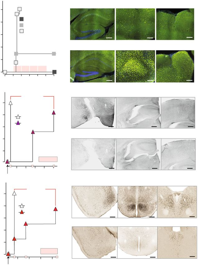
ARTICLE                                                              COMMUNICATIONS BIOLOGY | https://doi.org/10.1038/s42003-020-01538-4

N
          -methyl-D-aspartate receptors (NMDARs) play an                    unrelated young female patients who suffered from epileptic
          essential role in the survival, differentiation, and migra-       seizures, intellectual disability (ID), moderate hypotonia, and
          tion of neurons, as well as in the formation and stabili-         speech/language disorders26,27.
zation of synapses and neuronal circuits both during development               To unravel the functional contributions of the voltage-
and in adulthood1–5. The critical role of NMDARs is based on (I)            dependent Mg2+ block in neurological disease, brain physiol-
their slow response to the major excitatory neurotransmitter                ogy, and behavior, we generated and analyzed heterozygous and
L-glutamate, (II) the voltage-dependent current block by extra-             homozygous gene-targeted mice with global Grin2a(N615S)
cellular Mg2+, and (III) their high Ca2+ permeability6,7 (for a             expression (Grin2a+/S and Grin2aS/S, respectively). The viability
recent review see ref. 8). By combining these three features,               and good health of Grin2a+/S and Grin2aS/S mutant mice allowed
NMDARs provide a precise and elegant molecular mechanism for                us to resolve the functional consequences of this mutation, par-
the activation of Ca2+-dependent postsynaptic second messenger              ticularly for seizure susceptibility, hippocampal plasticity, hip-
cascades, which trigger specific intracellular responses9,10. In             pocampal oscillatory activity, and in cognitive performance
turn, these responses are necessary for the experience-dependent            during simple and complex associative learning tasks. Thus,
priming of neural networks11.                                               experimental results show that the voltage-dependent Ca2+ sig-
   For several decades the precise coincidence detection of pre- and        naling of GluN2A-type NMDARs is of particular importance for
postsynaptic activity by NMDAR-dependent Ca2+ signaling has been            the tight temporal control of attentional processes, which
postulated to be of crucial importance for learning and adapting to         becomes especially important when there are spatial and/or
environmental stimuli. However, this has rarely been tested directly        temporal discontiguities between relevant cues and behaviorally
at the behavioral level12,13. By introducing the well-characterized         relevant outcomes.
GluN2A(N615S) mutation (previously called N596)14,15 into the
mouse genome, we were able to study the effects of an inappropriate
                                                                            Results
glutamate-induced Ca2+ influx through GluN2A-type NMDARs,
                                                                            Generation of GluN2A(N615S)-expressing mice. Heterologous
even at resting potentials, on synaptic plasticity, activity-induced
                                                                            expression of GluN2A(N615S) (Fig. 1a) with GluN1 demon-
c-Fos expression, neuronal network activity in the hippocampus and,
                                                                            strated a reduced Mg2+ block of GluN1/2(N615S) receptors in
lastly, on behavior. This analysis had not been possible in previous
                                                                            the presence of 1 and 4 mM of Mg2+ at hyperpolarized mem-
studies with gene-targeted Grin2a(N614Q) mice that died for unknown
                                                                            brane potentials when compared with wild-type NMDARs
reasons 2 weeks after birth12.
                                                                            (Fig. 1b). In the absence of Mg2+, short glutamate applications
   The molecular components responsible for the Mg2+-regulated
                                                                            (20 ms) activated mutated and wild-type NMDAR channels with
Ca2+ influx through the channel are localized at the tip of the ion
                                                                            comparable current amplitudes and similar activation (rise time)
pore of heterotetrameric NMDARs. The immobile ion pore is
                                                                            and deactivation kinetics. During prolonged glutamate applica-
assembled from four P-loop structures in the M2 membrane
                                                                            tions (600 ms) slower desensitization kinetics were obvious for
segments of NMDAR subunits: i.e. two obligatory
                                                                            the GluN1/2A(N615S) compared to GluN1/2A heterodimeric
GluN1 subunits and two from the GluN2(A–D) or GluN3(A,B)
                                                                            receptors (Fig. 1b and Supplementary Table 1).
subunit families (for a review see ref. 16). Within this complexity,
                                                                                By classical gene-targeted replacement28 we inserted the
GluN1/2-receptors are the most abundant NMDAR subtypes
                                                                            c1844A>G mutation at the homologous position in exon 10,
throughout the central nervous system17–19. In these NMDAR
                                                                            and thus replaced the Grin2a asparagine codon (AAT, N615)
subtypes, an asparagine amino acid residue in the GluN1 subunits
                                                                            with a codon for serine (AGT) (Fig. 1c and Supplementary Fig. 1).
(N614, labeled previously N598 (ref. 20)) and two neighboring N
                                                                            The Grin2a cDNA sequence analysis of total brain mRNA of
residues in the GluN2 subunits (N614 and N615, labeled pre-
                                                                            heterozygous Grin2a+/S mice together with the comparable
viously N595 and N596 (ref. 15)) located at the tip of the P-loops,
                                                                            GluN2A immunosignals in forebrain extracts of Grin2a+/+ and
build the narrow constriction of the ion channel pore, and dif-
                                                                            Grin2aS/S littermates verified that adult mice expressed the
ferentially modulate Mg2+ block and Ca2+ permeability14,15,21.
                                                                            Grin2a(N615S) and Grin2a+ alleles at the same level (Fig. 1d, e).
   The amino acid substitutions GluN1(N614Q) and GluN1
                                                                            We also observed in forebrain extracts statistically comparable
(N614R) abolished or reduced both the Mg2+ block and Ca2+
                                                                            levels of the GluA1 subunit of the amino-3-hydroxy-5-methyl-4-
permeability of all NMDARs. This caused the premature death of
                                                                            isoxazolepropionic acid receptor (AMPAR), the postsynaptic
the respective mutant mice due to respiratory failure22, as
                                                                            marker protein PSD95 and the phosphorylated form of α-
described in mice completely lacking NMDARs (Grin1–/–)23. The
                                                                            CaMKII in Grin2aS/S, Grin2a+/s, and Grin2a+/+ mice. However,
importance of precise NMDAR signaling for the establishment of
                                                                            GluN2B levels were significantly higher in the membrane fraction
autonomic pattern activity in neuronal circuits is further
                                                                            but not in the total forebrain fraction from homozygous Grin2aS/S
emphasized by Grin2b knockout mice. In GluN2B-deficient pups,
                                                                            mouse brains when compared to heterozygous and wild-type
the trigeminal neuronal pattern formation is impaired and the
                                                                            littermates (Fig. 1e and Supplementary Fig. 2).
pups starve to death within the first days after birth due to the
lack of suckling responses24.
   The NMDAR function as a coincidence detector is generally                Regular glutamatergic signal transmission but increased
identified with the induction of long-term potentiation (LTP), the           GluN2B-type LTP in GluN2A(N615S)-expressing mice. Patch
dominant experimental model of synaptic plasticity25. The                   clamp recordings in CA1 pyramidal cells showed that the
voltage-controlled Mg2+ block is essential for this activity-               GluN2A(N615S) subunits are functionally incorporated in
dependent NMDAR signaling10. In recombinant GluN1/2A                        synaptic NMDARs. At –70 mV and in the absence of extracellular
NMDARs the Mg2+ block is predominantly determined by the                    Mg2+, the synaptic AMPA/NMDA current ratio and the indivi-
asparagine amino acid residue GluN2A(N615). In oocytes and                  dual peak of AMPAR and NMDAR currents were similar in
HEK293 cells expressing recombinant GluN1/GluN2A(N615S)                     CA1 cells from Grin2aS/S, Grin2a+/S, and Grin2a+/+ mice
heterodimeric receptors, the GluN2A(N615S) mutation led to a                (Fig. 2a, left). However, the presence of GluN2A(N615S) in
pronounced attenuation of the Mg2+ block and a 1.4-fold                     synaptic NMDAR was indicated by the decreased AMPAR/
increased Ca2+ permeability14,15. Notably, a similar Mg2+ block             NMDAR response ratio detected in Grin2aS/S mutants in the
attenuating point mutation (c.1845C>A, p.Asn615Lys) at the                  presence of 1 mM Mg2+ (Fig. 2a, right). This NMDAR-induced
identical position of the GluN2A subunit was found in two                   current increase was more pronounced at postnatal day 42 (P42)

2                         COMMUNICATIONS BIOLOGY | (2021)4:59 | https://doi.org/10.1038/s42003-020-01538-4 | www.nature.com/commsbio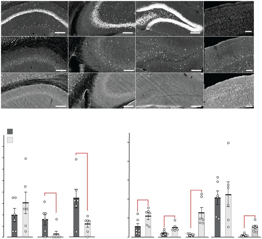
COMMUNICATIONS BIOLOGY | https://doi.org/10.1038/s42003-020-01538-4                                                                                                             ARTICLE

                    a                                              b                                           HEK293 cells
                           GluN2A(N615S)                                                    GluN1/2A                                          GluN1/2A(N615S)
                                                                                                        1.0 Inorm                                            1.0 Inorm
                                                NH2
                     (N615S)                    LBD               4 Mg2+                 - 80   - 40                     40              - 80        - 40                40
                                                                  1 Mg2+                                                V(mV)                                           V(mV)
                                                            out
                                                    4                                                  - 1.0                4 Mg2+                          - 1.0
                                                        1                                                                   1 Mg2+
                                                3       2   in                                         - 2.0                0 Mg2+                          - 2.0
                                                                  0 Mg2+

                                                                                     Glu                                                Glu
                               COOH                               0 Mg2+
                                                                                                               100pA                                            200pA

                        Grin2a+ GLVFNNSVPN                                                                 100pA                                        200pA
                                                                                                                                                                 100ms

                        Grin2as GLVFNSSVPN
                                                                                                                                                             100ms

                    c Grin2a gene-targeting
                                                                             N615                                                                     N615S
                                                      Grin2a+                                                                         Grin2aS
                            Chr. 16
                                                                           exon 10                                                                   exon 10 loxP
                   d Grin2a mRNA expression (forebrain)
                                                                              N615                      Grin2aS/S                                           N615S
                            Grin2a+/+                             A A C A A T T C T                                                                A A C A G T T C T
                                                                                                                        SpeI

                                                                                                                             N/S                    c-DNA sequence
                                                                                                                                  R                (7; source data)
                                                Grin2a+/S
                                                                       -14               -11                                      0 rel. pos.

                  e Protein expression (forebrain)
                                                Membrane fraction
                                                                                                                                                              Grin2a+/+ (3)
                                                                                                                                       p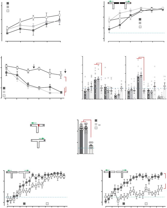
ARTICLE                                                                COMMUNICATIONS BIOLOGY | https://doi.org/10.1038/s42003-020-01538-4

Fig. 1 GluN2A(N615S) containing NMDAR expression in vitro and in vivo. a The position of N615 in the membrane segment M2 is depicted together with
the three channel-forming trans-membrane segments M1, 3, and 4. b In HEK293 cells, recombinantly expressed GluN1/GluN2A and GluN1/GluN2A
(N615S) channels were activated by fast glutamate application (1 mM; in the continuous presence of the co-agonist glycine, 10 µM) at holding potentials
from –100 to +40 mV in different extracellular Mg2+ concentrations. NMDAR-mediated peak currents were normalized to those obtained at +40 mV.
Data points represent mean ± SEM for n = 4–7 different HEK293 cells. Representative current traces evoked in 0 mM Mg2+ at – 60 mV, with 20 and 600
ms applications, are shown below the IV plots and were used to determine the current kinetics (Supplementary Table 1). c Schematic view of the A to G
replacement in exon 10 of the mouse Grin2a gene. d Reverse transcription PCR (RT-PCR)-sequence analyses of total brain mRNA show the A-to-G
mutation in pos. 0 and two diagnostic silent mutations at pos. –11 and –14 in the pore loop encoding gene segment in Grin2a+/+, Grin2aS/S, Grin2a+/S mice.
In Grin2a+/S mice, the overlay of two different colored “nucleotide” peaks, at position 0, –11, and –14 indicate equimolar amounts of mRNA from the
Grin2a+ and the targeted Grin2aS alleles. e Immunoblots of forebrain protein lysates of 4-week-old mice (Supplementary Fig. 2) indicate no genotype-
specific differences of GluN1, GluN2A, and the AMPAR subunit GluA1 expression relative to the β-actin levels (p > 0.05). The levels of PSD95 and αCaMKII
(in its phosphorylated state, pCaMKII) are also comparable between genotypes relative to the GAPDH expression. The GluN2B expression level in the
membrane fraction was significantly increased in Grin2aS/S mice when compared to Grin2a+/+ and Grin2a+/S mice but not in the levels of total protein
lysates (for statistics: Supplementary Statistics to Fig. 1). The number of mice is given in brackets. Error bars represent mean ± SEM.

expressing mice and showed only a trend towards lower                         Supplementary Fig. 4a), and decreased grip strength and climbing
fEPSP amplitudes at a given pre-volley amplitudes of 1.5 mv in                activity (Supplementary Fig. 4b–d). In marked contrast, the general
Grin2aS/S mice. Together with the similar paired-pulse ratio our              locomotor activity of Grin2aS/S mice that we recorded automatically
field recordings revealed no major alterations in CA3-to-CA1                   in the LABORAS home cage42 was significantly increased in the
synaptic transmission of Grin2aS/S and Grin2a+/S mice (Fig. 2b).              first 5 h of the night cycle (Supplementary Fig. 4d), although there
    To analyze whether these voltage-independent GluN1/2A                     was no difference in the maximum and mean running speed
(N615S) receptors can still induce synaptic plasticity, we analyzed           between genotypes. The number of rearing, grooming, and eating
field LTP (fLTP) at CA3-to-CA1 synapses in the different Grin2a                events was not affected (Supplementary Fig. 4d). Our analysis in the
genotypes. Here we found that the magnitude of hippocampal                    Catwalk test showed that Grin2aS/S mice exhibit a regular walking
fLTP in Grin2aS/S and Grin2a+/S mice was unaffected ex vivo and               pattern (Supplementary Fig. 4e) and can achieve normal balance
in vivo (Fig. 2c, d30), in contrast to the reduced fLTP found in              scores in the stationary rod test (Supplementary Fig. 4f). Some
GluN2A-deficient mice and in mice lacking the GluN2A                           minor alterations in the base support of the hind limbs (the distance
intracellular C-terminal domain of the GluN2A subunit31,32.                   of hind limbs during walking) of Grin2aS/S mice (Supplementary
This suggests that the coincidence signaling of GluN1/2(N615S)                Fig. 4e) might contribute to the slightly delayed acquisition in the
receptors is still operative. However, since the GluN2B antagonist            rotarod test (Supplementary Fig. 4g). In heterozygous Grin2a+/S
CP101,106 significantly reduced the fLTP in Grin2aS/S and                      mice, we found a trend towards reduced activity in the running
Grin2a+/S mice but not in Grin2a+/+ littermates (Fig. 2e), we                 wheel and climbing (Supplementary Fig. 4a, d), and 20–30% of
conclude that (i) pure GluN1/2A(N615S) receptors have a                       Grin2a+/S mice did show strong paw- and limb-clasping.
reduced contribution to the long-term synaptic enhancement                       The reduced Mg2+ block of GluN1/2A(N615S) receptors could
after tetanic stimulation and (ii) the fLTP recorded in Grin2aS/S             conceivably permit a glutamate-induced, voltage-uncontrolled
and Grin2a+/S is substantially mediated by GluN2B-containing                  Ca2+ influx into neurons that are sensitive to Ca2+-induced
receptors.                                                                    toxicity. However, we did not detect any signs of cytotoxicity or
    This conclusion was strengthened by using four tetanic                    neurodegeneration in Nissl-stained brain slices from adult
stimulations (4 × 100 Hz), which can induce GluN2B-dependent                  Grin2aS/S mice (Fig. 3d). Furthermore, no chromosomal DNA
LTP in the absence of functional GluN2A33,34. In comparison                   degradation could be detected in the terminal deoxynucleotidyl
with single tetanic stimulation, LTP was significantly increased               transferase dUTP nick end labeling (TUNEL) assay in the
40–45 min after the 4 × 100 Hz stimulation in hippocampal slices              hippocampus and adjacent cortical cell layers, suggesting that
of both Grin2aS/S and Grin2a+/S mice compared to WT control                   there was no apoptosis or necrosis in Grin2aS/S brains (Fig. 3e).
littermates. This LTP increase was reduced by CP101,606                       Our Timm staining of mossy fibers did not indicate hippocampal
(Supplementary Fig. 3a), an effect that is reminiscent of the one             sclerosis, a neuropathological marker of temporal lobe epilepsy in
described for LTP reduction in juvenile (P14) wild-type mice35.               humans and rodents32,43,44, not even in 6-months-old Grin2aS/S
This LTP was still completely NMDAR-dependent and could be                    mice (Fig. 3f). Lastly, the immunosignals of neuronal and
blocked by the NMDAR antagonist APV (Supplementary Fig. 3b).                  astrocytic markers NeuN and GFAP were comparable between
Together, these results show the incorporation of GluN2A                      controls and Grin2aS/S mice, as was the hippocampal layer-
(N615S) into synaptic NMDARs but reduced contribution of                      specific distribution of calbindin, the interneuronal protein
GluN2A(N615S) receptors in LTP.                                               parvalbumin, and the AMPAR subunit GluA1 (Fig. 3g).

GluN2A(N615S) homozygous mice show altered home cage                          Grin2aS/S mutant mice are highly sensitive to audiogenic sei-
behaviors but regular brain anatomy, no apoptosis or neuro-                   zures (AGSs). Considering the presence of an epileptic phenotype
degeneration. In contrast to other genetically modified mice with              in the patient with the analogous GluN2A(N615K) mutation27
altered NMDAR Ca2+ permeability and/or altered Mg2+ block12,22,               and the transient epileptiform discharges observed in GluN2A-
we found that Grin2aS/S and Grin2a+/S mice are viable and long-               deficient mice45, we analyzed the seizure susceptibility of GluN2A
living. However, Grin2aS/S mice can be recognized by their reduced            (N615S)-expressing mice. When exposed to a high-frequency
body weight. Moreover, Grin2aS/S mice showed poor nest building               acoustic stimulus (11 kHz), which is used for AGS induction in
and burrowing activities (Fig. 3a), which can be indicators of                DBA mice46, all Grin2aS/S mutants responded immediately after
impairments associated with hippocampal dysfunction36–38.                     tone onset with a stereotypic AGS response composed of wild
Grin2aS/S mice also exhibited the paw- and limb-clasping reflex                running followed by clonic seizures, tonic extension of limb
(Fig. 3b and Supplementary Data: Video 1)39–41, reduced muscle                extremities, and respiratory arrest (RA). In contrast, no seizures
strength, lower activity in the running wheel (Fig. 3c and                    were observed for Grin2a+/+ control mice, whereas in Grin2a+/S

4                           COMMUNICATIONS BIOLOGY | (2021)4:59 | https://doi.org/10.1038/s42003-020-01538-4 | www.nature.com/commsbio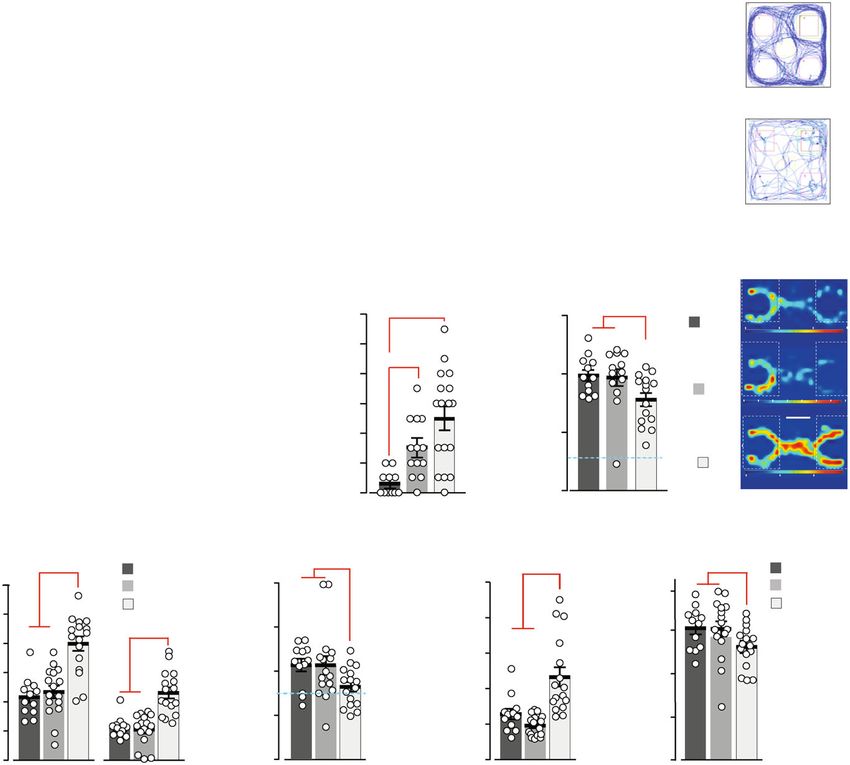
COMMUNICATIONS BIOLOGY | https://doi.org/10.1038/s42003-020-01538-4                                                                                                                                                                                                                                               ARTICLE

                                                                                                                                                                                                                                                                               P42
                   a                                   4.0
                                                                                                                      P42                                                                                             30
                                                                                                                                                                                                                                                                 p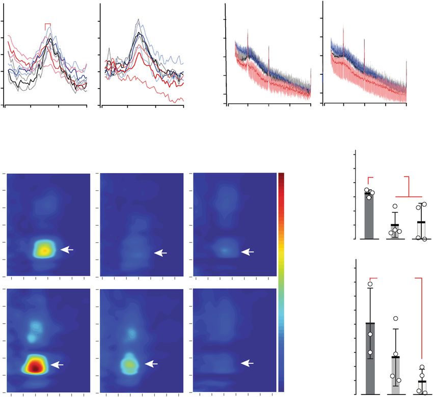
ARTICLE                                                                                                                           COMMUNICATIONS BIOLOGY | https://doi.org/10.1038/s42003-020-01538-4

                                                                                                                                                             Grin2a+/+   c                     Grin2a+/+
                    a                           Grin2a+/+                            Grin2a+/S                                   Grin2aS/S               b                                     Grin2a+/S
                                                                                                                                              p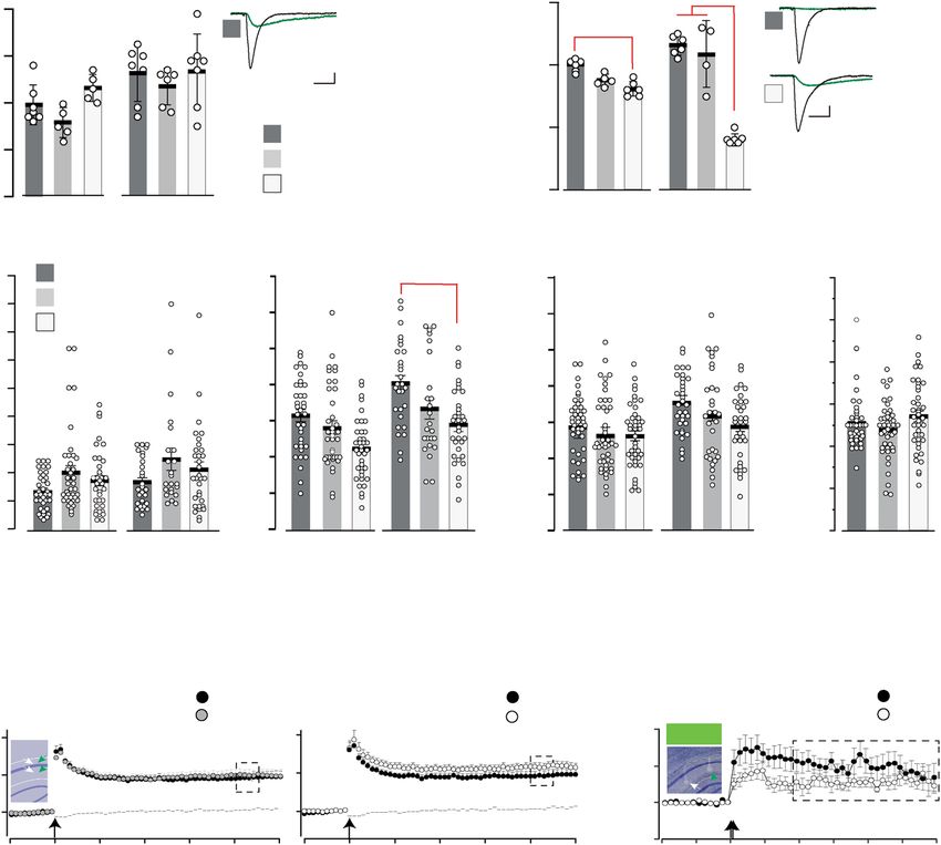
COMMUNICATIONS BIOLOGY | https://doi.org/10.1038/s42003-020-01538-4                                                                                         ARTICLE

                   a                                              p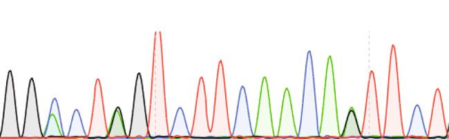
ARTICLE                                                                            COMMUNICATIONS BIOLOGY | https://doi.org/10.1038/s42003-020-01538-4

                  a       MK-801                                   CA1                 CA3                                       DG               Neo
                        (0.5 mg/kg)

                      Grin2a–/– (2)

                         WT (17)

                      Grin2aS/S (9)

                  b                                          WT (MK-801)
                                                             Grin2aS/S (MK-801)                                  200

                                                                                        c-FOS+ cells [n] / mm2
                                                    16                        p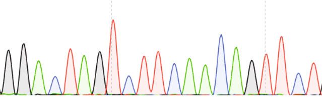
COMMUNICATIONS BIOLOGY | https://doi.org/10.1038/s42003-020-01538-4                                                                                                                                                                      ARTICLE

                  a                                                                Exploration                REM sleep                                           Exploration                                    REM sleep

                                  θ Power [log(µV2/Hz)]

                                                                                                                               γ Power [log(µV2/Hz)]
                                                                                       p
ARTICLE COMMUNICATIONS BIOLOGY | https://doi.org/10.1038/s42003-020-01538-4
Grin2a+/+ (10) Grin2a+/+ (3)
a Grin2a+/S (10) b Grin2a+/S (8) pCOMMUNICATIONS BIOLOGY | https://doi.org/10.1038/s42003-020-01538-4 ARTICLE
a Grin2a+/+ (6)
during odor learning [%]
Mean correct choices in
b
discrimination task [%]
100 Grin2a+/S (5)
Mean correct choices
100
Grin2aS/S (4)
in simple cue
80 Grin2a+/+ (9)
80
Grin2a+/S (9)
60 Grin2aS/S (10)
60 Ch.
40
100 200 300 400 500 1 2 3 4 5 6
Trials [n] Block [n] of 10 trials each
pARTICLE COMMUNICATIONS BIOLOGY | https://doi.org/10.1038/s42003-020-01538-4
mice were slightly faster at acquiring the task than controls (p < Grin2aS/S mice are impaired when there is spatiotemporal
0.05). Once the animals had acquired this simple visuo-tactile discontiguity. A potential feature of these spatial memory tasks
discrimination, we then reversed the floor-insert cue–reward which differentiates them from the simple associative learning
contingencies for each mouse (thus animals that had been trained tasks is the presence of a spatiotemporal discontiguity between
on A+/B− now experienced A−/B+ and vice-versa). There was the relevant environmental cues at the point at which a decision is
no effect of genotype during reversal learning, with all three made (e.g. to navigate in a particular direction) and the occur-
groups of mice acquiring the new cue–reward contingencies at rence of the unconditioned stimulus (e.g. the escape platform in
the same rate (Supplementary Fig. 7a). Thus, Grin2aS/S mice the watermaze, food rewards on the radial maze). To determine
could form simple cue/reward associations using cues from specifically whether Grin2aS/S mice were sensitive to spatio-
different sensory modalities, and could also perform more temporal discontiguities we returned to the appetitively motivated
complex, cognitively demanding tasks using these cues, for T-maze task, in which the presence of visuo-tactile cues in the
example, when cue–reward contingencies were reversed. form of floor inserts in the maze determined the location of a
sweet milk reward. We have shown that Grin2aS/S mice could
successfully acquire a simple visuo-tactile discrimination during
Grin2aS/S mice are impaired on a battery of spatial memory which floor inserts restricted to the goal arms of the maze indi-
tasks. We then assessed the spatial learning abilities of mice cated the presence or absence of reward (Fig. 8b). We next used
across a battery of spatial memory tasks. First, we investigated the entire floor structure of the T-maze as a conditional cue to
spatial reference memory (SRM) acquisition in the Morris indicate whether the reward was located in either the left or the
watermaze (MWM)71, during which mice were trained to find a right goal arm (Fig. 8e). Mice had to follow the rule that if floor
hidden platform that remained in the same, fixed location on insert A is present then reward is on the left whereas if floor insert
every trial, starting from different points around the perimeter B is present then reward is on the right. When the floor inserts
of the pool. Using the same protocol, with the same extramaze covered the entire T-maze, including the start arm and both goal
spatial cues and in the same testing laboratory, we have pre- arms, such that there was no discontiguity, then both Grin2a+/+
viously shown that the acquisition of this task is dependent on the and Grin2aS/S mice were able to acquire the task to a high level of
hippocampus72,73. Whereas both Grin2a+/+ littermates performance by the end of 19 blocks of training (Fig. 8e). There
and Grin2a+/S mice readily learned the location of the platform was a subtle impairment in the Grin2aS/S mice such that they
with training, Grin2aS/S mice displayed a large impairment, acquired the task slightly more slowly than the controls, but by
showing only a marginal improvement across training sessions 130 trials both groups were consistently scoring over 80% correct
(Fig. 8c and Supplementary Fig. 7b). In the probe tests during (Fig. 8e). Separate groups of Grin2a+/+ and Grin2aS/S mice were
which the platform was removed from the pool (performed after then trained on the discontiguous version of the task in which the
12 and 24 training trials), the Grin2a+/S and Grin2a+/+ litter- floor insert cues were now limited to the start arm only and thus
mates showed a strong and robust preference for the training absent from the goal arms and at the point when the mouse
quadrant that normally contained the escape platform. In con- received the reward (Fig. 8f). This time, while wild-type litter-
trast, the Grin2aS/S mice had no memory for the platform loca- mates readily acquired the task, Grin2aS/S mice failed to learn,
tion and exhibited chance levels of performance in both probe showing a significant impairment from training blocks 5 to 19.
test trials (Fig. 8c), in marked contrast to the successful acquisi- Thus, the Grin2sS/S mice were able to acquire the T-maze task but
tion that we have previously reported with Grin2a–/– mice in this only when there was no spatiotemporal discontiguity present.
very same MWM task74. Importantly, there was a highly significant genotype by task
To exclude the possibility that the impaired MWM perfor- interaction (F(1, 30) = 5.1; p < 0.05), and subsequent analysis of
mance of Grin2aS/S mice was caused by deficits in motivation or simple main effects showed that there was a significant effect of
sensorimotor abilities, a visible platform version of the task was discontiguity for the Grin2aS/S mice (F(1, 30) = 13.2; p < 0.005),
also conducted. In this task, the platform was raised above the but not for the wild-type controls (F < 1; p > 0.20).
water level and clearly marked with a black and white striped In summary, our behavioral analysis showed that Grin2aS/S
cylinder directly above. Mice of all three genotypes rapidly mice could form simple associations and thus perform discrimi-
learned to swim towards the visible platform and all mice reached nation tasks using cues from different sensory modalities,
nearly identical levels of performance by the third block of testing including during a more cognitively demanding reversal learning
(Supplementary Fig. 7c), consistent with our results from the task. However, the same Grin2aS/S animals were considerably
simple associative tasks using either odor- or visuo-tactile cues impaired in a battery of spatial learning paradigms, although this
(Fig. 8a, b). Thus, the spatial learning impairment in the MWM was not the case for heterozygous Grin2a+/S littermates. Grin2aS/S
was unlikely due to sensorimotor or motivational disturbances. mice were also impaired on conditional T-maze tasks, particularly
To test the generality of this spatial learning deficit in a when there was a spatiotemporal discontiguity between the
completely different experimental setting, with different sensor- relevant sensory cues present at the choice point and the
imotor and motivational demands, we next assessed acquisition presence/absence of reward.
of an appetitively motivated, SRM six-arm radial maze task. Here,
Grin2aS/S mice showed a substantial deficit in learning to
discriminate between the rewarded and non-rewarded arms. Discussion
While both the Grin2a+/S and Grin2a+/+ littermates learned the Here we provide the first direct experimental evidence that the
locations of the food rewards and showed a gradual improvement tightly controlled Mg2+ block of the GluN2A-type NMDAR, and
across days of training, making less reference memory errors as thus the voltage-dependent Ca2+ influx through its ion channel, is
training progressed, the Grin2aS/S mice showed little, if any, essential in different regions of the nervous system for coordinated
improvement (Supplementary Fig. 7d). Spatial working memory neuronal communication and its functional consequences.
(SWM) was also assessed using both the spontaneous and Our data obtained both on recombinant NMDARs and on
rewarded alternation versions of the T-maze task75,76. In both NMDARs from genetically modified animals shows that GluN2A
tasks, the Grin2aS/S mice were substantially impaired relative to (N615S) containing NMDARs have a remarkably reduced sen-
Grin2a+/S and Grin2a+/+ littermates (spontaneous: Supplemen- sitivity to external Mg2+ ions as measured by the linearized I/V
tary Fig. 7e; rewarded: Fig. 8d). relation of GluN1/2A(N615S) channels and their less reduced
12 COMMUNICATIONS BIOLOGY | (2021)4:59 | https://doi.org/10.1038/s42003-020-01538-4 | www.nature.com/commsbioCOMMUNICATIONS BIOLOGY | https://doi.org/10.1038/s42003-020-01538-4 ARTICLE voltage-controlled NMDARs currents in the presence of Mg2+ in block AGS in Grin2aS/S mice by pre-treatment with NMDAR HEK293 cells and hippocampal slices, respectively. Taking into antagonists, providing evidence that the NMDAR signaling account the 1.4-fold increased Ca2+ permeability15, no alterations directly promotes the brainstem-derived AGS induction. Notably, in the channel conductance and proven surface and synaptic Grin2a–/– mice are more resistant to electrical IC stimulation,50 expression of GluN2A(N615S)-containing NMDARs, all synaptic revealing a key role for GluN2A-type receptors in the modulation events are most likely associated with an increased NMDAR- of IC neuronal activity. Furthermore, these data indicate that in mediated Ca2+ entry at resting membrane potentials through the absence of GluN2A-type NMDARs, IC neurons are either less synaptic channels. Moreover, spill-over of glutamate might pro- excitable and/or under stronger inhibition. In Grin2aS/S mice, we duce additional Ca2+-influx through GluN1/2A(N615S) recep- have found exactly the opposite situation: the less-stringent vol- tors into dendrites located close to active synapses and will tage-controlled Ca2+ influx through GluN2A-containing amplify the impact of volume transmission to the network NMDAR leads to an over-excitation and/or reduced inhibition activity and the disturbance of synaptic plasticity in GluN2A of the neuronal populations that are activated by the audiogenic (N615S)-expressing mice. stimulus and trigger AGS. Thus, the constant glutamate-gated Alteration in synaptic plasticity could be monitored by an GluN1/2A(N615S) Ca2+ influx seems to mimic experimental increased LTP component of GluN2B-containing NMDARs in “kindling” models in sensitizing the network response to a strong hippocampi from GluN2A(N615S)-expressing mice. Very similar acoustic stimulus. MK-801 and memantine have an AGS pro- to our findings of an increased GluN2B component at CA3-to- tective effect in kindling models as well as in Grin2aS/S mice. CA1 synapses in young mice34, the LTP could be significantly Thus, the AGS network response is most likely triggered pre- enhanced by repeated tetanic stimulations and the enhancement dominantly by voltage-controlled NMDAR signaling through could be blocked by CP101,106 in adult Grin2a+/S and Grin2aS/S GluN1/2B or other GluN2A-independent NMDAR subtypes. A mice. This might indicate that the altered Ca2+ homeostasis by drug effect on GluN1/2A(N615S) receptors seems less likely, since the glutamate-triggered Ca2+ influx through the mutant MK-801 binding and memantine blockade of recombinant NMDARs keeps the plasticity mechanism of CA1 cells in an GluN1/2A(N615K) receptors is reduced55. immature state in GluN2A(N615S)-expressing mice. Opposite and contrasting results between Grin2a–/– and The homozygous Grin2aS/S mice exhibited a complex pheno- Grin2S/S mice were also found when analyzing MK-801 c-Fos type characterized by high sensitivity to AGSs and cognitive activity mapping in the hippocampus. In contrast to the IC deficits. In response to a strong acoustic stimulus, unregulated neurons, the glutamate-gated GluN1/2A(N615S) Ca2+ influx GluN2A receptor-mediated Ca2+ signaling is associated with was associated with reduced excitability of hippocampal principal over-excitation in the midbrain/brainstem, followed by strong cells. Although in Grin2aS/S mouse brains MK-801-induced c-Fos activation of some forebrain neuronal populations (mainly in the expressing cells were increased in several cortical areas, amygdala and hypothalamus), a response that cannot be com- they were much less visible in the hippocampus, in line with the pensated for and finds its end in generalized epileptic seizures lack of hippocampal activation observed after AGS. This suggests with respiratory arrest (AGS-RA). In contrast, the hippocampal that the hippocampus of Grin2aS/S mice is less active, which formation appears less excitable in these mice and hippocampal might be due to increased activity of GABAergic interneurons56, theta frequency and phase coupling between slow oscillations and or could reflect wider circuit effects. In contrast, in the absence of the amplitude of a fast oscillation is reduced during exploration. GluN2A in Grin2a–/– mice, the hippocampal pyramidal and These oscillatory changes correlated with impaired regulation of granular neurons are highly excitable following MK-801 chal- exploratory activity and attention, resulting in associative learning lenge. Thus, the regulation of NMDAR Ca2+ signaling via the deficits in situations in which the tight regulation of attentional voltage-dependent Mg2+ block plays a key role in the differential processes is likely to be important. Thus, our analysis of GluN2A priming of neural activity in separate and distinct neural (N615S)-expressing mice provides strong and direct evidence for networks. the divergent and brain region-specific role of GluN2A-type Changes in excitatory activity in the hippocampus of NMDAR Ca2+ signaling in the excitability of neuronal networks. behaving Grin2aS/S mice were associated with impaired neu- Opposite to what was observed previously for Grin2a–/– mice74, ronal network activity. We noticed a reduced phase–amplitude the voltage-independent GluN2A-type NMDAR Ca2+ signaling cross-frequency coupling in the lower gamma frequency range in Grin2aS/S mice generated increased excitability in neurons of during exploration and REM sleep, and a reduced theta fre- midbrain nuclei and decreased excitability of hippocampal prin- quency peak during exploration, which could potentially be cipal neurons. explained by the altered inhibition/excitation ratio of the neu- The AGS activity pattern in Grin2aS/S mice shows strong ronal networks82. This is in line with previous studies in similarity to the one described in subcortical/midbrain structures which NMDAR ablation in hippocampal parvalbumin-positive of epilepsy-prone DBA mice and GEPR-9s rats, which are fre- (PV+) interneurons showed disturbed theta-nested gamma quently used as animal models for sudden unexpected death in oscillations83–87. Interestingly, whereas NMDAR blockade leads epilepsy77. In those animal models, which rely on acoustic to hypersynchronous phase locking88, we observed the opposite kindling, the IC plays a key role in the initiation of epileptic when introducing the Grin2a(N615S) mutation. Thus, disrupted seizures, which also involves non-auditory brain structures such coincidence detection of the GluN2A-type NMDARs affects as the PAG and the substantia nigra pars lateralis78, consistent the coordinated oscillatory network activity and the slow with our c-Fos expression analyses of Grin2aS/S brains after AGS. inhibition of the hippocampal network during theta-related The central role of the IC is supported experimentally by the behaviors. increased AGS sensitivity seen in normal rats after inhibition of The hippocampal theta rhythm is of particular importance GABAA receptors in IC or after pharmacological NMDAR acti- during exploration and attention89,90. Notably, we observed clear vation of this nucleus79. EEG recordings and c-Fos expression behavioral impairments during exploration, including hyper- mapping have demonstrated that kindling causes permanent AGS activity and unregulated and exaggerated levels of attention to network expansion from the brainstem to forebrain structures, spatial, non-spatial and social cues in the Grin2aS/S mice. Despite most likely via the amygdala47,78,80, increasing the severity of the this, Grin2aS/S mice could still form simple associations in several seizures, and supported also by our results here in Grin2aS/S mice. different tests, but only if the relevant sensory cues and the In line with previous experiments81, we were able to effectively rewards were contiguous in space and time. Thus, basic COMMUNICATIONS BIOLOGY | (2021)4:59 | https://doi.org/10.1038/s42003-020-01538-4 | www.nature.com/commsbio 13
ARTICLE                                                             COMMUNICATIONS BIOLOGY | https://doi.org/10.1038/s42003-020-01538-4

associative learning mechanisms remained intact in Grin2aS/S               with other studies in heterozygous patients with the very same de
mice, in accordance with the behavior of Drosophila over-                  novo NMDAR mutations who displayed a similar (although not
expressing transgenic Mg2+ insensitive NMDARs13.                           identical) clinical manifestation92,94–96.
   Analogous to the above-mentioned transgenic flies, Grin2aS/S                Indeed, although two patients with the very same GluN2A
mice did exhibit pronounced learning deficits during a battery of           (N615K) mutation had early-onset epileptic encephalopathies,
spatial memory tests (e.g. MWM and radial maze). They were                 there were also differences in their clinical manifestations which
also impaired on the conditional T-maze learning task91, parti-            defied a clear genotype–phenotype correlation26. Correspond-
cularly when the relevant sensory cues at the choice point were            ingly, in our heterozygous Grin2a+/S mice the gain of function
separated in space and time from the reward outcomes. This                 Grin2a(N615S) mutation led—depending on the behavioral test
might reflect that the Grin2aS/S mice exhibit elevated, sustained,          and sometimes on the individual mouse—to either detectable or
and unregulated levels of attention, which can be particularly             non-detectable phenotypes (for phenotypes that were clearly
disruptive for tasks with inherent spatiotemporal discontiguity            evident in the homozygous mutant mice). Since glutamate sti-
during which appropriate and specific cues must be held in the              mulation—and thus neuronal activity—is an obligatory trigger for
forefront of attention to span or bridge any gaps between relevant         Ca2+ influx through mutated NMDARs, these gain of function
cues and their associated outcomes. Paying attention to irrelevant         NMDAR mutations may be particularly sensitive to non-genetic-
or inappropriate cues might be very disruptive on such tasks. This         factors that affect neuronal activity.
is in contrast to simple associative learning tasks, during which             To conclude, our experimental findings thus provide a detailed
sensory cues and rewards/outcomes are present at the same time             physiological explanation for the cognitive impairments combined
and in the same place, and thus credit assignment and appro-               with epileptic seizures, hypotonic muscle tone, and developmental
priate associations can be formed unimpaired. Thus, the strictly           delay in patients carrying similar de novo NMDAR mutations
controlled voltage-dependent Ca2+ signaling of GluN2A-type                 GRIN2A(N615K), GRIN2B(N615I), and GRIN2B(V618G). Notably, all
NMDARs appears to be of particular importance for the tight                three of these GluN2 subunit mutations were found to reduce the
temporal control of attentional processes which becomes espe-              Mg2+ block and to alter Ca2+ permeability in recombinant
cially important when there are spatial and/or temporal dis-               systems27,97. Our results demonstrate that the tightly regulated
contiguities between relevant cues and behaviorally relevant               Mg2+ block of the NMDAR, and thus the voltage-controlled Ca2+
outcomes.                                                                  influx, is essential in different regions of the nervous system for
   In recent years numerous de novo GRIN2 variants have been               coordinated neuronal communication. Consequently, its disrup-
identified in patients with neurological disorders. One set of rare         tion has profound consequences on brain function in numerous
mutations changes the activation profile of NMDARs and is                   different domains.
located in the ligand-binding domain (LBD) of the NMDAR. A
second set is located in the NMDAR channel pore of the channel             Methods
gate92. These mutations affect ion permeability and in particular          Ethical statement. Most animal experiments were performed at the Max Planck
the voltage-controlled Ca2+ influx. Carriers of these second set of         Institute of Medical Research in Heidelberg according to the institutional guide-
mutations suffer from neurological dysfunction with different              lines of the Max Planck Society, the “Interfakultäre Biomedizinische For-
degrees of severity. Up to 500 rare disease variants in NMDAR              schungseinrichtung” (IBF) animal core facility of Heidelberg University and the
                                                                           Interdisciplinary Neurobehavioral Core (INBC) of Heidelberg University. Genetic
genes have been identified in human patients. Most variants were            manipulations of mice were performed under the licenses of the Regional Board
found in GRIN2A and GRIN2B. The comparison of 249 indivi-                  Karlsruhe, Germany: Generation of mice (35-9185.81/G-4/02); mouse behavior
duals with pathogenic, or likely pathogenic, GRIN2A variants               and drug treatment of animals (35-9185.81/G-115/04, 35-9185.81/G-71/10; 35-
identified patients with severe developmental phenotypes asso-              9185.81/G-171/10; 35-9185.81G-105/16); in vivo (35-9185.81/G-273/12; 35-
                                                                           9185.81/G-171/10, 35-9185.81/G-44/16, 35-9185.81/G-100/16). Animals for
ciated with missense mutations in the ion pore or the linker               molecular, histological, and electrophysiological experiments were recorded under
domain. NMDAR mutations within the amino-terminal, LBD,                    the protocols MPI/T-6/06; 15/08; 20/9, 28/11. Ex vivo LTP experiments were
and null variants led to a less severe phenotype and were classified        conducted according to the Norwegian Animal Welfare Act and the European
as “loss of function mutations”93,94 whereas most of the severe            Union’s Directive 86/609/EEC. Behavioral experiments in the UK were conducted
                                                                           in accordance with the United Kingdom Animals Scientific Procedures Act (1986),
NMDAR mutations with altered Mg2+ block were considered as                 under the project license number PPL 30/2561 of the UK Home Office.
“gain of function” mutations94.
   A detailed study with 12 de novo GRIN missense variants from
                                                                           Statistics and reproducibility. In all figures, the number of independently
18 patients clearly showed that those missense mutations in the            recorded values is clearly indicated. The number of animals and the number of
P2 loop of GluN1, GluN2A, and GluN2B altered surface                       recorded data points are given as appropriate. In bar graphs, all data points used
expression, pharmacological properties, and other biophysical              are pictured together with the standard error of the mean. In the figure legends, the
characteristics. It also demonstrated that these variants can have         tests used for statistical evaluation (ANOVA, t-test, etc.) are stated together with
modest changes in agonist potency and proton inhibition. Fur-              the p values of the results. P values indicating a significant difference are given
                                                                           directly in the figures. Due to space limitations in the main figure legends, detailed
thermore, the voltage-dependent inhibition by Mg2+ was sig-                descriptions of the statistical analyses can be found in Supplementary Statistics to
nificantly reduced in all variants. Since the single channel                Figs. 1, 2, 3, 4, 7 and 8. In the Supplementary Figures, the details of the statistical
conductance and Ca2+ permeability can be altered to different              analyses are given in the figure legends. When multiple comparisons were used to
extents by different mutations, the degree of Ca2+ influx after             control the familywise error rate, we indicate the statistical test used (e.g. Bon-
                                                                           ferroni test). When appropriate, non-parametric analyses (e.g. Mann–Whitney U-
glutamate stimulation is specific for each mutation. This means             test) were conducted. Data records in vivo local field potential oscillations can be
that the severity of any “gain of function” mutation (like the             provided on request. Data were first recorded and analyzed in Excel (Microsoft),
Grin2a(N615S)) and thus the severity of the associated phenotype           and for the distribution blots, Graphpad Prism was used.
will be defined by the kind of mutation95. Li et al. summarize the
phenotypes of 4 GluN2A(N614S), 2 (N615K), and 1 GluN2B                     Generation of GluN2A(N615S)-expressing mice. For gene targeting at the Grin2a
(N615I), 1 (N615K) and 1(N616K) de novo mutations. Seven of                gene loci, we used the method which we described in detail for the targeting of the
these nine patients showed muscle hypertonia, six suffered from            Grin1 gene 22,28. A brief description of the targeting strategy to generate Grin2atm1.RSP
                                                                           (N615S) mice (herein abbreviated as Grin2aS/S) is given in Supplementary Fig. 1.
epileptic seizures, and all exhibited ID and developmental delay.
                                                                           Mice are genotyped by tail-PCR with specific primers. Primers used were: do: 2A-
All GluN2A mutations were associated with language problems                TM3do (5′-GTG TGG GCC TTC TTT GCY GTC-3′) and up: 2A-IN11UP1 (5′-CAT
and autism spectrum disorder was diagnosed in one GluN2A                   ATA TAC AAG CAT TGG AG-3′). Amplified gene fragments for the Grin2aS and
(N614S) patient and in one GluN2B(N615I) carrier. This is in line          Grin2a+ alleles are 559 and 482 bp, respectively. The Grin2aS/S mice were used

14                       COMMUNICATIONS BIOLOGY | (2021)4:59 | https://doi.org/10.1038/s42003-020-01538-4 | www.nature.com/commsbio
COMMUNICATIONS BIOLOGY | https://doi.org/10.1038/s42003-020-01538-4 ARTICLE
previously in a cellular LTP experiment induced by a low-frequency stimulation identified visually using IR-DIC microscopy. Identified cells were clamped at
pairing protocol30. The mouse line is available from the INFRAFRONTIER’S EMMA –70 mV35,103, either in the presence or in absence of 1 mM MgCl2. To evoke
mouse respiratory (EM:09319: B6.129-Grin2atmN615SRSP/kctt). synaptic currents, glass electrodes filled with aCSF were placed in the stratum
radiatum within 50–100 μm of the soma of the recorded neuron. Inhibitory
synaptic transmission was blocked during recordings by the addition of 10 μM
Expression analysis of Grin2a mRNA, cDNA synthesis, reverse transcription gabazine to the perfusion aCSF. The inter-sweep interval was 6 s. AMPAR- and
PCR, and sequencing. Mice were killed by decapitation, the total brain was NMDAR-mediated currents were pharmacologically dissected using the AMPAR
immediately isolated, and the forebrains (excluding cerebellum and olfactory bulb) and NMDAR antagonists, 2,3-dihydroxy-6-nitro-7-sulfamoyl-benzo[f]quinoxaline
were used as tissue input for the cDNA preparation using the RNeasy mini Kit (NBQX; 10 μM), and (2R)-amino-5-phosphonopentanoate (AP5; 100 μM),
(Qiagen, CD./ID: 74104). From the forebrain lysate (about 20 mg), total RNA was respectively. After recording the total current responses (containing both AMPAR
prepared using the RNeasy spin column, and the remaining DNA was digested by and NMDAR components, 100 sweeps), AMPAR channels were blocked by bath
DNAseI. First-strand cDNA synthesis was performed from about 1.5 μg total RNA application of NBQX (10 μM) and another 100 sweeps containing only NMDAR
using a Superscript II reverse transcriptase kit (SuperscriptTM III First-Strand responses were recorded. AMPA currents were obtained by subtraction of the
Synthesis System for RT-PCR. Cat. No 18080-51; now Thermo Fisher) primed by averaged NMDA response from the averaged total response. AMPA/NMDA ratios
random hexamers according to the manufacturer’s instructions. From this reaction, were calculated as the peak AMPAR-mediated current amplitudes divided by the
2 μl was directly used to amplify the M2 region spanning Grin2a cDNA fragment peak NMDAR-mediated current amplitudes.
by the Grin2a mRNA-specific primers NR2Aex9do (5′-CCT GTT GGA TAC AAC
AGA AAC TTA GC-3′) and NR2Aex11up (5′-CTG GTT GAA TTT GGT CAT
GTA CTG-3′) in 30 PCR cycles (Phusion® High-Fidelity PCR Kit; New England
Biolabs NEB; E0553S/L). The amplified 390 bp DNA fragment was gel-purified on Hippocampal field LTP recordings in acute brain slices. For local field potential
a 1.5% agarose gel in E-buffer (QIAquick Gel Extraction Kit; Qiagen, CatNo./ID: recordings at hippocampal CA3-to-CA1 synapses we used transverse, acute brain
28704) and primer NR2Aex11up was used for sequencing using a commercial slices35,103–106. In brief:
service (Eurofins, GATC Service). Sequence analyses were performed by Lasergene Slice preparation. Adult mice (2–4 months, 3–5 mice, per genotype and
(DNASTAR) and the chromatograms of the reverse strands (coding sequence) experiment) were sacrificed with Suprane (Baxter) and the brains were gently
were visualized with SnapGene viewer (SnapGene). removed from the skull. Transverse slices (400 μm) were cut from the middle and
dorsal portion of each hippocampus (Supplementary Data: Video 3) with a
vibroslicer (Campden Instruments NVSLM1) in cold aCSF (4 °C, bubbled with
Electrophysiological profile of GluN1/2A(N615S) receptors in HEK293 cells. 95% O2–5% CO2) containing (in mM): 124 NaCl, 2 KCl, 1.25 KH2PO4, 2 MgSO4,
For the transfection of human embryonic kidney cells (HEK293 cells; ATCC: CRL- 1 CaCl2, 26 NaHCO3, and 12 glucose. Slices were placed in an interface chamber
1573TM) we used the Ca2+ phosphate precipitation method of Chen and exposed to humidified gas at 28–32 °C and perfused with aCSF (pH 7.3) containing
Okayawa98. HEK293 cells were co-transfected with plasmids expressing the rat 2 mM CaCl2 for at least 1 h prior to the experiments. In some of the experiments,
GluN1-1a99 together with GFP and the rat GluN2A100 or GluN2A(N615S)14. Two DL-2-amino-5-phosphopentanoic acid (AP5, 50 μM; Sigma-Aldrich) was added to
days after transfection, GFP-labeled cells were lifted from the coverslip and whole- the aCSF in order to block NMDAR-mediated synaptic plasticity or a 10 µM
cell currents were recorded using an EPC-9 amplifier (HEKA, Lambrecht, Ger- concentration of GluN2B-specific antagonist CP101,606 (CP) (Pfizer) was added to
many) in the presence of 10 µM glycine by fast applying 1 mM glutamate from a the perfusion media.
Piezo-driven double-barrelled pipette. Solution exchange time, measured with an Synaptic excitability. Orthodromic synaptic stimuli (ARTICLE COMMUNICATIONS BIOLOGY | https://doi.org/10.1038/s42003-020-01538-4
electronic GmbH), and stored at 10 kHz (ITC-16, HEKA Elektronik) after 50 Hz home cage. Both at 2 and 18 h later the remaining food pellets in the burrowing
noise filtration by a Hum Bug Noise Eliminator (AutoMate Scientific, Inc.). tube were weighed to determine the burrowing activity.
Extracellular stimulation was generated with an isolated stimulator (A365, WPI).
The slopes of evoked LFPs were analyzed based on the middle one-third of the
Assessment of clasping reflex. For the assessment of the clasping reflex, the
rising phase (Fitmaster, HEKA Elektronik). At the beginning of each recording,
mouse was suspended by the end of its tail and elevated (10 cm) from the ground
two IO curves per mouse were generated, applying stimulation voltages with both
for 1 min. The behavior of paw- and limb-clasping was monitored39.
polarities. To evaluate changes in synaptic efficacy, a stimulus strength eliciting
35–40% of the maximum slope was used as the test pulse and was given every 30 s.
For LTP induction, two trains of high-frequency stimulation (50 × 100 Hz, 100 μs Acoustic stimulation and systemic drug administration. Mice were placed in an
pulse width, same intensity as test pulse) separated by 5 min were used. acrylic glass chamber (46 × 24 × 23 cm) with sawdust on the floor. The chamber
After recordings, mice were deeply anesthetized and electrical lesions were could be divided into two or four equal-sized compartments, which allowed
induced twice (20 µA, 10 s) for each single tungsten wire. Subsequently, mice were the simultaneous acoustic stimulation of different genotypes. Acoustic stimulation
perfused with phosphate-buffered saline (PBS) followed by 4% Paraformaldehyde (11 kHz, 110 dB) was delivered through speakers located on the top, center of the
(PFA, 16005 Sigma Aldrich). The mouse brains were sectioned at 80 µm thickness chamber. The stimulus was presented until the onset of AGS or for a maximum of
and classic Nissl staining was performed to verify the locations of electrodes. 80 s (4 × 20 s interrupted by 2-s breaks). Seizure responses were recorded by a
webcam. Pharmacological treatment with the NMDAR antagonists memantine
Nissl staining. Paraformaldehyde (PFA) fixed brains (4% in 0.1 M PBS) were (5 mg/kg; M9292 Sigma) or dizocilpine (MK-801: 0.2 mg/kg; No. 0924 Tocris), or
washed in 0.1 M PBS, dehydrated in ethanol solutions with increasing con- vehicle (PBS or saline) was by i.p administration. Drugs were injected 2–3 h before
centrations (70, 80, 90, 96, 100%, three times, 30 min each), cleared in xylol (three the first acoustic stimulation. If the AGS response was prevented the protocol was
times, 15 min and overnight), incubated (twice at 60 °C, overnight and 2 h), and repeated without any further pharmacological intervention after 24, 48, and 120 h.
finally embedded in paraffin. Sagittal brain slices (8 μm) were mounted on poly-L-
lysine-coated glass slides. Paraffin was removed by incubation of the slices with MK-801-induced c-Fos expression. To assess drug-induced c-Fos expression,
RotI-histol (2 × 10 min, Carl Roth GMBH) and subsequent treatment with ethanol mice were i.p.-injected with [+]-5-methyl-10,11-dihydro-5H-dibenzo-[a,d]-cyclo-
100, 96, and 70% (3 min, each) and rinsing in H2O. Staining was performed in 0.1% hepten-5,10-imine hydrogen maleate (MK-801; 0.5 mg/kg, Cat. No. 0924 Torcis).
cresyl violet solution for 5–10 min followed by short rinsing in H2O and two brief All homozygous Grin2aS/S mice injected with MK-801 exhibited similar hyper-
washes (in 96% ethanol). After dehydration in 100% ethanol (twice, 3 min each) activity and disturbed motion as Grin2a+/+ controls110. Animals were killed 2 h
slices were cleared in RotI-histol (twice, 3 min each) and mounted in a permanent after treatment and the brains were prepared for IHC analysis.
mounting medium (Eukit, Sigma-Aldrich).
Quantification of c-Fos expression. Bright-field images of DAB-stained slices
Apoptosis (TUNEL) assay. Mouse brains, perfused with PBS, were immersed in were taken with the Axio imager 2 (Zeiss), which was connected to an Axiocam
TissueTek (Sakura Finetek), rapidly frozen in liquid nitrogen and stored at –70 °C. (Zeiss). Images were acquired at a resolution of 4000–6000 pixels per inch and
The occurrence of apoptosis in 10-µm-thick brain sections was analyzed using the saved by Axiovision (Zeiss). With ImageJ (NIH) respective brain regions were
NeuroTacs II kit (Trevigen) according to the manufacturer’s instructions. Impor- selected manually and the c-Fos-positive cells within these areas were manually
tantly, with the exception of the terminal deoxynucleotidyl transferase TdT addi- counted by an observer blind with respect to genotype.
tion in some brain sections for the positive control, all slices were treated equally
throughout the assay procedure. Sections were then counterstained with cresyl
violet, dehydrated, and cleared in Histoclear (Thermo Fisher Scientific) before In vivo local field potential oscillations in the hippocampus. For in vivo neu-
being coverslipped. ronal network oscillations in the dorsal hippocampus of freely moving mice during
their night cycle, two octrodes (eight twisted wires) were implanted into the right
and left dorsal CA1 subregions111. One week after surgery, recordings were per-
Timm staining. For Timm staining of hippocampal slices, we used the method of formed for 5 h in the animal’s home cage. Extracellular signals were filtered (1–500
Denscher108 with minor mofiications106. Hz), amplified (RHA2116, Intan Technologies), digitized (2.5 kHz), and stored for
offline analyses with custom-written MATLAB routines (MathWorks). We
Immunohistochemical (IHC) analysis. Mice were anesthetized by isoflurane restricted our analyses to selected REM sleep phases and awake exploratory phases,
inhalation and transcardially perfused with ice-cold PBS (pH 7.5) and ice-cold 4% where both characteristic and robust theta-nested gamma oscillations (4–12 and
paraformaldehyde in PBS. Brains were post-fixed for 4 h at RT or overnight at 4 °C 30–140 Hz) occur. REM sleep was identified by the absence of movement on all
in the same fixative. For immunofluorescence (IF) assays, 50–70-µm-thick accelerometers accompanied by prominent regular theta oscillations while
Vibrotome brain slices were placed in 24-well plates free-floating in PBS. All exploration showed similar theta oscillation but exhibited prominent movement
incubation steps were performed gently shaking. Slices were blocked for 2 h at RT signals on the accelerometers. Power spectral density analysis was calculated by
in blocking buffer (5% normal goat serum, 0.5% Triton X-100, 1% bovine serum means of the Welch periodogram method using the pwelch function from the
albumin in PBS) followed by 5 min wash in 0.5% Triton X-100 in PBS73,105,106,109. “Signal Processing Toolbox” of MATLAB (50% overlapping, 4-s Hamming win-
Primary antibodies were applied in 0.5% Triton X-100, and 1% bovine serum dows). To measure the intensity of coupling between the phase of a slow oscillation
albumin in PBS at 4 °C. Slices were washed 3 × 10 min in 0.5% Triton X-100 in PBS and the amplitude of a fast oscillation, we computed the MI (for details see ref. 66).
before secondary antibody (dilutions see below) was applied and incubated for 2 h In brief, the MI is a measure to evaluate the divergence of phase–amplitude cou-
at RT. Slices were washed in PBS 2 × 10 min at RT and counterstained with DAPI pling from a uniform distribution, normalized such that values range between 0
diluted 1:10,000 in PBS for 15 min at RT. After a final wash in PBS, slices were (no coupling) and 1 (maximal coupling). We next selected the MI peaks to evaluate
mounted on glass slides (Menzel-Gläser), air-dried for 10 min, and embedded in the maximum MI values of low gamma and high gamma to theta coupling,
aqua polymount (Polyscience). Antibodies used: NeuN (1:1000, MAB337; Milli- respectively, for further statistical analyses.
pore; RRID: AB_2313673), anti-GFAP (1:400, Z0334; AgilantDako; RRID:
AB_10013382), anti-Calbindin (1:3000, CB38a; Swant; RRID:AB_10000347), anti-
Behavioral analysis. Before the behavioral experiments, mice were familiarized to
Parvalbunmin (1:3000, P3088; Sigma; RRID:AB_477329), anti-c-Fos (1:10000, (Ab-
the experimenter (who was blind to the genotypes) with repeated handling. Each
5)(4–7) Rabbit pAb, PC05; Millipore and 1:1000; Merk Millipore ABE457; RRID:
mouse was handled for at least two sessions per day for 3–5 days (2–3 min
AB_2631318), and anti-ARC (p16-Arc; 1:1000; sc-166760; Santa Cruz Bio-
per session) by the experimenter wearing the same lab coat for the entire behavioral
technology; RRID:AB_2060065) were used as primary antibodies. Secondary anti-
test block. For the familiarization, the mouse was taken out of its home cage and
mouse, either Cy3- (RRID:AB_2338683) or FITC-coupled (RRID:AB_2338594),
was allowed to explore the experimenter’s arms and pockets in the lab coat and to
and anti-rabbit, either Cy3- (RRID:AB_2338000) or FITC-coupled antibodies
get familiar with an open field before being moved back to its home cage.
(1:300 each, all purchased from Jackson Immuno Research Labs, RRID:
Embedding of the home cage was exchanged by the experimenter once a week. The
AB_2337972), were used for IF. Secondary anti-mouse (RRID:AB_2336176) and
behavioral experiments were performed during the light cycle between 9 a.m. to
anti-rabbit antibodies coupled to horseradish peroxidase (1:600, Vector; RRID:
5 p.m. For each experimental day, the mice were relocated in their home cages to
AB_2336176) were used for staining with diaminobenzidine (DAB). DAPI (1:5000)
the experimental room 1 h before the first experiment. For the operant olfactory
was added in some IF experiments before the final washing steps with PBS.
conditioning and for appetitively motivated maze tasks, mice were assigned to a
restricted water or food diet respectively, aimed at keeping the mice at 85–90% of
Nest building and burrowing. For the analysis of nesting37 and burrowing36 their free-feeding weight. Food was supplied ad libitum to water-deprived mice and
behavior, mice were individually housed in the home cage and had free access to water to food-deprived animals. Experiments were conducted with several coun-
food and water. First, a piece of paper tissue was placed in the home cage. On the terbalanced, gender- and age-matched mouse cohorts (3–10 months of age).
following day, the appearance of the nest was scored as follows: 1 = paper is During regular housing conditions, Grin2aS/S and Grin2a+/S mice showed no
untouched; 2 = paper is partially torn up; 3 = paper is shredded but there is no increased mortality. In 3 years of monitoring, the Grin2aS/S mice were born close to
identifiable nest site; 4 = flat nest; 5 = perfect nest. The test was done for three the expected Mendelian ratio from Grin2a+/S × Grin2a+/S matings (Grin2aS/S
consecutive days. For burrowing, 2.5 h before the start of the dark cycle a bur- 20.53%, Grin2a+/S 54.0%, Grin2a+/+ 25.47%; 263 animals total) and reached
rowing tube (L: 20 cm; Ø: 7 cm) filled with 200 g of food pellets was placed into the adulthood (6–12 months). Grin2a+/S and Grin2aS/S mice never showed any
16 COMMUNICATIONS BIOLOGY | (2021)4:59 | https://doi.org/10.1038/s42003-020-01538-4 | www.nature.com/commsbioYou can also read