Mood alterations in mouse models of Spinocerebellar Ataxia type 1 - Nature
←
→
Page content transcription
If your browser does not render page correctly, please read the page content below
www.nature.com/scientificreports
OPEN Mood alterations in mouse models
of Spinocerebellar Ataxia type 1
Melissa Asher1, Juao‑Guilherme Rosa1 & Marija Cvetanovic1,2*
Spinocerebellar ataxia type 1 (SCA1) is a fatal neurodegenerative disease caused by abnormal
expansion of glutamine-encoding CAG repeats in the Ataxin-1 (ATXN1) gene. SCA1 is characterized
by progressive motor deficits, cognitive decline, and mood changes including anxiety and depression,
with longer number of repeats correlating with worse disease outcomes. While mouse models have
been very useful in understanding etiology of ataxia and cognitive decline, our understanding of
mood symptoms in SCA1 has lagged. It remains unclear whether anxiety or depression stem from
an underlying brain pathology or as a consequence of living with an untreatable and lethal disease.
To increase our understanding of the etiology of SCA1 mood alterations, we used the elevated-plus
maze, sucrose preference and forced swim tests to assess mood in four different mouse lines. We
found that SCA1 knock-in mice exhibit increased anxiety that correlated with the length of CAG
repeats, supporting the idea that underlying brain pathology contributes to SCA1-like anxiety.
Additionally, our results support the concept that increased anxiety is caused by non-cerebellar
pathology, as Purkinje cell specific SCA1 transgenic mice exhibit decreased anxiety-like behavior.
Regarding the molecular mechanism, partial loss of ATXN1 may play a role in anxiety, based on our
results for Atxn1 haploinsufficient and null mice.
Spinocerebellar ataxia type 1 (SCA1) belongs to the group of the spinocerebellar ataxias (SCAs), autosomal domi-
nantly inherited cerebellar neurodegenerative diseases with progressive ataxia. SCA1 is caused by an abnormal
expansion of CAG repeats in the coding region of Ataxin-1 (ATXN1) gene, which leads to an expanded poly-
glutamine (polyQ) tract in the protein A TXN11. While the non-pathogenic ATXN1 allele contains 6–38 CAG
repeats interrupted by a CAT sequence, mutation carriers have 39–82 continuous CAG repeats. These repeats
are unstable and can undergo further germline expansion, often resulting in larger number of repeats in the
offspring that correlates with earlier onset and more severe disease in successive g enerations2,3.
SCA1 is primarily diagnosed by the onset of progressive movement deficits (ataxia) in the patient’s mid-
thirties on average4. In addition, many patients also experience cognitive symptoms5–7 and mood alterations,
including anxiety, depression, and suicidal t houghts8.
Based on a large EUROSCA study that included patients with SCA 1, 2, 3, and 6, approximately half of SCA
patients reported depression or anxiety using the quality of life (EQ-5D mood) questionnaire, and 17% and 15%
of patients reported depression and anxiety in the patient health questionnaire (PHQ) (21.9 and 15% in SCA1)9.
While follow-up studies using multivariate analysis found associations between ataxia and depression in S CA8,10,
other studies using different measures of depression (Beck Depression Inventory) found that motor and dominant
hand functions were more predictive of depressed mood than overall ataxia s core6. A longitudinal EUROSCA
Natural History study linked faster decline in mood alterations in patients with cognitive i mpairments11. While
the reported percentage of patients exhibiting depression, anxiety, and suicidal thoughts varied between stud-
ies (from 15 to 50%), these alterations in affect were found to significantly impair the quality of life and disease
outcomes of SCA patients even after accounting for the severity of a taxia9,12–15.
We currently have a good understanding of the pathological mechanisms of ataxia in S CA13. It is generally
accepted that cerebellar pathology caused by a toxic gain of mutant ATXN1 function is a major contributor to
motor deficits in S CA116. Recent studies also indicate that the cerebellum contributes to cognitive deficits in
SCA117, while brain stem and spinal cord pathologies contribute to premature lethality18. In contrast, very lit-
tle is known about the etiology of mood symptoms in SCA1. For example, it is not clear whether anxiety and
depression stem from the knowledge that patients suffer from untreatable, progressively debilitating and fatal
disease, or is caused by underlying pathology6,8,19. Moreover, it is unclear whether anxiety and depression in
patients with SCAs are caused by cerebellar neurodegeneration or pathology in other brain r egions20. In addition
to its well-known role in motor movement, cerebellar contribution to cognition and mood has been increasingly
1
Department of Neuroscience, University of Minnesota, 2101 6th Street SE, Minneapolis, MN 55455,
USA. 2Institute for Translational Neuroscience, University of Minnesota, 2101 6th Street SE, Minneapolis,
MN 55455, USA. *email: mcvetano@umn.edu
Scientific Reports | (2021) 11:713 | https://doi.org/10.1038/s41598-020-80664-9 1
Vol.:(0123456789)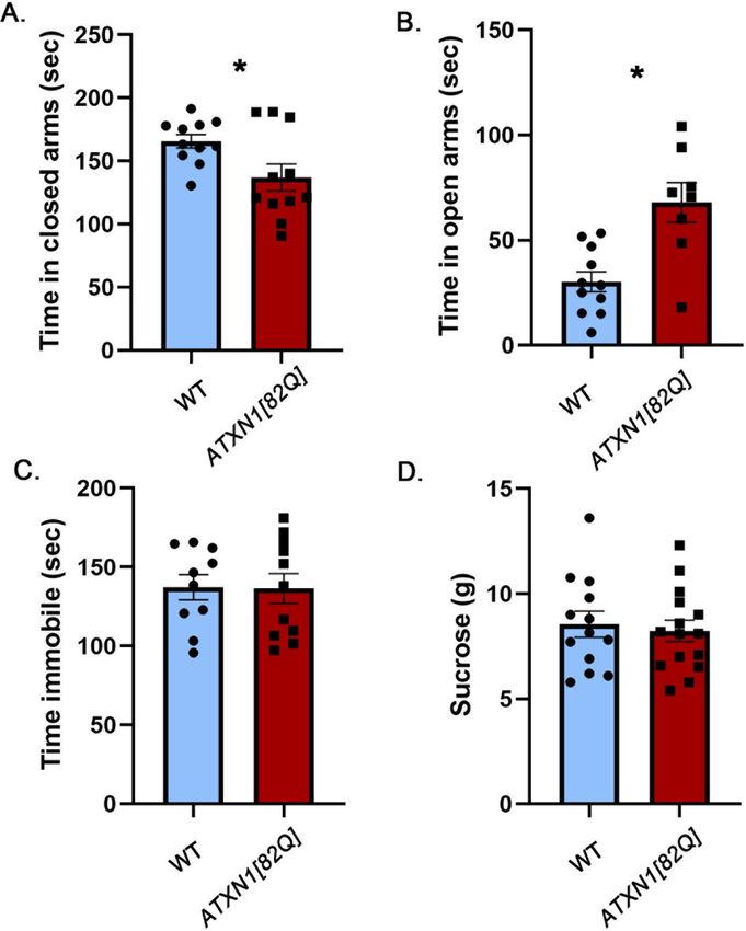
www.nature.com/scientificreports/
Figure 1. Atxn1154Q/2Q mice exhibit anxiety-like phenotype. 8–10 weeks old Atxn1154Q/2Q mice and their wild-
type littermates were tested on the Elevated Plus Maze, Forced Swim Test and Sucrose preference test. (A) Time
in closed and open arms. (B) Time spent immobile. (C) Amount of sucrose consumed. * indicates P < 0.05 using
unpaired, two-tailed t test, data are presented as mean ± SEM.
recognized21,22. Injury limited to the cerebellum can cause similar spectrum of cognitive and mood alterations,
including increased anxiety, and depression, termed Cerebellar Cognitive Affective Syndrome (CCAS)23,24. As
depression and anxiety impact the patient’s and their caregiver’s quality of life, it is important to understand their
causes so that comprehensive therapies can be developed to treat all aspects of S CA112.
Mouse models of SCA1 have been essential in implicating cerebellar pathology and gain-of-function mecha-
nisms in ataxia and cognitive deficits17,25–27. For instance, motor deficits in the transgenic SCA1 mouse line
ATXN1[82Q], which overexpress mutant ATXN1[82Q] protein selectively in cerebellar Purkinje cells, provided
support for the predominant role of cerebellar dysfunction in ataxia28. In addition, motor symptoms in two
SCA1 knock-in mouse lines (Atxn1154Q/2Q and Atxn178Q/2Q) correlate with the number of polyQ repeats, similar
to symptoms in patients with SCA126,29. Regarding the underlying molecular mechanism, the concept that polyQ
expansion causes ataxia largely through a toxic gain-of-function mechanism is supported by the lack of ataxia
ice27,30,31.
in Atxn1 null m
However, to date there has been no systematic investigation of mood in these mouse models. We wished
to determine (1) whether SCA1 mice exhibit mood alterations, (2) whether these alterations correlate with the
number of polyQ repeats, and whether (3) cerebellar pathology and (4) loss of ATXN1 function contribute to
mood alterations. This report represents our observations in four mouse lines whose performance on elevated
plus maze, forced swim test and sucrose preference was examined to address these questions.
Results
Ataxin‑1 knock‑in mice exhibit increased anxiety and sucrose consumption indicating that
mutant Ataxin‑1 induced pathogenesis may cause mood alterations. We first tested mood in
Atxn1154Q/2Q knock-in mice, in which one Atxn1 allele has a long CAG expansion (154 repeats)26, while the
other allele has 2 CAG repeats which is normal for wild-type mice. In these mice the mutant gene is under the
control of its endogenous promoter maintaining the brain wide expression of mutant ATXN1 at physiological
levels. Importantly, we started testing Atxn1154Q/2Q mice at 8 weeks of age, prior to detectable ataxia (at 12 weeks)
or failure to gain weight (11 weeks) to minimize potential interference of motor deficits or alterations in weight
with mood testing32.
The elevated plus maze (EPM) is a test used to assess anxiety-like responses of rodents33. The EPM relies upon
rodents’ proclivity toward dark, enclosed spaces and an unconditioned fear of heights/open spaces. It consists of
an elevated maze with four arms (two open and two enclosed) that are arranged to form a plus shape, and anxi-
ety behavior is assessed as increased time spent in the closed a rms33. Atxn1154Q/2Q mice spent more time in the
closed arms than their wild-type littermate controls (Fig. 1A left, 27.2% increase with 95% confidence interval of
(6.3%, 48.3%), wild-type (WT) average 158.3 ± 8.19 s, Atxn1154Q/2Q average 201.4 ± 15.96 s, P = 0.0138, unpaired,
two-tailed t-test, N = 17 WT and 11 Atxn1154Q/2Q mice), suggesting an anxiety-like phenotype. They also spent less
time in the open arms than their wild-type littermate controls but this difference was not statistically significant
(wild-type (WT) average 52.53 ± 13.52 s, Atxn1154Q/2Q average 40.26 ± 6.64 s, P = 0.3757, unpaired, two-tailed
t-test, N = 17 WT and 11 Atxn1154Q/2Q mice).
The forced swim test (FST) is commonly used for depression-like behavior. The FST involves the scoring
of active (swimming and climbing) versus passive (immobility) behavior when mice are forced to swim in a
cylinder from which there is no escape34. Atxn1154Q/2Q mice were indistinguishable from wild-type littermate
controls in immobility time (Fig. 1B, − 3.7% effect size with 95% confidence interval (− 17.8%, 10.4%), WT
average 156.6 ± 7.64 s, Atxn1154Q/2Q median 150.8 ± 6.7 s, P = 0.595 unpaired, two-tailed t-test, N = 17 WT and
12 Atxn1154Q/2Q mice).
Anhedonia is the inability to experience pleasure from rewarding or enjoyable activities and is a core symptom
of depression in humans. To measure anhedonia in mice, we used a sucrose preference test (SPT) based on a
Scientific Reports | (2021) 11:713 | https://doi.org/10.1038/s41598-020-80664-9 2
Vol:.(1234567890)
www.nature.com/scientificreports/
Figure 2. Atxn178/2Q mice exhibit increased sucrose consumption. 8–10 weeks old Atxn178Q/2Q mice and their
wild-type littermates were tested on the Elevated Plus Maze, Forced Swim Test and Sucrose preference test. (A)
Time in closed and open arms. (B) Time spent immobile. (C) Amount of sucrose consumed. * indicates P < 0.05
using unpaired, two-tailed t test, data are presented as mean ± SEM.
two-bottle choice paradigm in which mice were offered water in one bottle and sucrose in the other. SPT relies
on the ability of sucrose to act as a natural reward, and a reduction in sucrose preference indicates anhedonia and
depression35. However it is important to note that additional parameters, including alterations in metabolism36,
changes in weight or disruption of reward system37 may confound this interpretation. Contrary to our expecta-
tions, we found that Atxn1154Q/2Q mice consumed significantly more sucrose than wild-type littermates (Fig. 1C,
36.8% increase with 95% confidence interval (14.9%, 58.6%), WT average 9.46 ± 0.58 g, Atxn1154Q/2Q average
12.95 ± 0.84 g, N = 16 WT and 12 Atxn1154Q/2Q mice P = 0.0018, unpaired, two-tailed t-test), while water consump-
tion stayed the same (WT average 4.009 g, Atxn1154Q/2Q average 3.199 g, P = 0.6786). While this may be interpreted
as a lack of depression-like phenotype, alternatively mice may already exhibit alterations in m etabolism38 or
expression of genes or circuits important for the reward s ystem39,40 that affected their sucrose consumption. It
is also important to note that using anhedonia may not be the best way to evaluate depression in SCA1 as there
is no difference in depression prevalence between patients with SCA1 that have decreasing or increasing body
mass index (50% and 51.4% respectively)15.
There were no differences between female and male mice in any of the tests.
These results indicate that Atxn1154Q/2Q mice exhibit increased anxiety-like behavior but not a typical depres-
sive-like phenotype, as they had increased sucrose preference. They support the concept that anxiety in patients
with SCA1 may be caused, at least in part, by underlying brain pathology.
Anxiety‑like behavior correlates with number of polyQ repeats. The onset and severity of motor
deficits in patients with SCA1 correlate with the number of polyQ repeats in ATXN1, with higher numbers
of repeats causing earlier onset and more severe symptoms2. Two knock-in mouse models of SCA1 created
to express ATXN1 with 154Q and 78Q, Atxn1154Q/2Q and Atxn178Q/2Q respectively, exhibit a similar correla-
tion between the length of polyQ repeats and severity of motor deficits, albeit at longer polyQ repeats than
those found in humans. For example, onset of ataxia, as measured by decreased rotarod performance, occurs at
12 weeks in Atxn1154Q/2Q mice, while Atxn178Q/2Q mice show milder rotarod deficits only at 6 m
onths29. Therefore,
we next tested whether mood alterations in SCA1 knock-in mice similarly depend on the length of the polyQ
expansion by evaluating performance of Atxn178Q/2Q mice on mood tests.
There was no significant difference between Atxn178Q/2Q and wild-type mice on the elevated plus maze (Fig. 2A,
0.2% effect size with 95% confidence interval (− 11.5%, 11.8%), WT average time in closed arms = 122.3 ± 5.079 s,
Atxn178Q/2Q average time in closed arms = 122.5 ± 4.718 s, N = 22 WT and 19 Atxn178Q/2Q mice, P = 0.9724,
unpaired, two-tailed t-test, WT average time in open arms = 51.57 ± 6.73 s, Atxn178Q/2Q = 44.56 ± 4.5 s, P = 0.3765,
unpaired, two-tailed t-test), suggesting that knock-in SCA1 mice expressing mutant ATXN1 with fewer polyQ
repeats do not exhibit an anxiety-like phenotype at this age.
Atxn178Q/2Q mice were also indistinguishable from WT mice on the forced swim test (Fig. 2B, − 0.4% effect
size with 95% confidence interval (− 17.4%, 16.7%), WT average time immobile = 117.6 ± 6.13 s, Atxn178Q/2Q
average time immobile = 117.2 ± 7.73 s, N = 19 WT and 20 Atxn178Q/2Q mice, Student’s unpaired two-tailed t-test
P = 0.9661).
However, Atxn178Q/2Q mice consumed more sucrose than wild-type littermate controls (Fig. 2C, 24.4% effect
size with 95% confidence interval (4%, 44.8%), WT average 10.23 ± 0.52 g, Atxn178Q/2Q average 12.7 ± 0.38 g,
N = 23 WT and 20 Atxn178Q/2Q mice, P = 0.0194, unpaired, two-tailed t-test) with no statistically significant dif-
ference in water consumption (WT average 5.21 g, Atxn178Q/2Q average 6.13 g, P = 0.5498). This suggests that
increased sucrose consumption, unlike other features of the Atxn1154Q/2Q model, can be caused by the shorter
polyglutamine tract.
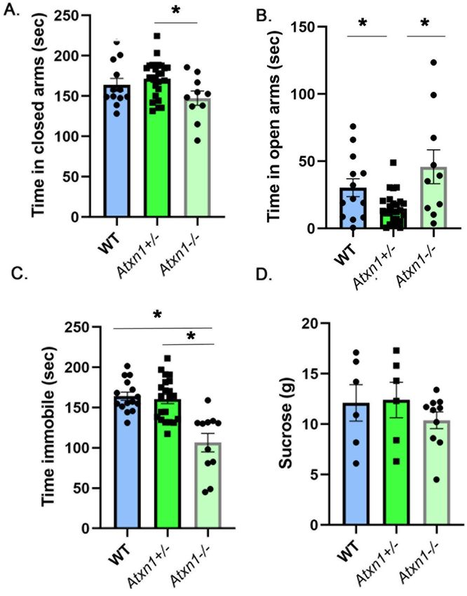
Purkinje neuron specific transgenic SCA1 mice exhibit reduced anxiety. One of the hallmarks of
SCA1 is pronounced cerebellar pathology, with significant loss of Purkinje neurons in the cerebellar cortex41.
Scientific Reports | (2021) 11:713 | https://doi.org/10.1038/s41598-020-80664-9 3
Vol.:(0123456789)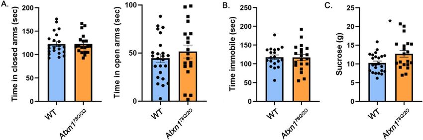
www.nature.com/scientificreports/
Figure 3. Cerebellar Purkinje cell-specific ATXN1[82Q] transgenic mice have attenuated anxiety. 8–10 weeks
old ATXN1[82] mice and their wild-type littermates were tested on the Elevated Plus Maze, Forced Swim Test
and Sucrose preference test. (A) Time in closed arms. (B) Time in open arms. (C) Time spent immobile. (D)
Amount of sucrose consumed. * indicates P < 0.05 using the unpaired, two-tailed t test, data are presented as
mean ± SEM.
Increasing evidence supports the role of the cerebellum in mood in addition to its role in motor c ontrol42. For
example, patients with the cerebellar injury exhibit disinhibition and other alterations in mood, collectively
termed Cerebellar Cognitive Affective Syndrome (CCAS)23,43.
To investigate the contribution of cerebellar pathology to the mood alterations caused by mutant ATXN1, we
examined an SCA1 transgenic mouse model, ATXN1[82Q] mice, which overexpress mutant ATXN1[82Q] only
romoter28.
in cerebellar Purkinje cells under the control of the Purkinje cell protein 2 (Pcp2) p
Surprisingly, we found a striking reduction in anxiety-like behavior in the ATXN1[82Q] mice on the elevated
plus maze: they spent significantly less time in the closed arms (Fig. 3A, − 17.3% effect size with 95% confidence
interval (− 32.5%, − 2.3%), WT average 165.6 ± 5.257 s, ATXN1[82Q] average 137 ± 10.58 s, N = 11 WT and 8
ATXN1[82Q] mice, P = 0.0253, unpaired, two-tailed t-test). This was accompanied by increased time in the open
arms, another sign of decreased anxiety (Fig. 3B, 125% effect size with 95% confidence interval (56.9%, 193%),
WT average 30.22 ± 4.745 s, ATXN1[82Q] average 67.99 ± 9.448 s, P = 0.0012, unpaired, two-tailed t-test).
We did not observe any differences between ATXN1[82Q] and wild-type mice in either the forced swim
test (Fig. 3C, − 0.5% effect size with 95% confidence interval (− 19.6%, 18.5%), WT average 137.3 ± 8.064 s,
ATXN1[82Q] average 136.5 ± 9.38 s, N = 11 WT and 10 ATXN1[82Q] mice, P = 0.9516, unpaired, two-tailed t-test)
or the sucrose preference test (Fig. 3D, − 3.75% effect size with 95% confidence interval (− 22.7%, 15.2%), WT
Scientific Reports | (2021) 11:713 | https://doi.org/10.1038/s41598-020-80664-9 4
Vol:.(1234567890)www.nature.com/scientificreports/
average 8.55 ± 0.6187 g, ATXN1[82Q] average 8.523 ± 0.5037 g, N = 13 WT and 15 ATXN1[82Q] mice, P = 0.6882,
unpaired, two-tailed t-test).
To test whether attenuated anxiety ATXN1[82Q] mice is caused by a reduced stress-induction, we examined
both basal and restraint stress-induced blood corticosterone levels in ATXN1[82Q] mice and wild-type lit-
termate controls. We found a trend towards decreased basal and stress-induced corticosterone levels, although
this difference was not statistically significant (Fig. 4, basal WT average 21.81 ± 6.64, basal ATXN1[82Q] average
12.55 ± 3.27, stress induced WT average 293 ± 23.54, stress-induced ATXN1[82Q] average 248 ± 28.35, two way
ANOVA significant effect of stress P < 0.0001 but not of genotype).
These results indicate that cerebellar dysfunction may attenuate anxiety in mice, akin to disinhibition seen
in patients with cerebellar injury. Moreover, these results are in contrast to the increased anxiety seen in SCA1
knock-in mice with brain-wide expression of mutant polyQ ATXN1, suggesting that non-cerebellar pathology
plays a dominant role in SCA1-like anxiety.
Partial and full loss of ataxin‑1 cause mood alterations in mice. PolyQ expansion in ATXN1 leads
to both gain and loss of ATXN1 function44,45. Gain-of-function is the predominant molecular mechanism of
motor deficits in SCA1, as supported by the lack of a strong motor phenotype in Atxn1 null m ice25,27, while loss-
of-function may contribute to cognitive deficits17,27. We therefore wanted to determine whether loss of ataxin-1
alters mood in Atxn1−/− mice. Additionally, patients with SCA1 have one normal and one mutant allele, and a
simple loss-of-function molecular mechanism would result in an approximately 50% decrease in ATXN1 func-
tion. To examine the effects of an approximately 50% loss of ataxin-1 function we also analyzed mood in het-
erozygous knockout (Atxn1+/−) mice.
On the elevated plus maze, heterozygous Atxn1+/− mice spent slightly more time in the closed arms than
wild-type or null mice (Fig. 5A, 9.45% and 16.4% effect sizes with 95% confidence intervals (− 3.2%, 21.3%)
and (2.3%, 31.3%) respectively comparing Atxn1+/− to WT and Atxn1-/− mice, WT mice average 156.3 ± 9.19 s,
Atxn1+/− average 171.6 ± 4.97 s, Atxn1−/− average 147.4 ± 5.72 s, N = 13 WT, 26 Atxn1+/−, and 10 Atxn1−/− mice,
One-way ANOVA, F (2,44) = 3.34, P = 0.044) but this difference was statistically significant only when compared
to null mice (P = 0.14 and P = 0.034 One-way ANOVA followed by post-hoc Tukey’s multiple comparison test
comparing Atxn1+/− to WT and Atxn1−/− mice respectively). Additionally, Atxn1+/− mice spent less time in the
open arms than wild-type or null mice (Fig. 5B, − 104.4% and − 210% effect sizes with 95% confidence intervals
(− 180.80%, − 29.3%) and (− 349%, − 70%) respectively comparing Atxn1+/− to WT and Atxn1−/− mice, WT mice
average 30.2 ± 6 s, Atxn1+/− average 14.81 ± 2.21 s, Atxn1−/− average 45.92 ± 12 s, F(2,44) = 7.04, P = 0.0021, One-
way ANOVA followed by post-hoc Tukey’s multiple comparison tests P = 0.01 and 0.022 comparing Atxn1+/− to
WT and Atxn1−/− mice). These results indicate an increased anxiety-like phenotype with 50% loss of ATXN1.
Full loss of ATXN1 resulted in slightly decreased anxiety in Atxn1 null mice compared to wild-type litter-
mates, i.e. increased time in open arms (effects size of 34.8% with 95% confidence interval (− 13.4%, 81.6%),
WT mice average 30.2 ± 6 s, Atxn1−/− average 45.92 ± 12 s) and decreased time in closed arms (− 10.2% effect size
with 95% confidence interval (− 5.6%, 25.9%), WT mice average 156.3 ± 9.19 s, Atxn1−/− average 147.4 ± 5.72 s),
but this was not statistically significant (one-way ANOVA with post-hoc Tukey’s test, P = 0.2745, and P = 0.2476
respectively).
This decrease in inhibition is consistent with the forced swim test, where Atxn1 null mice were significantly
less immobile (Fig. 5C, − 54.1% and − 50.8% effect sizes with 95% confidence intervals (− 77.4%, − 30.1%) and
(− 72.7%, − 28.9%) respectively comparing Atxn1−/− to WT and Atxn1+/− mice, WT average 164.2 ± 5.093 s,
Atxn1+/− average 160.6 ± 5.73 s, Atxn1−/− average 106.5 ± 11.39 s, N = 15 WT, 21 Atxn1+/−, and 11 Atxn1−/− mice,
one-way ANOVA F(2,44) = 17.17, followed by post-hoc Tukey’s multiple comparison tests P < 0.0001 for Atxn1−/− vs.
both WT and Atxn1+/−).
While Atxn1−/− mice consumed slightly less sucrose on average (Fig. 5D, − 16.5% and − 19.36% effect sizes with
95% confidence intervals (− 60.3%, 26.9%) and (− 62.9%, 24.1%), WT mice 12.12 ± 1.81 g, Atxn1+/− 12.4 ± 1.75 g,
Atxn1−/− 10.39 ± 0.83 g) these differences were not significant (one-way ANOVAF(2,19) = 0.687, P = 0.51, followed
by post-hoc Tukey’s multiple comparison tests P = 0.6369 and P = 0.5461 respectively for Atxn1−/− vs. WT and
Atxn1+/−).
Together these results indicate that full and partial loss of ATXN1 alter mood phenotype in mice in opposite
ways, with partial loss leading to increased anxiety and full loss resulting in decreased anxiety.
Discussion
In order to gain insight into the etiology of SCA1 mood alterations, we systematically (e.g. using the same tests
on the same genetic background at 8 weeks of age) investigated how mood in mice was affected by global or
cerebellum-limited expression of mutant ATXN1, polyQ length of ATXN1, and loss of ATXN1. In order to mini-
mize the confounding effect of impaired movement on behavioral tests, we have chosen 8 weeks as the testing
age because it precedes motor deficits. In addition, there is no detectable neurodegeneration at this age27,29,32,46–48
(e.g. reproducible dendritic atrophy and neuronal loss in the cerebellum are detected at 12 weeks and 24 weeks
respectively in Atxn1154Q/2Q and ATXN1[82Q] mice, whereas neuronal loss in the hippocampus is seen at 5 months
in Atxn1154Q/2Q mice. No neurodegeneration has been described in Atxn1−/− and Atxn178Q/2Q lines).
We found that Atxn1154Q/2Q mice, an SCA1 knock-in line with global expression of mutant ATXN1, exhibit
an anxiety-like phenotype on the elevated plus maze. This suggests that anxiety seen in patients with SCA1 may
be, at least in part, caused by underlying pathogenesis from mutant ATXN1.
This anxiety-like phenotype, like motor and cognitive deficits, depends on the length of the polyQ tract: we
did not observe increased anxiety in Atxn178Q/2Q mice, another SCA1 knock-in line that globally expresses mutant
ATXN1 with fewer polyQ repeats.
Scientific Reports | (2021) 11:713 | https://doi.org/10.1038/s41598-020-80664-9 5
Vol.:(0123456789)www.nature.com/scientificreports/
Figure 4. Basal and stress-induced blood corticosterone levels in ATXN1[82Q] and wild-type control mice.
Corticosterone levels were determined in total plasma from 7 pairs of ATXN1[82Q] mice and littermate WT
controls. Basal/minimally stressed plasma was obtained in the morning (9–12 am) at least three days before
stress induction. Stress level blood samples were collected after 15 min of restraint stress. * indicates P < 0.05
using the unpaired, two-tailed t test, data are presented as mean ± SEM.
Surprisingly, both lines of SCA1 knock-in mice consumed more sucrose than their wild-type littermate
controls in the sucrose preference test—the opposite of what we would expect from a depressive-like phenotype.
There are several possible explanations. First, SCA1 knock-in mice and some patients with SCA1 demonstrate
weight loss and increased metabolic consumption15,26. Thus, increased sucrose preference may be a way for the
SCA1 knock-in mice to meet their caloric needs. Alternatively, studies have also implicated the cerebellum, and
in particular granule neurons, in the encoding of reward in mice49, suggesting that pathological changes caused by
expression of mutant ATXN1 in granule neurons may alter the reward properties of sucrose. Patients with ataxia
have demonstrated increased impulsivity and compulsivity which correlated well with d epression50 indicating
that cerebellum contributes to reward processing in humans as w ell51,52. The effect of mutant ATXN1 on the role
of the cerebellum in regulating mood and encoding reward may account for the unexpected behavior of SCA1
knock-in mice in the sucrose preference test.
Knock-in mice are genetically similar to patients with SCA1 in that they have one normal and one expanded
allele and have physiological levels and a physiological spatial and temporal pattern of mutant ATXN1 expres-
sion. However, symptoms in mice only occur at very long repeat lengths, whereas in human patients over 39
uninterrupted polyQ repeats causes disease. The longest repeat length reported in a human is 82Q, in a patient
with juvenile SCA1 that is more severe than adult form of disease. While the longer polyQ requirement in mice
is likely due to their shorter lifespan and other species differences, an alternative interpretation is that knock-in
mouse models of SCA1 more accurately model juvenile form of SCA1. This would mean that any findings in
these mice, including our results presented here, might have translational value mostly for the juvenile form of
SCA1. In addition, the 154Q allele is longer than any found in humans, and the larger polyQ region could affect
the ATXN1 protein function in additional, unpredictable ways.
In addition to its well-described role in motor control, the cerebellum also contributes to regulation of cogni-
tion and affect53. However, in contrast to SCA1 motor and cognitive deficits, the anxiety-like phenotype seen in
Atxn1154Q/2Q mice does not seem to be caused by cerebellar pathology. In fact, ATXN1[82Q] mice-SCA1 trans-
genic line that overexpresses mutant ATXN1 only in cerebellar Purkinje cells-exhibited attenuated anxiety. As
levels of light can influence performance in EPM33, it is possible that increasing the light would prevent/moderate
this phenotype. However, similarly attenuated anxiety is reported in Lurcher mice, another mouse model in which
cerebellar Purkinje cells are a ffected54. In Lurcher mice, the attenuation of anxiety-like behavior was accompanied
by an increase in stress-induced blood corticosterone levels, suggesting that attenuated anxiety may be actually
caused by increased stress caused by t esting36. However, this does not seem to be the case in ATXN1[82Q] mice
as we have found that both basal and stress-induced blood corticosterone levels are decreased in ATXN1[82Q]
mice, although this difference was not statistically significant. The attenuation of anxiety in ATXN1[82Q] mice
further supports the finding that cerebellar damage can lead to disinhibition, reminiscent of the human cerebellar
cognitive affective syndrome (CCAS), which leads to disinhibition, impulsiveness and risky b ehavior23.
Purkinje cell limited overexpression of mutant ATXN1 attenuated anxiety in transgenic ATXN1[82Q] mice,
whereas global expression of mutant ATXN1 in knock-in mice increased anxiety. While interpreting these results,
it is important to keep in mind differences in the levels and patterns of mutant ATXN1 expression. Atxn1154Q/2Q
mice express mutant ATXN1 at physiologically-relevant levels under the endogenous promotor, while trans-
genic ATXN1[82Q] mice overexpress mutant ATXN1 protein ~ 30–60 f old25, which likely accounts for the more
severe Purkinje cell and cerebellar pathology in transgenic mice compared to knock-in Atxn1154Q/2Q mice55. This
Scientific Reports | (2021) 11:713 | https://doi.org/10.1038/s41598-020-80664-9 6
Vol:.(1234567890)www.nature.com/scientificreports/
Figure 5. Decreased immobility in homozygous (Atxn1−/−) and increased anxiety in heterozygous (Atxn1+/-)
mice. 8–10 weeks old wild-type, Atxn1+/−, and Atxn1−/− mice were tested on the Elevated Plus Maze, Forced
Swim Test and Sucrose preference test. (A) Time in closed arms. (B) Time in open arms. (C) Time spent
immobile. (D) Amount of sucrose consumed. * indicates P < 0.05 using the One way ANOVA followed by post-
hoc Tukey’s testing, data are presented as mean ± SEM.
severe Purkinje cell dysfunction may explain the attenuated anxiety in ATXN1[82Q] mice. On the other hand,
the opposite finding of increased anxiety in Atxn1154Q/2Q mice and altered sucrose preference in both Atxn178Q/2Q
and Atxn1154Q/2Q lines may suggest that dysfunction and/or pathology outside the cerebellar cortex contribute to
mood alterations in SCA1 knock-in mice. ATXN1 is expressed throughout the brain, including hippocampus,
cortex, cerebellum, and hypothalamus, and it is possible that pathology caused by polyQ expanded ATXN1 in any
of these regions contributes to SCA1-like mood alterations16,48,56. Indeed, a recent study implicated hippocampal
dysfunction in neurobehavioral deficits described in SCA1 knock-in m ice57. Future studies using conditional
SCA1 knock-in mice or viral-mediated approaches, in which polyQ ATXN1 can be selectively expressed or
deleted in distinct brain regions, will be able to identify the anatomical substrates of mood alterations in SCA1.
PolyQ expansion causes both gain and loss of ATXN1 function44,45. ATXN1 gain-of-function is thought
to underlie cerebellar neurodegenerative changes in SCA131, while ATXN1 loss-of-function—in particular in
complex with the transcriptional repressor Capicua (CIC)—causes a spectrum of neurobehavioral phenotypes
in mice and humans16. Our forced swim test results support and build upon the concept that loss of ATXN1
function causes mood alterations in mice, with decreased immobility in the full knock-out mice. In contrast,
the increased sucrose preference seen in global SCA1 knock-in SCA1 lines is likely due to a gain-of-function, as
we did not detect it in the ATXN1 null mice.
Scientific Reports | (2021) 11:713 | https://doi.org/10.1038/s41598-020-80664-9 7
Vol.:(0123456789)www.nature.com/scientificreports/
The effect of ATXN1 loss-of-function on anxiety in the elevated plus maze was more difficult to interpret—
a 50% decrease in ATXN1 expression resulted in a subtle anxiety-like phenotype in Atxn1+/− mice, while full
knock-out of ATXN1 slightly attenuated anxiety. Looking at the ranges covered by the 95% confidence intervals,
our results can support two main interpretations: either there is little to no effect of ATXN1 loss of function on
anxiety, or 50% and 100% loss of function cause opposite effects. The latter interpretation is consistent with a
recent study by Lu et al., which suggests that disrupting the ATXN1-CIC complex in development can cause
hyperactivity and decreased anxiety16. Together with that study, our results support the concept that complete loss
of ATXN1 causes developmental abnormalities that result in the hyperactivity and attenuated anxiety in mice.
There is evidence that this may also be the case in humans, as large deletions on chromosome 6p22 that span
ATXN1 gene have been reported in human patients that exhibit autism spectrum disorder (ASD), and attention
deficit hyperactivity disorder (ADHD)58,59.
On the other hand, 50% loss of ATXN1 function may be well tolerated during development but cause neu-
robehavioral problems, including anxiety-like behavior, in adulthood. This is consistent with recent studies indi-
cating loss of ATXN1 as a risk factor for Alzheimer’s disease and showing that a decrease in ATXN1 contributes
to Alzheimer’s disease pathology60,61. Future studies using conditional and constitutive knock-outs of ATXN1
would be able to address these questions.
In conclusion, we have found alterations in mood in existing SCA1 mouse lines, suggesting that mutant
ATXN1 pathology may contribute to anxiety and depression seen in patients with SCA1.
Methods
Mice. Atxn1−/− mice27 on a C57/Bl6 background were a gift from the laboratory of Dr. Huda Zoghbi.
Atxn1154Q/2Q, Atxn178Q/2Q and ATXN1[82Q] mice were a gift from the laboratory of Dr. Harry Orr. Atxn1154Q/2Q
mice are on C57/Bl6 background, while Atxn178Q/2Q and ATXN1[82Q] mice were originally on FVB background.
To minimize the effects of genetic background, Atxn178Q/2Q and ATXN1[82Q] mice were backcrossed onto a
C57/Bl6 background. Mice were housed in a temperature- and humidity-controlled room on a 12 h light/12 h
dark cycle with access to food and water ad libitum. 8- to 10-week-old mice went through a battery of tests, with
the same order of testing for all mice (in order: elevated plus maze, forced swim test, sucrose preference). When-
ever possible we tried to ensure similar numbers of male and female mice (Atxn1154Q/2Q (F:M = 5:8), Atxn178Q/2Q
(F:M = 8:12), ATXN1[82Q] (F:M = 5:8), Atxn1−/− (F:M = 5:6)).
All animal experiments were performed in ethical compliance with the National Institutes of Health’s Guide
for the Care and Use of Laboratory Animals and were approved by the ethical University of Minnesota Institu-
tional Animal Care and Use Committee (protocol number 1810-36435A).
Corticosterone measurements. Corticosterone concentration was determined in total plasma from 7
pairs of ATXN1[82Q] mice and littermate WT controls (F:M = 5:2). Basal/minimally stressed corticosterone con-
trol measurements were obtained through tail clip bleed procedure in the morning (9–12 am) at least three days
before stress induction. We collected samples in the morning due to lower corticosterone levels, and kept the
time consistent between groups. After mice were transferred into the procedure room, we collected blood using
tail snip within ~ 3 min using EDTA-coated capillary tubes that were then centrifuged for 25 min at 14.000 rpm
at 4 °C. Plasma was diluted 1:100 using diluent supplied in Corticosterone Double Antibody RIA Kit (MP Bio-
medicals). Mice were allowed three days to recover from stress before stress-level blood samples were collected.
Stress level blood samples were collected after 15 min of restraint stress. We collected blood after decapita-
tion. After centrifugation, plasma was diluted 1:200 in diluent. Corticosterone levels were determined by I125
radioimmunoassay using the Corticosterone Double Antibody RIA Kit in duplicate.
Mood testing. Elevated plus maze. The maze was approximately 76 cm across and consisted of two open
arms (6.3 cm × 34.2 cm) and two closed arms (6.3 cm × 34.2 cm with 19 cm tall opaque walls) extending from a
6.3 cm × 6.3 cm center area, raised 96 cm above the floor. The surrounding room was dark and the maze was lit
by overhead lights. Light intensity in the open arms was 45–50 lx, and in the closed arms was 3–6 lx. Each mouse
was placed in the center of the maze facing an open arm and allowed to explore for 5 min. During this time, the
mice were tracked automatically using AnyMaze software and the time spent in each zone (open arms, closed
arms, center area) was recorded. The maze was cleaned with 70% ethanol before each m ouse62.
Forced swim test. Mice were placed in a transparent cylindrical container (diameter 18.4 cm, height 23.5 cm)
filled to a depth of approximately 15 cm with water at 24–26 °C. They swam for 6 min before being removed from
the water and placed in a cage containing absorbent material on a heating pad to dry off. Time immobile was
tracked automatically using AnyMaze software (immobility was defined as a period of 85% or greater immobility
lasting 250 ms or longer). The time spent immobile during the last 4 min of the test is a nalyzed62.
Sucrose preference test. Mice were singly housed in cages with cotton nestlets in order to allow for accurate
measurement of each mouse’s water and sucrose consumption. Mice were acclimated to solitary housing for
3 days before the test. On the first day of testing, each mouse was given two bottles of water to test for any bias
towards one side of the cage. On each day following this, each mouse was given one bottle containing water and
another one containing either 2% or 4% sucrose. Bottles were weighed every 24 h to measure the amount of
liquid consumed. Results for the 4% sucrose day, for which we saw the greatest differences between groups, are
reported62.
All the results where we experienced technical difficulties, including poor video tracking during the EPM or
FST, and leaking bottles during the sucrose preference test, were excluded from the final analysis.
Scientific Reports | (2021) 11:713 | https://doi.org/10.1038/s41598-020-80664-9 8
Vol:.(1234567890)www.nature.com/scientificreports/
Statistical analysis. Wherever possible, sample sizes were calculated using power analyses based on the
standard deviations from our previous studies, significance level of 5%, and power of 90%. Statistical tests were
performed with GraphPad Prism. Data was analyzed using t-test or One-way ANOVA followed by post-hoc
Tukey’s testing, and estimation statistics to present effect sizes and 95% confidence intervals.
Data availability
All data will be freely available from authors upon request.
Received: 28 June 2020; Accepted: 18 December 2020
References
1. Orr, H. T. et al. Expansion of an unstable trinucleotide CAG repeat in spinocerebellar ataxia type 1. Nat. Genet. 4(3), 221–226
(1993).
2. Zoghbi, H. Y. et al. Spinocerebellar ataxia : variable age of onset and linkage to human leukocyte antigen in a large kindred. Ann.
Neurol. 23(6), 580–584 (1988).
3. Paulson, H. L., Shakkottai, V. G., Clark, H. B. & Orr, H. T. Polyglutamine spinocerebellar ataxias—from genes to potential treat-
ments. Nat. Rev. Neurosci. 18(10), 613–626 (2017).
4. Matilla-Dueñas, A., Goold, R. & Giunti, P. Clinical, genetic, molecular, and pathophysiological insights into spinocerebellar ataxia
type 1. Cerebellum 7(2), 106–114 (2008).
5. Bürk, K. et al. Cognitive deficits in spinocerebellar ataxia type 1, 2, and 3. J. Neurol. 250(2), 207–211 (2003).
6. Klinke, I. et al. Neuropsychological features of patients with spinocerebellar ataxia (SCA) types 1, 2, 3, and 6. Cerebellum 9(3),
433–442 (2010).
7. Fancellu, R. et al. Longitudinal study of cognitive and psychiatric functions in spinocerebellar ataxia types 1 and 2. J. Neurol.
260(12), 3134–3143 (2013).
8. Lo, R. Y. et al. Depression and clinical progression in spinocerebellar ataxias. Park. Relat. Disord. 22, 87–92 (2016).
9. Schmitz-Hubsch, T. et al. Self-rated health status in spinocerebellar ataxia—results from a European multicenter study. Mov. Disord.
25(5), 587–595 (2010).
10. Schmitz-hu, T. et al. Depression comorbidity in spinocerebellar ataxia. Mov. Disord. 26(5), 870–876 (2011).
11. Jacobi, H. et al. Long-term evolution of patient-reported outcome measures in spinocerebellar ataxias. J. Neurol. 265(9), 2040–2051
(2018).
12. Diallo, A. et al. Prediction of survival with long-term disease progression in most common spinocerebellar ataxia. Mov. Disord.
34(8), 1220–1227 (2019).
13. Kawai, M., Suenaga, Y., Watanabe, G. & Sobue, H. Cognitive impairment in spinocerebellar degeneration. Eur. Neurol. 61, 257–268
(2009).
14. Luo, L. et al. The initial symptom and motor progression in spinocerebellar ataxias. Cerebellum 16(3), 615–622 (2017).
15. Diallo, A. et al. Body mass index decline is related to spinocerebellar ataxia disease progression. Mov. Disord. 4(5), 689–697 (2017).
16. Lu, H. et al. Disruption of the ATXN1—CIC complex causes a spectrum of neurobehavioral phenotypes in mice and humans. Nat.
Genet. Gr. 49(4), 527–536 (2017).
17. Asher, M. et al. Cerebellar contribution to the cognitive alterations in SCA1: evidence from mouse models. Hum. Mol. Genet.
29(1), 117–131 (2020).
18. Friedrich, J. et al. Antisense oligonucleotide–mediated ataxin-1 reduction prolongs survival in SCA1 mice and reveals disease-
associated transcriptome profiles. J. Clin. Investig. 3(21), e123193 (2018).
19. Jacobi, H. et al. Biological and clinical characteristics of individuals at risk for spinocerebellar ataxia types 1, 2, 3, and 6 in the
longitudinal RISCA study: analysis of baseline data. Lancet Neurol. 12(7), 650–658 (2013).
20. Rüb, U. et al. Clinical features, neurogenetics and neuropathology of the polyglutamine spinocerebellar ataxias type 1, 2, 3, 6 and
7. Prog. Neurobiol. 104, 38–66 (2013).
21. Schmahmann, J. D., Guell, X., Stoodley, C. J. & Halko, M. A. The theory and neuroscience of cerebellar cognition. Annu. Rev.
Neurosci. 42, 337–364 (2019).
22. Kansal, K. et al. Structural cerebellar correlates of cognitive and motor dysfunctions in cerebellar degeneration. Brain 140, 707–720
(2017).
23. Schmahmann, J. D. & Sherman, J. C. The cerebellar cognitive affective syndrome. Brain 121(4), 561–579 (1998).
24. Liszewski, C. et al. Cognitive impairment and psychiatric symptoms in 133 patients with diseases associated with cerebellar
degeneration. J. Neuropsychiatry Clin. Neurosci. 16, 109–112 (2004).
25. Burright, E. N. et al. SCA1 transgenic mice: a model for neurodegeneration caused by an expanded CAG trinucleotide repeat. Cell
82(6), 937–948 (1995).
26. Watase, K. et al. A long CAG repeat in the mouse Sca1 locus replicates SCA1 features and reveals the impact of protein solubility
on selective neurodegeneration. Neuron 34(6), 905–919 (2002).
27. Matilla, A. et al. Mice lacking ataxin-1 display learning deficits and decreased hippocampal paired-pulse facilitation. J. Neurosci.
18(14), 5508–5516 (1998).
28. Clark, H. B. et al. Purkinje cell expression of a mutant allele of SCA1 in transgenic mice leads to disparate effects on motor behav-
iors, followed by a progressive cerebellar dysfunction and histological alterations. J. Neurosci. 17(19), 7385–7395 (1997).
29. Lorenzetti, D. et al. Repeat instability and motor incoordination in mice with a targeted expanded CAG repeat in the Sca1 locus.
Hum. Mol. Genet. 9(5), 779–785 (2000).
30. Zoghbi, H. Y. & Orr, H. T. Pathogenic mechanisms of a polyglutamine-mediated neurodegenerative disease, Spinocerebellar ataxia
type 1. J. Biol. Chem. 284(12), 7425–7429 (2009).
31. Rousseaux, M. W. C. et al. ATXN1-CIC complex is the primary driver of cerebellar pathology in spinocerebellar ataxia type 1
through a gain-of-function mechanism. Neuron 97(6), 1235–1243 (2018).
32. Cvetanovic, M., Patel, J. M., Marti, H. H., Kini, A. R. & Opal, P. Vascular endothelial growth factor ameliorates the ataxic phenotype
in a mouse model of spinocerebellar ataxia type 1. Nat. Med. 17(11), 1445–1447 (2011).
33. Walf, A. & Frye, C. The use of the elevated plus maze as an assay of anxiety-related behavior in rodents. Nat. Protoc. 2(2), 322–328
(2007).
34. Slattery, D. A. & Cryan, J. F. Using the rat forced swim test to assess antidepressant-like activity in rodents. Nat. Protoc. 7(6),
1009–1014 (2012).
35. Strekalova, T., Spanagel, R., Bartsch, D., Henn, F. A. & Gass, P. Stress-induced anhedonia in mice is associated with deficits in
forced swimming and exploration. Neuropsychopharmacology 29, 2007–2017 (2004).
36. Duca, F. A., Swartz, T. D. & Covasa, M. Effect of diet on preference and intake of sucrose in obese prone and resistant rats. PLoS
ONE 9(10), 1–8 (2014).
Scientific Reports | (2021) 11:713 | https://doi.org/10.1038/s41598-020-80664-9 9
Vol.:(0123456789)www.nature.com/scientificreports/
37. Avena, N. M. & Hoebel, B. G. Amphetamine-sensitized rats show sugar-induced hyperactivity ( cross-sensitization ) and sugar
hyperphagia. Pharmacol. Biochem. Behav. 74, 635–639 (2003).
38. Stucki, D. M. et al. Mitochondrial impairments contribute to Spinocerebellar ataxia type 1 progression and can be ameliorated by
the mitochondria-targeted antioxidant MitoQ. Free Radic. Biol. Med. 97, 427–440 (2016).
39. Goold, R. et al. Down-regulation of the dopamine receptor D2 in mice lacking ataxin 1. Hum. Mol. Genet. 16(17), 2122–2134
(2007).
40. Carta, I., Chen, C. H., Schott, A. L., Dorizan, S. & Khodakhah, K. Cerebellar modulation of the reward circuitry and social behavior.
Science https://doi.org/10.1126/science.aav0581 (2019).
41. Seidel, K. et al. Brain pathology of spinocerebellar ataxias. Acta Neuropathol. 124(1), 1–21 (2012).
42. Schmahmann, J. D. The role of the cerebellum in cognition and emotion: personal reflections since 1982 on the dysmetria of
thought hypothesis, and its historical evolution from theory to therapy. Neuropsychol. Rev. 20(3), 236–260 (2010).
43. Van Den, B. N. S. Impairments in emotion recognition and risk-takingbehavior after isolated, cerebellar stroke. Cerebellum 19,
419–425 (2020).
44. Lim, J. et al. Opposing effects of polyglutamine expansion on native protein complexes contribute to SCA1. Nature 452(7188),
713–718 (2008).
45. Crespo-Barreto, J., Fryer, J. D., Shaw, C. A., Orr, H. T. & Zoghbi, H. Y. Partial loss of ataxin-1 function contributes to transcriptional
dysregulation in spinocerebellar ataxia type 1 pathogenesis. PLoS Genet. 6(7), 1–17 (2010).
46. Kim, J. H., Lukowicz, A., Qu, W., Johnson, A. & Cvetanovic, M. Astroglia contribute to the pathogenesis of spinocerebellar ataxia
Type 1 (SCA1) in a biphasic, stage-of-disease specific manner. Glia 66(9), 1972–1987 (2018).
47. Cvetanovic, M., Hu, Y.-S. & Opal, P. Mutant ataxin-1 inhibits neural progenitor cell proliferation in SCA1. Cerebellum 16(2),
340–347 (2017).
48. Suh, J. et al. Loss of ataxin-1 potentiates Alzheimer ’ s pathogenesis by elevating cerebral BACE1 transcription. Cell 178, 1159–1175
(2019).
49. Wagner, M. J., Kim, T. H., Savall, J., Schnitzer, M. J. & Luo, L. Cerebellar granule cells encode the expectation of reward. Nature
544(7648), 96–100 (2017).
50. Amokrane, N. et al. Impulsivity in cerebellar ataxias: testing the cerebellar reward hypothesis in humans. Mov. Disord. 35(8),
1491–1493 (2020).
51. Miquel, M., Nicola, S. M., Gil-miravet, I., Guarque-chabrera, J. & Stanton, M. A working hypothesis for the role of the cerebellum
in impulsivity and compulsivity. Front. Behav. Neurosci. 3, 1–14 (2019).
52. Vazquez-Sanroman, D. et al. The cerebellum on cocaine: plasticity and metaplasticity. Addict. Biol. 20, 941–955 (2015).
53. Koziol, L. F. et al. Consensus paper : the cerebellum ’s role in movement and cognition. Cerebellum 13(1), 151–177 (2014).
54. Monnier, C. & Lalonde, R. Elevated + -maze and hole-board exploration in lurcher mutant mice. Brain Res. 702, 169–172 (1995).
55. Emir, U. E., Clark, H. B., Vollmers, M. L., Eberly, L. E. & Ӧz, G. Non-invasive detection of neurochemical changes prior to overt
pathology in a mouse model of spinocerebellar ataxia type 1. J. Neurochem. 127(5), 660–668 (2013).
56. Asher, M., Johnson, A., Zecevic, B., Pease, D. & Cvetanovic, M. Ataxin-1 regulates proliferation of hippocampal neural precursors.
Neuroscience 322, 54–65 (2016).
57. Tichanek, F. et al. Hippocampal mitochondrial dysfunction and psychiatric-relevant behavioral deficits in spinocerebellar ataxia
1 mouse model. Sci. Rep. https://doi.org/10.1038/s41598-020-62308-0 (2020).
58. Di Benedetto, D. et al. 6P22.3 Deletion: report of a patient with autism, severe intellectual disability and electroencephalographic
anomalies. Mol. Cytogenet. https://doi.org/10.1186/1755-8166-6-4 (2013).
59. Celestino-Soper, P. B. et al. Deletions in chromosome 6p22.3-p24.3, including ATXN1, are associated with developmental delay
and autism spectrum disorders. Mol. Cytogenet. 5(1), 5–17 (2012).
60. Bertram, L. et al. Genome-wide association analysis reveals putative alzheimer’s disease susceptibility loci in addition to APOE.
Am. J. Hum. Genet. 83(5), 623–632 (2008).
61. Zhang, C. et al. Loss of function of ATXN1 increases amyloid beta-protein levels by potentiating beta-secretase processing of
amyloid precursor protein. J. Biol. Chem. 285(12), 8515–8526 (2010).
62. Asher M. Master Thesis Ataxin-1 in cognition and mood. Univ Minnesota Digit Conserv (accessed May 2020); http://hdl.handl
e.net/11299/193407 (2017).
Acknowledgements
We are grateful to Drs. Huda Zoghbi and Harry T. Orr for the generous gift of mouse lines and to all the members
of Cvetanovic and Orr laboratories for suggestions. We are grateful to Dr. William Engeland for the help with
the corticosterone test. This work was funded by grant from the Bob Allison Ataxia Research Center and the
start-up funds for Dr. Cvetanovic.
Author contributions
M.C. conceived the study. M.A. and J.G.R. performed the experiments. M.C., M.A., and J.G.R. analyzed the data.
All authors prepared the figures and wrote the manuscript.
Competing interests
The authors declare no competing interests.
Additional information
Correspondence and requests for materials should be addressed to M.C.
Reprints and permissions information is available at www.nature.com/reprints.
Publisher’s note Springer Nature remains neutral with regard to jurisdictional claims in published maps and
institutional affiliations.
Scientific Reports | (2021) 11:713 | https://doi.org/10.1038/s41598-020-80664-9 10
Vol:.(1234567890)www.nature.com/scientificreports/
Open Access This article is licensed under a Creative Commons Attribution 4.0 International
License, which permits use, sharing, adaptation, distribution and reproduction in any medium or
format, as long as you give appropriate credit to the original author(s) and the source, provide a link to the
Creative Commons licence, and indicate if changes were made. The images or other third party material in this
article are included in the article’s Creative Commons licence, unless indicated otherwise in a credit line to the
material. If material is not included in the article’s Creative Commons licence and your intended use is not
permitted by statutory regulation or exceeds the permitted use, you will need to obtain permission directly from
the copyright holder. To view a copy of this licence, visit http://creativecommons.org/licenses/by/4.0/.
© The Author(s) 2021
Scientific Reports | (2021) 11:713 | https://doi.org/10.1038/s41598-020-80664-9 11
Vol.:(0123456789)You can also read