Role of vasoactive intestinal peptide in seasonal encoding by the suprachiasmatic nucleus clock
←
→
Page content transcription
If your browser does not render page correctly, please read the page content below
European Journal of Neuroscience

European Journal of Neuroscience, Vol. 35, pp. 1466–1474, 2012                                               doi:10.1111/j.1460-9568.2012.08054.x

    BEHAVIORAL NEUROSCIENCE

Role of vasoactive intestinal peptide in seasonal encoding
by the suprachiasmatic nucleus clock

Eliane A. Lucassen,1 Hester C. van Diepen,1 Thijs Houben,1 Stephan Michel,1 Christopher S. Colwell1,2,*
and Johanna H. Meijer1,*
1
 Laboratory of Neurophysiology, Department of Molecular Cell Biology, Leiden University Medical Center, PO Box 9600,
2300 RC Leiden, The Netherlands
2
 Laboratory of Circadian and Sleep Medicine, Department of Psychiatry and Biobehavioral Sciences, University of California, Los
Angeles, CA, USA

Keywords: circadian, electrophysiology, mouse, photoperiod

Abstract
The neuropeptide vasoactive intestinal peptide (VIP) is critical for the proper functioning of the neural circuit that generates circadian
rhythms. Mice lacking VIP show profound deficits in the ability to generate many behavioral and physiological rhythms. To explore
how the loss of VIP impacts on the intact circadian system, we carried out in vivo multiunit neural activity (MUA) recordings from the
suprachiasmatic nucleus of freely moving VIP knockout (KO) mice. The MUA rhythms were largely unaltered in the VIP KO mice, with
no significant differences being seen in the amplitude or phase of the rhythms in light–dark conditions. Robust differences between
the genotypes were revealed when the mice were transferred from light–dark to constant darkness conditions. In addition, the ability
of the VIP KO mice to encode changes in photoperiod was examined. Strikingly, the behavioral and physiological rhythms of VIP KO
mice showed no adaptation to short or long photoperiods. The data indicate that the intact circadian system can compensate for
some of the consequences of the loss of VIP, whereas this peptide is indispensable for endogenous encoding of seasonal
information.

Introduction
Circadian rhythms are present in almost all organisms, and have              Meijer et al., 2010). Thus, the SCN is critically involved in both
evolved as an adaptation to the rotation of the earth around its axis. In    circadian and seasonal changes in physiology and behavior.
mammals, these rhythms are generated in the suprachiasmatic nucleus             In order to generate a coherent output signal, it is important for
(SCN) of the hypothalamus. The SCN consists of approximately                 individual neurons within the SCN to be mutually synchronized. A
20 000 neurons that produce an endogenous circadian rhythm in                major candidate implicated in interneuronal SCN synchronization is
multiunit neuronal activity (MUA), reaching its peak during the day          vasoactive intestinal peptide (VIP). Anatomically, VIP is expressed in
and its trough at night. In order to function adaptively, the SCN neural     neurons in the ventral, core SCN region that receive light input
activity rhythms need to be synchronized to the environment, and a           through the retinohypothalamic tract (RHT). These VIP-expressing
variety of evidence indicates that the light–dark (LD) cycle is the main     cells send projections to the dorsal, shell SCN, as well as leaving the
environmental signal responsible. In addition to its role in the             SCN and innervating SCN targets such as the subparaventricular zone
generation of daily rhythms, the SCN is critical for the body’s              (Abrahamson & Moore, 2001; Antle et al., 2009; Golombek &
response to seasonal changes. In particular, the neural activity patterns    Rosenstein, 2010). Application of VIP alters the firing rate of SCN
in the SCN change as a function of day-length (VanderLeest et al.,           neurons (Reed et al., 2002), induces Per1 expression (Nielsen et al.,
2007, 2009; Brown & Piggins, 2009; Meijer et al., 2010). These               2002), and causes phase shifts of the SCN circadian rhythm (Reed
photoperiod-induced changes in the patterns of SCN neural activity           et al., 2001; Meyer-Spasche & Piggins, 2004; An et al., 2011). The
are thought to underlie seasonal changes in mammals’ physiology and          loss of VIP or its receptor [VIP receptor 2 (VIPR2)] disrupts neural
behavioral activity patterns (Goldman, 2001; Ebling & Barrett, 2008;         activity rhythms of the SCN measured in vitro (Cutler et al., 2003;
                                                                             Brown et al., 2007), at least partly because of loss of synchrony of the
                                                                             SCN cell population (Aton et al., 2005; Ciarleglio et al., 2009).
Correspondence: Johanna H. Meijer, as above.                                 Behaviorally, all VIP-deficient and VIP2R-deficient mice exhibit
E-mail: j.h.meijer@lumc.nl                                                   disruptions in their ability to express a coherent circadian rhythm in
                                                                             constant darkness (DD) as well as alterations in their ability to
*C.S.C. and J.H.M. contributed equally to this work.
                                                                             synchronize to the LD cycle (Harmar et al., 2002; Colwell et al.,
Received 18 August 2011, revised 19 January 2012, accepted 30 January 2012   2003; Aton et al., 2005; Ciarleglio et al., 2009).

ª 2012 The Authors. European Journal of Neuroscience ª 2012 Federation of European Neuroscience Societies and Blackwell Publishing Ltd
VIP is essential for seasonal encoding in circadian clock 1467
Synchronization of neurons within the SCN is also important for 10-s epochs. MUA data were smoothed with a penalized least-squares
seasonal encoding mechanisms of the SCN (VanderLeest et al., 2007; algorithm (Eilers, 2003) to describe the waveform of the rhythm, and
Brown & Piggins, 2009; Meijer et al., 2010). In short days, the SCN to determine peak and trough time, and peak width. Behavioral
ensemble rhythm is compressed and shows a narrow peak, whereas in activity was measured simultaneously with passive infrared sensors.
long days, it is decompressed and shows a broad peak. The changes Mice were initially held for 5 days under an LD12 : 12 cycle followed
in the waveform pattern of the electrical SCN rhythm arise from by DD. The peak width was determined per mouse in the smoothed
changes in phase relationships among activity patterns of single curve at half-maximum values and averaged.
neurons. In short days, subpopulations of SCN neurons are synchro- The time of the peak and trough were determined with zeitgeber
nized, whereas in long days, these subpopulations show a broader time (ZT) 12 defined as lights-off. The MUA data were smoothed, and
distribution (VanderLeest et al., 2007). An important attribute of the the mean period of MUA was calculated by determining peak-to-peak
seasonal encoding system is its ‘memory’ for photoperiod. Even time in DD per mouse. The mean amplitude was calculated per cycle,
when animals are transferred from the photoperiod into DD, the phase per mouse, by subtraction of trough height from peak height. The
relationships are preserved, and animals continue to show the mean amplitude decrease after transfer to DD was calculated by
characteristic behavioral patterns for days–weeks. It is not yet dividing the mean amplitude levels in DD by the mean amplitude
understood how the seasonal information is stored within the SCN, levels in the LD cycle. Phase advances in MUA were calculated by
and an immediate question is which transmitter system is involved in comparing the time of the 50% level of the final downward slope in
this process. the LD cycle and the half-maximum level of the first downward slope
In the present study, we first carried out in vivo MUA recordings in DD, and were corrected for endogenous period.
from the SCN of freely moving VIP knockout (KO) mice and Mice were entrained to short and long photoperiods for 25 days
littermate wild-type (WT) controls. We then exposed the VIP KO before recordings, as shorter entrainment periods did not lead to
mice to long (16 : 8 LD) and short (8 : 16 LD) photoperiods, and consistent waveform changes in the MUA rhythm in WT mice
measured the behavioral and physiological responses. (VanderLeest et al., 2007). Mice exposed to long and short days were
recorded for at least 5 days in the LD cycle and for approximately
5 days in DD. The peak width and phase of MUA were calculated. For
mice that were exposed to long photoperiods, the quality of the rhythm
Materials and methods
decreased, and not all peaks could be used for the analysis. We only
Animals and housing used peaks that had an amplitude of at least 30% of the amplitude in
The VIP ⁄ PHI) ⁄ ) mouse model was originally generated in the the LD cycle.
Department of Psychiatry and Biobehavioral Sciences at the Univer-
sity of California in Los Angeles. Mouse genotype was confirmed
with a triple-primer PCR assay (see Colwell et al., 2003 for more Behavioral data collection and analysis
information). Littermate VIP+ ⁄ + mice were used as WT control
We used passive infrared sensors to register behavioral activity during
animals in this experiment. The Animal Experiments Ethical Com-
the MUA recording (Figs 1, 2, and 4; Fig. S2). We made activity
mittee of Leiden University Medical Center approved all of the
profiles by adding behavioral activity in 10-min bins per 24 h, and we
experiments.
made MUA profiles by adding smoothed MUA data per 24 h
(Fig. S2). For all other behavioral analyses, behavioral activity was
monitored by the wheel-running activity of individually housed mice,
Electrode implantation
recorded in 1-min bins (Clocklab; Actimetrics, Wilmette, IL, USA).
Male mice were housed under LD12 : 12, LD18 : 6 (long day) or VIP KO mice and their WT littermates were recorded for 30 days in
LD6 : 18 (short day) cycles. At a minimum age of 12 weeks, the mice either short or long days, and the recordings were continued for at least
were anesthetized with Hypnorm ⁄ Dormicum, and implanted with 14 days after release into DD. The duration of the active phase of the
tripolar stainless steel electrodes (0.125 mm; Plastics One, Roanake, activity rhythm (alpha) was determined manually in individual mice
VA, USA) with a stereotaxic instrument. Two electrodes (polyimide- by estimation of the activity onsets and offsets on each day.
insulated) were used for differential recordings of the MUA, and one Amplitudes of behavioral rhythms were determined in individual
was placed in the gray matter for reference. Aiming for the SCN under mice by F periodogram analysis over the first 10 days of wheel-
a 5 angle in the coronal plane, the following stereotaxic coordinates running activity in DD. The phase advance in behavioral activity after
were used: 0.4 mm anterior of bregma, 0.48 mm lateral to the midline, release into DD was determined by fitting a straight line through all
and 5.44 mm ventral to the surface of the cortex (Paxinos & Franklin, activity onsets, and was corrected for endogenous period.
2008). At the end of the experiment, the position of the electrode was
histologically confirmed by passing a small electric current through
the electrode, enabling the resulting iron deposition from the tip to be Statistical analysis
stained with potassium ferrocyanide.
Independent samples two-sided t-tests were performed in spss, and
differences were considered to be significant if P < 0.05. To assess the
normality of the data obtained from the in vivo MUA recordings,
In vivo MUA recordings Shapiro–Wilk tests were performed. All data were normally distrib-
All mice had a recovery period after surgery of at least 1 week before uted, except for the phase advance after lights off in WT mice
placement in the recording cage with a Plexiglas front, and were (P = 0.01), so we performed a Mann–Whitney test. All values are
connected to the recording system with a counterbalanced swivel shown as mean ± standard error of the mean. For analysis of
system that allowed the mice to move freely. After appropriate behavioral activity patterns, calculations of differences in alpha, in
amplification and filtering (0.5–5 kHz) of the signal, the action total behavioral activity and in amplitude, we used independent
potentials were detected with window discriminators and counted in samples two-sided t-tests.
ª 2012 The Authors. European Journal of Neuroscience ª 2012 Federation of European Neuroscience Societies and Blackwell Publishing Ltd
European Journal of Neuroscience, 35, 1466–14741468 E. A. Lucassen et al.
A C
B D E
Fig. 1. SCN MUA rhythms recorded from the SCN of freely moving mice. Representative examples of MUA rhythms recorded from the SCN of a WT mouse (A)
and a VIP KO (B) mouse. The mice were initially held in an LD12 : 12 cycle, and subsequently placed into DD. Individual data points represent 10-min epochs.
Smoothed data are indicated by a white line. For this and subsequent figures, a white background indicates light exposure, and a gray background represents
darkness. Behavioral activity was measured with a passive infrared sensor, and is depicted at the bottom of each plot. (C) The mean peak (upward triangles) and
trough (downward triangles) in the MUA rhythms recorded when mice were held in LD12 : 12. Symbols show means ± standard errors of the mean in this and
subsequent figures. (D) Mean period of MUA in DD in WT and VIP KO mice. (E) The mean (%) decrease in amplitude of the MUA level when mice were switched
from LD12 : 12 to DD as a percentage of baseline MUA levels in each genotype. The asterisk indicates a significant difference at P < 0.01 measured with t-tests.
Results mice tended to have a shorter free-running period of the MUA rhythm
Behavioral rhythms (VIP KO, 21.92 ± 0.86 h; WT, 23.45 ± 0.41 h), but this difference
was not significant (P = 0.16, t-test). The mean free-running periods
Wheel-running activity was measured under LD12 : 12 and DD of MUA rhythms were similar to the periods in wheel-running activity
conditions to confirm the VIP-deficient phenotype observed in in VIP KO mice (P = 0.11) and in WT mice (P = 0.55). The width of
previous studies (see Fig. S1 for examples). All of the VIP KO the peak in MUA was almost identical between WT mice
(n = 8) and WT littermate (n = 7) mice showed clear rhythms of (11.14 ± 0.34 h) and VIP KO mice (11.31 ± 0.41 h; P = 0.76). There
locomotor activity in LD conditions. When transferred to DD, VIP KO were two robust differences between the genotypes when they were
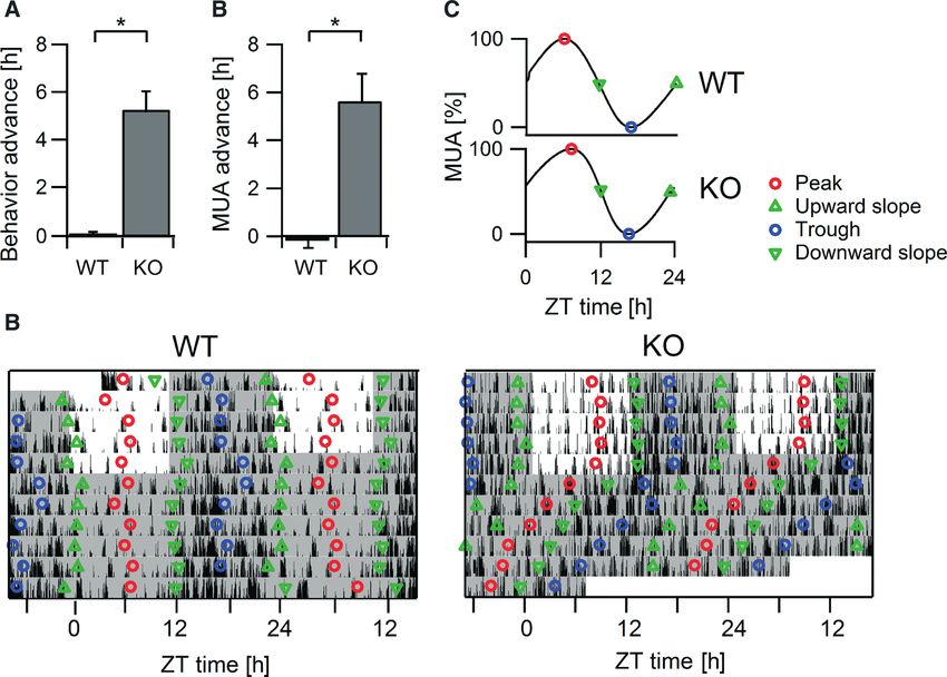
mice, in contrast to WT mice, began behavioral activity 5.59 ± 0.78 h transferred from LD conditions to DD. First, the amplitude of the
earlier than what would be expected on the basis of their previous MUA rhythm decreased more in VIP KO mice (48% ± 8%) than in
activity rhythm under LD conditions. All VIP KO mice remained WT mice (22 ± 4%, P < 0.01, t-test). Second, the MUA activity
rhythmic during the first 10 days in DD, as measured by F rhythm was dramatically phase advanced (Fig. 2B). The half-maxi-
periodogram analysis. Their free-running period was shorter mum level of the downward slope in MUA, which is strongly
(23.31 ± 0.38 h) than that of WT mice (23.73 ± 0.06 h), but this correlated with activity onset, started 5.68 ± 1.42 h before project
difference was not significant (P = 0.33). lights-off in VIP KO mice and at )0.19 ± 0.58 h in WT mice
(P < 0.01, Mann–Whitney test). The start of behavioral activity of the
same VIP KO mice was earlier than expected on the basis of
MUA rhythms endogenous period when lights were switched off (Fig. 2A). Average
Long-term MUA rhythms were recorded from the SCN of VIP KO waveforms in MUA under LD conditions did not differ between the
(n = 5) and WT littermate (n = 6) mice (Fig. 1A and B). Recordings groups (Fig. 2C). We observed that the downward slope of MUA was
were performed for five cycles in LD12 : 12, and then for five cycles strongly correlated with activity onset in both strains of mice
in DD. All of the VIP KO and WT mice showed diurnal rhythms in (Fig. 2D). We made activity profiles of total behavioral activity per
MUA when exposed to the LD cycle, with a peak in MUA in the 24 h and 24-h profiles of concurrently measured MUA of both
middle of the day, and a trough in the middle of the night. The mean genotypes (Fig. S2). We calculated the level of MUA at which half of
times to peak after lights-on (ZT 0) were 8.34 ± 1.05 h in WT mice the total behavioral activity had occurred. On average, this occurred at
and 8.22 ± 1.17 h in VIP KO mice (P = 0.94, t-test). MUA reached the same level of MUA in WT mice (1 ± 0%) and in VIP KO mice
its trough at ZT 17.42 ± 0.76 h and ZT 17.21 ± 0.94 h in WT and (7 ± 4%, P = 0.28, t-test).
VIP KO mice, respectively (P = 0.79, t-test). Under these conditions,
the durations of the width of the peak in MUA measured at half-
maximum levels were 11.81 ± 0.32 h in WT mice and 12.17 ± 0.41 h Behavioral and MUA rhythms measured in long and short
in VIP KO mice (P = 0.50, t-test). Thus, in LD conditions, VIP KO photoperiods
mice were able to maintain a robust rhythm in electrical activity with The wheel-running activity of VIP KO and WT mice was measured in
characteristics and phasing comparable to WT rhythms. short (LD8 : 16; KO, n = 16; WT, n = 17) and long (LD16 : 8; KO,
All of the VIP KO (n = 5) and WT (n = 6) mice showed circadian n = 11; WT, n = 16) photoperiods, respectively. Mice were held in
rhythms in MUA when the mice were held in DD. In DD, VIP KO the LD cycle for 30 days, and were then released into DD (Fig. 3;
ª 2012 The Authors. European Journal of Neuroscience ª 2012 Federation of European Neuroscience Societies and Blackwell Publishing Ltd
European Journal of Neuroscience, 35, 1466–1474VIP is essential for seasonal encoding in circadian clock 1469 Fig. 2. Phase advance in behavior and MUA after release in DD. Bar graphs compare the phase advance in behavior (A) and MUA (B) when a mouse was released into DD from the LD cycle. The asterisk indicates a significant difference at P < 0.01 measured with t-tests (A) or Mann–Whitney tests (B). (C) The average waveforms of MUA in WT and VIP KO mice in LD12 : 12, made by adding smoothed MUA activity waveforms of all mice used for analysis. Peaks and troughs in MUA are displayed as red and blue circles, respectively. Half-maximum values of MUA are indicated by green triangles. (D) Representative example of behavioral activity of WT (left panel) and VIP KO (right panel) mice measured with a passive infrared sensor. Each horizontal row represents a double-plotted 24-h day, with successive days plotted from top to bottom. Reference points (see legend) from the recorded MUA rhythm are superimposed on the locomotor activity plot. Table 1). All of the mice showed a robust daily rhythm in LD Finally, long-term MUA rhythms were recorded from the SCN of conditions, with activity increasing sharply after lights-off. The VIP KO mice held in long days (n = 6) or short days (n = 6; Fig. 4). amplitude of the daily rhythm in locomotor activity was reduced Recordings were performed for five cycles in the LD condition, (P < 0.01) in VIP KO mice as compared with WT controls, with the followed by five cycles in DD. All of the VIP KO mice showed mutant mice in the long days exhibiting the lowest amplitude diurnal rhythms in MUA, whether exposed to a short or a long (Table 1). Total behavioral activity was significantly lower in the VIP photoperiod. The average width of the MUA peak measured at half- KO mice (P < 0.01) than in controls, but did not differ between long maximum levels was significantly (P < 0.001, t-test) decreased when and short photoperiod exposure in either genotype. Following the mice were held in short day (9.82 ± 0.38 h) as compared with long entrainment to long days and release into DD, VIP KO mice showed day (14.40 ± 0.45 h) conditions. In three mice, MUA was measured a phase advance of behavioral activity of 12.74 ± 0.61 h as compared in more than one photoperiod consecutively (Table S1). The peak with the time of expected behavioral onset based on endogenous width of MUA lengthened with longer photoperiod exposure. When period. After short day exposure and release into DD, VIP KO mice mice were transferred to DD, all of the VIP KO mice (6 ⁄ 6) continued started activity 3.85 ± 0.28 h earlier than expected. The behavioral to show clear circadian rhythms in MUA after short day exposure. In phase advance in DD was significantly (P < 0.05) altered by exposure contrast, after exposure to long photoperiods, some of the VIP KO to the different photoperiods (Fig. 3). In DD, VIP KO mice showed mice (2 ⁄ 6) lost the circadian rhythmicity in MUA, and the remaining lower amplitude rhythms than WT controls (Table 1). mice showed weak rhythms. Therefore, we could only reliably One of the important changes in circadian behavior observed in determine period and phase advance in MUA after short day exposure: mice held in different photoperiods is an adaptive alteration in the 22.96 ± 0.13 and 1.97 ± 0.81 h. Importantly, after exposure to DD, duration of the night activity interval (alpha) (Fig. 3). WT mice the prior photoperiod had no significant impact on the MUA peak showed a significantly (P < 0.05) longer alpha when held in short width (Fig. 4). The mean widths of the peak were 10.93 ± 0.83 and days (12.9 ± 0.3 h) than when held in long days (7.7 ± 0.0 h). Similar 12.16 ± 0.77 h after short and long day exposure (P = 0.37, t-test). differences in alpha (P < 0.05) were observed in VIP KO mice held in Thus, for both the behavioral and MUA rhythms, the VIP KO mice short days (12.8 ± 0.3 h) or long days (6.4 ± 0.2 h). As expected, lost the photoperiod-driven plasticity in output. when WT animals were released into DD, alpha remained significantly (P < 0.05) longer after short days (12.6 ± 0.3 h) than after long days Discussion (9.0 ± 0.3 h). This history-dependent change in alpha (after-effects) was lost in the mutant mice. When VIP KO mice held in short days MUA rhythms were released into DD, the alpha of the activity rhythm was only We implanted electrodes into the SCN of VIP KO mice and littermate 9.3 ± 0.5 h, and those that were in long days showed an alpha of WT controls, and continuously recorded MUA in freely moving mice. 11.4 ± 0.5 h. Under LD12 : 12 conditions, VIP KO mice showed strong daily ª 2012 The Authors. European Journal of Neuroscience ª 2012 Federation of European Neuroscience Societies and Blackwell Publishing Ltd European Journal of Neuroscience, 35, 1466–1474
1470 E. A. Lucassen et al.

                            A                                                     B

                            C                            D                                         E

Fig. 3. Behavior of VIP KO mice exposed to short and long photoperiod conditions. Representative wheel-running activity records of WT and VIP KO mice under
(A) short (LD6 : 18) and (B) long (LD18 : 6) photoperiod exposure. Each horizontal row represents wheel-running activity over a 24-h cycle. Successive days are
plotted from top to bottom. (C) Bar graphs plotting the behavior phase advance measured at the LD ⁄ DD transition for VIP KO mice in each photoperiod. Bar graphs
showing the duration of the active phase (alpha) in each genotype under LD (D) and DD (E) conditions. The asterisk indicates a significant difference at P < 0.05
measured with t-tests.

rhythms in MUA that peaked during the second half of the day and                   example, in DD, the wheel-running activity in VIP KO mice is
reached their trough in the middle of the night (Fig. 1). The                      reduced in amplitude and coherence, and declines into arrhythmicity
amplitudes and the phase of the rhythms could not be distinguished                 over the course of several weeks (Colwell et al., 2003), whereas
from those recorded from the SCN of WT mice. When VIP KO mice                      VIPR2 KO mice are apparently arrhythmic from the start of constant
were transferred to DD, the MUA rhythm continued, but displayed a                  conditions (Harmar et al., 2002). Rhythms in body temperature
reduction in amplitude that was significantly larger than the amplitude            (Sheward et al., 2010; Hannibal et al., 2011; Schroeder et al., 2011),
drop observed in WT mice (Fig. 1). Also, upon release into DD, the                 feeding and metabolism (Bechtold et al., 2008) are phase advanced
MUA rhythm of VIP KO mice showed a phase advance of about 6 h                      but remain relatively intact. Rhythms in sleep (non-rapid eye
(Fig. 2), consistent with behavioral findings in earlier reports (Colwell          movement and rapid eye movement) show reduced day–night
et al., 2003; Aton et al., 2005; Ciarleglio et al., 2009). We analysed             amplitude and a phase advance in LD conditions (Sheward et al.,
the half-maximum levels of the downward slope of MUA, as this                      2010; Hu et al., 2011). Rhythms in heart rate in both VIP and VIPR2
phase of the rhythm is closely correlated with behavioral onset                    mutants are low-amplitude or arrhythmic in LD and DD conditions
(Houben et al., 2009). We found that the phase advance of the                      (Sheward et al., 2010; Schroeder et al., 2011). Similarly, rhythms in
electrical activity rhythm was similar to the phase advance of the                 the blood levels of adrenocorticotropic hormone and cortisol are all
behavioral activity rhythm. We considered whether this shift reflects              lost in VIP KO mice in both LD and DD conditions (Loh et al., 2008).
an unmasking of the rhythm in DD. As the electrical activity rhythm                With the present work, we see for the first time what is occurring at the
of the SCN peaked at the middle of the day under LD conditions, we                 level of the SCN in vivo, and find that the overall SCN neural activity
have no evidence for masking of the SCN rhythm by the light. Our                   rhythm is normal in LD conditions (Fig. 1). This suggests that we may
results indicate that, in both LD and DD conditions, the VIP KO mice               need to look to the output pathways to explain the variability in the
show a stable phase relationship between the SCN and behavioral                    impact of the loss of VIP on SCN-driven behaviors. Presumably, the
activity, and start their activity at the 50% level of the falling slope, as       SCN projects to extra-SCN tissues and peripheral outputs via
do WT mice.                                                                        interneurons, particularly those that regulate autonomic outflow from
                                                                                   the paraventricular nucleus or the brainstem, and ⁄ or those that drive
                                                                                   the pituitary output and hence adrenal or other endocrine rhythms
The impact of VIP deficiency varies with output                                    (Buijs et al., 2006). Many of these interneurons are VIPergic, and VIP
As a result of recent work, it is becoming clear that the impact of the            may therefore be required for communication between the SCN and
loss of VIP or VIPR2 varies with the output being measured. For                    peripheral tissues to drive rhythms in behavior and other biological

                 ª 2012 The Authors. European Journal of Neuroscience ª 2012 Federation of European Neuroscience Societies and Blackwell Publishing Ltd
                                                                                                     European Journal of Neuroscience, 35, 1466–1474
VIP is essential for seasonal encoding in circadian clock 1471
A B
C
Fig. 4. MUA rhythms recorded from the SCN of VIP KO mice exposed to short and long photoperiods. Representative examples of MUA recordings from the SCN
of VIP KO mice in (A) short (LD8 : 16) or (B) long (LD16 : 8) photoperiods and after release into DD. For visibility of the data, the first 3.5 days in DD are shown,
but traces are part of a longer recording. Note the loss of circadian rhythmicity in the mice (B, top panel) after they had been released into DD. The white line shows
the smoothed data (10-min bins). Behavioral activity as recorded with passive infrared sensors is depicted below. (C) The width of the peak that was measured at
half-maximum electrical activity levels from each genotype. After exposure to long days, the MUA rhythms were disturbed and not all peaks could be used for the
measurements. The asterisk indicates a significant difference at P < 0.05 measured with t-tests. As MUA rhythms became sloppy after long day exposure, not all
peaks could be used for peak width determination.
processes (Abrahamson & Moore, 2001; Antle & Silver, 2005;
Table 1. Behavioral parameters of mice in short (LD8 : 16) and long
(LD16 : 8) photoperiods Kalsbeek et al., 2006; Vosko et al., 2007).
Short day Long day
Cellular consequences
VIP KO WT VIP KO WT
The quality of the rhythm in MUA is remarkable, given the
Alpha in 12.8 ± 0.3 12.9 ± 0.3 6.4 ± 0.2 7.7 ± 0.1
compelling evidence that VIP plays a critical role in coupling cells
LD (h)*, in the SCN circuit (Welsh et al., 2010). Within the SCN, genetic loss
Alpha in 9.3 ± 0.5 12.6 ± 0.3 11.2 ± 0.6 9.0 ± 0.3 of VIP or VIPR2 disrupts circadian rhythms in neural activity in the
DD (h)*,à,§ SCN population (Cutler et al., 2003; Brown et al., 2007), where the
Period in 23.9 ± 0.2 23.8 ± 0.2 23.8 ± 0.4 23.9 ± 0.2 loss of VIP decreases the number of electrically rhythmic SCN
DD (h)
Amplitude 0.6 ± 0.1 0.9 ± 0.0 0.4 ± 0.2 0.9 ± 0.0 neurons (Aton et al., 2005) and weakens the synchrony among the
in DD ,à,§ rhythmic SCN neurons (Aton et al., 2005; Ciarleglio et al., 2009).
Phase advance 3.9 ± 0.3 0.2 ± 0.1 12.7 ± 0.6 0.0 ± 0.1 Even at the molecular level, the temporal expression of Per1, Per2 and
(h) ,à,§ Bmal1 is disrupted in the SCN (Loh et al., 2011). SCN explants from
VIP or VIPR2 KO mice showed low-amplitude rhythms in
*P < 0.05 between short and long days in WT mice; P < 0.05 between short
and long days in VIP KO mice; àP < 0.05 between genotypes in short days; PER2::LUC and PER1::LUC bioluminescence that were not altered
§P < 0.05 between genotypes in long days. in phase as compared with controls (Maywood, et al., 2006; Hughes
ª 2012 The Authors. European Journal of Neuroscience ª 2012 Federation of European Neuroscience Societies and Blackwell Publishing Ltd
European Journal of Neuroscience, 35, 1466–14741472 E. A. Lucassen et al.
et al., 2011; Loh et al., 2011). Recent work by Maywood et al. (2011) cannot explain this phenomenon. We therefore conclude that seasonal
has demonstrated that paracrine signaling is sufficient to induce encoding is greatly compromised in VIP KO mice. Similar to
rhythmicity in VIP-deficient SCN slices, and that VIP plays a observations in LD12 : 12 conditions, VIP KO mice exposed to
predominant role in mediating this coupling as measured by different photoperiods showed a rapid and large phase advance with
PER2::LUC bioluminescence. All of the analyses of the VIP and the transition to DD, but the amplitude of the phase advance was
VIPR2 KO mice indicate that the coupling within the SCN network is dependent on the previous photoperiods (Fig. 3). The phase advance
greatly reduced by the loss of VIP ⁄ VIPR2 signaling, and that the VIP was particularly large after exposure to long photoperiods, and was
KO mice can be considered to have a ‘broken’ SCN network. much smaller, and almost the same as, the behavior of WT mice
following exposure to short photoperiods. The fact that the behavior of
mice after short days resembled the behavior of WT mice may indicate
System-level compensation that the short photoperiod has acted as a strong synchronizer, thereby
The present results raise the question of what is occurring in the intact normalizing the behavior of VIP KO mice.
system that enables the SCN to maintain strong rhythms in MUA in To test whether the impairment in seasonal encoding in VIP KO
the presence of reduced cellular synchrony resulting from the loss of mice is based on deficiencies within the SCN clock, we investigated
VIP. The most obvious explanation is that light directly stimulates the impact of photoperiod on SCN MUA waveform (Fig. 4). The
SCN activity when animals are kept in an LD cycle (Meijer et al., waveform of the SCN rhythm shows major differences in response to
1996, 1998). The decreased behavioral amplitude in DD is also long and short photoperiods that are brought about by changes in
explainable by an absence of entraining light signals. The melanopsin- synchrony among molecular and electrical activity rhythms of SCN
expressing retinal ganglion cells are both directly light-sensitive and neurons (Hazlerigg et al., 2005; Inagaki et al., 2007; Naito et al.,
receive information from rods and cones (Panda, 2007; Ecker et al., 2008; Sosniyenko et al., 2009; VanderLeest et al., 2007; Brown &
2010; Lall et al., 2010). These melanopsin-expressing retinal ganglion Piggins, 2009). A major question that follows from these results
cells thus encode ambient lighting (Berson et al., 2002) and generate concerns the underlying mechanism of phase synchrony vs. desyn-
action potentials that travel down the RHT and reach the SCN. The chrony in seasonality. To investigate the putative role of VIP, we
RHT terminals release glutamate (Antle et al., 2009) and, under measured the electrical activity patterns from SCN neurons in VIP KO
certain conditions, the neuropeptide pituitary adenylate cyclase- mice exposed to short and long days (Fig. 4). VIP KO mice showed a
activating peptide (Hannibal, 2002). The net result of RHT stimulation broadening of their MUA patterns when they were kept in long days,
is an increase in the firing rate of SCN neurons. Prior work with VIP and a lengthening when kept in short days. The broadness of the peaks
KO mice demonstrated that the direct effects of light in suppressing at half-maximum levels differed significantly between the photoperi-
activity and of darkness in enhancing activity are completely unaltered ods (14.6 vs. 9.8 h), which is similar to what is seen in WT animals
by the loss of VIP (Colwell et al., 2003). Therefore, in an LD cycle, (VanderLeest et al., 2007). Importantly, the MUA of three mice was
the direct effects of light and dark on MUA are likely to play a major recorded in multiple photoperiods. The results from these three
role in maintaining the rhythmicity in SCN physiology and behavior. recordings showed the same photoperiod-driven changes in peak
Previous work has also provided evidence that SCN neural activity is width that we observed in the population (Table S1). Interestingly,
influenced by activity in other brain regions (Deboer et al., 2003; when VIP KO mice were transferred to DD, the width of the MUA
Vansteensel et al., 2003), which may explain why, in VIP KO profiles was indistinguishable between the groups from long and short
animals, the rhythms in DD are maintained in vivo, whereas the photoperiods. It is well accepted that the ‘after-effects’ in DD indicate
rhythms measured in vitro are severely decreased (Aton et al., 2005; the storage of photoperiodic information in the SCN clock. In other
Brown et al., 2007). Prior studies with the VIPR2 KO mice have also words, to test the encoding capacity of the SCN for photoperiod
found behavioral (Power et al., 2010) and physiological (Sheward information, the waveform of the rhythm should be evaluated after
et al., 2010) evidence that activity is a strong regulator of rhythmicity termination of the light cycle. In our results, the peak width in long
in these mice. A variety of earlier studies have noted that locomotor days did not exceed the peak width in short days, and in fact it became
activity itself can modify SCN activity (Meijer et al., 1997; Yamazaki narrower. The absence of any after-effect on the electrical activity
et al., 1998; Schaap & Meijer, 2001; Nakamura et al., 2011). The rhythm therefore suggests a total absence of seasonal encoding
direct regulation of SCN firing by lighting and activity levels would capability of the SCN in the absence in VIP.
not be detected in the in vitro recording, and could be viewed as a Although photoperiod did not affect the width of the peak, it did
systems-level compensation for the loss of coupling in the VIP KO affect the phase advancing response of the MUA rhythm when the
mice. mice were transferred to DD (Fig. 3E). The phase advance was about
10 h after exposure to long days and about 2.5 h after exposure to
short days. These findings were consistent with the magnitude of the
behavioral advances that we observed in VIP KO mice. Finally, we
Role of VIP in seasonal encoding note that the overall quality of the rhythm in both behavior and in
The SCN serves as a seasonal clock through its ability to encode day- MUA activity, as indicated by the amplitude, was better after exposure
length (Goldman, 2001; Beersma et al., 2008; Meijer et al., 2010). In to short photoperiods than after exposure to long photoperiods.
line with previous literature (Refenetti, 2002; VanderLeest et al., Possibly, the short days functioned as a strong synchronizer, resulting
2007), the duration of behavioral activity (alpha) was broadened or in an absence of a strong phase advancing response and a diminished
compressed when WT mice were exposed to short and long days, attenuation of the rhythm in darkness.
respectively (Fig. 3). Interestingly, alpha in VIP KO mice was shorter
under long than under short photoperiods. When mice were transferred
to DD, alpha remained highly correlated with the previous length of
photoperiod in WT mice. In VIP KO mice, however, the duration of Conclusion
alpha was not shorter after long day exposure following release into The robustness of the rhythm in electrical impulse frequency of the
darkness. In fact, alpha was even longer after long days, and we SCN in vivo, as compared with previous in vitro recordings (Aton
ª 2012 The Authors. European Journal of Neuroscience ª 2012 Federation of European Neuroscience Societies and Blackwell Publishing Ltd
European Journal of Neuroscience, 35, 1466–1474VIP is essential for seasonal encoding in circadian clock 1473
et al., 2005; Brown et al., 2007), indicate that communication An, S., Irwin, R.P., Allen, C.N., Tsai, C. & Herzog, E.D. (2011) Vasoactive
between neuronal networks can compensate for the absence of VIP- intestinal polypeptide requires parallel changes in adenylate cyclase and
phospholipase C to entrain circadian rhythms to a predictable phase. J.
induced synchrony among SCN neurons. The SCN MUA is acutely Neurophysiol., 105, 2289–2296.
increased by retinal input (Meijer et al., 1998), and we speculate that Antle, M.C. & Silver, R. (2005) Orchestrating time: arrangements of the brain
the direct effect of light boosts the magnitude of the MUA rhythms. circadian clock. Trends Neurosci., 28, 145–151.
Possibly, light input maintains phase coherence via the RHT or Antle, M.C., Smith, V.M., Sterniczuk, R., Yamakawa, G.R. & Rakai, B.D.
through behavioral feedback effects, via the geniculohypothalamic (2009) Physiological responses of the circadian clock to acute light exposure
at night. Rev. Endocr. Metab. Disord., 10, 279–291.
input or the median raphe. Additional regulatory effects of behav- Aton, S.J., Colwell, C.S., Harmar, A.J., Waschek, J. & Herzog, E.D. (2005)
ioral activity and sleep from other brain regions on SCN activity are Vasoactive intestinal polypeptide mediates circadian rhythmicity and
also likely (Meijer et al., 1997; Yamazaki et al., 1998; Deboer et al., synchrony in mammalian clock neurons. Nat. Neurosci., 8, 476–483.
2003), and need to be further explored. Prior work has provided Bechtold, D.A., Brown, T.M., Luckman, S.M. & Piggins, H.D. (2008)
Metabolic rhythm abnormalities in mice lacking VIP-VPAC2 signaling. Am.
elegant evidence that SCN intercellular coupling provides robustness J. Physiol. Regul. Integr. Comp. Physiol., 294, R344–R351.
in the circadian system against genetic perturbations (Liu et al., Beersma, D.G., van Bunnik, B.A., Hut, R.A. & Daan, S. (2008) Emergence of
2007). Here, we show that the intact SCN circuit can overcome the circadian and photoperiodic system level properties from interactions among
dysfunction caused by the loss of intercellular coupling in the SCN. pacemaker cells. J. Biol. Rhythms, 23, 362–373.
To put it another way, as important as VIP is for the coupling of Berson, D.M., Dunn, F.A. & Takao, M. (2002) Phototransduction by retinal
ganglion cells that set the circadian clock. Science, 295, 1070–1073.
SCN neurons, the intact system appears to be able to compensate for Brown, T.M. & Piggins, H.D. (2009) Spatiotemporal heterogeneity in the
its loss, at least under some lighting conditions. However, our electrical activity of suprachiasmatic nuclei neurons and their response to
findings also indicate that there are some functions that require VIP- photoperiod. J. Biol. Rhythms, 24, 44–54.
mediated intercellular coupling, for example the reorganization of the Brown, T.M., Colwell, C.S., Waschek, J.A. & Piggins, H.D. (2007) Disrupted
neuronal activity rhythms in the suprachiasmatic nuclei of vasoactive
SCN circuit that is part of the adaptation to changes in photoperiod. intestinal polypeptide-deficient mice. J. Neurophysiol., 97, 2553–2558.
Without VIP, both the behavior and the physiology of the SCN Buijs, R.M., Scheer, F.A., Kreier, F., Yi, C., Bos, N., Gonchurak, V.D. &
appear to be unable to adapt to seasonal changes, and the ‘memory’ Kalsbeek, A. (2006) Organization of circadian functions: interactions with
for photoperiod is lost. This is an important finding in the search for the body. Prog. Brain Res., 153, 341–360.
mechanisms underlying the consolidation of phase synchrony within Ciarleglio, C.M., Gamble, K.L., Axley, J.C., Strauss, B.R., Cohen, J.Y.,
Colwell, C.S. & McMahon, D.G. (2009) Population encoding by circadian
the SCN. clock neurons organizes circadian behavior. J. Neurosci., 29, 1670–1676.
Colwell, C.S., Michel, S., Itri, J., Rodriguez, W., Tam, J., Lelievre, V., Hu, Z.,
Liu, X. & Waschek, J.A. (2003) Disrupted circadian rhythms in VIP- and
Supporting Information PHI-deficient mice. Am. J. Physiol., 285, R939–R949.
Additional supporting information can be found in the online version Cutler, D.J., Haraura, M., Reed, H.E., Shen, S., Sheward, W.J., Morrison, C.F.,
Marston, H.M., Harmar, A.J. & Piggins, H.D. (2003) The mouse VPAC2
of this article: receptor confers suprachiasmatic nuclei cellular rhythmicity and responsive-
Fig. S1. Behavior of WT and VIP KO mice in LD12 : 12 and in DD. ness to vasoactive intestinal polypeptide in vitro. Eur. J. Neurosci., 17, 197–
Fig. S2. Correlation between behavior and MUA in WT and VIP KO 204.
mice in LD12 : 12. Deboer, T., Vansteensel, M.J., Détári, L. & Meijer, J.H. (2003) Sleep states
alter activity of suprachiasmatic nucleus neurons. Nat. Neurosci., 6, 1086–
Table S1. Width of the peaks in MUA in a subpopulation of VIP KO 1090.
mice that were measured in multiple photoperiods. Ebling, F.J. & Barrett, P. (2008) The regulation of seasonal changes in food
Please note: As a service to our authors and readers, this journal intake and body weight. J. Neuroendocrinol., 20, 827–833.
provides supporting information supplied by the authors. Such Ecker, J.L., Dumitres, O.N., Wong, K.Y., Alam, N.M., Chen, S.K., LeGates,
materials are peer-reviewed and may be re organized for online T., Renna, J.M., Prusky, G.T., Berson, D.M. & Hattar, S. (2010)
Melanopsin-expressing retinal ganglion-cell photoreceptors: cellular diver-
delivery, but are not copy-edited or typeset by Wiley-Blackwell. sity and role in pattern vision. Neuron, 67, 49–60.
Technical support issues arising from supporting information (other Eilers, P. (2003) A perfect smoother. Anal. Chem., 75, 3631–3636.
than missing files) should be addressed to the authors. Goldman, B.D. (2001) Mammalian photoperiodic system: formal properties
and neuroendocrine mechanisms of photoperiodic time measurement. J. Biol.
Rhythms, 16, 283–301.
Golombek, D.A. & Rosenstein, R.E. (2010) Physiology of circadian entrain-
Acknowledgements ment. Physiol. Rev., 90, 1063–1102.
We thank Hans Duindam, Jan Janse and Sander van Berloo for excellent Hannibal, J. (2002) Pituitary adenylate cyclase-activating peptide in the rat
technical support. The work was supported by a grant to J. H. Meijer from the central nervous system: an immunohistochemical and in situ hybridization
EC FP6 integrated project ‘EUCLOCK’ (contract number 018741), by the study. J. Comp. Neurol., 453, 389–417.
TOPGO.L.10.035 grant from NWO (number 91210064), and by CHDI Hannibal, J., Hsiung, H.M. & Fahrenkrug, J. (2011) Temporal phasing of
Foundation grant A-2702 to C. S. Colwell. locomotor activity, heart rate rhythmicity, and core body temperature is
disrupted in VIP receptor 2-deficient mice. Am. J. Physiol. Regul. Integr.
Comp. Physiol., 300, R519–R530.
Harmar, A.J., Marston, H.M., Shen, S., Spratt, C., West, K.M., Sheward, W.J.,
Abbreviations Morrison, C.F., Dorin, J.R., Piggins, H.D., Reubi, J.C., Kelly, J.S., Maywood,
DD, constant darkness; KO, knockout; LD, light–dark; MUA, multiunit neural E.S. & Hastings, M.H. (2002) The VPAC(2) receptor is essential for circadian
activity; RHT, retinohypothalamic tract; SCN, suprachiasmatic nucleus; VIP, function in the mouse suprachiasmatic nuclei. Cell, 109, 497–508.
vasoactive intestinal peptide; VIPR2, vasoactive intestinal peptide receptor 2; Hazlerigg, D.G., Ebling, F.J. & Johnston, J.D. (2005) Photoperiod differentially
WT, wild-type; ZT, zeitgeber time. regulates gene expression rhythms in the rostral and caudal SCN. Curr. Biol.,
15, R449–R450.
Houben, T., Deboer, T., Oosterhout, F. & Meijer, J.H. (2009) Correlation with
behavioral activity and rest implies circadian regulation by SCN neuronal
References activity levels. J. Biol. Rhythms, 24, 477–487.
Abrahamson, E.E. & Moore, R.Y. (2001) Suprachiasmatic nucleus in the Hu, W.P., Li, J.D., Colwell, C.S. & Zhou, Q.Y. (2011) Decreased REM sleep
mouse: retinal innervation, intrinsic organization and efferent projections. and altered circadian sleep regulation in mice lacking vasoactive intestinal
Brain Res., 916, 172–191. polypeptide. Sleep, 34, 49–56.
ª 2012 The Authors. European Journal of Neuroscience ª 2012 Federation of European Neuroscience Societies and Blackwell Publishing Ltd
European Journal of Neuroscience, 35, 1466–14741474 E. A. Lucassen et al.
Hughes, A.T., Guilding, C. & Piggins, H.D. (2011) Neuropeptide signaling Nielsen, H.S., Hannibal, J. & Fahrenkrug, J. (2002) Vasoactive intestinal
differentially affects phase maintenance and rhythm generation in SCN and polypeptide induces per1 and per2 gene expression in the rat suprachiasmatic
extra-SCN circadian oscillators. PLoS ONE, 6, e18926. nucleus late at night. Eur. J. Neurosci., 15, 570–574.
Inagaki, N., Honma, S., Ono, D., Tanahashi, Y. & Honma, K. (2007) Separate Panda, S. (2007) Multiple photopigments entrain the mammalian circadian
oscillating cell groups in mouse SCN couple photoperiodically to the onset oscillator. Neuron, 53, 619–621.
and end of daily activity. Proc. Natl. Acad. Sci. USA, 104, 7664–7669. Paxinos, G. & Franklin, K.B.J. (2008) The Mouse Brain in Stereotaxic
Kalsbeek, A., Palm, I.F., La Fleur, S.E., Scheer, F.A., Perreau-Lenz, S., Ruiter, Coordinates. Academic Press, Waltham, MA.
M., Kreier, F., Cailotto, C. & Buijs, R.M. (2006) SCN outputs and the Power, A., Hughes, A.T.L., Samuels, R.E. & Piggins, H.D. (2010) Rhythm-
hypothalamic balance of life. J. Biol. Rhythms, 21, 458–469. promoting actions of exercise in mice with deficient neuropeptide signaling.
Lall, G.S., Revell, V.L., Momiji, H., Al Enezi, J., Altimus, C.M., Güler, A.D., J. Biol. Rhythms, 25, 235–246.
Aguilar, C., Cameron, M.A., Allender, S., Hankins, M.W. & Lucas, R.J. Reed, H.E., Meyer-Spasche, A., Cutler, D.J., Coen, C.W. & Piggins, H.D.
(2010) Distinct contributions of rod, cone and melanopsin photoreceptors to (2001) Vasoactive intestinal polypeptide (VIP) phase-shifts the rat suprach-
encoding irradiance. Neuron, 66, 417–428. iasmatic nucleus clock in vitro. Eur. J. Neurosci., 13, 839–843.
Liu, A.C., Welsh, D.K., Ko, C.H., Tran, H.G., Zhang, E.E., Priest, A.A., Buhr, Reed, H.E., Cutler, D.J., Brown, T.M., Brown, J., Coen, C.W. & Piggins, H.D.
E.D., Singer, O., Meeker, K., Verma, I.M., Doyle, F.J., Takahashi, J.S. & (2002) Effects of vasoactive intestinal polypeptide on neurones of the rat
Kay, S.A. (2007) Intercellular coupling confers robustness against mutations suprachiasmatic nuclei in vitro. J. Neuroendocrinol., 14, 639–646.
in the SCN circadian clock network. Cell, 129, 605–616. Refenetti, R. (2002) Compression and expansion of circadian rhythm in mice
Loh, D.H., Abad, C., Colwell, C.S. & Waschek, J.A. (2008) Vasoactive under long and short photoperiods. Integr. Physiol. Behav. Sci., 37, 114–
intestinal peptide is critical for circadian regulation of glucocorticoids. 127.
Neuroendocrinology, 88, 246–255. Schaap, J. & Meijer, J.H. (2001) Opposing effects of behavioural activity and
Loh, D.H., Dragich, J.M., Kudo, T., Schroeder, A.M., Nakamura, T.J., light on neurons of the suprachiasmatic nucleus. Eur. J. Neurosci., 13, 1955–
Waschek, J.A., Block, G.D. & Colwell, C.S. (2011) Effects of vasoactive 1962.
intestinal peptide genotype on circadian gene expression in the suprachias- Schroeder, A., Loh, D.H., Jordan, M.C., Roos, K.P. & Colwell, C.S. (2011)
matic nucleus and peripheral organs. J. Biol. Rhythms, 26, 200–209. Circadian regulation of cardiovascular function: a role for
Maywood, E.S., Reddy, A.B., Wong, G.K.Y., O’ Neill, J.S., O’ Brien, J.A., vasoactive intestinal peptide. Am. J. Physiol. Heart Circ. Physiol., 300,
McMahon, D.G., Harmar, A.J., Okamura, H. & Hastings, M.H. (2006) H241–H250.
Synchronization and maintenance of timekeeping in suprachiasmatic circa- Sheward, W.J., Naylor, E., Knowles-Barley, S., Armstrong, J.D., Brooker,
dian clock cells by neuropeptidergic signaling. Curr. Biol., 16, 599–605. G.A., Seckl, J.R., Turek, F.W., Holmes, M.C., Zee, P.C. & Harmar, A.J.
Maywood, E.S., Chesham, J.E., O’ Brien, J.A. & Hastings, M.H. (2011) A (2010) Circadian control of mouse heart rate and blood pressure by the
diversity of paracrine signals sustains molecular circadian cycling in suprach- suprachiasmatic nuclei: behavioral effects are more significant than direct
iasmatic nucleus circuits. Proc. Natl. Acad. Sci. USA, 108, 14306–14311. outputs. PLoS ONE, 5, e9783.
Meijer, J.H., Watanabe, K., Détàri, L. & Schaap, J. (1996) Circadian rhythm in Sosniyenko, S., Hut, R.A., Daan, S. & Sumova, A. (2009) Influence of
light response in suprachiasmatic nucleus neurons of freely moving rats. photoperiod duration and light–dark transitions on entrainment of Per1 and
Brain Res., 741, 352–355. Per2 gene and protein expression in subdivisions of the mouse SCN. Eur. J.
Meijer, J.H., Schaap, J., Watanabe, K. & Albus, H. (1997) Multiunit activity Neurosci., 30, 1802–1814.
recordings in the suprachiasmatic nuclei: in vivo versus in vitro models. VanderLeest, H.T., Houben, T., Michel, S., Deboer, T., Albus, H., Vansteensel,
Brain Res., 753, 322–327. M.J., Block, G.D. & Meijer, J.H. (2007) Seasonal encoding by the circadian
Meijer, J.H., Watanabe, K., Schaap, J., Albus, H. & Détári, L. (1998) Light pacemaker of the SCN. Curr. Biol., 17, 468–473.
responsiveness of the suprachiasmatic nucleus: long-term multiunit and VanderLeest, H.T., Rohling, J.H., Michel, S. & Meijer, J.H. (2009) Phase
single-unit recordings in freely moving rats. J. Neurosci., 18, 9078–9087. shifting capacity of the circadian pacemaker determined by the SCN
Meijer, J.H., Michel, S., Vanderleest, H.T. & Rohling, J.H. (2010) Daily and neuronal network organization. PLoS ONE, 4, e4976.
seasonal adaptation of the circadian clock requires plasticity of the SCN Vansteensel, M.J., Yamazaki, S., Albus, H., Deboer, T., Block, G.D. & Meijer,
neuronal network. Eur. J. Neurosci., 2, 2143–2151. J.H. (2003) Dissociation between circadian Per1 and neuronal and behavioral
Meyer-Spasche, A. & Piggins, H.D. (2004) Vasoactive intestinal polypeptide rhythms following a shifted environmental cycle. Curr. Biol., 13, 1538–
phase-advances the rat suprachiasmatic nuclei circadian pacemaker in vitro 1542.
via protein kinase A and mitogen-activated protein kinase. Neurosci. Lett., Vosko, A.M., Schroeder, A., Loh, D.H. & Colwell, C.S. (2007) Vasoactive
358, 91–94. intestinal peptide and the mammalian circadian system. Gen. Comp.
Naito, E., Watanabe, T., Tei, H., Yoshimura, T. & Ebihara, S. (2008) Endocrinol., 152, 165–175.
Reorganization of the suprachiasmatic nucleus coding for day length. J. Biol. Welsh, D.K., Takahashi, J.S. & Kay, S.A. (2010) Suprachiasmatic nucleus: cell
Rhythms, 23, 140–149. autonomy and network properties. Annu. Rev. Physiol., 72, 551–577.
Nakamura, T.J., Nakamura, W., Yamazaki, S., Kudo, T., Cutler, T., Colwell, Yamazaki, S., Kerbeshian, M.C., Hocker, C.G., Block, G.D. & Menaker, M.
C.S. & Block, G.D. (2011) Age-related decline in circadian output. J. (1998) Rhythmic properties of the hamster suprachiasmatic nucleus in vivo.
Neurosci., 31, 10201–10205. J. Neurosci., 18, 10709–10723.
ª 2012 The Authors. European Journal of Neuroscience ª 2012 Federation of European Neuroscience Societies and Blackwell Publishing Ltd
European Journal of Neuroscience, 35, 1466–1474You can also read