Effect of Reduced Retinal VLC-PUFA on Rod and Cone Photoreceptors
←
→
Page content transcription
If your browser does not render page correctly, please read the page content below
Retina

Effect of Reduced Retinal VLC-PUFA on Rod and Cone
Photoreceptors
Lea D. Bennett,1,2 Richard S. Brush,1,2 Michael Chan,1,2 Todd A. Lydic,3 Kristen Reese,3
Gavin E. Reid,3,4 Julia V. Busik,5 Michael H. Elliott,2,6,7 and Robert E. Anderson1,2,6,7
1
  Department of Cell Biology, University of Oklahoma Health Sciences Center, Oklahoma City, Oklahoma, United States
2
  Dean McGee Eye Institute, Oklahoma City, Oklahoma, United States
3Department of Chemistry, Michigan State University, East Lansing, Michigan, United States
4
  Department of Biochemistry and Molecular Biology, Michigan State University, East Lansing, Michigan, United States
5
  Department of Physiology, Michigan State University, East Lansing, Michigan, United States
6
  Department of Ophthalmology, University of Oklahoma Health Sciences Center, Oklahoma City, Oklahoma, United States
7Oklahoma Center for Neuroscience, University of Oklahoma Health Sciences Center, Oklahoma City, Oklahoma, United States

Correspondence: Robert E. Ander-            PURPOSE. Autosomal dominant Stargardt-like macular dystrophy (STGD3) is a juvenile-onset
son, University of Oklahoma Health          disease that is caused by mutations in Elovl4 (elongation of very long fatty acids-4). The Elovl4
Sciences Center, 608 Stanton L.             catalyzes the first step in the conversion of C24 and longer fatty acids (FAs) to very long-chain
Young Boulevard, Oklahoma City,             FAs (VLC-FAs, ‡C26). Photoreceptors are particularly rich in VLC polyunsaturated FAs (VLC-
OK 73104, USA;
Robert-Anderson@ouhsc.edu
                                            PUFA). To explore the role of VLC-PUFAs in photoreceptors, we conditionally deleted Elovl4
                                            in the mouse retina.
Submitted: January 21, 2014
Accepted: April 1, 2014                     METHODS. Proteins were analyzed by Western blotting and lipids by gas chromatography (GC)–
                                            mass spectrometry, GC-flame ionization detection, and tandem mass spectrometry. Retina
Citation: Bennett LD, Brush RS, Chan
M, et al. Effect of reduced retinal VLC-
                                            function was assessed by electroretinography (ERG), and structure was evaluated by bright
PUFA on rod and cone photorecep-            field, immunofluorescence, and transmission electron microscopy.
tors. Invest Ophthalmol Vis Sci.            RESULTS. Conditional deletion (KO) of retinal Elovl4 reduced RNA and protein levels by 91%
2014;55:3150–3157. DOI:10.1167/             and 96%, respectively. Total retina VLC-PUFAs were reduced by 88% compared to the wild
iovs.14-13995                               type (WT) levels. Retinal VLC-PUFAs incorporated in phosphatidylcholine were less abundant
                                            at 12 months compared to 8-week-old levels. Amplitudes of the ERG a-wave were reduced by
                                            22%, consistent with photoreceptor degeneration (11% loss of photoreceptors). Analysis of
                                            the rod a-wave responses gave no evidence of a role for VLC-PUFA in visual transduction.
                                            However, there were significant reductions in rod b-wave amplitudes (>30%) that could not
                                            be explained by loss of rod photoreceptors. There was no effect of VLC-PUFA reduction on
                                            cone ERG responses, and cone density was not different between the WT and KO mice at 12
                                            months of age.
                                            CONCLUSIONS. The VLC-PUFAs are important for rod, but not cone, function and for rod
                                            photoreceptor longevity.
                                            Keywords: VLC-PUFA, Elovl4, STDG3

P   atients with autosomal dominant Stargardt-like macular
    dystrophy (STGD3), a juvenile form of macular degenera-
tion, develop a loss of central vision at an early age. Mutations
                                                                                    Retinal VLC-PUFAs are incorporated into phosphatidylcho-
                                                                                line in photoreceptor outer segment membranes15 and have
                                                                                been suggested to have a role in disk curvature and plasma
in the elongation of very long-chain fatty acids-4 (Elovl4) gene                membrane fluidity, thus aiding in phototransduction.6 The role
have been associated with the disease in patients with                          of VLC-PUFAs in the neural retina still is under debate.7,8 The
STGD3.1–5 These mutations cause a frame shift in the Elovl4                     purpose of this study was to elucidate the role of VLC-PUFA in
transcript, introducing a premature stop codon, resulting in the                the retina by conditionally deleting Elovl4 expression in rod
synthesis of a truncated protein that has lost an ER retention/                 and cone photoreceptor cells, thus, removing VLC-PUFAs from
retrieval signal. The truncated protein is not targeted to the                  both cell types. Here we showed that these FAs are important
endoplasmic reticulum, the site of synthesis of very long-chain                 for rod, but not cone, survival in 12-month-old mice. We found
polyunsaturated fatty acids (VLC-PUFAs; 26–40 carbons6–8).                      no evidence from our electroretinographic (ERG) analysis to
Because the mutant protein has no enzymatic activity,9 the loss                 support a role for VLC-PUFA in visual transduction in rod or
of VLC-PUFAs may be involved in the STGD3 disease                               cone photoreceptors. However, we did find significant
pathogenesis. Expression of the Elovl4 gene is limited mainly                   reduction in rod b-wave amplitudes in conditional deletion
to the brain, testis, skin, and retinal photoreceptor cells.10                  (KO) mice that could not be explained by rod cell death. Also,
While the skin contains very long chain saturated fatty acids                   there were significant reductions in the oscillatory potentials
(VLC-FAs),11,12 sperm cells and the retina are enriched in VLC-                 (OPs) and scotopic threshold responses in KO mice. These
PUFAs.13,14                                                                     findings are presented and discussed in the companion paper.

Copyright 2014 The Association for Research in Vision and Ophthalmology, Inc.
www.iovs.org j ISSN: 1552-5783                                                                                                            3150
Retinal VLC-PUFA in Rod and Cone Photoreceptors                                             IOVS j May 2014 j Vol. 55 j No. 5 j 3151

MATERIALS     AND   METHODS                                        Immunofluorescence
Materials                                                          Immunofluorescence labeling was performed on frozen sections
                                                                   using standard procedures described previously16 to visualize
Primary antibodies used were anti-Elovl46 (1:1000) and anti-b-     the Elovl4 (1:100) expression. Anti-rabbit-594 (Invitrogen) was
actin (1:1000; ABCAM, Cambridge, MA, USA). The Elovl4              used at 1:1000 and DAPI was used at 1:5000. Slides were viewed
antibody used here has been shown by Agbaga et al.6 to label       with an FV-500 Olympus Confocal Microscope (Olympus,
protein only in the inner segments and outer nuclear layer of      Tokyo, Japan) and the images were acquired with Flow View
whole rat retina. Specificity of the antibody was shown by         software (Olympus America, Center Valley, PA, USA). Contrast
preadsorbtion of the Elovl4 antigen by immunohistochemistry        and brightness were adjusted with Photoshop (Adobe Systems,
and by Western blotting resulting in the absence of Elovl4         Mountain View, CA, USA). Immunofluorescence for cone
staining or the absence of the 32 kDa immunospecific band,         quantification was performed on paraffin-embedded sections
respectively.6 Horseradish-conjugated secondary antibodies         stained with PNA (1:100), visualized with a Nikon epifluor-
(rabbit polyclonal and mouse monoclonal) were from Pierce          escence microscope (Nikon Eclipse E800 microscope; Nikon,
Scientific (Rockford, IL, USA). Fluorescein-conjugated antibod-    Tokyo, Japan), and processed with ImageJ (National Institutes of
ies were anti-rabbit antibody 488 (Invitrogen, Grand Island, NY,   Health [NIH], available in the public domain at http://imagej.nih.
USA), and peanut agglutinin-594 (PNA) and 4 0 6-dimidino-2-        gov/ij/). Cones were quantified by the threshold of PNA
phenylindole (DAPI; Vector Laboratories, Burlingame, CA,           fluorescence relative to the total area of the photoreceptor
USA). All solvents for lipid analysis were HPLC grade. Lipid       nuclei and presented as number of cones per unit area.
internal standards used for tandem MS analysis were 14:0/14:0
phosphatidylcholine (PC), 14:0/14:0 phosphatidylethanol-
amine (PE), and 14:0/14:0 phosphatidylserine (PS; Avanti Polar     Quantitative RT-PCR (qRT-PCR)
Lipids, Alabaster, AL, USA).                                       RNA was extracted from whole retinas using the Trizol reagent
                                                                   (Sigma-Aldrich, St. Louis, MO, USA). The cDNA was reverse
Animals
                                                                   transcribed from 1 lg of RNA using the Reverse Transcription
Mice with the Elovl4 gene containing LoxP sites flanking exons     kit (Promega, Madison, WI, USA). The qRT-PCR was performed
2 and 3 were mated with a transgenic mouse line expressing         in triplicate according to standard procedures. The Elovl4
Cre recombinase driven by the ChX10 promoter (Jackson              message expression was normalized to Hprt expression.
Laboratories, Bar Harbor, ME, USA) to delete Elovl4 before         Primers used to detect Elovl4 were 5 0 -TCCAGAAA
photoreceptor differentiation (Elovl4 floxed mice were a gift      TATCTTTGGTGG-3 0 and 5 0 -GTTAAGGCCCAGTTCAATT-3 0 .
from Kang Zhang, University of Southern California, San Diego,     The primers used to detect Hprt were 5 0 -CTTTGCTGACCTG
CA, USA).7 Three genotypes of mice were generated after            CTGGATTAC-3 0 and 5 0 -TTGGGGCTGTACTGCTTAACC-3 0 .
several backcrosses: Cre//Elovl4f/f (wild type [WT]), Cre6/
Elovl4f/f (KO), and Cre6/Elof/WT (Het). The Hets were used as a    Fatty Acid Analysis
control for Cre expression. The mice were housed in a facility
with a 12-hour light on/off cycle, with on light equal to 5 lux.   Retinas were pooled from three mice of the same genotype,
    Mice were killed with CO2 inhalation or perfusion.             and extracted by the method of Bligh and Dyer.17 Fatty acids
Perfusion involved placing a single mouse in a bell jar            were transesterified to methyl esters18 and analyzed using gas
containing isoflurane until the mouse was deeply anesthetized      chromatography-mass spectrometry (GC-MS) and gas chroma-
and unresponsive to intense pinching of the footpad. The heart     tography-flame ionization detection (GC-FID).19 The chromato-
was exposed, a cannula was placed in the left ventricle, and an    graphic peaks were integrated and processed with
incision was made in the right atrium to allow fluid to escape.    ChemStation software (Agilent Technologies, Santa Clara, CA,
Paraformaldehyde (2%) and glutaraldehyde (2.5%) in phos-           USA). Each genotype had 4 distinct samples containing 6
phate buffer (PB, 0.1 M, pH 7.4) were perfused at 120 mm Hg        retinas each (n ¼ 4). Lipids were measured in 16-week-old mice.
for 5 minutes. Eyeball orientation was marked by cauterization,
harvested intact, and submerged in the same perfusion fixative     Tandem Mass Spectrometry Analysis of Retina
cocktail for later analysis.
    All procedures were performed according to the Associa-        Lipids (MS/MS)
tion for Research in Vision and Ophthalmology (ARVO)               Retinal lipids were extracted as described previously.20 The PC
Statement for the Use of Animals in Ophthalmic and Vision          lipids were identified by using precursor ion (PI) scanning of
Research, and the University of Oklahoma Health Sciences           m/z 184. The PE lipids were identified by scanning for a
Center Guidelines for Animals in Research. All protocols were      neutral loss (NL) of 141 m/z, and PS lipids were identified
reviewed and approved by the Institutional Animal Care and         using an NL of 185 m/z. The isolation windows of quadrupoles
Use Committees of the University of Oklahoma Health Sciences       1 and 3 were maintained at 0.5 Da, while the collision gas
Center and the Dean A. McGee Eye Institute.                        pressure of quadrupole 2 was maintained at 0.5 mtorr of argon.
Western Blot Analysis                                              Collision energies were optimized for the lipid class of interest.
                                                                   Automated peak finding, correction for 13C isotope effects, and
Whole retinas were homogenized in T-Per (Pierce Scientific)        quantitation of lipid molecular species against internal
buffer and 30 lg of protein were loaded from three different       standards were performed by using the Lipid Mass Spectrum
mice per genotype for verification of Elovl4 deletion. SDS-        Analysis (LIMSA) software version 1.021 peak model fit
PAGE and Western blotting (WB) were performed using                algorithm.
standard methods. Primary antibodies were used at 1:500 or
1:1000 dilutions, and secondary antibodies were applied at         ERG Studies
1:5000 dilution. Blots were visualized with a Kodak Imager
(Kodak Image Station 4000R; Eastman Kodak Company,                 Mice were dark-adapted overnight and ERGs were performed
Rochester, NY, USA). Carestream imaging software (Carestream       as described previously.22 Rod a-wave amplitude was deter-
Health, Inc., Rochester, NY, USA) was used to densitometrically    mined at 8 ms after the flash to exclude intrusion from inner
quantify protein expression relative to b-actin expression.        retinal neuron responses. The b-waves were calculated by
Retinal VLC-PUFA in Rod and Cone Photoreceptors                                               IOVS j May 2014 j Vol. 55 j No. 5 j 3152

subtracting the minimum voltage (trough) from maximum                with a 95% confidence interval to determine statistical
voltage (peak) that was generated after the flash. To obtain the     significance at P < 0.05.
cone b-waves, mice were light-adapted for 5 minutes to bleach
the rod response and given 15 consecutive flashes at 2000
cd.s/m2 from white, green, and blue light. Cone flicker ERG          RESULTS
was performed on light-adapted mice at 5, 10, 20, and 30 Hz.
The implicit times for the b-waves were determined as the time       Elovl4 Was Deleted in Photoreceptors
(t) after the flash minus an intrinsic delay of 1 ms (t  1) in      We conditionally deleted Elovl4 in mouse rod and cone
which the maximum amplitude was recorded.                            photoreceptors using floxed Elovl4 mice bred to mice
    The ERG responses were obtained over 6 log units of retinal      expressing Cre-recombinase driven by the Chx10 promoter.
illuminances and fit to a mathematical model developed by            Chx10 is expressed briefly, but uniformly, in neuroprogenitor
Hood and Birch,23 based on the biochemistry of photo-                cells28 (E9.5–E16.5) and is retained in only a subset of bipolar
transduction, to determine the saturated response of an              cells in the adult mouse retina. Thus, we used Chx10-driven
individual rod (Rmp3) and the sensitivity (s), which represents      Cre to efficiently delete Elovl4 from photoreceptors without
amplification of the phototransduction cascade.24 The td is the      constitutively expressing Cre in these cells. Since Chx10 is
time delay after flash onset and was predetermined by pairwise       expressed in neuroprogenitor cells, which precedes differen-
optimization of the variables Rmp3, S, and td.25 S was set to 25     tiation to the other retinal cell types (horizontal, ganglion,
s2(td – s)1 and td was determined from 9 different 12-month-       amacrine, Müller glial, and bipolar cells), the Chx10-cre would
old WT mice. The average of the optimized td was 2.84                delete Elovl4 if it were expressed in those cells as well.
seconds and was used thereafter as a fixed parameter for all 12-     Immunofluorescence microscopy (Fig. 1A) shows that Elovl4
month-old mice to determine S and Rmp3. Parameters of this           protein expression (red) was located primarily in the inner
analysis exclude time at which the b-wave intrusion occurs so        segments and around the photoreceptor nuclei of Cre/
that the derived a-max is exclusively reflective of the              Elovl4f/f (WT) mice, whereas only background staining was
photoreceptor response. Data were analyzed by using The              detected in the photoreceptor ONL in the conditional KO
MathWorks software (MatLab, Inc., Natick, MA, USA).                  (Creþ/Elovl4f/f) retinas.
    The maximum rod b-wave (Vmax) amplitude was deter-                   We measured Elovl4 protein and RNA expression in the WT
mined as the least squares fit by nonlinear log intensity versus     and KO retinas to assess the efficiency of Cre-recombinase
amplitude response analysis. To avoid underestimation of Vmax,       excision of the Elovl4 gene. Representative immunoblots of
responses from light intensities greater than 0.5 cd.s/m2 were      protein extracts from whole retinas showed that Elovl4 protein
fitted according to the Naka-Rushton equation.26 The sensitiv-       levels in the KO retina were barely detectable compared to the
ity of the b-wave, k, is the retinal illuminance at ½Vmax. The       protein in the WT retina (Fig. 1B). Quantification of Elovl4
implicit time (IT) was calculated by linear regression analysis of   deletion indicated that protein expression was reduced by 96%
b-wave response time as a function of light intensity, where IT      in the KO mouse retina compared to the WT (Fig. 1C). The
was determined at ½Vmax. Data were analyzed with GraphPad            Elovl4 mRNA levels were reduced by 91% in the KO compared
Prism (GraphPad Software, Inc., La Jolla, CA, USA).                  to the WT retina (Fig. 1C).
                                                                         To determine if this effective deletion of Elovl4 resulted in
Histology                                                            changes in retinal lipid biochemistry, we analyzed the content
                                                                     of VLC-PUFAs in total lipids from KO and control retinas by GC-
Intact eyeballs were prepared as described previously27 for          MS, GC-FID, and MS/MS. GC-MS was run first to identify the
quantifying the photoreceptor nuclei in the outer nuclear layer      VLC-PUFA, which then were quantified by GC-FID. The most
(ONL). One eye was used per mouse.                                   abundant retinal fatty acid, docosahexaenoic acid (DHA;
                                                                     22:6n3), was not significantly affected by Elovl4-deletion
Electron Microscopy                                                  (Fig. 2A). However, VLC-PUFAs and 24:6n3 were significantly
                                                                     lower in the KO retina compared to control retinas (Fig. 2B).
Plastic-embedded eyes were prepared after perfusion using            The Elovl4 deletion resulted in 88% less VLC-PUFAs in the total
reagents purchased from Electron Microscopy Sciences (EMS;           lipid content of the KO retinas compared to WT. Mice
Hatfield, PA, USA). After cauterization to mark orientation, the     heterozygous for the Elovl4 floxed allele (Creþ/Elovl4f/w,
cornea and lens were removed, and the eyeball was placed in          Het) had VLC-PUFA levels indistinguishable from WT mice,
fresh fixative for 2 hours. Eyes were incubated in 1% osmium         demonstrating that this reduction in VLC-PUFAs was specific
tetroxide in 0.1 M phosphate buffer (pH 7.4) for 1 hour              for Elovl4 deletion and not a consequence of Cre transgene
followed by dehydration in a graded series of ethanol up to          expression (Fig. 2B). However, VLC-PUFA precursor 24:6n3
100%. Eyes were embedded in epoxy resin Epon-Araldite (5:3)          was increased in the heterozygous (Het) mice compared to WT
plus accelerators BDMA and DP30. Ultra-thin (100 nm)                 and KO mouse retina.
sections were lead-stained and viewed with and Hitachi H-                Analysis of the molecular species of PC, PE, and PS by
7600 transmission electron microscope (Hitachi High Technol-         tandem mass spectrometry confirmed and expanded the GC-
ogies America, Inc., Pleasanton, CA, USA). Images were taken         FID results. Comparing 8-week-old WT and KO mice, there
at 310,000.                                                          were significantly less retinal VLC-PUFA in PC species in Elovl4
                                                                     KO retinas (Fig. 3), while lipid species containing fatty acids
Statistical Analyses                                                 with less than 24 carbons were not grossly altered (Supple-
                                                                     mentary Fig. S1A). As expected,11 there were no VLC-PUFAs in
Statistical analyses were performed by using GraphPad Prism          PE or PS (Supplementary Figs. S1B, S1C, respectively). After 1
5.0 software (GraphPad Software, Inc.). Student’s 2-tailed t-test    year, all VLC-PUFA PC species in the Elovl4 KO retinas trended
was performed for GC-FID, Rmp3, Vmax, ONL area, cone                 toward further reductions relative to their levels at 8 weeks of
percentage, and implicit time analysis. Two-way ANOVAs were          age, whereas VLC-PUFA PC species ‡ 54 total fatty acyl
performed for rod and cone ERG analysis. One-way ANOVA               carbons trended to increase in the WT retinas at 12 months of
was performed for analysis of lipid species by MS/MS                 age. The net effect of the observed age-related changes in VLC-
Bonferroni’s multiple comparison post hoc test was performed         PUFA abundances was an exacerbation of the disparity in VLC-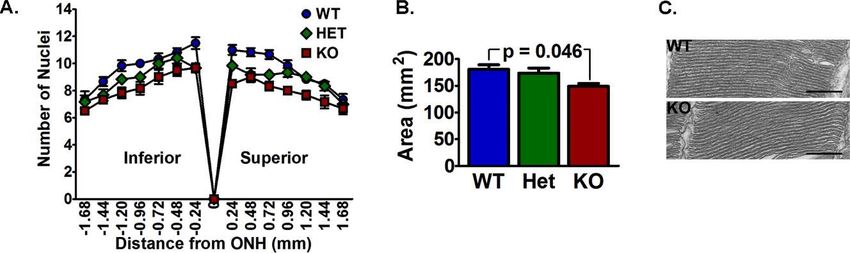
Retinal VLC-PUFA in Rod and Cone Photoreceptors                                                     IOVS j May 2014 j Vol. 55 j No. 5 j 3153

FIGURE 1. Conditional deletion of Elovl4. (A) Immunofluorescence of WT and KO retinas showed Elovl4 (red) was located in the perinuclear area
and the inner segments (IS) in WT, with only background fluorescence in the KO retinas. Scale bars: 20 lm. (B) Immunoblots showed that Elovl4
was deleted in the KO, but not in the WT retina. (C) Elovl4 mRNA in KO retinas relative to Hprt was 9% of the mRNA levels in WT retinas. Elovl4
protein expression relative to b-actin in the KO retina was 4% of the WT levels (n ¼ 7 WT mRNA and 7 WT protein; n ¼ 4 KO mRNA and 5 KO
protein). Data are expressed as mean 6 SEM.

PUFA PC levels between WT and Elovl4 KO with age.                         is important to note that the a-wave was measured at a time (8
Histograms of detected PC, PE, and PS are included in                     ms) before intrusion of the b-wave, so that voltage contribution
Supplementary Figure S1.                                                  from secondary neurons would not mask the rod response.
                                                                          The ERG responses were fit to a model of phototransduction
Rod Function Was Impaired in Retinal VLC-PUFA–                            that also excludes b-wave intrusion to determine the maximum
                                                                          rod photocurrent and the sensitivity of the phototransduction
Deficient Mice
                                                                          cascade.23 The VLC-PUFA–deficient mice had a 22% decrease in
To determine the effects of VLC-PUFAs on rod function, we                 maximum rod response amplitude (Rmp3) compared to WT
measured rod responses by ERG. The rod a-wave is a measure                and Het mice (Fig. 4C). The sensitivity (S) of Het rod response
of phototransduction cascade triggered by photon capture in               was higher compared to WT and KO mice, but was not
the rod outer segments and is indicative of rod function. The             different between the WT and KO mice (Fig. 4D).
rod b-wave reflects the summed bipolar cells response to rod-                 The b-wave was analyzed to determine the maximum
mediated glutamate concentration in the synaptic cleft.                   bipolar cell amplitude (Vmax) and the implicit time (IT), which
Although there were no differences in rod responses of 5-                 is time after the flash when the maximum b-wave amplitude
week-old WT, Het, and KO mice (Supplementary Fig. S2A), we                occurs. At 5 weeks, there were no differences in Vmax,
found reductions in rod-mediated function (a- and b-waves) in             sensitivity, or IT (Supplementary Fig. S2B). At 12 months, the
12-month-old KO mice compared to congenic controls (Fig.                  KO mice had a 32% lower Vmax than WT and 39% lower Vmax
4A). The KO responses to increasing light intensities failed to           amplitude than Het mice (Fig. 4E). At 12 months of age, the
reach the same amplitudes as the WT and Het mice (Fig. 4B). It            VLC-PUFA–deficient mice had a 16.9 and 24.4 msec delay in

FIGURE 2. Elovl4 KO mice had reduced VLC-PUFAs. (A) GC-FID showed that C16 to C22 FAs in whole retinas were not affected by Elovl4 ablation
(n ¼ 4). (B) The FAs containing more than 26 carbons were barely detectable in KO retinas. Precursor 24:6n3 in KO retina was lower compared to
WT (n ¼ 4). Data expressed as the mean 6 SD.
Retinal VLC-PUFA in Rod and Cone Photoreceptors IOVS j May 2014 j Vol. 55 j No. 5 j 3154
responses (Rmp3 and Vmax) were used to calculate the b/a-
wave ratio. Additionally, the b-wave response appears 2 orders
of magnitude before the a-wave response so ratios to specific
light intensities would be limited by the occurrence of the a-
wave response. The reduction of b-wave in the KO mice was
observed at low light intensities before the a-wave amplitude
was evident and exceeded the reduction of the a-wave
indicated by the b-/a-wave ratio (Fig. 4B). These results
indicated that the reduced rod-mediated bipolar cell responses
could not be attributed to the loss of rod cells, because the a-
and b-waves would have decreased proportionally if this were
the case.
Mice With Reduced VLC-PUFA Had a Loss of
Photoreceptors
To determine if the loss of rod cells could explain the
decreased rod responses in the VLC-PUFA–deficient mice, we
FIGURE 3. The VLC-PUFA PC is not replenished in 12-month-old Elovl4 examined the retinal morphology and found no apparent
KO retina. The PC molecular species containing VLC-PUFAs (52:12 to defects in the younger mice (Supplementary Fig. S4). There
56:11) were not replenished in the retinas of KO mice at 12 months of was, however, a small but significant loss of rod photoreceptor
age compared to 8-week-old KO mice. The VLC-PUFA values were nuclei in the 12-month-old KO mice (Fig. 5A). The total area
lower in the KO mice than in the age-matched WT mice. occupied by photoreceptor nuclei was 11% smaller in the
Elovl4 KO retina than in the control retinas at 12 months (Fig.
5B), which could account for the smaller a-wave responses in
the IT compared to WT and Het mice, respectively (Supple- the KO mice.
mentary Fig. S3). At 12 months, the KO mice had significantly The VLC-PUFAs have been suggested to provide disk
reduced b/a-wave ratio (1.92 6 0.11) compared to WT (2.19 6 structure and membrane fluidity to photoreceptor outer
0.06) and Het (2.45 6 0.13) mice (Fig. 4F), which indicated segments (OS) and, thus, influence phototransduction efficien-
that the KO b-wave had decreased disproportionately to the cy.6,14 Therefore, we examined rod outer segment disk
decreased a-wave. To avoid b-wave intrusion, the maximum ultrastructure in 12-month-old WT and KO mice. Micrographs
FIGURE 4. Rod-mediated function deteriorated in 12-month-old VLC-PUFAs–deficient mice. (A) Representative ERG response to Log 2.0 cd.s/m2
from WT, Het, and KO mice. (B) The KO rod (a-wave) and bipolar (b-wave) responses to increasing light intensities had lower amplitudes compared
to WT and Het mice. (C) Maximum rod response (Rmp3) from the KO (201.3 6 11.21) mice was lower than the responses from the WT (259.5 6
20.73) and Het (257.0 6 19.58) mice. (D) Rod Rmp3 sensitivity was increased in Het (91.6 6 12.61) mice, but was not different between the WT
(44.2 6 3.57) and KO (52.8 6 4.8) mice. (E) Maximum bipolar response (Vmax) from the KO mice was lower than the responses from WT and Het
mice. (F) The b-/a-wave ratio was lower in KO (1.92 6 0.11) compared to WT (2.19 6 0.06) and Het (2.45 6 0.13) mice (n ¼ 8 WT, 6 Het, and 6
KO). Data are represented as the mean 6 SEM. ns, not significant.Retinal VLC-PUFA in Rod and Cone Photoreceptors IOVS j May 2014 j Vol. 55 j No. 5 j 3155
FIGURE 5. The VLC-PUFA–deficient mice had a loss of photoreceptors. (A) Histogram of the number of photoreceptor nuclei in a column at
indicated distances from the optic nerve head to the peripheral retina. (B) Area occupied by photoreceptor nuclei measured from hematoxylin and
eosin–stained sections was smaller in the KO mice than in the WT mice, but was not different compared to the Het retina (n ¼ 5 WT, 7 Het, and 6
KO). (C) Representative micrographs of rod outer segments were similar between the WT and KO mice. Scale bars: 500 nm.
showed that the disks appeared similar between the two responses for all groups of mice to white, blue, and green light
groups (Fig. 5C). This supported that the reduction of the a- stimuli (Figs. 6A, 6B). The KO cone response time to white
wave was due to a loss of photoreceptor cells. light was increased slightly compared to the Het mice, but all
other cone response times were comparable between the mice
Reduced VLC-PUFA Did Not Affect Cone (Fig. 6B). Additionally, we measured cone responses to
Photoreceptors different frequencies of light stimuli (flicker) in 12–month-
old mice at 3, 10, 20, and 30 Hz pulse frequencies (Fig. 6C). We
To investigate the role of VLC-PUFAs in cone photoreceptors, found no effects of retinal VLC-PUFA depletion on cone
we used ERG to test the function of light-adapted Elovl4 WT, function. Furthermore, the presence and distribution of cones
HET, and KO mice at 12 months of age. We found similar cone were similar between the groups of mice as indicated by PNA
FIGURE 6. Analysis of cones in Elovl4 WT and KO mice. (A) Responses to white light, medium wavelength green light, and short wavelength blue
light were similar in age-matched WT and KO mice at 5 weeks and 12 months of age. (B) Cone response times were not different between age-
matched WT and KO mice. (C) Cone flicker responses (b-wave) to indicated frequencies showed no differences between WT and KO mice at 12
months of age. (D) The PNA immunolabeling of cone outer segment sheaths and cone pedicles indicated similar cone distribution between 12-
month-old WT and KO mice. Scale bars: 50 lm; 5-week (n ¼ 10 WT, 5 Het, and 10 KO). 12-month (n ¼ 8 WT, 7 Het, and 8 KO). The ERG response
data are expressed as mean 6 SEM.Retinal VLC-PUFA in Rod and Cone Photoreceptors IOVS j May 2014 j Vol. 55 j No. 5 j 3156
immunofluorescence (white) labeling of cone OS sheaths and months of age. Barabas et al.29 did not find photoreceptor
the cone pedicles located in the outer plexiform layer (OPL; degeneration in R-cKO mice, but their mice were younger,
Fig. 6D). The percent of cones relative to total area occupied which may explain the difference between the studies, since
by the photoreceptor nuclei (density of fluorescence per mm2) time exacerbates the detrimental effects of VLC-PUFA reduc-
in the 12-month-old mice was comparable between the Elovl4 tion as we have shown here (Figs. 4, 5).
WT and KO mice (8.284 6 0.3637, n ¼ 4 and 8.859 6 0.6373, Loss of rod-mediated bipolar cell responses (b-wave; Vmax)
n ¼ 5, respectively). first became evident at 6 months of age (not shown), and was
greater in Elovl4 KO mice by 12 months of age compared to
WT and Het mice (Fig. 4). These results agreed with those of
DISCUSSION Harkewicz et al.,7 who reported that R-cKO mice had
decreased b-wave responses, but disagreed with those of
Photoreceptor-specific deletion of Elovl4 significantly reduced Barabas et al.,8 who did not find rod-mediated deficits in their 6-
the levels of VLC-PUFAs in the retina, but did not alter the month-old R-cKO mice. There are several possible explanations
overall composition of the major retinal fatty acids (Figs. 2, 3; for the contradictory results. First, the genotype of the mice
Supplementary Figs. S2, S3). These results supported previ- used in the previous studies was Rod Opsin-Creþ/Elovl4f/f,
ously published work showing that Elovl4 elongates long chain which resulted in Elovl4 ablation only from rod cells (with a
PUFAs to VLC-PUFA in Elovl4-transduced cells and in vivo 77% Cre-efficiency32), whereas the Chx10-Creþ/Elovl4f/f mice
where Elovl4 was conditionally deleted independently in used here had Elovl4 deleted in rods and cones (with more
either rod or cone cells.6,7,29 In contrast with these reports, than 95% Cre-efficiency33). Additionally, in the previous
however, we have demonstrated that reduction of retinal VLC- studies, Cre expression remained in the photoreceptors of
PUFA PC in Elovl4 KO mice is maintained over time, and by 12 adult mice, unlike the Chx10-Cre mice that did not express Cre
months the differences in the abundance of retinal PC VLC- in adult photoreceptors. The Elovl4f/f mice used in all three
PUFAs between the WT and Elovl4 KO mice were amplified studies were from the same founders and had intronic LoxP
(Fig. 3). Our results implied that these fatty acids are sites.7,8 In the absence of Cre-recombinase, WT Elovl4 protein
synthesized locally and remain stable over time, and that the would be expressed and, therefore, is considered a good
effects of retinal Elovl4 deletion are likely to be compounded generic control.8 However, this strategy does not control for
with age. Furthermore, the very low levels of VLC-PUFAs at 12 potential consequences of Cre transgene expression itself. In
months indicated that other Elovl4-expressing tissues are not the current study, we included mice that expressed Cre and
able to replenish photoreceptors deficient in these fatty acids. were heterozygous for the floxed allele. Given that VLC-PUFAs
It currently is unclear as to why the Het mice did not have half were not reduced in these mice and other measured
the total amount of VLC-PUFAs compared to WT or twice as parameters were not different, our results unequivocally
much as the KO because they express one functional copy of showed the consequences of tissue-specific Elovl4 ablation,
Elovl4. One explanation could be that elongation catalysis is and assured that the resulting structural and functional
regulated by specific fatty acid concentration.19,30 Reducing abnormalities are not due to off-target effects of Cre
elongase expression could alter fatty acid concentrations expression. Whether there is a masking effect and Elovl4 is
resulting in compensentory upregulation of Elovl4 catalytic expressed elsewhere in the retina still could be the case and
activity. the Chx10 deletion would result in loss of Elovl4 in these cells.
Extensive cone function analyses at multiple time points However, our antibody can be peptide blocked as shown by
(not shown) revealed no differences in cone b-wave ampli- Agbaga et al.6 and does not recognize a protein in the Elovl4
tudes to different wavelengths or frequencies of light KO retina. Since we do not detect Elovl4 in other retinal cells it
stimulation (Figs. 6A, 6B). There were only minor changes in is unlikely that the phenotypes presented here result from
cone b-wave implicit time and no signs of cone loss even after deletion of the gene in other retinal cells.
12 months of VLC-PUFA deprivation (Fig. 6C). These results The Elovl4 protein has been well established as the
agreed with those of Barabas et al.,29 but are contrary to those elongase involved in the initial rate limiting step in the
of Harkewicz et al.,7 who found that cone-specific deletion of production of VLC-PUFAs.6–8 There has been controversy,
Elovl4 resulted in the dysfunction of cone flicker responses at however, surrounding the exact role of VLC-PUFAs in the
7 months of age compared to generic WT mice. We did not find retina. The results presented here clearly showed that VLC-
cone flicker dysfunction in our 12-month-old KO mice in PUFAs are vital for rod function and rod longevity. In the
which rods and cones were targeted for Elovl4 deletion, companion paper, we showed a reduction in rod ERG
compared to congenic control WT and Het mice. It currently is oscillatory potentials and scotopic threshold responses in KO
unclear why cone-only deletion of Elovl4 would result in cone mice, and presented biochemical and morphologic evidence
dysfunction in one study, while our mice with Elovl4 deletion that the ERG changes are correlated with reduced VLC-PUFAs
in rods and cones did not have cone structural or functional and synaptic architecture.
deficits. One possibility for the difference could be different
mouse backgrounds and/or the absence of Cre-expressing
controls in the other studies. A recent study reported that Acknowledgments
transgenic mice expressing a mutated Elovl4 had comparable The authors thank members of the Dean Bok laboratory
lipid profiles with WT mice, but had rod loss beginning at 2 (University of California, Los Angeles [UCLA], Los Angeles, CA,
months followed by a late onset of cone degeneration at 24 USA) for their help in the perfusion experiments, and Mark Dittmar
months.31 Thus, we cannot rule out cone degeneration in the for his invaluable assistance with the animals.
VLC-PUFA–deficient mice as late onset cone degeneration or as Supported by NIH Grants EY00871, EY04149, P30EY021725, and
a secondary late event resulting from the rod degeneration. P20RR017703 (REA); and EY019494 (MHE); and by the Founda-
We found that mice with reduced VLC-PUFAs had dimin- tion Fighting Blindness (REA); Research to Prevent Blindness
ished rod function (a-wave, Rmp3) at 12 months of age (Departmental); and Grants GM103508 (GER, JVB) and EY016077
compared to controls, which correlated with a loss of (JVB, GER).
photoreceptor nuclei (Figs. 4, 5), in agreement with Harkewicz Disclosure: L.D. Bennett, None; R.S. Brush, None; M. Chan,
et al.,7 who also reported that rod-specific Elovl4 conditional None; T.A. Lydic, None; K. Reese, None; G.E. Reid, None; J.V.
KO (R-cKO) adult mice had retinal degeneration at 10 and 15 Busik, None; M.H. Elliott, None; R.E. Anderson, NoneRetinal VLC-PUFA in Rod and Cone Photoreceptors IOVS j May 2014 j Vol. 55 j No. 5 j 3157
References with protection from light damage. J Neurochem. 2008;105:
784–796.
1. Edwards AO, Donoso LA, Ritter R III. A novel gene for 17. Bligh EG, Dyer WJ. A rapid method of total lipid extraction and
autosomal dominant Stargardt-like macular dystrophy with purification. Canad J Biochem Physiol. 1959;37:911–917.
homology to the SUR4 protein family. Invest Ophthalmol Vis 18. Morrison WR, Smith LM. Preparation of fatty acid methyl
Sci. 2001;42:2652–2663. esters and dimethylacetals from lipids with boron fluoride–
2. Zhang K, Kniazeva M, Han M, et al. A 5-bp deletion in ELOVL4 methanol. J Lipid Res. 1964;5:600–608.
is associated with two related forms of autosomal dominant 19. Yu M, Benham A, Logan S, et al. ELOVL4 protein preferentially
macular dystrophy. Nat Genet. 2001;27:89–93. elongates 20:5n3 to very long chain PUFAs over 20:4n6 and
3. Donoso LA, Frost AT, Stone EM, et al. Autosomal dominant 22:6n3. J Lipid Res. 2012;53:494–504.
Stargardt-like macular dystrophy: founder effect and reassess- 20. Julia V, Busik GER, Lydic TA. Global analysis of retina lipids by
ment of genetic heterogeneity. Arch Ophthalmol. 2001;119: complementary precursor ion and neutral loss mode tandem
564–570. mass spectrometry. Methods Mol Biol. 2009;579:33–70.
4. Griesinger IB, Sieving PA, Ayyagari R. Autosomal dominant 21. Haimi P, Uphoff A, Hermansson M, Somerharju P. Software
macular atrophy at 6q14 excludes CORD7 and MCDR1/PBCRA tools for analysis of mass spectrometric lipidome data. Anal
loci. Invest Ophthalmol Vis Sci. 2000;41:248–255. Chem. 2006;78:8324–8331.
5. Kniazeva M, Chiang MF, Morgan B, et al. A new locus for 22. Li F, Marchette LD, Brush RS, et al. DHA does not protect
autosomal dominant stargardt-like disease maps to chromo- ELOVL4 transgenic mice from retinal degeneration. Mol Vis.
some 4. Am J Hum Genet. 1999;64:1394–1399. 2009;15:1185–1193.
6. Agbaga MP, Brush RS, Mandal MNA, Henry K, Elliott MH, 23. Hood DC, Birch DG. Rod phototransduction in retinitis
Anderson RE. Role of Stargardt-3 macular dystrophy protein pigmentosa: estimation and interpretation of parameters
(ELOVL4) in the biosynthesis of very long chain fatty acids. derived from the rod a-wave. Invest Ophthalmol Vis Sci.
Proc Nat Acad Sci U S A. 2008;105:12843–12848. 1994;35:2948–2961.
7. Harkewicz R, Du H, Tong Z, et al. Essential role of ELOVL4 24. Lamb TD, Pugh EN Jr. A quantitative account of the activation
protein in very long chain fatty acid synthesis and retinal steps involved in phototransduction in amphibian photore-
function. J Biol Chem. 2012;287:11469–11480. ceptors. J Physiol. 1992;449:719–758.
8. Barabas P, Liu A, Xing W, et al. Role of ELOVL4 and very long- 25. Weymouth AE, Vingrys AJ. Rodent electroretinography:
chain polyunsaturated fatty acids in mouse models of Stargardt methods for extraction and interpretation of rod and cone
type 3 retinal degeneration. Proc Natl Acad Sci U S A. 2013; responses. Prog Retin Eye Res. 2008;27:1–44.
110:5181–5186. 26. Naka KI, Rushton WA. S-potentials from luminosity units in the
9. Logan S, Agbaga MP, Chan MD, et al. Deciphering mutant retina of fish (Cyprinidae). J Physiol. 1966;185:587–599.
ELOVL4 activity in autosomal-dominant Stargardt macular 27. Marchette LD, Wang H, Li F, Babizhayev MA, Kasus-Jacobi A.
dystrophy. Proc Natl Acad Sci U S A. 2013;110:5446–5451. Carcinine has 4-hydroxynonenal scavenging property and
10. Mandal MNAR, Wong PW, Gage PJ, Sieving PA, Ayyagari R. neuroprotective effect in mouse retina. Invest Ophthalmol Vis
Characterization of mouse orthologue of ELOVL4: genomic Sci. 2012;53:3572–3583.
organization and spatial and temporal expression. Genomics. 28. Liu IS, Chen JD, Ploder L, et al. Developmental expression of a
2004;83:626–635. novel murine homeobox gene (Chx10): evidence for roles in
11. Vasireddy V, Uchida Y, Salem N Jr, et al. Loss of functional determination of the neuroretina and inner nuclear layer.
ELOVL4 depletes very long-chain fatty acids (> or ¼C28) and Neuron. 1994;13:377–393.
the unique omega-O-acylceramides in skin leading to neonatal 29. Barabas P, Liu A, Xing W, et al. Role of ELOVL4 and very long-
death. Hum Mol Genet. 2007;16:471–482. chain polyunsaturated fatty acids in mouse models of Stargardt
12. Li W, Sandhoff R, Kono M, et al. Depletion of ceramides with type 3 retinal degeneration. Proc Natl Acad Sci U S A. 2013;
very long chain fatty acids causes defective skin permeability 110:5181–5186.
barrier function, and neonatal lethality in ELOVL4 deficient 30. Hubbard AF, Askew EW, Singh N, Leppert M, Bernstein PS.
mice. Int J Biol Sci. 2007;3:120–128. Association of adipose and red blood cell lipids with severity
13. Furland NE, Oresti GM, Antollini SS, Venturino A, Maldonado of dominant Stargardt macular dystrophy (STGD3) secondary
EN, Aveldano MI. Very long-chain polyunsaturated fatty acids to an ELOVL4 mutation. Arch Ophthalmol. 2006;124:257–
are the major acyl groups of sphingomyelins and ceramides in 263.
the head of mammalian spermatozoa. J Biol Chem. 2007;282: 31. Kuny S, Filion M-A, Suh M, Gaillard F, Sauvé Y. Long-term
18151–18161. retinal cone survival and delayed alteration of the cone mosaic
14. Aveldaño MI, Sprecher H. Very long chain (C24 to C36) in a transgenic mouse model of Stargardt-like dystrophy
polyenoic fatty acids of the n-3 and n-6 series in dipolyunsa- (STGD3). Invest Ophthalmol Vis Sci. 2013;55:424–439.
turated phosphatidylcholines from bovine retina. J Biol Chem. 32. Le YZ, Zheng L, Zheng W, et al. Mouse opsin promoter-
1987;262:1180–1186. directed Cre recombinase expression in transgenic mice. Mol
15. Aveldaño MI. A novel group of very long chain polyenoic fatty Vis. 2006;12:389–398.
acids in dipolyunsaturated phosphatidylcholines from verte- 33. Rowan S, Cepko CL. Genetic analysis of the homeodomain
brate retina. J Biol Chem. 1987;262:1172–1179. transcription factor Chx10 in the retina using a novel
16. Ueki Y, Wang J, Chollangi S, Ash JD. STAT3 activation in multifunctional BAC transgenic mouse reporter. Dev Biol.
photoreceptors by leukemia inhibitory factor is associated 2004;271:388–402.You can also read