Anti-inammatory protein TNFα-stimulated gene-6 (TSG-6) reduces inammatory response after brain injury in mice
←
→
Page content transcription
If your browser does not render page correctly, please read the page content below
Anti-inflammatory protein TNFα-stimulated gene-6
(TSG-6) reduces inflammatory response after brain
injury in mice
Kazadi Nadine Mutoji
University of Houston
Mingxia Sun
University of Houston
Amanda Nash
University of Houston
Sudan Puri
University of Houston
Vincent Hascall
Cleveland Clinic
Vivien J Coulson-Thomas ( vcoulsonthomas@gmail.com )
University of Houston https://orcid.org/0000-0002-5848-0225
Research article
Keywords: TSG-6, glial scar, astrocytes, glycosaminoglycans and inflammation
Posted Date: November 12th, 2020
DOI: https://doi.org/10.21203/rs.3.rs-23682/v3
License: This work is licensed under a Creative Commons Attribution 4.0 International License.
Read Full License
Version of Record: A version of this preprint was published at BMC Immunology on August 4th, 2021. See
the published version at https://doi.org/10.1186/s12865-021-00443-7.
Page 1/24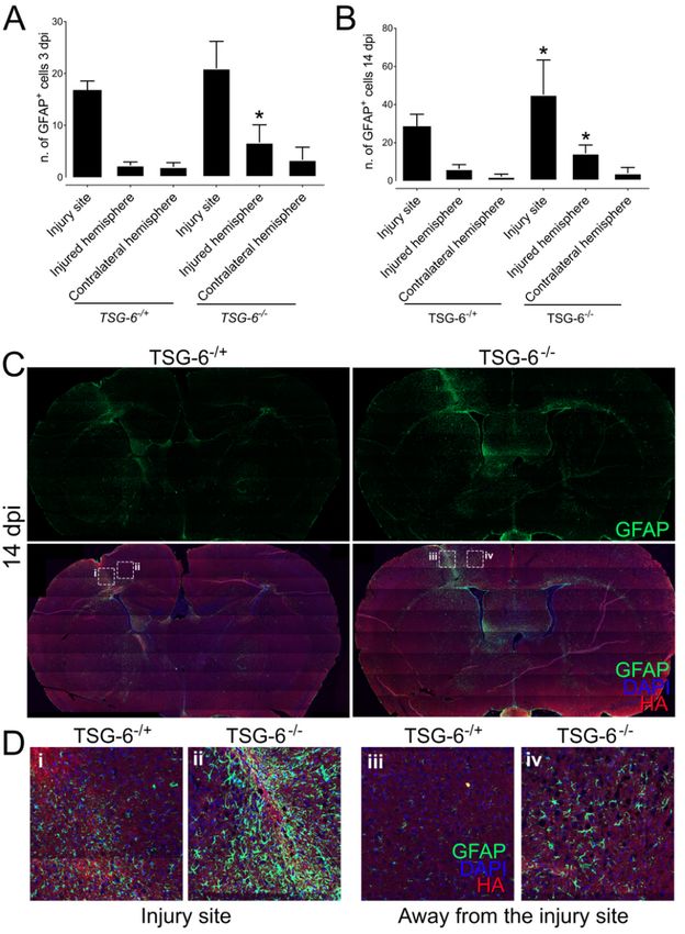
Abstract
Background: Current research suggests that the glial scar surrounding penetrating brain injuries is
instrumental in preserving the surrounding uninjured tissue by limiting the inflammatory response to the
injury site. We recently showed that tumor necrosis factor (TNF)-stimulated gene-6 (TSG-6), a well-
established anti-inflammatory molecule, is present within the glial scar. In the present study we
investigated the role of TSG-6 within the glial scar.

Methods: TSG-6 null and littermate control mice were subjected to penetrating brain injuries (2 mm
puncture wound to the frontal cortex), after which both the injury site and remaining injured hemisphere
were analyzed. The presence of activated astrocytes, inflammatory markers and glial scar components
was evaluated by real-time PCR and immunofluorescence.

Results: Our findings show that mice lacking TSG-6 present a more severe inflammatory response after
injury, which was correlated with an enlarged area of astrogliosis beyond the injury site.

Conclusion: Our data provides evidence that TSG-6 has an anti-inflammatory role within the glial scar.

Background
Traumatic brain injury (TBI) is a major medical concern that affects over 10 million people in the world
each year (1,2) . A variety of injuries can cause TBI leading to a range of injury severities (3–7). With
improved medical interventions over the years, the mortality rate due to TBI has decreased, resulting in a
significant number of people living with the long-term effects of TBI. It is well accepted that in addition to
the immediate effects of TBI there are also multiple potential long-term gradually evolving sequelae that
are influenced by the type of injury, severity of the injury and medical interventions at the time of injury
(8,9). Additionally, a link between mild traumatic brain injuries and Alzheimer’s disease or chronic
traumatic encephalopathy has long been suspected (10). At present, long-term effects of repeated TBI
have been seen in multiple sports-related injuries, including post-traumatic parkinsonism, post-traumatic
dementia and chronic post-concussion syndrome (11–14). Thus, studying the short- and long-term
consequences of TBI at a cellular and molecular level may lead to a new understanding and perhaps
better long-term management of such injuries via new and/or refined treatment strategies.

Astrogliosis is a hallmark of TBI, which commences hours after injury and leads to an abnormal increase
in the number of activated astrocytes in and around the injury site (15,16). Immediately after injury (acute
phase), astrocytes are activated, becoming highly proliferative and up-regulating the production of
extracellular proteins (17–19). These astrocytes and their deposited extracellular matrix in and around
the injury site form a glial scar. Over the years, a significant body of evidence has demonstrated that the
glial scar contains molecules, such as chondroitin sulfate proteoglycans (CSPGs), that impede axonal
growth, thus inhibiting neuronal regeneration (15,20–23). The intensity of the acute and chronic reactive
astrogliosis, including the quantity and composition of the glial scar, affects immediate and long-term
effects of TBI (6,16,24,25). Penetrating brain injuries (PBIs) cause direct parenchymal laceration, neuronal
                                                   Page 2/24
cell loss and hemorrhage, which lead to focal tissue damage at the injury site. Astrogliosis is triggered
after TBIs forming a glial scar in and around the injury site (26–29). Importantly, uninjured tissue
bordering the injury site is also subject to astrogliosis, and the process of glial scarring therefore extends
beyond the injury site (30). Given the fact that glial scarring limits regeneration after injury, many studies
have investigated whether limiting astrogliosis after injury, with particular focus on limiting deposition,
could potentially promote regeneration (23,27,31–34). Although many studies were able to demonstrate
beneficial effects of limiting glial scarring on neuronal regeneration, many others were inconclusive or
actually found there was an increased inflammatory response culminating in tissue damage beyond the
injury site and an increase in neuronal loss. Thus, mounting evidence indicates that reactive astrocytes
surrounding the injury site are instrumental in preserving the surrounding uninjured tissue by forming scar
borders, which separate damaged and inflamed tissue from adjacent viable neural tissue (15,16,24,35–
40). Sofroniew and colleagues elegantly demonstrated that targeting astrocytes after brain and spinal
cord injury leads to increased inflammation, delayed recovery and increased neuronal loss (39,41–44).
Moreover, the inhibition of astrocyte proliferation prolongs the healing period following central nervous
system (CNS) injury (45). Data from Hermann et al. show that GFAP-driven ablation of STAT3 in
astrocytes leads to the loss of lesion demarcation and subsequent glial scar formation, and, in turn,
results in increased invasion of inflammatory cells into adjacent viable tissue and further spread of
inflammation (46). This suggests that early glial scar formation by astrocytes restricts movement of
inflammatory cells located within the lesion site into adjacent healthy tissue, thereby restricting tissue
damage to the injury site. Thus, a recent body of evidence suggests scar tissue bordering the injury site is
necessary for limiting inflammation and tissue damage to the injury site (37,41). There is currently a
significant number of studies investigating how reactive astrocytes regulate and limit inflammation to the
injury site, and which cellular components and major pathways could play a role in this process
(20,35,41,45,47).
We recently found that tumor necrosis factor (TNF)-stimulated gene-6 (TSG-6) is secreted by astrocytes
after injury and is a major constituent of the glial scar, but the role it plays within the glial scar remains to
be established (48). TSG-6 is a 35-kDa protein that is secreted by a wide range of cell types in response to
inflammatory mediators and growth factors (49), and was originally identified as a gene product induced
in fibroblasts by TNF (12). TSG-6 contains a link module domain that mediates its interaction with the
glycosaminoglycans (GAGs) hyaluronan (HA) and CS (49–51). Our recent study identified that TSG-6 is
expressed in the CNS, where it catalyzes the transfer of heavy chains (HCs) from Inter-a-Inhibitor (IaI, also
known as ITI) onto HA, forming a specialized HA/HC/TSG-6 matrix within the glial scar, but the role of
this specialized matrix within the glial scar remains to be established (48,52–56). This specific
HA/HC/TSG-6 matrix has previously been shown to be monocyte-adhesive in other tissues and is
believed to be present in most, if not all, inflammatory processes (57,58). These TSG-6 modified HA
matrices bind inflammatory cells, and the interaction of these cells with this matrix modulates their
responses, which are central to pathological inflammation (59–65). The main objective of this study was
to investigate the role TSG-6, a constituent of the glial scar, has in astrogliosis after a PBI. Given the well-
characterized anti-inflammatory role of TSG-6 in other sites, the premise of this study was that TSG-6
Page 3/24could participate in the formation of an immunosuppressive environment within the glial scar. Our
findings show that TSG-6 null mice present a more severe inflammatory response and increased glial scar
deposition after injury when compared to littermate control mice. This increased inflammatory response
in TSG-6 null mice was correlated with an enlarged area of astrogliosis beyond the injury site.
Methods
TSG6 null (TSG6-/-) or heterozygous (TSG6+/-) mice and animal maintenance
Transgenic Tsg-6 null mice (Tnfip6D/D), hereafter referred to as Tsg-6-/- mice, and heterozygous mice,
hereafter referred to as Tsg-6+/- mice, were maintained as previously described (56). Our previously
published work demonstrated that Tsg-6+/- mice present a similar distribution of astrocytes throughout
the brain to wild-type (wt) mice (48). Moreover, Tsg-6+/- mice have previously been shown not to display
a phenotype and present similar TSG-6 expression levels as wt mice, and were therefore used as
littermate controls in our study (56). Experimental procedures for handling the mice were approved by the
Institutional Animal Care and Use Committee (IACUC), University of Houston under protocol 16-036.
Brain Injury
Mice (7 to 8 weeks old) were anesthetized with ketamine (80-100 mg/kg - Vedco INC, Catalog# 07-890-
8598) and xylazine (5-10 mg/kg, Akorn INC, Catalog# 07-808-1947) by IP injection and allowed to go into
full anesthetic state. A sterile surgical drill (Precision Tools, Model Craft PPV2237) was used to make a
hole of approximately 1.5 mm in diameter in the skull over the right frontal cortex at the stereotaxic
coordinates AP: 1.0mm, ML: 1.5mm, and DV: 1.5mm, according to Franklin and Paxinos (66). A 30-gauge
needle (Exel, Catalog# 26437) was then used to make a puncture wound at a depth of 2 mm. After injury,
the skin at the surgical site was closed with two sutures. The area was then cleaned with 70% ethanol,
and mice were placed on a heating pad and monitored until they regained consciousness prior to being
transferred to a clean cage. All surgeries were carried out at the same time of day to minimize bias. Mice
were monitored daily and did not show any decrease in weight ≥ 15% when compared to their pre-
surgical weight. Mice were euthanized at 1, 3 and 5 days post injury to study the acute effects of brain
injury, and at 10 and 14 days to study long-term/chronic effects. Five mice per experimental group were
used for the real-time PCR analysis and at least seven mice per experimental group were used for
immunofluorescence analysis.
Perfusion fixation and brain tissue processing
Brain samples were collected at 1, 3, 5, 10, and 14 days post injury for immunofluorescence analyses.
Briefly, mice were initially injected with a lethal dose of combined anesthetics, ketamine and xylazine.
Once mice were under deep anesthesia, abdominal excisions were performed to expose the heart, which
was used to perfuse 2% formalin (Fisher Scientific, Catalog# SF100-4) throughout the whole body via a
gravity-driven flow system for whole body fixation. Subsequently, the brain was isolated from the skull
Page 4/24and further immerse fixed for 2 days in 2% paraformaldehyde (Electron Microscopy Sciences, Catalog#
15710). For cryosection processing, brains were immersed in 30% sucrose for 2 days, embedded in OCT
embedding medium (Fisher Healthcare, Catalog# 4585) and frozen. Sections 10 μm thick were obtained,
mounted on superfrost slides (VWR, Catalog# 48311-703) and stored at -20°C until use.
Immunofluorescence
Upon use, the slides were heated at 65oC for 30 minutes and, subsequently, sections were washed with
PBS to remove tissue freezing medium. Sections were then treated with 0.1% glycine (Fisher Chemical,
Catalog# G46-500), blocked with 5% FBS (Seradigm, Catalog# 3100-500) and permeabilized with 0.1%
saponin prepared in PBS. Sections were then incubated with the primary antibodies anti-Tenascin
(Abcam, Ab108930), anti-GFAP (Abcam, Ab4647), anti-CD68 (Abcam, Ab31630) and anti-b III tubulin
(Covance, PRB-435P-100). Sections were washed and incubated with appropriate secondary donkey
antibodies conjugated with Alexa Fluor® 488 (Life Technologies) or Alexa Fluor® 555 (Life
Technologies) for one hour at 18oC. For HA staining, tissues were incubated with biotinylated HA binding
protein (385911, Millipore) followed by NeutrAvidin®Alexa 555 (Life Technologies). The tissues were then
washed and nuclei stained with 4’,6-diamidino-2-phenylindole (DAPI, Sigma-Aldrich). Sections were
mounted in Prolong®Gold (Molecular Probes) and imaged using a ZEISS LSM 800 Confocal microscope
with Airyscan. Secondary controls were done with a goat IgG isotype control (ab37388; Abcam) in place
of the primary antibody and did not yield any significant staining (results not shown). For imaging,
multiple z-stack tiles were captured of entire brain sections and frames were processed together into a
single image (using the stitching mode followed by full orthogonal projection) using Zen Software
(Zeiss). The number of GFAP+ and CD68+ cells in and around the injury site were counted by two
independent investigators in a blinded manner and the relative Fluorescent intensity was measured using
the Zen Software (Zeiss). At least 2 sections were scanned and analyzed from each animal for each set
of antibodies and representative images shown in the figures.
RNA extraction from brains and real-time PCR analysis
Brains collected from injured mice at 1, 5 and 10 days post injury were used for RNA extraction. At least 5
mice were used per experimental group and each animal was analyzed separately. Briefly, mice were
euthanized and brain tissue was immediately isolated from each mouse. Injury sites (A samples) were
dissected from the rest of the injured right hemisphere, transferred into a labeled Eppendorf tube and
immediately immersed in liquid nitrogen. The remaining right hemisphere brain tissue (B samples) from
each animal was transferred into a different tube and also frozen as described. The samples were kept
at -80oC until RNA extraction. Total RNA was isolated from these tissue samples using Trizol® Reagent
(Invitrogen, Carlsbad, CA) and chloroform extraction (Sigma-Aldrich, Catalog# 650498). First strand cDNA
was reverse transcribed using 1.5 to 2 μg of total RNA and the high capacity cDNA Reverse Transcription
kit (Applied Biosystems, catalog# 4368814, lot 00593854) according to the manufacturer’s instructions.
Quantitative real-time PCR amplification was performed on 1 μg or 50 ng of the cDNA (1:5) using the
PowerUp SYBR Green Master Mix kit (Applied Biosystems, Catalog# A25918) in a CXF Connect Real-time
Page 5/24System from BIO-RAD, using an activation cycle of 95°C for 10 min, 40 cycles of 95°C for 15 seconds and
60°C for 1 min. A complete list of primers used in this study is shown in Table 1. Gene expression levels
were normalized against Actb and Gapdh using the 2−ΔCt and/or 2−ΔΔCt methods.
Statistical analysis
All values are presented as the mean ± standard deviation of the mean. The difference between the two
groups was compared by means of the Student’s t-test. P ≤ 0.05 was considered to be statistically
significant. Statistical analysis was performed using the GraphPad Prism version 7 software package
(GraphPad Software, San Diego, CA, USA). * was used to indicate statistical differences of ≤ 0.05. Unless
indicated otherwise, * indicates the statistical difference of Tsg-6-/- mice compared to Tsg-6+/- mice for
each time point.
Results
TSG-6 expression after PBI
In order to investigate whether TSG-6 is present in the glial scar after brain injury, we analyzed the
expression profile of Tsg6 in the injury site and injured hemisphere before and after a PBI in Tsg-6+/- mice
(Figure 1A). There was a 2-fold increase in Tsg-6 expression 5 days after injury when compared to
uninjured mice. There was a further increase in Tsg-6 expression over time after injury, with expression
increasing 2 fold from 5 to 10 days after injury (Figure 1A). Interestingly, we did not find a difference in
the expression levels of Tsg-6 between the injury site and the remaining hemisphere, indicating that Tsg-6
expression is not contained solely to the injury site (Figure 1A). Therefore, there is also an increase in Tsg-
6 expression in the surrounding tissue after injury. No Tsg-6 expression was identified in any of the
samples from Tsg-6-/- mice confirming that these mice are indeed null for Tsg-6.
Analysis of astrocyte recruitment after PBI
We assessed the level of astrogliosis in the injury site and in the remaining injured hemisphere by
quantifying the levels of GFAP+ astrocytes using real-time PCR (Figure 1B and C). For such, we isolated
mRNA from the injury site and remaining injured hemisphere 1 and 5 days after injury of Tsg-6-/- and Tsg-
6+/- mice. Both Tsg-6-/- and Tsg-6+/- mice presented an increase in the levels of GFAP expression in the
injury site when compared to the remaining injured hemisphere, which corroborates literature (36,63,64).
Tsg-6-/- mice showed a significant increase in GFAP levels within the injury site at both 1 and 5 days post-
injury when compared to Tsg-6+/- mice (Figure 1B and C). This data indicates that Tsg-6-/- mice have more
astrocytes in the injury site when compared to Tsg-6+/- mice. At 5 days after injury, there was a significant
increase in GFAP expression in the injured hemisphere of Tsg-6-/- mice compared to Tsg-6+/- mice,
indicating that Tsg-6-/- mice present astrogliosis beyond the injury site at 5 days post-injury.
The effect of TSG-6 on the secretion of inflammatory markers after PBI
Page 6/24The inflammatory response was also assessed in Tsg-6-/- and Tsg-6+/- mice 1, 5 and 10 days post-injury
by quantifying the expression levels of NFkB, Rantes and IL1b (Figure 2). Higher expression levels of
NFkB, Rantes and IL1b were detected in Tsg-6-/- mice when compared to Tsg-6+/- mice during the acute
phase after injury. Specifically, a ~2.5-fold and 3-fold increase in NfkB expression was found in the injury
site and remaining injured hemisphere, respectively, in Tsg-6-/- mice compared to Tsg-6+/- mice 5 days
after injury (Figure 2B). 10 days after injury there was still a significant increase in NfkB expression in the
surrounding hemisphere of Tsg-6-/- mice when compared to Tsg-6+/- mice (Figure 2C). No significant
differences were found in the expression of NfkB between Tsg-6-/- and Tsg-6+/- mice 1 day after injury
(Figure 1A). The levels of Ccl5 (Rantes) were also assessed 1, 5 and 10 days post-injury. There was a
significant increase in the expression of Rantes in the injured hemisphere of Tsg-6-/- mice when compared
to Tsg-6+/- mice (a 4-fold increase) 5 days after injury; however, no difference was found between Tsg-6-/-
and Tsg-6+/- mice 1 and 10 days post-injury (Figure 2D-F). IL1b levels were increased in the injury site of
TSG-6-/- mice when compared to Tsg-6+/- mice at 1 day post-injury (Figure 2G). At 5 days post-injury a 3-
fold and 4-fold increase in the expression of IL1b were noted in the injury site and remaining injured
hemisphere, respectively, of Tsg-6-/- mice when compared to Tsg-6+/- mice (Figure 2H). At 10 days post-
injury, a 2.5-fold increase in the expression of IL1b was noted in the injury site of Tsg-6-/- mice when
compared to Tsg-6+/- mice (Figure 2I).
The effect of TSG-6 on the activation of microglia and infiltration of macrophages into the injury site after
PBI
In order to assess the inflammatory response in Tsg-6-/- and Tsg-6+/- mice, we also evaluated the number
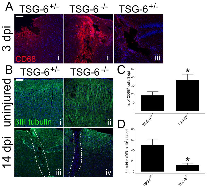
of CD68+ cells present within the injury site at 3 days post-injury (Figure 3). CD68 is routinely used as a
marker for macrophages and activated microglia. There was a significant increase in the number of
CD68+ cells in and around the injury site of Tsg-6-/- mice when compared to Tsg-6+/- mice (Figure 3A
panels i and ii). Importantly, even when analyzing deeper regions of the injury site of Tsg-6+/- mice, the
level of CD68+ cell infiltration was not as intense as that observed in Tsg-6-/- mice (Figure 3A panel iii).
The combined number of CD68+ cells in the injury site and within a range of 100 mm from the wound
edge was counted from images obtained from 2 different sections from at least 5 mice from each
experimental point. A 2-fold increase in CD68+ cells was found in Tsg-6-/- mice when compared to Tsg-6+/-
mice.
Correlation between increased inflammatory response and neuronal damage
In order to verify whether the increased inflammatory response observed in Tsg-6-/- mice correlates with
neuronal loss, the distribution of neurons in and around the injury site was analyzed in Tsg-6-/- and Tsg-
6+/- mice 14 days post-injury (Figure 3B). For such, b III tubulin was used as a tissue-specific marker for
identifying neurons within injured and non-injured brains. The distribution of b III tubulin can be seen in
the equivalent region of uninjured Tsg-6-/- and Tsg-6+/- mice (Figure 3 B panels i and ii). A significant
Page 7/24increase in the area devoid of b III tubulin staining can be observed in and around the injury site of Tsg-6-/-
mice when compared to Tsg-6+/- mice 14 days post-injury (Figure 3B iii and iv). The relative fluorescence
units (RLU) were quantified from an image of the injury site captured from at least 3 mice per
experimental point. There was a 4-fold decrease in b III tubulin staining in and around the injury site of
Tsg-6-/- mice when compared to Tsg-6+/- mice 14 days post-injury (Figure 3D).
The effect of TSG-6 on the secretion of glial scar components after PBI
We also evaluated glial scar secretion within the injury site and injured hemisphere by evaluating the
expression levels of the biosynthetic enzymes responsible for HA and CS chain elongation, specifically
hyaluronan synthase 2 (Has2), carbohydrate (chondroitin 4) sulfotransferase (chst 11) and carbohydrate
(chondroitin 4) sulfotransferase 12 (chst 12) (Figure 4). Has2 expression increased in the injury site when
compared to the remaining injured hemisphere 5 days post injury in both Tsg-6+/- and Tsg-6-/- mice,
confirming the numerous previously published reports showing that HA is an integral component of the
glial scar. Interestingly, there was a 2-fold increase in Has2 expression in the injury site of Tsg-6-/- mice
when compared to Tsg-6+/- mice 5 days after injury, indicating that there is a higher rate of glial scar
production in Tsg-6-/- mice when compared to Tsg-6+/- mice (Figure 4A). At 10 days post-injury, Has2
expression was still increased by 2-fold in Tsg-6-/- mice when compared to Tsg-6+/- mice, but at this time
point there was also an increase in Has2 expression in the remaining injured hemisphere of Tsg-6-/- mice
when compared to Tsg-6+/- mice (Figure 4B). Thus, at 10 days after injury, in Tsg-6-/- mice, the expression
of glial scar components was no longer limited to the injury site, but was also present within the
remaining injured hemisphere. Interestingly, this was also true for the expression of Chst11 and Chst12,
which showed a 5-fold and 4-fold increase, respectively, within the injured hemisphere of Tsg-6-/- mice at
5 days post injury when compared to Tsg-6+/- mice (Figure 4C and E). The increase in Chst11 and Chst12,
in both the injury site and injured hemisphere, was maintained through to 10 days post-injury (Figure 4D
and F).
The effect of TSG-6 on astrocyte activation and recruitment after PBI
In order to further investigate the process of astrogliosis in Tsg-6+/- and Tsg-6-/- mice, injured brains were
harvested and processed for histology. Sections were stained for GFAP in order to assess the distribution
of astrocytes in and around the injury site, and also throughout the remaining brain tissue. The number of
astrocytes (GFAP+ cells) was counted within the injury site, throughout the injured hemisphere, and also
throughout the contralateral hemisphere 3 and 14 days post-injury (Figure 5A and B). At 3 and 14 days
post-injury there was a significant increase in the number of astrocytes within the injury site when
compared to the injured hemisphere and contralateral hemisphere in both Tsg-6+/- and Tsg-6-/- mice. At 3
days post-injury there was no significant difference between the number of astrocytes within the injury
site between Tsg-6+/- and Tsg-6-/- mice; however, there was a significant increase in the number of
astrocytes within the injured hemisphere in Tsg-6-/- mice when compared to Tsg-6+/- mice (Figure 5A). At
14 days post-injury there was a significant increase in the number of astrocytes within the injury site and
Page 8/24injured hemisphere in Tsg-6-/- mice when compared to Tsg-6+/- mice (Figure 5B and D). The increase in
astrocytes can be seen beyond the injury site in Tsg-6-/- mice (Figure 5C and D panel iv).
Discussion
Chondroitin sulfate proteoglycans (CSPGs) are well established as major extracellular matrix
components in the central nervous system (69). Over a decade ago, Silver et al. identified that CSPGs
within the glial scar inhibit axonal growth, and this triggered a great deal of interest in targeting CS within
the scar tissue as a means to promote axonal regeneration (32,70–72). Over the years, strategies utilizing
the enzymes chondroitinase ABC (ChABC) and ChAC have been used to remove the CS component of the
glial scar as a means to promote axonal growth and regeneration (50,73–77). Many studies have shown
that specifically removing CS within the glial scar is enough for axons to grow across the injury site
(32,70,78,79). However, significant regeneration was never observed in these studies, and many groups
found limited or no improvement after targeting CS within the glial scar (70). One unique characteristic of
TSG-6 is its known ability to bind to a number of ligands including HA, CS and core proteins of
proteoglycans (i.e. versican and aggrecan), forming specific HA/HC/TSG-6 and/or CS/HA/HC/TSG-6
matrices with immunosuppressive characteristics (61,80–84). Our previous study suggests these
HA/HC/TSG-6 matrices are also present within the glial scar (48). Therefore, given that TSG-6 directly
binds to both HA and CS to form specific anti-inflammatory matrices, the ChABC and ChAC treatments
used over the years to target the glial scar as a means to promote regeneration would also have removed
TSG-6, a known anti-inflammatory molecule that is also a component of the glial scar (82). The loss of
TSG-6 by these treatments could, in part, explain why significant functional recovery was never obtained
after ChABC and/or ChAC treatments.
To explore the role of TSG-6 in TBI, specifically astrogliosis, we compared the differences in injury
outcomes in Tsg-6-/- and Tsg-6+/- mice after PBIs. Our data show an increase in TSG-6 expression in the
injured hemisphere of Tsg-6+/- mice after TBI. This increase in expression of TSG-6 after CNS insults
supports our earlier findings in a rat model that astrocytes secrete high levels of TSG-6 upon injury, which
aids in the formation of a specialized HA/HC/TSG-6 matrix as part of an inflammatory response (48).
Since TSG-6 is known for having anti-inflammatory properties, to further study whether high levels of
TSG-6 serve a purpose of rapidly suppressing inflammation after injury, we performed similar penetrating
injuries in Tsg-6-/- mice. We used immunofluorescence and RNA expression analyses of inflammatory
and glial scar markers to elucidate the outcome during the acute phase and chronic phase of TBI. During
the acute phase after injury, the observed increase in astrocyte activation, inflammatory cell infiltration
and expression of inflammatory cytokines in Tsg-6-/- mice indicate that the loss of TSG-6 results in a
greater inflammatory response. Moreover, during the chronic phase of injury, unrestricted inflammatory
response was observed throughout the injured hemisphere and was not limited to the injury site, as is
seen after normal glial scar formation. Thus, injured Tsg-6-/- mice appear to experience more severe
tissue damage than their Tsg-6+/- counterpart, both within and around the injury site. Thus, the loss of
TSG-6 allows the damage to spread from the injury site to neighboring healthy tissues. We postulate that
Page 9/24the cause of such widespread damage is due to the lack of the specialized HA-TSG6 or HA/HC/TSG-6
matrix, which could possibly serve to stabilize the glial scar and form an immunosuppressive
environment, thereby protecting adjacent tissue from further damage. This hypothesis is further
supported by the increase in CSPG and HA biosynthesis, both glial scar components, in Tsg-6-/- mice.
Specifically, these mice show increased Has2, Chst11 and Chst12 expression levels in tissues collected
after the onset of glial scarring, and also during the chronic phase of astrogliosis, indicating an increase
in scar tissue formation. This increase in expression was not only observed at the injury site, but also
throughout the whole injured hemisphere, suggesting that the tissue damage spreads beyond the injury
site in the absence of TSG-6. Collectively, these results demonstrate that the loss of TSG-6 leads to a
more severe inflammatory response and, consequently, increased scarring after TBI. Thus, our results
support the hypothesis put forward by many groups over the past decade that preventing the formation
of the glial scar leads to inflammation and damage beyond the injury site. We also provide experimental
evidence that shows that the glial scar functions to restrict the damage to the injury site. Importantly,
these findings should be taken into account when attempts are made to disrupt the glial scar as a means
to promote neuronal regeneration, since preventing formation of the glial scar may not have the beneficial
outcomes as previously presumed.
Conclusion
Our results show that TSG-6 has an anti-inflammatory role in the glial scar. Our study further supports the
hypothesis that the glial scar forms a protective border surrounding the injury site thereby preventing the
spread of inflammation and damage beyond the injury site.
Abbreviations
TBI: Traumatic brain injury
TNF: Tumor necrosis factor
TSG-6: TNF-stimulated gene-6
GFAP: Glial Fibrillary Acidic Protein
VIM: Vimentin
CSPGs: Chondroitin sulfate proteoglycans
STAT3: Signal transducer and activator of Transcription 3
HA: Hyaluronan
GAGs: Glycosaminoglycans
Page 10/24CNS: Central nervous system
HC: Heavy Chain
ITI or IαI: Inter-alpha inhibitor
hUMSCs: Human umbilical cord mesenchymal stem cells
PTX3: Pentraxin-3
NFKB: Nuclear factor kappa-light-chain-enhancer
IL1β: Interleukin 1 beta
Ccl5: Chemokine (C-C motif) ligand 1 or Rantes
HAS: Hyaluronan synthase
Chst: Carbohydrate (chondroitin 4) sulfotransferase
Declarations
Ethics approval
Experimental procedures for handling the mice and animal care were in accordance to regulations of the
National Institute of Health and were approved by the Institutional Animal Care and Use Committee,
University of Houston.
Consent for publication
Not applicable
Availability of data and materials
The datasets used and/or analyzed during the current study are available from the corresponding author
on reasonable request.
Competing interests
The authors declare that they have no competing interests.
Funding
This study was supported by start-up funds from the University of Houston to VJCT, The Mizutani
Foundation grant to VJCT and the National Institute of Health/National Eye Institute R01 EY029289 to
VJCT and Core grant P30 EY07551.
Page 11/24Authors' contributions
VH and VCT generated the hypothesis and experimental design. VCT, KNM and MS contributed to the
experimental design. KNM, MS, AN and VCT conducted the experiments and helped with the data
analysis. All authors read and approved the final manuscript.
Acknowledgements
The authors would like to thank Denise Lerma and Nicole Grimmes who participated in the study. The
authors are in debt to Tarsis F. Gesteira and Yvette May Coulson- Thomas for their invaluable discussions
and suggestions throughout the study. Transgenic Tsg-6 null mice (Tnfip6D/D) were kindly provided by Dr.
Mark Lauer.
Author Details
1College of Optometry, University of Houston, Houston, Texas, USA.
2
Cleveland Clinic, Cleveland, Ohio, USA
Tables
References
1. Hyder AA, Wunderlich CA, Puvanachandra P, Gururaj G, Kobusingye OC. The impact of traumatic
brain injuries: a global perspective. NeuroRehabilitation. 2007;
2. Bose P, Hou J, Thompson FJ. Traumatic Brain Injury (TBI)-Induced Spasticity: Neurobiology,
Treatment, and Rehabilitation. Brain Neurotrauma: Molecular, Neuropsychological, and Rehabilitation
Aspects. 2015.
3. Blennow K, Brody DL, Kochanek PM, Levin H, McKee A, Ribbers GM, et al. Traumatic brain injuries.
Nat Rev Dis Prim. 2016;
4. Graham DI, Mcintosh TK, Maxwell WL, Nicoll JAR. Recent advances in neurotrauma. Journal of
Neuropathology and Experimental Neurology. 2000.
5. Zaninotto ALC, Costa BT, Ferreira IS, French M, Paiva WS, Fregni F. Traumatic brain injury. In:
Neuromethods. 2018.
6. Burda JE, Bernstein AM, Sofroniew M V. Astrocyte roles in traumatic brain injury. Experimental
Neurology. 2016.
7. Gugliandolo E, D’Amico R, Cordaro M, Fusco R, Siracusa R, Crupi R, et al. Neuroprotective effect of
artesunate in experimental model of traumatic brain injury. Front Neurol. 2018;
8. Kovacs SK, Leonessa F, Ling GSF. Blast TBI models, neuropathology, and implications for seizure
risk. Frontiers in Neurology. 2014.
Page 12/249. Sharp DJ, Scott G, Leech R. Network dysfunction after traumatic brain injury. Nature Reviews
Neurology. 2014.
10. Fakhran S, Alhilali L. Neurodegenerative changes after mild traumatic brain injury. In: Concussion.
2012.
11. NAGAHIRO S, MIZOBUCHI Y. Current Topics in Sports-related Head Injuries: A Review. Neurol Med
Chir (Tokyo). 2014;
12. Jordan BD. The clinical spectrum of sport-related traumatic brain injury. Nature Reviews Neurology.
2013.
13. Costanza A, Weber K, Gandy S, Bouras C, Hof PR, Giannakopoulos P, et al. Review: Contact sport-
related chronic traumatic encephalopathy in the elderly: Clinical expression and structural substrates.
Neuropathology and Applied Neurobiology. 2011.
14. McAllister T, McCrea M. Long-Term Cognitive and Neuropsychiatric Consequences of Repetitive
Concussion and Head-Impact Exposure. J Athl Train. 2017;
15. Sofroniew M V., Vinters H V. Astrocytes: Biology and pathology. Acta Neuropathologica. 2010.
16. Sofroniew M V. Astrogliosis. Cold Spring Harb Perspect Biol. 2015;
17. George N, Geller HM. Extracellular matrix and traumatic brain injury. Journal of Neuroscience
Research. 2018.
18. Siracusa R, Fusco R, Cuzzocrea S. Astrocytes: Role and functions in brain pathologies. Front
Pharmacol. 2019;
19. Zhou Y, Shao A, Yao Y, Tu S, Deng Y, Zhang J. Dual roles of astrocytes in plasticity and reconstruction
after traumatic brain injury. Cell Communication and Signaling. 2020.
20. Sofroniew M V. Reactive astrocytes in neural repair and protection. Neuroscientist. 2005.
21. Smith PD, Coulson-Thomas VJ, Foscarin S, Kwok JCF, Fawcett JW. “GAG-ing with the neuron”: The
role of glycosaminoglycan patterning in the central nervous system. Experimental Neurology. 2015.
22. McGraw J, Hiebert GW, Steeves JD. Modulating astrogliosis after neurotrauma. Journal of
Neuroscience Research. 2001.
23. Kawano H, Kimura-Kuroda J, Komuta Y, Yoshioka N, Li HP, Kawamura K, et al. Role of the lesion scar
in the response to damage and repair of the central nervous system. Cell and Tissue Research. 2012.
24. Burda JE, Sofroniew M V. Reactive gliosis and the multicellular response to CNS damage and
disease. Neuron. 2014.
25. Sofroniew M V. Molecular dissection of reactive astrogliosis and glial scar formation. Trends in
Neurosciences. 2009.
26. Okada S, Hara M, Kobayakawa K, Matsumoto Y, Nakashima Y. Astrocyte reactivity and astrogliosis
after spinal cord injury. Neuroscience Research. 2018.
27. Davies SJA, Goucher DR, Doller C, Silver J. Robust Regeneration of Adult Sensory Axons in
Degenerating White Matter of the Adult Rat Spinal Cord. J Neurosci. 2018;
Page 13/2428. McKeon RJ, Jurynec MJ, Buck CR. The Chondroitin Sulfate Proteoglycans Neurocan and
Phosphacan Are Expressed by Reactive Astrocytes in the Chronic CNS Glial Scar. J Neurosci. 2018;
29. Okada S, Nakamura M, Katoh H, Miyao T, Shimazaki T, Ishii K, et al. Conditional ablation of Stat3 or
Socs3 discloses a dual role for reactive astrocytes after spinal cord injury. Nat Med. 2006;
30. Fitch MT, Doller C, Combs CK, Landreth GE, Silver J. Cellular and molecular mechanisms of glial
scarring and progressive cavitation: In vivo and in vitro analysis of inflammation-induced secondary
injury after CNS trauma. J Neurosci. 1999;
31. Vogelaar CF, König B, Krafft S, Estrada V, Brazda N, Ziegler B, et al. Pharmacological suppression of
CNS scarring by deferoxamine reduces lesion volume and increases regeneration in an in vitro model
for astroglial-fibrotic scarring and in rat spinal cord injury in vivo. PLoS One. 2015;
32. Silver J, Miller JH. Regeneration beyond the glial scar. Nature Reviews Neuroscience. 2004.
33. Galtrey CM, Fawcett JW. The role of chondroitin sulfate proteoglycans in regeneration and plasticity
in the central nervous system. Brain Research Reviews. 2007.
34. C.-M. L, J.-W. L, Y.-C. C, H.-H. S, L. W, Y.-S. Y, et al. Hyaluronic acid inhibits the glial scar formation
after brain damage with tissue loss in rats. Surg Neurol. 2009;
35. Gesteira TF, Coulson-Thomas YM, Coulson-Thomas VJ. Anti-inflammatory properties of the glial
scar. Neural Regeneration Research. 2016.
36. Voskuhl RR, Peterson RS, Song B, Ao Y, Morales LBJ, Tiwari-Woodruff S, et al. Reactive Astrocytes
Form Scar-Like Perivascular Barriers to Leukocytes during Adaptive Immune Inflammation of the
CNS. J Neurosci. 2009;
37. Wanner IB, Anderson MA, Song B, Levine J, Fernandez A, Gray-Thompson Z, et al. Glial Scar Borders
Are Formed by Newly Proliferated, Elongated Astrocytes That Interact to Corral Inflammatory and
Fibrotic Cells via STAT3-Dependent Mechanisms after Spinal Cord Injury. J Neurosci. 2013;
38. Faulkner JR. Reactive Astrocytes Protect Tissue and Preserve Function after Spinal Cord Injury. J
Neurosci. 2004;
39. Myer DJ, Gurkoff GG, Lee SM, Hovda DA, Sofroniew M V. Essential protective roles of reactive
astrocytes in traumatic brain injury. Brain. 2006;
40. Fitch MT, Silver J. CNS injury, glial scars, and inflammation: Inhibitory extracellular matrices and
regeneration failure. Experimental Neurology. 2008.
41. Bush TG, Puvanachandra N, Horner CH, Polito A, Ostenfeld T, Svendsen CN, et al. Leukocyte
infiltration, neuronal degeneration, and neurite outgrowth after ablation of scar-forming, reactive
astrocytes in adult transgenic mice. Neuron. 1999;
42. Wilhelmsson U. Absence of Glial Fibrillary Acidic Protein and Vimentin Prevents Hypertrophy of
Astrocytic Processes and Improves Post-Traumatic Regeneration. J Neurosci. 2004;
43. Pekny M, Johansson CB, Eliasson C, Stakeberg J, Wallén Å, Perlmann T, et al. Abnormal reaction to
central nervous system injury in mice lacking glial fibrillary acidic protein and vimentin. J Cell Biol.
1999;
Page 14/2444. Pekny M. Astrocytic intermediate filaments: Lessons from GFAP and vimentin knock-out mice. Prog
Brain Res. 2001;
45. Faulkner JR, Herrmann JE, Woo MJ, Tansey KE, Doan NB, Sofroniew M V. Reactive Astrocytes Protect
Tissue and Preserve Function after Spinal Cord Injury. J Neurosci. 2004;
46. Herrmann JE, Imura T, Song B, Qi J, Ao Y, Nguyen TK, et al. STAT3 is a critical regulator of
astrogliosis and scar formation after spinal cord injury. J Neurosci. 2008;
47. Fan H, Zhang K, Shan L, Kuang F, Chen K, Zhu K, et al. Reactive astrocytes undergo M1
microglia/macrohpages-induced necroptosis in spinal cord injury. Mol Neurodegener. 2016;
48. Coulson- Thomas VJ, Lauer ME, Soleman S, Zhao C, Hascall VC, Day AJ, et al. TSG-6 is constitutively
expressed in adult CNS and associated with astrocyte-mediated glial scar formation following spinal
cord injury. J Biol Chem. 2016;
49. Milner CM, Day AJ. TSG-6: A multifunctional protein associated with inflammation. J Cell Sci.
2003;116(10):1863–73.
50. Bradbury EJ, Moon LDF, Popat RJ, King VR, Bennett GS, Patel PN, et al. Chondroitinase ABC
promotes functional recovery after spinal cord injury. Nature. 2002;
51. Massey JM, Hubscher CH, Wagoner MR, Decker JA, Amps J, Silver J, et al. Chondroitinase ABC
digestion of the perineuronal net promotes functional collateral sprouting in the cuneate nucleus
after cervical spinal cord injury. J Neurosci. 2006;
52. Carrette O, Nemade R V., Day AJ, Brickner A, Larsen WJ. TSG-6 Is Concentrated in the Extracellular
Matrix of Mouse Cumulus Oocyte Complexes Through Hyaluronan and Inter-Alpha-Inhibitor
Binding1. Biol Reprod. 2001;
53. Salustri A, Yanagishita M, Hascall VC. Synthesis and accumulation of hyaluronic acid and
proteoglycans in the mouse cumulus cell-oocyte complex during follicle-stimulating hormone-
induced mucification. J Biol Chem. 1989;
54. Salustri A, Yanagishita M, Underhill CB, Laurent TC, Hascall VC. Localization and synthesis of
hyaluronic acid in the cumulus cells and mural granulosa cells of the preovulatory follicle. Dev Biol.
1992;
55. Camaioni A, Hascall VC, Yanagishita M, Salustri A. Effects of exogenous hyaluronic acid and serum
on matrix organization and stability in the mouse cumulus cell-oocyte complex. J Biol Chem. 1993;
56. Fulop C. Impaired cumulus mucification and female sterility in tumor necrosis factor-induced protein-
6 deficient mice. Development. 2003;
57. Stober VP, Johnson CG, Majors A, Lauer ME, Cali V, Midura RJ, et al. TNF-stimulated gene 6 promotes
formation of hyaluronan-inter-α-inhibitor heavy chain complexes necessary for ozoneinduced airway
hyperresponsiveness. J Biol Chem. 2017;
58. Lauer ME, Glant TT, Mikecz K, DeAngelis PL, Haller FM, Husni ME, et al. Irreversible heavy chain
transfer to hyaluronan oligosaccharides by tumor necrosis factor-stimulated gene-6. J Biol Chem.
2013;
Page 15/2459. Petrey AC, De La Motte CA. Thrombin Cleavage of Inter-α-inhibitor Heavy Chain 1 Regulates
Leukocyte Binding to an Inflammatory Hyaluronan Matrix. J Biol Chem. 2016;
60. Hill DR, Rho HK, Kessler SP, Amin R, Homer CR, McDonald C, et al. Human milk hyaluronan enhances
innate defense of the intestinal epithelium. J Biol Chem. 2013;
61. Baranova NS, Foulcer SJ, Briggs DC, Tilakaratna V, Enghild JJ, Milner CM, et al. Inter-α-inhibitor
impairs TSG-6-induced hyaluronan cross-linking. J Biol Chem. 2013;
62. Kessler SP, Obery DR, De La Motte C. Hyaluronan Synthase 3 Null Mice Exhibit Decreased Intestinal
Inflammation and Tissue Damage in the DSS-Induced Colitis Model. Int J Cell Biol. 2015;
63. Lim Y, Bendelja K, Opal SM, Siryaporn E, Hixson DC, Palardy JE. Correlation between Mortality and
the Levels of Inter‐Alpha Inhibitors in the Plasma of Patients with Severe Sepsis. J Infect Dis. 2003;
64. Schmidt EP, Overdier KH, Sun X, Lin L, Liu X, Yang Y, et al. Urinary glycosaminoglycans predict
outcomes in septic shock and acute respiratory distress syndrome. Am J Respir Crit Care Med. 2016;
65. Coulson-Thomas VJ, Gesteira TF, Hascall V, Kao W. Umbilical cord mesenchymal stem cells suppress
host rejection: The role of the glycocalyx. J Biol Chem. 2014;
66. Franklin KBJ, Paxinos G. The Mouse Brain in Stereotaxic Coordinates (map). Academic Press. 2007.
67. Pelinka LE, Kroepfl A, Schmidhammer R, Krenn M, Buchinger W, Redl H, et al. Glial fibrillary acidic
protein in serum after traumatic brain injury and multiple trauma. J Trauma - Inj Infect Crit Care.
2004;
68. Eng LF, Ghirnikar RS, Lee YL. Glial Fibrillary Acidic Protein: GFAP-Thirty-One Years (1969-2000).
Neurochem Res. 2000;
69. S.M. D, S. K-A. Chondroitin sulfate proteoglycans: Key modulators in the developing and pathologic
central nervous system. Exp Neurol. 2015;
70. McKeon RJ, Höke A, Silver J. Injury-induced proteoglycans inhibit the potential for laminin-mediated
axon growth on astrocytic scars. Exp Neurol. 1995;
71. Bradbury EJ, Carter LM. Manipulating the glial scar: Chondroitinase ABC as a therapy for spinal cord
injury. Brain Research Bulletin. 2011.
72. McKeon RJ, Schreiber RC, Rudge JS, Silver J. Reduction of neurite outgrowth in a model of glial
scarring following CNS injury is correlated with the expression of inhibitory molecules on reactive
astrocytes. J Neurosci. 1991;
73. Foerster AP. Spontaneous regeneration of cut axons in adult rat brain. J Comp Neurol.
1982;210(4):335–56.
74. Brückner G, Bringmann A, Härtig W, Köppe G, Delpech B, Brauer K. Acute and long-lasting changes in
extracellular-matrix chondroitin-sulphate proteoglycans induced by injection of chondroitinase ABC
in the adult rat brain. Exp Brain Res. 1998;121(3):300–10.
75. Filous AR, Miller JH, Coulson-Thomas YM, Horn KP, Alilain WJ, Silver J. Immature astrocytes promote
CNS axonal regeneration when combined with chondroitinase ABC. Dev Neurobiol.
2010;70(12):826–41.
Page 16/2476. Moon LDF, Asher RA, Rhodes KE, Fawcett JW. Regeneration of CNS axons back to their target
following treatment of adult rat brain with chondroitinase ABC. Nat Neurosci. 2001;
77. Elkin BS, Shaik MA, Morrison B. Chondroitinase ABC reduces brain tissue swelling in vitro. J
Neurotrauma. 2011;28(11):2277–85.
78. Busch SA, Silver J. The role of extracellular matrix in CNS regeneration. Current Opinion in
Neurobiology. 2007.
79. Klapka N, Hermanns S, Straten G, Masanneck C, Duis S, Hamers FPT, et al. Suppression of fibrous
scarring in spinal cord injury of rat promotes long-distance regeneration of corticospinal tract axons,
rescue of primary motoneurons in somatosensory cortex and significant functional recovery. Eur J
Neurosci. 2005;
80. Milner CM, Tongsoongnoen W, Rugg MS, Day AJ. The molecular basis of inter-α-inhibitor heavy chain
transfer on to hyaluronan: Figure 1. Biochem Soc Trans. 2007;
81. Getting SJ, Mahoney DJ, Cao T, Rugg MS, Fries E, Milner CM, et al. The link module from human
TSG-6 inhibits neutrophil migration in a hyaluronan- and inter-α-inhibitor-independent manner. J Biol
Chem. 2002;
82. Baranova NS, Nilebäck E, Haller FM, Briggs DC, Svedhem S, Day AJ, et al. The inflammation-
associated protein TSG-6 cross-links hyaluronan via hyaluronan-induced TSG-6 oligomers. J Biol
Chem. 2011;
83. Higman VA, Briggs DC, Mahoney DJ, Blundell CD, Sattelle BM, Dyer DP, et al. A refined model for the
TSG-6 link module in complex with hyaluronan: Use of defined oligosaccharides to probe structure
and function. J Biol Chem. 2014;
84. Martin J, Midgley A, Meran S, Woods E, Bowen T, Phillips AO, et al. Tumor necrosis factor-stimulated
gene 6 (TSG-6)-mediated interactions with the inter-α-inhibitor heavy chain 5 facilitate tumor growth
factor β1 (TGFβ1)-dependent fibroblast to myofibroblast differentiation. J Biol Chem. 2016;
Tables
Table 1
Page 17/24Gene (Mus accession Forward (5’ 3’) Reverse (5’ 3’)
musculus) number
Tenascin C (tnc) NM_011607.3 CCAGGGTTGCCACCTATTT GTCTAGAGGATCCCACTCTACTT
Gfap NM_001131020.1 AACAACCTGGCTGCGTATAG TCTCGAACTTCCTCCTCATAGAT
Tsg-6 /Tnfaip6 NM_009398.2 CCCACATGCAAAGGAGTGTG TGAGCCGAATGTGCCAGTAG
Chst1 NM_021439.2 CACCCAGTCATGCGGAGGAA GCAGGATGGCAGTGTTGGAT
Chst12 NM_021528.3 GAGCTGGAGAACGAAGAGTTT CAGGAGGTACTGGATGAAGTTG
IL1b NM_008361.4 GTGCAAGTGTCTGAAGCAGC CTCATCACTGTCAAAAGGTGGC
Cspg4 NM_139001.2 TCTACAGCTCCTGCCTCCTT ATGTGGAGAACTGGAGCAGC
Ccl5 (Rantes) NM_013653.3 CCTCACCATATGGCTCGGAC ACGACTGCAAGATTGGAGCA
Nfkb1 NM_008689.2 GTCACCCATGGCACCATAAA CCTTCACCTTCAGTTTCCTTCTC
Has1 NM_008215.2 CTA TGC TAC CAA GTA TAC CTC TCT CGG AAG TAA GAT TTG
G GAC
Has2 CGG TCG TCT CAA ATT CAT CTG ACA ATG CAT CTT GTT CAG
CTC
NM_008216.3
Has 3 GAT GTC CAA ATC CTC AAC AAG CCC ACT AAT ACA TTG CAC AC
NM_008217.4
Itih1 CCA CCC CAT CGG TTT TGA AGT TGC CAC GGG TCC TTG CTG
GTC T TAG TCT
Itih2 ATG AAA AGA CTC ACG TGC TTT ATT TGC CTG GGG CCA GT
TTC
Itih3 TGA GGA GGT GGC CAA CCC CGC TTC TCC AGC AGC TGC TC
ACT
Actb NM_007393.5 CACTGTCGAGTCGCGTCC TCATCCATGGCGAACTGGTG
Gapdh NM_001289726.1 AACAGCAACTCCCACTCTTC CCTGTTGCTGTAGCCGTATT
Page 18/24Figures
Figure 1
Page 19/24TSG-6 and GFAP expression after PBI. TSG-6 and GFAP mRNA expressions were quantified in the injury
site and the injured hemisphere after PBI. (A) TSG+/- mice were subjected to PBI, and the injury site and
remaining injured hemisphere were collected 5 and 10 days after injury for analysis of TSG-6 expression.
(B and C) TSG+/- and TSG-6-/- mice were subjected to PBI, and the injury site and remaining injured
hemisphere were collected 1 day (B) and 5 days (C) after injury for analysis of GFAP expression. * =
p≤0.05 comparing TSG-6-/+ and TSG-6-/- mice.
Figure 2
Page 20/24Analysis of inflammatory markers after PBI. NFB, RANTES and IL1 mRNA expressions were quantified
in the injury site and the injured hemisphere after PBI. TSG+/- mice and TSG-6-/- mice were subjected to
PBI and the injury site and remaining injured hemisphere were collected 1, 5 and 10 days after injury.
mRNA was extracted and subjected to real-time PCR analysis for NFB (A, B and C), RANTES (D, E and F)
and IL1 (G, H and I) mRNA expression. * = p≤0.05 comparing TSG-6-/+ and TSG-6-/- mice.
Figure 3
Analysis of inflammatory cell infiltration and neuronal cell loss after PBI. The distribution of
macrophages and activated microglia was evaluated within the injury site of TSG+/- and TSG-6-/- mice 3
days post-injury (dpi) by anti-CD68 immunostaining (red) (A). Neuronal cells were immunostained with
anti- III tubulin (green) in the equivalent area of uninjured TSG+/- (i) and TSG-6-/- (ii) mice and within the
Page 21/24injury site of TSG-6-/+ (iii) and TSG-6-/- (iv) mice 14 days post-injury (dpi). The number of CD68+ cells
was counted in the injury site and within 100 m of the wound edge of TSG-6-/+ and TSG-6-/- mice 3
days post-injury (C). The relative fluorescent units (RFU) of anti- III tubulin staining were quantified in
and around the injury site of TSG-6-/+ and TSG-6-/- mice 14 days post-injury (D). Nuclei were
counterstained with DAPI. Scale bar represents 100 m. * = p≤0.05 comparing TSG-6-/+ and TSG-6-/-
mice.
Figure 4
Page 22/24Analysis of glial scar extracellular matrix components after PBI. HAS2, Chst 11 and Chst 12 mRNA
expression levels were quantified in the injury site and the injured hemisphere after PBI. TSG+/- and TSG-
6-/- mice were subjected to PBI, and the injury site and remaining injured hemisphere were collected 5 and
10 days after injury. mRNA was extracted and subjected to real-time PCR analysis for HAS2 (A and B),
Chst11 (C and D) and Chst 12 (E and F) mRNA expression. * = p≤0.05 comparing TSG-6-/+ and TSG-6-/-
mice.
Figure 5
Page 23/24Analysis of astrocyte activation and recruitment after PBI. Brain sections from TSG-6+/- and TSG-6-/-
mice were analyzed by immunofluorescence. Astrocytes were identified with anti-GFAP (green) and the
glial scar with HABP (red). Nuclei were counterstained with DAPI (blue). Z-stacks were captured of the
entire brain section using the tilling mode, and images were stitched together using Zen software.
Thereafter, the number of astrocytes was counted within the injury site, within the injured hemisphere and
in the contralateral hemisphere of brains 3 (A) and 14 dpi (B) in a double blinded manner. The distribution
of astrocytes throughout the brain sections shows that in TSG-6-/- mice the increase in astrocytes is not
restricted to the injury site (C). Magnified images of the areas demarcated in (C) can be seen in (D). At
least 3 mice were analyzed per genotype for each time point. * = p≤0.05 comparing TSG+/- and TSG-6-/-
mice.
Supplementary Files
This is a list of supplementary files associated with this preprint. Click to download.
NC3RsARRIVEGuidelinesChecklistMutoji.pdf
Page 24/24You can also read