Aging, Immunity, and COVID-19: How Age Influences the Host Immune Response to Coronavirus Infections?
←
→
Page content transcription
If your browser does not render page correctly, please read the page content below
REVIEW
                                                                                                                                              published: 12 January 2021
                                                                                                                                        doi: 10.3389/fphys.2020.571416

                                                Aging, Immunity, and COVID-19: How
                                                Age Influences the Host Immune
                                                Response to Coronavirus Infections?
                                                Varnica Bajaj 1,2† , Nirupa Gadi 1,2† , Allison P. Spihlman 1,2† , Samantha C. Wu 1,2† ,
                                                Christopher H. Choi 1,2 and Vaishali R. Moulton 1*
                                                1
                                                 Division of Rheumatology and Clinical Immunology, Department of Medicine, Beth Israel Deaconess Medical Center,
                                                Harvard Medical School, Boston, MA, United States, 2 School of Medicine, Boston University, Boston, MA, United States

                                                The novel coronavirus severe acute respiratory syndrome coronavirus 2 causing the
                                                Coronavirus disease (COVID-19) pandemic has ravaged the world with over 72
                                                million total cases and over 1.6 million deaths worldwide as of early December
                                                2020. An overwhelming preponderance of cases and deaths is observed within
                                                the elderly population, and especially in those with pre-existing conditions and
                                                comorbidities. Aging causes numerous biological changes in the immune system,
                            Edited by:
                                                which are linked to age-related illnesses and susceptibility to infectious diseases. Age-
                     Roberto Paganelli,
   University of Studies G. d’Annunzio          related changes influence the host immune response and therefore not only weaken
               Chieti and Pescara, Italy        the ability to fight respiratory infections but also to mount effective responses to
                        Reviewed by:            vaccines. Immunosenescence and inflamm-aging are considered key features of the
                        Daniela Frasca,
   University of Miami, United States
                                                aging immune system wherein accumulation of senescent immune cells contribute
                         Roberto Nisini,        to its decline and simultaneously increased inflammatory phenotypes cause immune
 National Institute of Health (ISS), Italy
                                                dysfunction. Age-related quantitative and qualitative changes in the immune system
                *Correspondence:
                                                affect cells and soluble mediators of both the innate and adaptive immune responses
                 Vaishali R. Moulton
        vmoulton@bidmc.harvard.edu              within lymphoid and non-lymphoid peripheral tissues. These changes determine not
     † These  authors have contributed          only the susceptibility to infections, but also disease progression and clinical outcomes
    equally to this work and share first        thereafter. Furthermore, the response to therapeutics and the immune response to
                            authorship
                                                vaccines are influenced by age-related changes within the immune system. Therefore,
                    Specialty section:          better understanding of the pathophysiology of aging and the immune response will
          This article was submitted to         not only help understand age-related diseases but also guide targeted management
 Clinical and Translational Physiology,
                a section of the journal        strategies for deadly infectious diseases like COVID-19.
                Frontiers in Physiology
                                                Keywords: aging, immunity, coronavirus, SARS-CoV, COVID-19, infection, immune response
            Received: 21 July 2020
       Accepted: 16 December 2020
        Published: 12 January 2021
                                                INTRODUCTION
                              Citation:
        Bajaj V, Gadi N, Spihlman AP,           From infecting exotic wild animals to royalty, the novel coronavirus severe acute respiratory
    Wu SC, Choi CH and Moulton VR               syndrome coronavirus 2 (SARS-CoV-2) has ravaged the world with the coronavirus disease
          (2021) Aging, Immunity, and
                                                2019 (COVID-19) pandemic (Guan et al., 2020; Shi et al., 2020). While SARS-CoV-2
     COVID-19: How Age Influences
          the Host Immune Response
                                                is related and similar in structure to its older relatives SARS-CoV and Middle eastern
            to Coronavirus Infections?          respiratory syndrome coronavirus (MERS-CoV) of the Coronavirus family, which caused
            Front. Physiol. 11:571416.          the SARS and MERS epidemics, respectively, this novel strain has rapidly spread across
    doi: 10.3389/fphys.2020.571416              continents causing the worst pandemic of the 21st century. A striking feature of COVID-19

Frontiers in Physiology | www.frontiersin.org                                         1                                        January 2021 | Volume 11 | Article 571416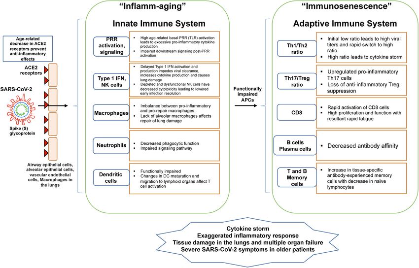
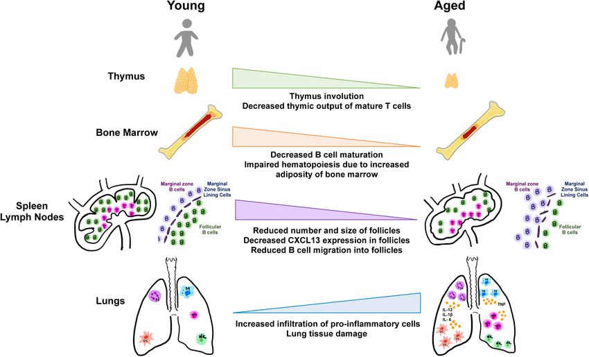
Bajaj et al.                                                                                                            Aging, Immunity, and COVID-19

is the demographics of the populations afflicted. While men                   hypermutation to shift production of antibodies from the IgM
suffer worse outcomes and higher mortality rates than women,                  to more specific high affinity neutralizing IgG isotypes. Notably,
and individuals of black and hispanic race/ethnic minorities are              the immune response is a double edged sword which on one
disproportionately affected (Gadi et al., 2020; Kopel et al., 2020;           hand mediates protective immunity and is necessary, but on the
Muñoz-Price et al., 2020), most notably, elderly individuals >60              other hand, excess and inappropriate production of inflammatory
years of age have been most severely afflicted as evidenced by                cytokines can lead to cytokine storm syndrome (Zhou Y. et al.,
the significantly high morbidity and mortality in this group                  2020) causing organ immunopathology leading to complications
(Santesmasses et al., 2020). A majority of cases and deaths in                and death, and this scenario could not be more true for
every country worldwide involves the aging population, and                    COVID-19.
numbers correlate with increasing age. In addition, the presence                 As with every system in the body, natural aging is
of comorbidities including obesity, diabetes, hypertension,                   accompanied by progressive biological changes in the immune
cardiovascular, and lung disease and cancer accompanied by                    system (Figure 1), some of which lead to its declining functions
immunocompromised states and tissue damage predispose to the                  as evidenced by increased susceptibility to respiratory infections
significantly higher rates of mortality in this group.                        such as influenza and novel coronaviruses. On the other
   Severe acute respiratory syndrome coronavirus 2 is a large                 hand, age-related immune-mediated inflammation or inflamm-
(27.9–31 kb) enveloped positive sense single stranded RNA virus,              aging, and associated inflammatory diseases increase with aging
which enters the human host most commonly through the                         (Franceschi et al., 2000; Shaw et al., 2013; Weyand and Goronzy,
respiratory tract and attaches to cells via its spike (S) protein using       2016; Fuentes et al., 2017; Fulop et al., 2018). These changes
the angiotensin converting enzyme 2 (ACE2) receptor on alveolar               in concert with comorbidities together render older individuals
epithelial type II pneumocytes in the lung (Zhang H. et al., 2020).           vulnerable to latent or novel infections and lead to the observed
Once inside cells, it replicates and new viral particles release              increases in morbidity and mortality of COVID-19. In younger
from cells and invade neighboring cells and extracellular spaces              individuals, a larger repertoire of naive immune cells endow the
and cause tissue damage leading to clinical signs and symptoms.               ability to fight new infections and respond to foreign antigens
Besides the lung, the ACE2 receptor is highly expressed in                    successfully thus resulting in milder forms of the disease or
epithelial cells of the gastrointestinal tract and in other organs            even asymptomatic infection, as is observed in a majority of
including the kidneys, vascular endothelial cells, and other tissues          younger individuals who test positive for the novel coronavirus.
including high expression in adipose tissues (Hamming et al.,                 Increased combined diversity of the antigen receptors in T and
2004; Al Heialy et al., 2020; Al-Benna, 2020; Albini et al., 2020;            B lymphocytes enables a larger robust pool of precursors able
Gu et al., 2020; Li et al., 2020; Thum, 2020; Verdecchia et al.,              to recognize a wider range of foreign organisms and better
2020; Verma et al., 2020). Accordingly, the clinical presentation             able to fend off new diseases such as those caused by the
can vary with respiratory symptoms being most common such                     novel coronavirus.
as cough, shortness of breath, and fever, but GI symptoms                        This review will focus on concepts of immunosenescence
such as diarrhea, nausea vomiting are not uncommon. Recently                  and inflamm-aging as they pertain to age-related physiologic
clinical signs of vascular endothelial dysfunction and abnormal               changes in the immune system, and how these influence the host
coagulopathy-related manifestations have been reported leading                immune response to viral infections including coronaviruses.
to thromboembolism and strokes (Oxley et al., 2020). Severe                   We discuss how aging impacts the innate and adaptive immune
disease complications include pneumonia and acute respiratory                 systems specifically focusing on cellular components and soluble
distress syndrome (ARDS) requiring mechanical ventilation                     secretory mediators such as antibodies and cytokines. We further
and can be fatal.                                                             discuss tissue-specific aspects of immunity related to aging
   The normal immune response to viral infections including                   including physiologic changes in lymphoid tissues and peripheral
coronaviruses involves both the innate and adaptive immune                    non-lymphoid tissue sites of infection such as the lung (Figure 2).
systems. Innate mechanisms include the recognition of viral                   Finally we review therapeutic and preventive vaccine approaches
nucleic acids via the toll-like receptors (TLR) expressed by                  underway including antivirals and immune-modulating agents,
dendritic cells (DC) and macrophages, followed by intracellular               and immune response to vaccines influenced by aging with
signaling and production of antiviral type I interferon (IFN)                 relevance to COVID-19.
in antiviral defense. Natural killer (NK) cells are important in
the early antiviral defense mechanisms. In addition, monocyte-
macrophage mediated production of proinflammatory cytokines                   IMMUNOSENESCENCE AND
IL-6, IL-1β, tumor necrosis factor (TNF) and chemokines enable                INFLAMM-AGING
the recruitment of neutrophils and other inflammatory immune
cells to the site of infection. The adaptive immune response is               Immunosenescence describes the age-associated shift in both
initiated upon viral antigen presentation to CD4 helper T cells               innate and adaptive immune systems that leads to the
and CD8 cytotoxic T cells. Cytokines such as IFN-γ are important              reduced ability to fight novel infections and contributes to the
to activate macrophages and cellular immunity, while antigen-                 development of a chronic state of inflammation (Stahl and
specific CD8 T cells are important for killing viral infected cells.          Brown, 2015). These alterations of the immune system lead
CD4 helper T (Th) cells are important in providing cognate                    to higher rates of infection and disease. Changes observed in
help to B cells, which undergo class switching, and somatic                   immunosenescence are not simply a decline of immune cells but

Frontiers in Physiology | www.frontiersin.org                             2                                   January 2021 | Volume 11 | Article 571416
Bajaj et al.                                                                                                                            Aging, Immunity, and COVID-19

  FIGURE 1 | Schematic shows age-related changes in the innate and adaptive immune system with relevance to COVID-19. As the SARS-CoV-2 enters
  parenchymal cells expressing the ACE2 receptor, it activates PRR of the innate immune system. With aging, there is an imbalance in cellular functions and signaling,
  decreasing innate activation of an already weakened adaptive immune system. These imbalances lead to “inflammaging” from proinflammatory innate immune
  responses and immunosenescence of the adaptive immune response.

reflect a more complex change in action across various immune                          Immunosenescence represents a remodeling of the immune
responses. Several hallmarks are commonly characteristic of                            system and reflects the plasticity of this system for change with
immunosenescence. The immune system is compromised in                                  age (Alves and Bueno, 2019).
its ability to respond effectively to new antigens, linked to a                            Chronic low-grade inflammation, termed “inflamm-aging”
decrease in peripheral naive T and B cells (Stahl and Brown,                           is associated with immunosenescence, driven by a reduced
2015). Accumulation of memory T cells is also associated with                          ability to endure inflammatory triggers as well as an increased
immunosenescence. The antigen load experienced within a                                production of pro-inflammatory cytokines, acute phase proteins,
lifetime coupled with decreased lymphopoiesis, decreases naive                         and oxidative stressors (Aiello et al., 2019). The age-associated
T cell populations and leads to an accumulation of memory cell                         increase in the number of senescent cells in conjunction with
subsets. Accumulation of CD4 and CD8 T cells that have lost the                        the decrease in the immune system’s ability to remove these
expression of costimulatory surface protein CD28 is associated                         cells results in an environment rich with pro-inflammatory
with aging and this loss of CD28 is considered a key feature of                        cytokines and reactive oxygen species (ROS) and further drives
senescent T cells (Weyand and Goronzy, 2016). These CD28null                           neighboring cells into senescence (Stahl and Brown, 2015).
T cells express other markers including CD27, another co-                              Inflammaging is associated with chronic stimulation of the
stimulatory molecule, and are characterized by short telomeres,                        innate immune system and increased secretion of inflammatory
a lack of telomerase, and inhibitory signaling receptors. This                         mediators as evidenced by increased proinflammatory NF-κB
shorter leukocyte telomere length is associated with stress and                        signaling (Salminen et al., 2018). However, under stimulation
several stress and aging-related disorders (de Punder et al.,                          with in vivo/vitro exogenous antigens or mitogens, inflammaging
2019). CD28 signaling is necessary for optimal telomerase up-                          is also associated with down-regulated innate immune responses,
regulation, and experiments in vitro have demonstrated that loss                       driven by regulatory subtypes of T and B cells as well as
of telomerase activity occurs concurrently with loss of CD28                           regulatory subtypes of macrophages, dendritic, NK, and type
expression in T cells (de Punder et al., 2019). Senescent cells often                  II NK T cells (Salminen, 2020). This chronic inflammatory
undergo changes in gene expression, making them less likely to                         environment therefore induces immunosuppression through
undergo apoptosis, and many secrete molecular factors such as                          downregulation of both innate and adaptive immune responses
cytokines, which impact their surrounding microenvironment.                            (Salminen et al., 2019). Myeloid-derived suppressor cells (MDSC)

Frontiers in Physiology | www.frontiersin.org                                      3                                         January 2021 | Volume 11 | Article 571416
Bajaj et al.                                                                                                                           Aging, Immunity, and COVID-19

  FIGURE 2 | Aging and tissues in immunity. Aging is accompanied by the decline in function of lymphoid and non-lymphoid tissues involved in the host immune
  response. Degeneration of primary lymphoid organs lowers production of naive T and B lymphocytes which results in reduced migration to secondary lymphoid
  organs and to sites of antigen encounter. Additionally, the lungs and extrapulmonary organs are susceptible to accumulation of proinflammatory cells and mediators.

also regulate the differentiation and function of several other                       specificity. More specifically, this impaired ability to produce
immune cell types to prevent inflammation (Salminen et al.,                           high affinity immunoglobulin molecules is the result of a shift
2019). MDSCs induce the differentiation of regulatory T cells                         from immunoglobulin production by naive B cells (IgD, IgM) to
and regulatory B cells, which both have immunosuppressive                             those produced by memory B cells (IgG, IgA) (Aiello et al., 2019).
effects via secretion of anti-inflammatory cytokines such as IL-                      The deregulation theory proposes that immunosenescence
10 and TGF-β. It has been observed that the numbers of both                           results from changes to ratios of different expressed gene
MDSCs and regulatory T cells are significantly increased during                       isoforms caused by changes in RNA processing with age resulting
the inflammaging process (Salminen et al., 2018). While the                           in atypical or abnormal secondary immune cell activation
functions of these cells have not been fully elucidated, they have                    (Fuentes et al., 2017). Infection with latent viruses such as
been associated with many aging-related morbidities such as                           cytomegalovirus (CMV) has also been shown to potentially play
infection, cancer, and autoimmune disease (Alves and Bueno,                           a role in immunosenescence by amplification of aging-associated
2019). These cells may potentially have a role in the generation                      inflammation, and both CMV seropositivity and elevated CMV
of immunosenescence. Additionally, it has been found that there                       IgG antibody titers are associated with phenotypic/functional
are elevated plasma levels of interleukin (IL)-6, IL-1, and TNF in                    alterations to adaptive immunity and an increased risk for
the elderly population (Martín et al., 2017). These factors lead to                   all-cause mortality (Solana et al., 2012). Studies to understand
constant activation of immune response, leading to a constant,                        the full extent of its involvement are still underway.
continued inflammatory response.                                                          While the numerous immunological correlates and
    Several current theories exist to explain how                                     consequences of COVID-19 have yet to be determined, it
immunosenescence occurs. The autoimmune theory describes                              is clear that immune processes are involved. When in the
immunosenescence as the result of production of autoantibodies                        lungs, SARS-CoV-2 induces cytokine storm and a strong
secondary to age-associated thymus involution, which leads                            immune response characterized by the increased presence of
to a deficiency of naive T cells in addition to, increased                            many inflammatory cytokines (Nieman, 2020). SARS-CoV-2
activation of T cells to “neoantigens” (Fuentes et al., 2017). The                    shares almost 80% RNA sequence homology with SARS-CoV,
immunodeficiency theory simply postulates immunosenescence                            making it possible that it uses similar strategies to evade
as the impaired ability to mount defenses against new infections                      immune system responses as SARS-CoV (Felsenstein et al.,
due to decreased availability of naive T cells (Fuentes et al.,                       2020). The changes associated with immunosenescence leaves
2017). In addition, with age, there are decreased levels of plasma                    older adults particularly vulnerable to SARS-CoV-2 and more
B cells but increased levels of circulating immunoglobulin                            severe COVID-19 infection (Nikolich-Zugich et al., 2020). T
from antibody-producing B lymphocyte cells with low antigen                           lymphocytes, particularly CD4+ T cells, CD8+ T cells, and B cell

Frontiers in Physiology | www.frontiersin.org                                     4                                         January 2021 | Volume 11 | Article 571416
Bajaj et al. Aging, Immunity, and COVID-19
activation are critical in the host defense against viral infections in murine models and humans (Chen J. et al., 2020; Xie et al.,
(Felsenstein et al., 2020). Thus, the depletion of these cells 2006; Yoon et al., 2016). Specifically, aging-related decrease in
combined with the expression of pro-inflammatory cytokines ACE2 expression levels were observed in lung epithelial cells
likely play a role in triggering the hyper-inflammatory reaction of aged rats compared to young rats (Xie et al., 2006), and in
seen among COVID-19 patients. the thoracic aorta of old versus young mice (Yoon et al., 2016).
Furthermore, in a bioinformatic analysis of publicly available
human genomics and transcriptomics gene expression data, age-
INNATE IMMUNITY associated decrease in ACE2 expression levels was observed in
multiple tissues including blood, kidneys, and adrenal glands,
SARS-CoV-2 Entry and ACE2 Receptors and also found to be lower in type 2 diabetes patients compared
Coronaviruses use the surface spike (S) glycoprotein to enter to healthy individuals (Chen J. et al., 2020). Therefore, a
human host cells via the ACE2 receptor on the host cells decrease in ACE2 receptor expression coupled with aging-related
(Nikolich-Zugich et al., 2020; Wan et al., 2020). In humans, immune inflammation and comorbidities may compromise the
three common strains of coronaviruses, SARS-CoV, Human anti-inflammatory response and predispose older individuals
Coronavirus NL-63 (NL63), and SARS-CoV-2 target airway to exaggerated inflammatory responses, which is one of the
epithelial cells, alveolar epithelial cells, vascular endothelial cells, hallmarks of COVID-19 (Ciaglia et al., 2020; Verdecchia et al.,
and macrophages in the lungs as they express abundant ACE2 2020). Binding of SARS-CoV-2 to ACE2 receptors further lowers
receptors (Tay et al., 2020; Verdecchia et al., 2020). As compared the ACE2 expression (Banu et al., 2020; Verdecchia et al., 2020),
to SARS-CoV, the SARS-CoV-2 evolved by mutating part of its which increases the angiotensin 2 activity that contributes to
S glycoprotein to have a 10–20× higher affinity for the ACE2 an acute exaggerated pro-inflammatory response (Kuba et al.,
receptor (Nikolich-Zugich et al., 2020; Wrapp et al., 2020). The 2005; AlGhatrif et al., 2020; Ciaglia et al., 2020; Verdecchia et al.,
main respiratory cells targeted by SARS-CoV and SARS-CoV- 2020). However, there has been some controversy regarding the
2 for ACE2 receptor-mediated entry are type 2 pneumocytes variability in ACE2 expression with aging as ACE2 expression in
and ciliated bronchial epithelial cells (Albini et al., 2020). the lungs were shown to decrease in aged rats (Xie et al., 2006)
ACE2 receptors are also present in the heart (cardiomyocytes but the expression levels in aged male and female mice (Nozato
and pericytes), gut (enterocytes of the small intestine and et al., 2019) and humans have been variable based on the cohort
colon), kidneys (renal tubular and intestinal epithelial cells), (Fernández-Atucha et al., 2017; Li et al., 2020). Altogether, despite
testis (spermatogonia, Leydig cells, and Sertoli cells), brain the lower rates of infection in older individuals, the prognosis of
(neurons and glial cells), arterial smooth muscle cells, arterial COVID-19 may be worse due to a decrease in ACE2 expression
and venous endothelial cells, myofibroblasts and adipocytes, levels and a decrease in anti-inflammatory response.
that can be affected in patients with co-morbidities such as A decrease in ACE2 expression has been reported in
cardiovascular disease, chronic obstructive pulmonary disease, individuals with hypertension and diabetes (Li et al., 2020;
pulmonary hypertension, and diabetes (Hamming et al., 2004; Xia Verdecchia et al., 2020) making them susceptible to increased
and Lazartigues, 2008; Albini et al., 2020; Gu et al., 2020; Li et al., inflammation via the angiotensin 2 pro-inflammatory pathway,
2020; Thum, 2020; Verdecchia et al., 2020; Verma et al., 2020). By while treatment with ACE inhibitors (ACEI) and angiotensin
binding to the ACE2 receptor on endothelial cells, SARS-CoV-2 receptor blockers (ARB) tends to increase ACE2 expression (Li
can lead to endothelial cell inflammation, vasculitis, disseminated et al., 2017). This ACE2 upregulation can be physiologically
intravascular coagulation, and thromboembolism (Albini et al., restorative given its anti-inflammatory roles, however, may
2020). SARS-CoV-2 can also infect enterocytes via ACE2 causing predispose them to a greater risk of SARS-CoV-2 infection (Fang
diarrhea and associated pain and cramping (Gu et al., 2020). et al., 2020; Li et al., 2020). Because ACE2 is upregulated in
Higher ACE2 receptor expression levels were found in the patients with diabetes and cardiovascular disease who are treated
nasal epithelium of young adults less than 60 years compared to with ACEI and ARBs, this creates a controversy regarding these
those in children (Bunyavanich et al., 2020). This higher ACE2 treatments (Albini et al., 2020; AlGhatrif et al., 2020; Fang
expression in young adults may predispose them to an increased et al., 2020; Onweni et al., 2020). Hence, the treatment of older
risk for contracting coronavirus infections. Accordingly, based hypertensive and diabetic individuals with ACEIs and ARBs has
on epidemic trajectories mapped from epidemiological data from been a dilemma (AlGhatrif et al., 2020). However, the WHO and
South Korea, the highest numbers of daily new SARS-CoV- others suggest continuing the use of these drugs in COVID-19
2 positive cases were among younger individuals in the 20– patients since no risk has been shown,1 including in the recent
39 and 40–59 year age groups compared to people over 60 first randomized BRACE Corona trial,2 and also because of their
years (Yu et al., 2020). However, while it has been coopted beneficial role (Meng et al., 2020; Yan et al., 2020).
as the entry point for the SARS-CoV-2 virus on host cells, In addition to ACE2 receptors, dipeptidyl peptidase 4 (DPP4)
the ACE2 enzyme also plays a critical anti-inflammatory role might serve as a binding target for SARS-CoV-2 entry (Solerte
in the renin-angiotensin-aldosterone-system (RAAS) signaling et al., 2020; Valencia et al., 2020). DPP4 expression is enhanced
pathway by converting the proinflammatory angiotensin 2 to
anti-inflammatory angiotensin 1–7 (Rodrigues Prestes et al., 1
www.who.int/news-room/commentaries/detail/covid-19-and-the-use-of-
2017; Albini et al., 2020; AlGhatrif et al., 2020). Moreover, aging angiotensin-converting-enzyme-inhibitors-and-receptor-blockers
has been associated with decline in levels of ACE2 expression 2
www.clinicaltrials.gov/ct2/show/NCT04364893
Frontiers in Physiology | www.frontiersin.org 5 January 2021 | Volume 11 | Article 571416Bajaj et al. Aging, Immunity, and COVID-19
in type 2 diabetic patients, obese individuals and senescent cells neurodegenerative disease and increased tissue damage (Weyand
which increases proliferation of human smooth muscle cells and and Goronzy, 2016). This phenomenon is known as “inflamm-
upregulation of pro-inflammatory cytokines such as MCP-1, IL- aging,” which is coupled with other changes in the immune
6, and IL-8 via NF-kB activation (Röhrborn et al., 2015; Valencia system with age known as “immunosenescence” (Fulop et al.,
et al., 2020), thus potentially leading to worse outcomes of SARS- 2018). This age-associated chronic basal inflammation is in
CoV-2 in diabetic patients. Therefore, the use of DPP4 inhibitors part due to the continuous engagement of pattern recognition
may be beneficial as they significantly lower the levels of IL- receptor (PRRs) on innate immune cells with pathogen associated
6, which plays a major role in the inflammatory response in molecular patterns (PAMPs) via reactivation of latent viral
SARS-CoV-2 patients (Solerte et al., 2020; Valencia et al., 2020). infections or the release of endogenous DAMPs that are released
from senescent and necrotic cells (Shaw et al., 2013; Frasca et al.,
Innate Pro-inflammatory Responses 2017; Rea et al., 2018). TLRs are the main PRRs that are highly
Once the SARS-CoV-2 enters the host cells via the ACE2 expressed on monocytes, macrophages, neutrophils, DC, and
receptors, it actively replicates followed by release of viral some lymphocytes (Rea et al., 2018; Moreno-Eutimio et al., 2020;
particles (Tay et al., 2020). This causes the host cell to undergo Onofrio et al., 2020; Plenge, 2020; Sallenave and Guillot, 2020).
pyroptosis and release damage-associated molecular patterns TLRs that play a major role in SARS-CoV-2 are TLR-7 and TLR-8,
(DAMPs) such as ATP, nucleic acid, ASC oligomers, and which sense the ssRNA of SARS-CoV-2 and are expressed in the
cytokines (Lee et al., 2020; Tay et al., 2020). These DAMPs endosomal compartments of respiratory epithelial cells (Moreno-
trigger the neighboring epithelial cells, endothelial cells, and Eutimio et al., 2020; Onofrio et al., 2020; Plenge, 2020; Sallenave
alveolar macrophages to generate pro-inflammatory cytokines and Guillot, 2020). TLR-7 and TLR-8 induce the production of
and chemokines such as IL-6, IP-10, macrophage inflammatory IFN-regulated cytokines and production of pro-inflammatory
protein (MIP) 1-alpha and 1-beta (Lee et al., 2020; Tay et al., cytokines, respectively (Moreno-Eutimio et al., 2020; Onofrio
2020). These proteins further attract monocytes, macrophages, et al., 2020). The stimulator of IFN gene (STING) pathway,
and T cells, which also produce IFN-γ, creating a pro- involved in the production of IFNs and NF-kB in response to
inflammatory feedback loop (Tay et al., 2020). However, in case cytosolic DNA and RNA viruses, is also believed to be involved
of a defective immune response, there is an increased build up of in SARS-CoV-2 infection and is upregulated in senescent cells
immune cells in the lungs, overproduction of proinflammatory (Berthelot and Lioté, 2020). Innate immune cell migration and
cytokines leading to tissue damage in the lungs, ARDS and signaling events downstream of PRR activation are impaired with
multi-organ damage due to the resulting cytokine storm which aging leading to increased cytokine secretion and dysregulation,
can be fatal (Tay et al., 2020). Excessive inflammation and also known as a cytokine storm (Shaw et al., 2013; Onofrio et al.,
cytokine storm are characteristics of cytokine release syndrome, 2020). Age-related alterations in TLR protein expression in innate
which is elicited by SARS-CoV-2 in patients with dysregulated immune cells can induce cytokine production (Shaw et al., 2013;
immune responses (Acharya et al., 2020; Tay et al., 2020). The Onofrio et al., 2020). For example, a lowered surface expression
innate immune system tends to dysregulate with aging and of TLR1 associated with lowered TLR1/TLR2 can induce cytokine
acquires proinflammatory phenotypes accompanied by tissue production in human monocytes (Shaw et al., 2013; Onofrio et al.,
inflammation (Acharya et al., 2020). 2020). This defect is primarily post-translational that worsens
There are several patterns of immune responses to with age (Shaw et al., 2013; Onofrio et al., 2020). Over-production
coronaviruses, primarily SARS-CoV and SARS-CoV-2 (Du of cytokine reflects high basal TLR activation, which cannot be
and Yuan, 2020; Nieman, 2020; Nikolich-Zugich et al., 2020). further activated in response to a pathogen, contributing to a
Severe symptoms are associated with intense pro-inflammatory failure of innate immune system response (Shaw et al., 2013;
responses and cytokine storm that can cause tissue damage Onofrio et al., 2020).
especially in the lungs (Nieman, 2020; Nikolich-Zugich et al., Age-related impairment in PRR signaling can affect macro-
2020). In SARS-CoV, the non-survivors displayed a state termed autophagy and vice versa (Shaw et al., 2013; Rea et al., 2018).
as “stuck in innate immunity” in which there was an increased Defects in either can stimulate the inflammatory cascade (Shaw
innate activation of IFN-α and IFN-γ, CXCL10, CCL2, and et al., 2013; Rea et al., 2018). Autophagy is a process that
downstream IFN stimulated genes, but a lack of antibody removes damaged proteins and large aggregates to regenerate
production (Nikolich-Zugich et al., 2020). This state was more new and healthy cells. These autophagic responses to intracellular
prevalent in the older individuals (Nikolich-Zugich et al., 2020). pathogens are impaired in older individuals (Shaw et al., 2013;
Some studies have shown that SARS-CoV-2 may induce an early Rea et al., 2018). Impaired autophagy can in turn affect PRR
activation of the adaptive immune response system, which may signaling via increased ROS, which increases with age (Shaw et al.,
interfere with the innate immune response to eliminate the virus 2013; Rea et al., 2018). ROS is unregulated and overproduced
quickly (Du and Yuan, 2020). This may lead to an overreaction as a by-product of mitochondrial energy production, active
of the immune system causing a cytokine storm that could be immunological phagocytic processes and prostaglandin pathways
damaging to the tissues and possibly lead to two waves of the through COX enzyme production that begin to dysfunction with
disease (Du and Yuan, 2020). age (Rea et al., 2018). In addition to changes in redox balance,
With age, the innate inflammatory responses gain in age-related increase in senescent cells, senescence-associated
intensity and duration leading to many inflammatory diseases secretory phenotype (SASP), excess oxidative state, DNA damage
such as rheumatoid arthritis, cardiovascular disease, and and decline in successful autophagy can trigger inflammasome
Frontiers in Physiology | www.frontiersin.org 6 January 2021 | Volume 11 | Article 571416Bajaj et al. Aging, Immunity, and COVID-19
by stimulating an inflammatory cascade involving NF-κB, IL-1α, Shaw et al., 2013). Additionally, IFN production from
TGF-β, and IL-6 pathways (Shaw et al., 2013; Rea et al., 2018). plasmacytoid DCs declines with age in response to virus
Excessive secretion of ROS, along with the secretion of other (Agrawal, 2013). Therefore, the impaired and delayed production
proteases, can cause an uninhibited infiltration of inflammatory of type I IFN in older individuals along with impaired production
cells damaging the lungs in addition to the damage caused by of NK cells affect the first-line antiviral response to SARS-CoV-2
SARS-CoV-2 virus (Rea et al., 2018; Tay et al., 2020). invasion and lowers the chances of infection resolution at an
The anti-viral innate immune response also includes soluble earlier stage as compared to younger individuals (Shaw et al.,
components of the complement and coagulation system, such 2013; Aiello et al., 2019; Acharya et al., 2020).
as mannose-binding lectin (MBL), and natural antibodies such
as IgM, and IgA (Matricardi et al., 2020). Reduction of these Neutrophils, Macrophages, and Dendritic
mediators may contribute to the increased susceptibility of older
Cells
people to SARS-CoV-2. Age-dependent decrease of natural IgM
Neutrophils and macrophages are phagocytic cells of the first-
(Muthana and Gildersleeve, 2016) and MBL (Terai et al., 1993;
line of defense whose phagocytic functions decrease with aging
Tomaiuolo et al., 2012) have been described. MBL is higher in
(Shaw et al., 2013; Jackaman et al., 2017). Furthermore, age-
those under 20 years compared to those over 20, and is noted
related changes in macrophages and neutrophils contribute
to decrease with age (Tomaiuolo et al., 2012), and an age-related
to chronic low-grade inflammation and dysregulation of
decline in IgM begins between the 40s and 50s (Terai et al.,
immunosuppression mediated by macrophages (Shaw et al.,
1993; Matricardi et al., 2020). Therefore, the decrease in MBL
2013; Jackaman et al., 2017). In older individuals, neutrophils
and IgM with age may compromise the anti-viral innate immune
migrate inaccurately and spread further in response to stimuli
response and increase the susceptibility of older individuals to
due to constitutive activation of the PI3K pathway, have reduced
COVID-19 (Terai et al., 1993; Tomaiuolo et al., 2012; Muthana
phagocytosis and decreased intracellular killing activity (Shaw
and Gildersleeve, 2016; Matricardi et al., 2020).
et al., 2013; Jackaman et al., 2017). Age-related defects in
neutrophils are primarily due to impaired signal transduction
Type I IFN and NK Cells
pathways such as impaired anti-apoptotic responses to GM-CSF
During a pathogenic invasion, PAMPs and DAMPs activate PRRs
mediated through the JAK-STAT tyrosine kinase and PI3K-
that initiate a signaling cascade, which leads to the expression of
AKT pathways (Shaw et al., 2013; Jackaman et al., 2017).
type I IFN and other inflammatory factors (Acharya et al., 2020;
Even though many viruses can stimulate the production of
Nikolich-Zugich et al., 2020). An early activation and production
neutrophil extracellular traps (NETs) from neutrophils, there is
of type I IFN is protective in antiviral defense (Acharya et al.,
no significant evidence on the role of neutrophils or neutrophil
2020; Nikolich-Zugich et al., 2020). Type I IFN acts as an
NETs in SARS-CoV-2 pathogenesis (Mozzini and Girelli, 2020).
immediate antiviral response via autocrine and paracrine type
In healthy elderly adipose and hepatic tissue, the macrophages
I IFN receptor (IFNAR) signaling to restrict viral replication
are highly present as a pro-inflammatory M1 phenotype and as an
and spread (Acharya et al., 2020; Nikolich-Zugich et al., 2020).
immunosuppressive M2 phenotype in elderly lymphoid tissues,
However, a delay in production can lead to tissue damage and
lung, and muscle (Jackaman et al., 2017). Macrophages can be
inflammatory cytokine storm (Acharya et al., 2020; Nikolich-
easily activated in older individuals as a result of accumulation
Zugich et al., 2020). This delay in type I IFN production increases
of immune complexes, elevated cytokines, hormones, free fatty
with age and was seen in older individuals with SARS-CoV
acid, oxidized low-density lipoproteins and immunoglobulins
(Acharya et al., 2020; Nikolich-Zugich et al., 2020). Additionally,
(Acharya et al., 2020). This activation promotes low-grade
the evolved immune evasive mechanisms of the SARS-CoV virus
inflammation negatively affecting the effectiveness of immune
prevent early induction of the type I IFN, which was initially
response (Acharya et al., 2020). In patients with SARS-CoV-2,
assumed to be the case in SARS-CoV-2 (Acharya et al., 2020;
there is an imbalance between proinflammatory macrophages
Nikolich-Zugich et al., 2020).
and pro-repair airway macrophages due to impaired IFN
In addition to the delay in local IFN responses, which impedes
production (Acharya et al., 2020). As a result of age-related
viral clearance and leads to the development of cytokine release
decrease in IL-12 production by DCs and microenvironment
syndrome, type I IFN are minimally detected in patients with
alterations from changes in splenic marginal zones, antigen
severe SARS-CoV-2 symptoms (Aiello et al., 2019; Acharya
presenting cells become functionally impaired (Shaw et al., 2013).
et al., 2020). Due to this impaired IFN production, there is
Alterations in the lung microenvironment also lead to changes in
an imbalance between proinflammatory macrophages and
DC maturation and migration to lymphoid organs, thus affecting
reparative airway macrophages (Aiello et al., 2019; Acharya
T cell activation in SARS-CoV-2 patients (Tay et al., 2020).
et al., 2020). Additionally, other immune cells such as NK cells
are regulated by IFN during coronavirus infections, which
are also depleted and dysfunctional in patients with severe Comorbidities and Innate Immune
SARS-CoV-2 symptoms (Aiello et al., 2019; Acharya et al., 2020). Responses
Impaired NK cell function is associated with higher rates of Elderly patients (over the age of 60) and patients with
infection and mortality in older adults (Shaw et al., 2013). As comorbidities experience more severe symptoms of SARS-CoV-
per in vivo models, cytotoxicity of NK cells induced by type 2 partially due to hyperactive chronic innate inflammatory
I IFN decreases in aged mice (Plett and Murasko, 2000; responses as well as structural and functional changes
Frontiers in Physiology | www.frontiersin.org 7 January 2021 | Volume 11 | Article 571416Bajaj et al. Aging, Immunity, and COVID-19
in organs (Frasca et al., 2017). Obesity increases the responders of adaptive immunity and their function through
complications associated with the virus and aging in part life is critical for prevention of host morbidity and mortality.
due to structural/functional impairments (Iacobellis et al., They are especially important in discussions of COVID-19, as
2020a; Malavazos et al., 2020) and also because visceral fat may their effector roles for viral protection must be balanced with
serve as a reservoir for the virus and amplify inflammatory their regulatory roles to prevent host pathogenesis (Grifoni et al.,
responses (Ryan and Caplice, 2020). Obese tissues can affect the 2020; Ye et al., 2020). An imbalance in this ratio as is seen in
mechanics of the respiratory system by reducing the compliance serious cases of COVID-19 could be owed to T cell or B cell
and lung volumes (Littleton, 2012) and pose as a risk factor for dysfunction, with the latter being reliant on T cell responses
asthma (Tashiro and Shore, 2019). Computed tomographic (CT) (Grifoni et al., 2020). Therefore, it is vital that the various roles
imaging showed evidence of increased fatty liver and epicardial of T cells in the context of SARS-CoV-2 infection are better
adipose tissues in older COVID-19 patients who were critically understood, especially in regards to the aging immune system,
ill (Iacobellis et al., 2020b), and also in younger patients under the most apparent targets of severe disease.
40 with severe disease compared to those with mild disease At puberty, there is a 90% drop in T cell production
(Deng et al., 2020), suggesting obesity as a potential predictor of via involution of the thymus, which turns to adipose tissue
COVID-19 severity. Adipose tissues also serve as an endocrine (Fulop et al., 2018; Nikolich-Zugich et al., 2020). T lymphocyte
organs and secrete hormones like leptin, adiponectin, and production is again decreased around 40–50 years of age where
inflammatory cytokines such as IL-6 and TNF (Banerjee et al., the remainder of the thymus further degenerates (Nikolich-
2020). ACE2 expression levels are higher in visceral adipose Zugich et al., 2020). This secondary atrophy causes production
tissue compared to subcutaneous fat (Zhang et al., 2018), and of naïve T cells to drop to only 1% of its output before primary
since obese and diabetic patients have more adipose tissue, involution. Additionally, lymph nodes become less capable of
they have increased numbers of ACE2 expressing adipocytes, storage, maintenance, and support of naïve T cells with advancing
potentially increasing their susceptibility to an elevated viral age (Nikolich-Zugich et al., 2020). To illustrate, TCR excision
load in the adipose tissue (Banerjee et al., 2020), which could circles are used as markers of naïve T cell output and decreased
lead to increased severity of SARS-CoV-2 infection (Fajnzylber exponentially by 95% in subjects 60 years of age compared to
et al., 2020). Aging is also associated with metabolic dysfunction 25 years (Ponnappan and Ponnappan, 2011). However, total
in correlation with dysfunction of adipose tissue, as evidenced numbers of T cells show little decline with age except in the very
by correlation between mitochondrial and arterial dysfunction elderly, and this is due to a compensatory increased peripheral T
in epididymal white adipose tissue and metabolic dysfunction cell proliferation, called homeostatic proliferation accompanied
in aged B6D2F1 mice (Donato et al., 2014). Additionally in by acquisition of activated/memory cell phenotypes (Ponnappan
obese individuals, innate immune cells such as macrophages, and Ponnappan, 2011; Sprent and Surh, 2011). This means
NK cells, and neutrophils as well as adaptive immune cells that while the elderly may have similar lymphocyte levels to
infiltrate the adipose tissue and induce inflammatory responses younger adults, their ability to mount an adaptive response
via pro-inflammatory cytokine secretion, metabolic switches, and to a novel antigen is significantly reduced from replacement
phenotypic and functional changes (Frasca et al., 2017). Chronic of naïve T lymphocytes with antigen-independent experienced
low-grade inflammation promoted by excess nutrients and cells (Ponnappan and Ponnappan, 2011). This has serious
elevated pro-inflammatory cytokines can cause insulin resistance implications in the context of a novel pandemic, as the elderly are
in both mice and humans, which can in turn produce systemic less likely to be able to mount a rapid, high-affinity T cell effector
and local inflammation (Frasca et al., 2017). Furthermore, and regulatory response.
obesity is a risk factor for endothelial dysfunction (Engin, 2017)
and when combined with SARS-CoV-2 induced endothelial Th1/Th2
cell injury, could potentially exacerbate endothelial dysfunction The levels and proportion of differentiated CD4 T cells into
leading to further complications (Amraei and Rahimi, 2020). Th1 and Th2 types and their associated cytokines are vital
The combined age-related dysfunctional patterns of different for elimination of a pathogen without incurring damage to
aspects of the innate immune inflammatory responses prevent the host tissue (Ponnappan and Ponnappan, 2011; Biasi et al.,
resolution of this viral infection (Frasca et al., 2017). Hence, older 2020; Ye et al., 2020). Th1 cells are notably proinflammatory
individuals over 60 years and individuals with co-morbidities are and secrete cytokines such as IFN-γ, IL-1β, IL-2, and most
at a higher risk for SARS-CoV-2 severity as they are more likely pathologically in COVID-19, IL-6 (Biasi et al., 2020; Ye et al.,
to develop a defective immune response and suffer higher rates of 2020). Th2 cells have primarily anti-inflammatory function via
morbidity and mortality. IL-4 and IL-10 and attempt to attenuate Th1 pathways to
avoid uncontrolled attack on the host (Biasi et al., 2020; Ye
et al., 2020). The optimal ratio of Th1/Th2 differs by the
ADAPTIVE IMMUNITY pathogen, and failure of the host to adequately regulate these
pathways may result in prolonged disease states or mortality (Ye
CD4 T Cells et al., 2020). While the original SARS-CoV produced and was
CD4 T helper cells are a key lymphocyte population of adequately eliminated by a predominately Th1 response, host
the adaptive immune system. By recognizing MHC Class success over the novel coronavirus seems to prefer attenuation
II molecules on antigen-presenting cells, they are the first of these proinflammatory processes (Ye et al., 2020). While
Frontiers in Physiology | www.frontiersin.org 8 January 2021 | Volume 11 | Article 571416Bajaj et al. Aging, Immunity, and COVID-19
patients of COVID-19 have elevated Th1 and Th2 cytokines, be involved in proinflammatory activation of the complement
patients who ultimately survived had significantly higher levels cascade which activates mast cell degranulation in the lungs
of anti-inflammatory Th2 cytokines than proinflammatory Th1 and is predictive for ARDS development (Nikolich-Zugich et al.,
cytokines (Biasi et al., 2020). To complement, patients who died 2020). Additionally, IL-17 is positively regulated by IL-6 which
of COVID-19 had features of the infamous “cytokine storm” with is significantly increased in serious states of COVID-19, adding
highly upregulated levels of a plethora of mostly proinflammatory to the fatal cytokine storm (Ponnappan and Ponnappan, 2011;
Th1 cytokines (Ye et al., 2020). Biasi et al., 2020; Ye et al., 2020). In a Chinese population, an
The literature on levels and ratio of Th1 and Th2 CD4+ cells IL-17 polymorphism analysis showed that patients predisposed
and associated cytokine levels in healthy older populations is to lower levels of IL-17 secretion had greater 30-day survival for
conflicting. However, most sources report that Th2 responses are ARDS compared to patients with genetically higher levels of IL-17
upregulated in healthy elderly populations and also predominate (Xie et al., 2019).
in early disease states, which is associated with elderly Studies have shown that Th17 cells and IL-17 are significantly
susceptibility to tuberculosis, influenza, and other infections higher in human and murine elderly populations (Ponnappan
(Ponnappan and Ponnappan, 2011; Har-Noy and Or, 2020). and Ponnappan, 2011; Schmitt et al., 2013; Lim et al., 2014). In
What is troubling with SARS-CoV-2 is that like many viruses, it a mouse model, Th17-associated proinflammatory cytokines IL-
has evolved mechanisms for evading the host immune system, 17, IL-1β, CCR6, CCL20, ROR-γt, and IL-22 were significantly
replicating insidiously (Har-Noy and Or, 2020; Nikolich-Zugich upregulated even in healthy aged mice compared to young
et al., 2020). This initial Th2 predominance in elderly populations subjects (Lim et al., 2014). This increase in Th17 and IL-17 levels
initially aids coronavirus replication until host mechanisms are is especially present in elderly patients with autoimmune and/or
rapidly overwhelmed by the pathogen and high viral titers (Har- inflammatory diseases, especially obesity (Liu and Nikolajczyk,
Noy and Or, 2020). In response, patients that progress to critical 2019). Evidence shows that adipocytes, reservoirs for Th1, IL-
illness, in particular the elderly, are believed to experience a 6, and TNF, can directly activate differentiation of CD4 T cells
rapid switch to Th1-predominating pathways, overloading the into Th17 via the STAT3 pathway (Liu and Nikolajczyk, 2019).
host with inflammatory cytokines that inadvertently harm host These Th17 cells are involved with a feed forward loop of adipose
tissue (Ye et al., 2020). This initial Th2 preference followed by tissue inflammation which over the years can be a primary driver
a rapid upregulation of Th1 pathways is the molecular basis of of systemic inflammation, heart disease, diabetes, fatty liver, and
inflammaging and the increase in secretion of proinflammatory cancer (Liu and Nikolajczyk, 2019; Maffetone and Laursen, 2020).
cytokines in an activated, aging immune system (Ye et al., 2020). These conditions, especially type II diabetes, in turn provide
In fact, most critically ill patients and those who succumbed, did unique positive feedback and support for Th17 production (Ip
not initially report severe symptoms in early stages of the disease; et al., 2016). This presents an interesting overlap, as a growing
the conditions of these patients deteriorated suddenly with the proportion of adults over 65 are overweight or obese, and up
appearance of a rapid and extreme Th1 response (Ye et al., 2020). to 94% of patients with serious cases of COVID-19 had at
This situation is incredibly interesting, as children, women, least one underlying disorder where adiposity could be a trigger
and especially pregnant women are known to have higher levels (Maffetone and Laursen, 2020). The immune responses of obese
of Th2 cytokines in relation to COVID-19 (Molloy and Bearer, COVID-19 patients are largely unexplored and present a rich area
2020). However, it is believed that these groups are better able to for research development.
coordinate the transition and attenuation of Th1 responses once
the infection is detected, preventing unnecessary host damage Treg Cells
(Molloy and Bearer, 2020). Unfortunately, elderly populations Immunosuppressive regulatory T (Treg) lymphocytes function
appear to lose the ability to smoothly transition and coordinate to inhibit inflammatory T cells including Th1, Th2, and Th17
this ratio once viruses have been detected, which may explain the cells. To protect host tissue, they secrete immune-suppressive
rapid cytokine storm and deterioration of hospitalized patients cytokines such as IL-10 and TGF-β which antagonize the effects
(Ponnappan and Ponnappan, 2011). of the proinflammatory IL-6 and IL-17 that are implicated in
COVID-19 (Ponnappan and Ponnappan, 2011; Schmitt et al.,
Th17 2013; Ye et al., 2020). The optimal presence of Treg lymphocytes
Another component of the adaptive immune system of interest varies by disease but has been necessary for mild aftermath of
in immune senescence is the Th17 cell type. These cells mainly MHV infections, while Treg diminution is associated with severe
produce proinflammatory cytokines such as IL-17 and IL-22 morbidity. Treg levels have not been sufficiently studied in the
and are involved in the stimulation of IL-6 and TNF (Schmitt context of COVID-19, but a recent study reports an inverse
et al., 2013; Nikolich-Zugich et al., 2020). In cases of MERS, correlation between naïve Treg levels and COVID-19 disease
SARS, and COVID-19, secretion of Th17’s hallmark cytokine IL- severity (Chen G. et al., 2020). Whether this relation has a
17 is directly related to disease severity (Nikolich-Zugich et al., causative role in disease outcome is yet unknown.
2020). This is likely because in many coronavirus infections, The literature on Treg function in the elderly is complicated.
IL-17 functions to recruit neutrophils; it additionally activates IL- It seems that overall numbers of Treg cells in those over 65 years
22 and associated cytokines that cause pathological states and are significantly reduced compared to a 50 to 65-year age group
pulmonary edema in the host (Grifoni et al., 2020; Nikolich- (Schmitt et al., 2013). This difference is especially significant for
Zugich et al., 2020). In SARS, IL-17 has also been found to elderly patients suffering from episodes of ischemic stroke (Dolati
Frontiers in Physiology | www.frontiersin.org 9 January 2021 | Volume 11 | Article 571416Bajaj et al. Aging, Immunity, and COVID-19
et al., 2018). This overall decline in Treg numbers is in line developed some type of CD8 proinflammatory cytokine response
with the second wave of thymic degeneration, which may explain to the coronavirus infection (Grifoni et al., 2020). Based on
these numbers (Ponnappan and Ponnappan, 2011; Schmitt et al., several hospital studies, it is believed the SARS-CoV-2 replicates
2013; Nikolich-Zugich et al., 2020). Foxp3 is a key transcription within host cells due to functional exhaustion of these cytotoxic
factor and marker of Treg cells, necessary for Treg function and cells (Diao et al., 2020; Zheng et al., 2020). Clinically, this
inhibition of all CD4+ subtypes (Ponnappan and Ponnappan, often presents as lymphopenia and expression marker studies
2011; Schmitt et al., 2013). While overall Treg numbers are have shown that the remaining cytotoxic lymphocytes manifest
lower, it seems that expression of Foxp3 is upregulated, giving markers of exhaustion (Diao et al., 2020; Zheng et al., 2020). For
the remaining Treg cells such strong CD4+ immunosuppressive example, PD1 and Tim3 are fatigue markers of CD8 T cells that
potential that even anti-inflammatory cytokines such as IL- were upregulated in ICU patients compared to mild cases (Diao
10 are inhibited (Ponnappan and Ponnappan, 2011; Schmitt et al., 2020). These markers are also highly expressed in patients
et al., 2013). This increase in Treg immunosuppression is quite progressing from prodromal to active signs of COVID-19 disease
counterintuitive when realizing its effects on CD4 pathways that (Diao et al., 2020). Yet another study supported these findings of
already function to attenuate inflammation (Schmitt et al., 2013). cellular exhaustion by study of the NKG2A receptor on CD8 cells
Additionally, although there is higher functioning of Treg cells, and NK cells (Zheng et al., 2020). Expression of this receptor was
it seems that inhibition of the Th17 subtype is lost in elderly upregulated in both cells and correlated with significantly lower
patients, producing a high, proinflammatory ratio of Th17/Treg percentages of IFN-γ+ CD8, IL-2+CD8 T cells, and granzyme
cells (Schmitt et al., 2013). B+ CD8 T cells which are secretion-specific subtypes (Zheng
In parallel to the relationship between Th17-associated et al., 2020). In addition, studies report upregulated levels of
inflammation and adiposity, there is also a rich association cytotoxic granules such as granzyme B and perforin markers
between Treg populations and fat accumulation (Liu and (Grifoni et al., 2020). This data altogether implies that the novel
Nikolajczyk, 2019). With the progression of obesity, the coronavirus stimulates extreme activation of CD8 T cells and
proportion of Tregs in adipose tissue wanes (Liu and Nikolajczyk, their cytokines with resultant fatigue.
2019). The expression profiles and phenotypes of these As related to cell exhaustion, there is evidence that the PD-
lymphocytes also differ across fat levels in the host (Liu and 1 and Tim-3 exhaustion molecules are upregulated in CD8
Nikolajczyk, 2019). Visceral adipose tissue-resident Tregs of lymphocytes from healthy aged mice compared to younger
lean individuals produce high levels of anti-inflammatory IL-10 controls (Lee et al., 2016). A result of this exhaustion was the
(Liu and Nikolajczyk, 2019), which upregulates Th2 pathways, inability to proliferate under antigen stimulation of the TCR,
antagonizing effects of IL-6 and TNF, a mechanism also supposed indicating the lower functioning of aged cytotoxic cells (Lee
to occur in COVID-19 patients (Ye et al., 2020). However, this et al., 2016). CD8 T cell numbers as they pertain to age have
anti-inflammatory cytokine production decreases with increasing been largely unexplored in humans in the context of COVID-
levels of adiposity (Liu and Nikolajczyk, 2019). What is also 19. However, results from a study of the original SARS-CoV in
interesting is that high fat levels in the diet may alter expression a monkey model may lend interesting insights. It was found that
of Treg cells towards proinflammatory states (Carlsen et al., peripheral lymphocytes, specifically CD8 T cells, were overall
2009). To elucidate, mice models have shown that a high fat significantly lower in elderly monkeys both before and after
diet, especially in males, increases expression of NF-κb, a protein SARS-CoV infection (Clay et al., 2014). Younger monkeys had
critically involved in DNA production and proinflammatory higher CD8 counts for the entirety of the study, indicating that
cytokine release in the aging immune system (Carlsen et al., they were better able to sustain these levels (Clay et al., 2014).
2009). NF-κb levels significantly increase in the elderly, and Elderly monkeys had a drop in CD8 counts after five days post-
increased expression of this transcription factor occurs through infection, resulting in higher viral titers (Clay et al., 2014). Flow
several pathways (Ponnappan and Ponnappan, 2011; Polesso cytometry analysis additionally confirmed that not only were the
et al., 2017). Non-canonical upregulation has been shown to cell numbers lower, but their function and responsiveness were
reduce suppressive function in Tregs and actively gear them also reduced (Clay et al., 2014).
towards proinflammatory states (Polesso et al., 2017). From this
association, elderly patients with high adiposity seem particularly
susceptible to low levels of functional, immunosuppressive Tregs B cells, Plasma Cells, and Antibody
(Carlsen et al., 2009; Polesso et al., 2017; Liu and Nikolajczyk, Response
2019). Their Tregs are likely prone to inflammatory cytokine Differences in function of B cells and plasma cells have yet to be
release, although more studies need to be done to establish a studied in the context of COVID-19 or coronavirus infections,
causal relationship. but there are relevant changes in this part of the adaptive
immunity that are relevant for any pathogen. From production
CD8 T Cells of antibodies to the secretion of cytokines, B lymphocytes are
Cytotoxic CD8 T cells are lymphocytes that recognize peptides vital mediators of the adaptive immune system (Ponnappan
presented in the context of MHC class I to identify infected and Ponnappan, 2011; Nikolich-Zugich et al., 2020; Ye et al.,
host cells, and therefore are a vital component of the adaptive 2020). Of these two functions, degeneration in the quality of
immune system (Ponnappan and Ponnappan, 2011; Grifoni et al., secreted antibodies has been better studied in the context of
2020). In fact, a majority of recovered COVID-19 patients had aging (Ponnappan and Ponnappan, 2011; Nikolich-Zugich et al.,
Frontiers in Physiology | www.frontiersin.org 10 January 2021 | Volume 11 | Article 571416You can also read