Review Article An In Vitro and In Vivo Comparison of Osteogenic Differentiation of Human Mesenchymal Stromal/Stem Cells - Hindawi.com
←
→
Page content transcription
If your browser does not render page correctly, please read the page content below
Hindawi
Stem Cells International
Volume 2021, Article ID 9919361, 23 pages
https://doi.org/10.1155/2021/9919361

Review Article
An In Vitro and In Vivo Comparison of Osteogenic
Differentiation of Human Mesenchymal Stromal/Stem Cells

 Jamie Mollentze , Chrisna Durandt , and Michael S. Pepper
 Institute for Cellular and Molecular Medicine, Department of Immunology; SAMRC Extramural Unit for Stem Cell Research
 and Therapy, Faculty of Health Sciences, University of Pretoria, Pretoria, South Africa

 Correspondence should be addressed to Michael S. Pepper; michael.pepper@up.ac.za

 Received 16 March 2021; Revised 23 July 2021; Accepted 20 August 2021; Published 8 September 2021

 Academic Editor: LAURA DE GIROLAMO

 Copyright © 2021 Jamie Mollentze et al. This is an open access article distributed under the Creative Commons Attribution
 License, which permits unrestricted use, distribution, and reproduction in any medium, provided the original work is
 properly cited.

 The use of stem cells in regenerative medicine, including tissue engineering and transplantation, has generated a great deal of
 enthusiasm. Mesenchymal stromal/stem cells (MSCs) can be isolated from various tissues, most commonly, bone marrow but
 more recently adipose tissue, dental pulp, and Wharton’s jelly, to name a few. MSCs display varying phenotypic profiles and
 osteogenic differentiating capacity depending and their site of origin. MSCs have been successfully differentiated into
 osteoblasts both in vitro an in vivo but discrepancies exist when the two are compared: what happens in vitro does not
 necessarily happen in vivo, and it is therefore important to understand why these differences occur. The osteogenic process is a
 complex network of transcription factors, stimulators, inhibitors, proteins, etc., and in vivo experiments are helpful in
 evaluating the various aspects of this osteogenic process without distractions and confounding variables. With that in mind,
 the results of in vitro experiments need to be carefully considered and interpreted with caution as they do not perfectly
 replicate the conditions found within living organisms. This is where in vivo experiments help us better understand
 interactions that might occur in the osteogenic process that cannot be replicated in vitro. Potentially, these differences could
 also be exploited to develop an optimal MSC cell therapeutic product that can be used for bone disorders. There are many
 bone disorders, most of which cause a great deal of discomfort. Clinically acceptable protocols could be developed in which
 MSCs are used to aid in bone regeneration providing relief for patients with chronic pain. The aim of this review is to examine
 the differences between studies conducted in vitro and in vivo with regard to the osteogenic process to better define the gaps in
 current osteogenic research. By better understanding osteogenic differentiation, we can better define treatment strategies for
 various bone disorders.

1. Introduction multiorgan systems. All of these in vitro developments have
 allowed us to better understand the mechanisms of how cells
In vitro experiments have increased in complexity over the operate under set experimental conditions at relatively low
past several years from the use of omic technologies to study cost. These in vitro models have helped us to study a wide
the cellular activity of primary cells to immortalized cells range of diseases and have provided the basis for many treat-
that overexpress telomerase allowing one to create human ment strategies. However, many of these treatment strategies
cell lines with normal or abnormal phenotypes. They have have also fallen short when tested in animal/human studies,
been also allowed for the use of stromal/stem cells that have as in vitro studies isolate specific process which do not rep-
the ability to differentiate into various tissues under the resent what is truly happening within an organism. In vivo
influence of diverse stimuli outside the living organism. models should bridge this gap. For an in vivo model to be
There have also been advances in bioengineering/material successful, it should reflect the physiology and biomechanics
science that have allowed for the development of in vitro of certain aspects of what happens within the human body.
2 Stem Cells International

 True stem cells are defined as undifferentiated cells ion mixture that dissolves the calcium phosphate in the
which simultaneously possess self-renewal and differentia- defective bone tissue, a process known as bone resorption
tion ability. Their capacity to differentiate into several cell [13]. Once the defective bone tissue is cleaned out,
types has solicited a great deal of interest in the fields of cell- macrophage-like cells smooth the resorbed bone tissue in
and gene-based therapy, and regenerative medicine [1]. preparation for matrix deposition [12]. Osteoclasts then
Langer and Vacanti [2] defined tissue engineering as “an recruit bone-forming cells termed osteoblasts before they
interdisciplinary field that applies the principles of engineer- undergo apoptosis. Osteoblasts are responsible for synthesiz-
ing and life sciences toward the development of biological ing components of the bone matrix, such as type I collagen,
substitutes that restore, maintain, or improve tissue func- proteoglycan, and alkaline phosphatase (ALP), to name a
tion”. Regenerative medicine, therefore, largely depends on few [7]. By balancing bone resorption and bone formation,
the ability of stem cells to differentiate into the cell type of bone homeostasis is maintained (Figure 1).
interest, replacing damaged or dysfunctional cells at the site
of injury, and in so doing, restoring structure and function 3. Regulation of Osteogenesis
to the damaged tissue or organ [3]. The development of tis-
sue engineering that specifically focusses on bone regenera- Osteogenesis is controlled by a wide range of stimulators and
tion is important for bony defects and also when a inhibitors, which occur both at the transcriptional level and
fractured bone does not heal resulting in a nonunion [4]. through extracellular signalling pathways. Runt-related tran-
Bone regeneration is a lengthy and complicated process, scription factor 2 (RUNX2) is an essential transcription fac-
and orthopaedic surgeons often face bone regeneration that tor that controls the differentiation of MSCs into osteoblasts
is suboptimal. The ability of mesenchymal stromal/stem [14]. Additionally, osteogenesis is regulated through changes
cells (MSCs) to differentiate into osteoblasts has prompted in the osteoprotegerin (OPG)/RANKL ratio. RANKL binds
surgeons and researchers to investigate the use of MSCs, in to RANK, found on the surface of preosteoclasts, to induce
combination with a biomaterial scaffold, to improve bone differentiation of preosteoclasts into mature osteoclasts in
repair and regeneration [5]. The use of MSCs in vitro to the presence of M-CSF, leading to bone resorption [7]. Oste-
study the osteogenic process can help simplify this complex oclast differentiation needs to be blocked in order for osteo-
process and help us study individual processes that occur blast differentiation to occur; this happens through the
throughout osteogenesis. However, in vivo osteogenic exper- secretion of OPG that acts as a soluble decoy receptor, which
iments are equally if not more important to understand the binds to RANKL, blocking RANKL/RANK interactions and
osteogenic process as a whole. thereby inhibiting osteoclast differentiation (Figure 1).

2. Osteogenesis 3.1. RUNX2: Master Regulator of Osteogenic Transcription.
 RUNX2 is the main molecular regulator responsible for the
Osteogenesis can be divided into intramembranous and differentiation of MSCs into preosteoblasts and is expressed
endochondral ossification processes. Intramembranous ossi- early to promote osteogenesis and inhibit adipogenesis and
fication occurs in the craniofacial bones and clavicle and chondrogenesis [15]. RUNX2 regulates many downstream
involves the direct differentiation of MSCs into osteocytes osteogenic genes such as Osterix (Osx), osteocalcin (Ocn),
to form bone, while endochondral ossification involves the ALP, β-catenin, core-binding factor-1α (CBF-1α), bone sia-
differentiation of MSCs into chondrocytes to form cartilage, loprotein (BSP), osteonectin, osteopontin (Opn), and type I
which then forms a template for bone formation. Endochon- collagen, to name a few (Figure 2). Furthermore, activatio-
dral ossification is responsible for the formation of the long, n/overexpression of RUNX2 results in a significant decrease
short, and irregular bones that form part of the axial and in adipogenic-related transcription factors and enzymes,
appendicular skeleton [6]. peroxisome proliferator-activated receptor (PPARγ), and
 Bone is a highly dynamic tissue and involves the con- lipoprotein lipase (LPL) [16]. RUNX2 is downregulated dur-
stant build up and breakdown of bone tissue known as bone ing the later stages of bone maturation [15]. In vitro studies
remodelling. Bone is composed of both cells (osteocytes, show that RUNX2 also directly regulates synthesis of both
osteoblasts, and osteoclasts) and an extracellular matrix that OPG and RANKL [17, 18]. These findings have been con-
is mineralized by the deposition of calcium hydroxyapatite firmed in vivo [16, 19]. Otto et al. [19] showed that a muta-
[7]. Bone matrix homeostasis is monitored and maintained tion in the RUNX2 gene resulted in a complete absence of
by mature bone cells known as osteocytes [8]. When matrix osteoblasts, which resulted in turn in a cartilaginous skele-
microdamage occurs, such as in a fracture, disruption of ton; RUNX2-deficient mice also die shortly after birth. A
osteocyte canaliculi leads to the paracrine release of cyto- later study done by Adhami et al. [20] demonstrated that
kines and other mediators by osteocytes, attracting osteo- RUNX2 null mice were born alive and were identical to
clasts to the site of injury/defect [9, 10]. Osteoclastogenesis wildtype mice and only after a month did the RUNX2 null
(osteoclast differentiation) from mononuclear osteoclast mice display poor growth, weighing 20-25% less than their
precursors can also be induced by the secretion of receptor wildtype counterparts. With closer inspection, they found
activator of nuclear factor kappa-B ligand (RANKL) and there was a 50% decrease in trabecular number and a 20%
macrophage-colony stimulating factor (M-CSF) by sur- decrease in trabecular thickness indicating that the loss of
rounding stromal and osteoblast cells [11, 12]. Osteoclasts RUNX2 led to significant growth deficits. They also noticed
secrete a collagen-digesting enzyme and an acidic hydrogen impaired bone mineralization due to a decrease in the
Stem Cells International 3

 Resorption Formation

 Osteoclast RANK RANKL
 progenitor

 MSC

 Growth factors OPG

 Growth factors
 Bone defect

 Osteoclast Osteoblast Osteoid

 Macrophage

 Osteocyte

Figure 1: Schematic representation of the bone remodelling process. Solid lines indicate differentiation, and dotted lines indicate
stimulation. Osteocytes within bone tissue stimulate osteoclast progenitor cells to differentiates into osteoclasts. Osteoblasts can also
stimulate osteoclast progenitor cells through RANK/RANKL binding. Once the defective bone tissue is cleared, macrophage-like cells
smooth the resorbed bone tissue. Before undergoing apoptosis, osteoclasts recruit osteoblasts for matrix deposition. Osteoblasts stimulate
the release of osteoprotegerin (OPG) that acts as a soluble decoy and inhibits osteoclast differentiation. Adapted from Wittkowske et al.
[12], bone remodelling cycle, https://creativecommons.org/licenses/by/4.0/legalcode http://creativecommons.org/licenses/by/4.0/. MSC:
mesenchymal stroma/stem cell; RANK: receptor activator of nuclear factor kappa-Β; RANKL: receptor activator of nuclear factor kappa-
Β ligand; OPG: osteoprotegerin.

 Wnt BMP Notch OPN
 CON
 BMP
 BSP
 PPAR RUNX2 Col1 Wnt
 -catenin Dkk2
 Msx2
 CBF-1 BSP
 Osx
 Vitamin D ALP -catenin
 RUNX2 RUNX2 Atf4
 CBF-1
 Mx2 IGF-1 HOX-B7
 OCN OPN
 Wnt Osx Msx2 Osx ALP
 BMP
 IGF-1

 Mineralization

 Mesenchymal stem cell Pre-osteoblast Osteoblast Osteocyte
 RUNX2

 BMP Dlx5

 HOX-B7
 BMP RUNX2
 RUNX2
 Mx2
 Osx
 Noggin BMP Foxc1 Dlx5
 Osteonectin BSP
 Col1
 ALP

 Stimulates
 Inhibits

Figure 2: Regulation of MSC osteogenic differentiation. Green arrows indicate positive regulation while red lines indicate negative
regulation. This figure illustrates the complex network of cells and mediators involved in bone formation.
4 Stem Cells International

average density of hydroxyapatite. Together, these findings acidic phosphoprotein 1 (Dmp1), an osteocyte differentia-
indicate that RUNX2 is important in bone formation. The tion marker. Consistent with Nakamura et al.’s [37]
difference between the two studies could be explained by dif- in vitro study, Narisawa et al. [38] demonstrated that the
ferences in the strain of mice. On the other hand, Zhang overexpression of ALP by osteoblasts resulted in an increase
et al. [16] demonstrated that the overexpression of RUNX2 in bone mineralization in vivo. ALP-/- mice exhibit long
in 4-week-old nude mice resulted in increased mineral bone and skull fusion defects, and by administering exoge-
deposition. nous ALP, the authors were able to increase bone density
 and the life span of these mice [38–42]. Another early-
3.2. Early-Stage Osteogenic Regulators. Msx2 is a homeobox stage osteogenic marker is COL1A1. Mutations in COL1A1
transcription factor that mainly controls the early stages of have been studied extensively in osteogenesis imperfecta, a
osteogenic differentiation but also plays a role in the later genetic disorder that results in bone fragility and multiple
stages of osteoblastic mineralization. Ex vivo studies have fractures. COL1A1 is important for the synthesis of collagen
shown that the expression of Msx2 promoted upregulation type I which is a major component of bone extracellular
of Osx and ALP, but did not influence the expression of matrix (ECM) and is expressed in all osteoblastic cells
RUNX2 [21]. Cheng et al. [21] demonstrated the ability of throughout osteogenic differentiation, and mutations lead
Msx2 to regulate osteogenesis through the suppression to ineffective or absent differentiation [43, 44].
PPARγ. Ocn, a late stage osteogenic marker, is downregu-
lated in the early stages of osteogenesis through protein- 3.3. Late-Stage Osteogenic Regulators. Transcription factors
protein interactions between Msx2 and Ocn [22]. Satokata involved in the later stages of osteogenesis regulate terminal
et al. [23] reported osteoblast deficiency leading to osteopo- differentiation and are involved in mineralization. Some of
rosis syndromes in Msx2 null mice, supporting the idea that the most important late-stage transcription factors are
Msx2 plays an important role in osteogenic differentiation. Opn, distal less homeobox 5 (Dlx5), Ocn, OPG, and BSP,
The insulin-like growth factor (IGF) axis regulates both oste- to name a few. Opn is a matricellular protein that belongs
oblast and osteoclast differentiation and is one of the most to the small integrin-binding ligand N-linked glycoprotein
abundant growth factors in bone tissue [24]. Osteocytes (SIBLING) family and is involved in mineralization in
upregulate IGF-1 in response to mechanical loading; IGF-1 response to mechanical stress. Chen et al. [45] observed that
is thus considered to be an early osteogenic marker [25]. Opn-/- MSCs form considerably less bone tissue in vitro
The knockout of IGF-1 in MSCs compromises the osteo- compared to their wild-type counterparts; however, the
genic process in vitro [26]. This study was corroborated by same is not true in vivo. Chen et al. [45] suggest that the dif-
Zhang et al. [27] who showed that bone formation was ference between in vitro and in vivo studies may reflect func-
completely blocked by disrupting the Igf1 gene in mature tional redundancy and that other members of the SIBLING
osteoblasts. Similarly, in vivo, a disruption in the Igf1 gene family can compensate for Opn deficiency. Interestingly,
inhibited periosteal expansion resulting in rodents with however, Opn-/- mice did show a higher fat weight/body
smaller body features [28]. weight ratio. Dlx5 is another bone inducing transcription
 Osx and activating transcription factor 4 (Atf4) are factor that plays a role in the later stages of osteogenesis.
located downstream of RUNX2 and are both important In vitro studies show that by inhibiting Dlx5, RUNX2 and
transcription factors in osteogenesis. Atf4 regulates osteo- Osx expression was blocked, suggesting that Dlx5 may be
genesis through its ability to regulate Ocn and collagen type an upstream regulator of RUNX2 and Osx. Dlx5 is also a
I. Deletion of Atf4 in mice led to impaired terminal osteo- downstream target of BMP signalling [46]. Additionally,
blast differentiation and resulted in severe osteopenia and upregulation of Dlx5 did not increase the osteogenic
other defects during skeletal development [29, 30]. Osx is a markers ALP and Ocn in vitro. Other cell culture studies
potent bone forming stimulator that is part of the specificity demonstrated however that overexpression of Dlx5 increases
protein 1 family [31]. Osx stimulates osteoblastic differenti- expression of Ocn [47]. Dlx5 null osteoblasts display a
ation in MSCs through the repression of PPARγ, which higher RANKL/OPG ratio, suggesting that Dlx5-deficient
inhibits adipogenesis [32]. Several in vivo studies have dem- osteoblasts are able to induce osteoclastogenesis [48]. Dlx5-
onstrated the indispensable function of Osx in osteogenic deficient mice displayed delayed and abnormal osteogenesis,
differentiation [31, 33–35]. The importance of Osx was dem- resulting in severe craniofacial abnormalities as well as a
onstrated by Hilton et al. [33]: inhibition of Osx impairs decrease in RUNX2, Osx, Ocn, and BSP expression [48,
osteoblast mineralization of cartilage into bone. In vitro 49]. An increase in the number of osteoclasts was observed
studies suggest that Osx is modulated by IGF-I, BMPs, in the femurs of Dlx5 null mice [50]. Bone defects were also
Msx2, and the Wnt signalling pathway [31, 34, 35]. Overex- present in Dlx5/Dlx6 double knockout mice, further indicat-
pression of Osx in C2C12 cells resulted in increased expres- ing that Dlx5 plays an important role in bone mineralization
sion of ALP and Ocn, leading to the calcification of bone [50]. Interestingly, the forced overexpression of Dlx5 in vivo
tissue [31]. ALP plays an important role in phosphate also resulted in reduced bone mineralized matrix deposition
metabolism by hydrolysing inorganic phosphate to promote despite high levels of RUNX2 and BSP expression, suggest-
matrix calcification, thus playing a key role during osteogen- ing a block in the later stages of osteogenesis [51].
esis [36]. Nakamura et al. [37] overexpressed ALP in wild- OPG is expressed by osteoblasts, MSCs, and endothelial
type osteoblast cells which resulted in increased expression cells and can enhance osteogenesis by acting as a decoy
of osteogenic genes RUNX2, Osx, Ocn, and dentin matrix receptor for RANKL, inhibiting osteoclastogenesis [52, 53].
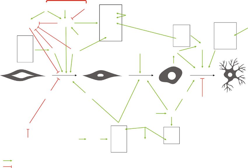
Stem Cells International 5

Both in vitro and in vivo models have demonstrated that ing to the promotion of osteogenesis. In contrast, when
OPG levels are inversely related to osteoclastogenesis [54, HOXB7 was inhibited, these transcription factors were
55]. In an in vitro study, the treatment of undifferentiated downregulated resulting in a decrease in ALP activity that
MSCs with OPG resulted in the enhancement of osteogen- led to a decrease in mineralization. Other HOX genes
esis [56]. Furthermore, OPG knockout mice demonstrate involved in osteogenesis are HOXa2 and HOXd9. In vivo
an increase in bone resorption due to increased osteoclast studies showed that during bone regeneration, HOXa2 is
activity [57]. upregulated after bone fracture while HOXd9 is downregu-
 Ocn and BSP are both noncollagenous proteins found in lated [70].
bone tissue. Ocn is the most abundant, noncollagenous pro- The β-catenin protein is multifunctional. One important
tein in bone tissue and is used as a biochemical marker for function is its ability to regulate the transduction of Wnt sig-
bone formation in vitro and in vivo: an increase in Ocn levels nalling [71]. The inhibition of β-catenin leads to the inhibi-
has been associated with an increase in bone mineral density tion of osteogenesis and the promotion of chondrogenesis
[58]. BSP is found in mineralized tissue such as bone, calci- [72]. β-catenin is activated by the Wnt signalling pathway;
fied cartilage, and dentin and makes up approximately 8% of β-catenin then interacts with LEF/TCF which together
the noncollagenous protein of bone [59]. Although the func- increase bone mineralization [73]. Ex vivo studies have dem-
tion of BSP is not yet fully known, it is suspected to play a onstrated the importance of β-catenin in osteoblast mineral-
role in the formation of hydroxyapatite (essential compo- ization through its downstream regulation of BMP2 [74]. In
nent of healthy bone tissue) [60]. In the absence of BSP an in vivo study, Hill et al. [75] knocked-down β-catenin
in vitro, osteogenic differentiation is negatively impacted. from head and limb mesenchyme in mouse embryos. In
BSP overexpression leads to an increase in osteoblast- the absence of β-catenin, the mutant mice did not form cor-
related gene expression as well as enhanced mineralization. tical or trabecular bone. Interestingly, the overexpression of
The opposite is also true; when BSP expression is reduced, β-catenin does not result in an increase in osteoblast num-
there is both a reduction in osteoblast-related gene expres- ber, but rather inhibits chondrogenesis and allows for MSC
sion and bone mineralization [61]. In vitro studies have sug- osteogenesis [75].
gested that a lack of BSP reduces osteoprogenitor cell There are several other transcription factors, not dis-
numbers and has a compensatory role on Opn. The BSP-/- cussed in this review, that are involved in osteogenesis.
phenotype is associated with the upregulation of Opn in an These include matrix extracellular phosphoglycoprotein
attempt to rescue the cells. However, the overexpression of (MEPE), human high-temperature requirement protein 1
Opn is not enough to rescue the cells, and thus, bone forma- (HTRA1), IGFBP-2, and secreted protein acidic and rich in
tion and mineralization do not occur [62, 63]. BSP-/- mice cysteine (SPARC), TMEM119, sclerostin, and hypoxia-
demonstrate normal skeletal development; however, they inducible factor-1α (HIF-1α); for further information, please
display undermineralization of long bones [63, 64]. refer to [76–83].
 The osteogenic process involves a complex network of
3.4. Additional Osteogenic Transcription Factors. Other tran- cells and mediators, and even the slightest disruption of
scription factors that are involved in osteogenic differentia- the network leads to defective bone formation (Figure 2).
tion are frizzled-related protein (FRZB), dickopf (Dkk) 2,
homeobox protein Hox-B7 (HOXB7), β-catenin, and others. 3.4.1. Signalling Pathways. Successful translation of in vitro
FRZB is a Wnt modulator that increases the expression of findings to clinical applications in vivo requires a good
osteogenic-related markers and calcium deposition. The understanding of potential differences in events during
overexpression of Frzb in MC3T3-E1 cells increases osteo- in vitro and in vivo regulation of osteogenic differentiation.
genic activity while the loss of Frzb results in a decrease in The BMP pathway and the Wnt/β-catenin signalling path-
osteogenic activity [65]. However, Frzb null mice show an way are two important extracellular signalling pathways
increase in cortical bone thickness [66]. These contrasting involved in osteogenic differentiation [72, 84]. Several stud-
results may be explained by the deficiency of FRZB leading ies have investigated the role of the BMP pathway during
to supraphysiological levels of other Wnt modulators such in vitro and in vivo osteogenic differentiation and reported
as Dkk1 and Dkk2 that stimulate osteogenesis. Dkk1 and on the differences and similarities in extracellular signalling
Dkk2 work antagonistically in vivo, where the increased pathways regulating events in these settings. Tsialogiannis
expression of Dkk1 results in a decrease in bone mass while et al. [85] concluded that the BMP pathway plays an impor-
an increase in Dkk2 expression positively stimulates bone tant role during both in vitro and in vivo osteogenic differen-
formation [67, 68]. tiation. The majority of studies looking at the relationship
 When the transcription factor HOXB7 is over expressed, between the Wnt/β-catenin signalling pathway and bone
osteogenesis is enhanced through the upregulation of formation have been done in vivo. Other extracellular signal-
RUNX2 [69]. Gao et al. [69] performed both in vitro and ling pathways that play a role in osteogenesis are the Notch
in vivo studies to investigate the role of HOXB7 during oste- signalling pathway, the hedgehog pathway, fibroblast growth
ogenic differentiation. In their in vitro studies, the overex- factor (FGF), vascular endothelial growth factor (VEGF),
pression of HOXB7 enhanced bone mineralization through and extracellular signal-regulated kinase [86] (Figure 3).
activation of ALP. HOXB7 overexpression also had an effect
on other osteogenic transcription factors and proteins such 3.5. BMP Signalling Pathway. BMP binds to its receptor,
as RUNX2, osteonectin, collagen type I, BSP, and Ocn, lead- BMPR, found on epithelial cells, which in turn activates
6 Stem Cells International

 Gremlin-1
 Chordin

 Noggin BMP BMPR

 ALP
 Smad
 Foxc1 BMP4 Col1

 COX2 BMP9 RUNX2

 OPG/RANKL
 Wnt/ –
 catenin
 Osteogenesis

 PPAR Notch

Figure 3: Illustration of how various signalling pathways regulate osteogenesis through the master regulator of osteogenesis, RUNX2. Green
arrows indicate positive regulation while red lines indicate negative regulation.

the intracellular transcription factor Smad. Smad binds to expression of RUNX2 and Dlx-5. Similarly, in vivo studies
the master regulator, RUNX2. The Smad-RUNX2 complex showed that COX-2 knockout mice displayed 98% and
induces osteogenesis [87] (Figure 3). Using various BMP 86% reduction in bone formation when they received a bone
antagonists in vitro, Tsialogiannis et al. [85] demonstrated graft from other COX-2 knockout mice or wild type mice,
that inhibition of BMP function affects multiple downstream respectively [99].
factors, such as RUNX2, BSP, and Ocn. The investigators Foxc1 is another important osteogenic regulator that
extended their investigation by overexpressing noggin, a interacts with an osteogenic factor, BMP4. Foxc1 mutant
BMP antagonist, in transgenic mice and reported a signifi- mice display numerous abnormalities related to bone devel-
cant decrease in bone density and bone formation in these opment. The calvarial bones and sternum are absent, the ribs
animals [88]. In contrast, complete knockout of noggin led are deformed, and the skull base is reduced in size [100,
to irregularly thickened bones and death shortly after birth 101]. The ectopic expression of Foxc1 in C2C12 myoblasts
[85, 89]. Other BMP antagonists include chordin and grem- resulted in the rescue of osteogenesis by increasing ALP
lin. Multiple in vitro studies have demonstrated that chordin activity and inducing early osteogenic markers such as
is a strong endochondral ossification stimulator [90–93]. RUNX2 and type I collagen [102]. Furthermore, Hopkins
Zhang et al. [94] examined the role of chordin in vivo and et al. [103] demonstrated that a downregulation of Foxc1
their results show that BMP-2 enhances maturation of chon- in C2C12 cells resulted in the inhibition of RUNX2, Msx2,
drocytes resulting in growth of the growth plate of and ALP activity (Figure 3). These investigators suggested
Hamburger-Hamilton stage 25-27 embryonic chick limbs. that Foxc1 is required for the initiation of osteogenesis but
When chordin (a BMP antagonist) was expressed ectopi- not for the later stages, as they observed a decrease in Foxc1
cally, it resulted in a delayed growth rate of the growth plate levels as differentiation proceeded.
by binding to BMP to inhibit BMP’s function. From previ-
ous in vitro studies it is known that when gremlin-1 is sup- 3.6. Wnt/β-Catenin Signalling Pathway. Wnt/β-catenin sig-
pressed, the expression of osteoblastic genes ALP, BSP, nalling, also known as the classical or canonical Wnt path-
MSX2, OC, OPN, and RUNX2 is significantly increased way, is of particular importance as it can either induce or
[95]. It was only recently that the role of gremlin was inves- inhibit osteogenesis. This pathway can regulate the expres-
tigated in vivo. Rowan et al. [96] explored the effect of Grem sion of RUNX2 to induce osteogenesis. Alternatively, the
1 deletion in ROSA26CreER-Grem1 flx/flx mice. Although Wnt pathway inhibits osteogenesis by altering the
these mice demonstrated normal bone structure, there were OPG/RANKL ratio. The expression of PPARγ is also con-
other abnormalities present including severe bowel disrup- trolled by the Wnt pathway. PPARγ is the main transcrip-
tion as well as abnormal haematopoiesis. Cyclooxygenase 2 tion factor in adipogenesis, and therefore, its expression
(COX-2) enhances in vitro osteogenic differentiation needs to be inhibited in order for osteogenesis to occur
through initiating the BMP signalling pathway via a positive [35] (Figure 3).
regulatory loop with BMP9, a potent osteogenic stimulator Mice lacking the Lrp5 gene, which codes for a Wnt cor-
[97, 98]. Wang et al. [98] demonstrated that COX-2 is criti- eceptor, developed osteopenia, while the overexpression of
cal for orchestrating the BMP/Smad signalling pathway Lrp5 resulted in high-bone-mass syndromes [104, 105].
in vitro (Figure 3). Silencing Cox2 downregulated the Genome-wide association studies in humans revealed an
Stem Cells International 7

association between multiple mutations in Wnt1 and Wnt16 therefore MSCs can either be positive or negative for CD34
and early onset osteogenesis imperfecta and osteoporosis; [126–128]. Currently, there is no cell surface protein specific
both bone disorders result in brittle bones as well as an to MSCs, and MSCs isolated from different sources may dif-
increased risk of fractures [106, 107]. Hilton et al. [33] fer regarding cell surface protein expression profiles. Table 1
removed all the components of the Notch network in mice, summarizes the different cell surface markers that are asso-
and this resulted in increased bone mass and a depleted pool ciated with MSCs isolated from different tissue sites.
of MSCs in the bone marrow [33]. The Notch network
inhibits osteogenesis through the expression of HEY1 and 6. Osteogenic Potential of MSCs In Vitro
HEYL transcription factors that directly inhibit RUNX2
(Figure 3). Overexpression of Notch-1 in mice inhibited In vitro, MSCs are induced to undergo osteogenic differenti-
osteogenesis through the inhibition of the Wnt/β-catenin ation following exposure to compounds such as β-glycero-
signalling pathway [108]. It is clear that extracellular signal- phosphate, dexamethasone, and ascorbate-2-phosphate,
ling pathways play a major role in osteogenesis via a com- that promote cell proliferation and osteogenic differentia-
plex network of transcription factors. It is therefore tion. Although these 3 compounds (β-glycerophosphate,
important to examine the network as a whole and not sepa- dexamethasone, and ascorbate-2-phosphate) are present in
rate out specific interactions, as would occur in an in vitro all in vitro osteogenic media, there is a lack of consensus
setting. regarding the optimal medium for in vitro osteogenic differ-
 entiation of MSCs, particularly regarding the concentration
4. Mesenchymal Stromal/Stem Cells of dexamethasone, which varies significantly between stud-
 ies. Table 2 summarises the composition of the osteogenic
MSCs contain a population of multipotent adult stem cells media used most often. Ascorbic acid and dexamethasone
capable of differentiating into cell types of mesodermal ori- are the main osteogenic inducing factors, and together
gin [109]. MSCs were initially isolated from bone marrow increase the activity of ALP. Upregulation of ALP activity
(BM) and are in this setting referred to as bone marrow- increases the speed at which bone differentiation occurs
derived MSCs (BM-MSC) [110]. Since then, human MSCs [129]. Ascorbate-2-phosphate is responsible for the synthe-
have been isolated from various foetal and adult tissues, such sis of collagen in the early stages of osteogenesis, while β-
as adipose tissue [111], the amniotic membrane [112], amni- glycerophosphate is responsible for mineralization in the
otic fluid [113], placental and foetal membranes [114], later stages [130, 131]. Along with increasing ALP activity,
umbilical cord lining membrane [115], the endometrium dexamethasone also regulates the osteogenesis-related gene
[116], dental tissue [117], menstrual blood [118], peripheral RUNX2 [132].
blood [119], skin [120], synovial fluid [121], and Wharton’s Various spectrophotometric assays are used to deter-
jelly [122]. mine the extent of in vitro osteogenic differentiation. Both
 It is well accepted that isolated MSC populations are het- the Von Kossa assay and the Alizarin Red S (ARS) assay
erogeneous, containing both stem cells and mature stromal stain for calcium deposits that are present in bone tissue.
cells. Even though the terms mesenchymal stem cells and The Von Kossa assay is a qualitative assay in which calcium
mesenchymal stromal cells are used interchangeably [109], is replaced with silver ions (source: silver nitrate solution) to
there are distinct differences between the two. Mesenchymal form black/brown deposits that can be analysed under a
stem cells possess the ability to self-renew and differentiate, microscope [133]. The ARS assay is semiquantitative in
demonstrating the functionality of true stem cells, while which ARS reacts with calcium to form a red deposit which
mesenchymal stromal cells refers to a heterogeneous popula- is extracted using acetic acid. The extracted dye is spectro-
tions of progenitor cells at various stages of maturation. photometrically quantified at 405 nm [134]. Another assay
Directly after isolation, the isolated MSC population may that is often used to quantify osteogenesis is the ALP assay
also contain differentiated cells present in the tissue micro- that also uses spectrophotometry to measure the level of
environment such as endothelial cells, pericytes, fibroblasts, ALP activity. In short, 4-nitrophynylphosphate is used as a
and immune cells, as well as elements of circulating blood phosphate substrate for ALP which dephosphorylates 4-
[123–125]. nitrophenylphosphate which then turns yellow. This colour
 change is measured at 405 nm [135].
5. Characterization of MSCs MSCs isolated from various tissues also differ in their
 differentiation capabilities [152–155]. This may be due to
All MSCs, independent of their source, should adhere to DNA methylation of key transcription factors. Xu et al.
minimal criteria recommended by the International Society [152] demonstrated that MSCs retain their epigenetic mem-
for Cellular Therapy (ISCT). These include (a) the ability ory and favour either of adipogenic or osteogenic differenti-
to adhere to plastic; (b) the expression of a specific set of cell ation, depending on their tissue of origin. In BM-MSCs, the
surface markers such as cluster of differentiation (CD)73, CpG island in the RUNX2 promoter is hypomethylated
CD90, CD105 or CD13 and the lack of CD14, CD19, while the CpG island in PPARγ is hypermethylated. The
CD31, CD45, and human leukocyte antigen (HLA)-DR; opposite is true in adipose tissue-derived stromal/stem cells
and (c) the ability to differentiate into at least adipocytes, (ASCs): the PPARγ promoter is hypomethylated while the
osteoblasts, and chondrocytes in vitro [126]. Many studies RUNX2 promoter is hypermethylated. Pérez-Silos et al.
have suggested that the expression of CD34 is variable and and McLeod et al. [156, 157] suggest that MSCs consist of
8 Stem Cells International

 Table 1: Cell surface markers expressed by MSCs isolated from different tissues.

 Cell surface marker
Source Reference
 Positive Negative
 CD10, CD13, CD29, CD34, CD44, CD49e, CD59, CD71, CD11b, CD14, CD19, CD31, CD34, CD45, [111,
Adipose tissue
 CD73, CD90, CD105, CD166, CD200, HLA-ABC CD56, CD146, CD235a, Stro1, HLA-DR 136–139]
Amniotic CD11b, CD10, CD14, CD19, CD20, CD34, [113,
 CD29, CD44, CD73, CD90, CD105, SH2-4 HLA-ABC
membrane and fluid CD45, CD79a, HLA-DR 140–142]
 [57, 126,
Bone marrow CD29, CD44, CD73, CD90, CD105, CD271, Stro-1 CD14, CD34, CD45, HLA-DR
 137, 143]
 CD29, CD34, CD44, CD73, CD90, CD105, CD105, CD11b, CD14, CD19, CD31, CD34, CD45, [117,
Dental tissue
 CD117, CD166, Stro1 CD79a, CD146, HLA-DR 143–145]
Endometrium CD44, CD49d, CD479f, CD73, CD90, CD105, CD146 CD14, CD19, CD34, CD45, HLA-DR [116, 146]
 [119, 147,
Peripheral blood CD29, CD73, CD90, CD105, CD106, CD146, CD166, CD34, CD45, CD133
 148]
Placental and foetal
 CD29, CD73, CD90, CD105 CD34, CD45 [114]
membrane
Skin CD29, CD44, CD73, CD90, CD105, CD166 CD14, CD34, CD45, HLA-DR [120, 149]
 CD11b, CD14, CD19, CD31, CD34, CD45,
Synovial fluid CD44, CD73, CD90, CD105, CD147, Stro-1 [121, 141]
 CD79a, CD106, HLA-DR
Umbilical cord [111, 115,
 CD29, CD44, CD73, CD90, CD105, CD106, HLA-I CD14, CD31, CD34, CD45, HLA-DR
lining membrane 150]
Wharton’s jelly
within umbilical CD73, CD90, CD105 CD14, CD19, CD34, CD45, CD79, HLA-DR [122, 151]
cord

subpopulations that share common features while varying in significantly decreased compared to controls, while the oste-
the expression profile of their cell surface proteins, which ogenic potential was similar when BM-MSCs from OA
can be related to differences in differentiation potential. Can- patients and MSCs from the control group were com-
tentin et al. [158] found that UC-MSCs produced signifi- pared [161].
cantly more ECM, while stronger staining for type I He et al. [174, 175] demonstrated that the extracellular
collagen was observed for BM-MSCs indicating that BM- matrix is important in directing MSCs down a specific line-
MSCs have enhanced osteogenic potential when compared age: a hydroxyapatite- (HA-) collagen matrix was found to
to UC-MSCs. UC-MSCs produced molecules that BM- be superior to a HA-synthetic hydrogel for osteogenic differ-
MSCs did not such as type X collagen and the HtrA1 gene entiation. For chondrogenesis, the HA-synthetic hydrogel
product. UC-MSCs additionally displayed a higher propor- was preferred over the HA-collagen matrix. The HA-
tion of CD73+ cells. The authors suggest that the difference collagen matrix imitated the natural composition of bone
in CD73 expression and the production of these atypical and resembled the physical and chemical microenvironment
molecules are the major reason for differences in chondro- found in the human body, thus favouring osteogenesis. The
genic differentiation potential between BM-MSCs and UC- reason why the HA-synthetic hydrogel was favoured for
MSCs. chondrogenesis is not fully understood, as the HA-
 Other factors that may influence the differentiation synthetic hydrogel does not imitate natural cartilage. Over-
capabilities of MSCs include the age of the donor, the health all, the use of a matrix increased cell proliferation, adhesion,
of the donor, culture conditions, and method of isolation. migration, and differentiation. The biomechanics of the
Barboni et al. and Xin et al. [159, 160] both demonstrated MSC microenvironment also has an effect on differentiation
a positive correlation between age and DNA methylation capabilities. Gungordo et al. [176] concluded that rat BM-
status. Barboni et al. [159] observed a correlation between MSCs progress to an adipogenic lineage under unstrained
gestational age of amniotic-derived MSCs and global DNA conditions on a softer polyacrylamide hydrogel film, while
methylation status, which resulted in a decrease in osteo- rat BM-MSCs seeded on a stiffer polyacrylamide hydrogel
genic differentiation potential. Xin et al. [160] extensively and under strained conditions are driven down the osteo-
compared DNA methylation status and multilineage differ- genic lineage. The use of animal serum which contains
ential capabilities. An age-related decline in ASC osteogenic xenoantigens is another culture condition that can affect dif-
differentiation was observed when ASCs from young and old ferentiation potential, specifically osteogenic differentiation
donors were compared. In another study, the differentiation [177, 178]. Okajcekova et al. [179] compared three different
potential of BM-MSCs from patients with osteoarthritis osteogenic induction media and their differentiation capabil-
(OA) was compared to MSCs isolated from a control group ities, of which one was xeno-free. Not only did the xeno-free
of a similar age: both the chondrogenic and adipogenic dif- induction medium result in significantly greater osteogenic
ferentiation potential of BM-MSCs from OA patients were differentiation potential compared to the other two, but the
Table 2: Summary of different osteogenic differentiation media reported in the literature.

 Induction Basal culture Dexamethasone Ascorbate-2- B-glycero-
Reference Cell density Assays/stains Passage FBS Antibiotics
 Stem Cells International

 time (days) medium (μM) phosphate (μM) phosphate (mM)
 Alizarin Red S DMEM-low
Cai et al. 2014 [162] NI 2 21 10% 100 units/mL 0.01 155.26 1 × 10⁻⁵
 and ALP glucose (lg)
Vieira et al. 2010
 NI Von Kossa 3 21 NI 10% NI 0.1 50 1 × 10⁻⁵
[163]
Nishimura et al.
 5 × 10⁵ 5 14 DMEM 10% 100 units/mL 0.05 0.0002 10
2015 [129]
Bieback et al. 2004
 3:1 × 10³ /cm2 Von Kossa NI 21 Cell systems 10% NI 0.1 50 10
[164]
Waterman et al. 3 × 10⁴ cells/well (6-
 Alizarin Red S NI NI NI NI NI 0.1 50 1 × 10⁻⁵
2010 [165] well)
Elashrya et al. 2019 2 × 10⁴ cells/well (6-
 Alizarin Red S 2-3 NI DMEM 10% 100 U/mL 0.1 60 10
[166] well)
 DMEM-high
Li et al. 2015 [167] 5 000 cm2 ALP NI NI 10% 100 U/mL 0.01 155.26 10
 glucose
Sotiropoulou et al. 50 μg/mL
 NI Von Kossa NI NI DMEM-lg 10% 1 50 10
2006 [168] Gentamicin
Rada et al. 2011
 NI Alizarin Red S NI 21 α-MEM 10% 1% 0.1 155.26 10
[169]
Meuleman et al.
 NI Von Kossa 1 14 α-MEM NI NI 0.1 60 10
2006 [170]
Sasaki et al. 2008
 NI Von Kossa NI NI DMEM 10% 0.1 μM - 50 10
[171]
Zuk et al. 2001 ALP or Von
 NI 1 14 DMEM 10% 1% 1 50 10
[172] Kossa
Bunnell et al. 2008
 NI Alizarin Red S NI 14 α-MEM 20% 1% 0.001 50 2
[173]
Wagner et al. 2005 ALP or Von
 1 − 2 × 10⁴ cells/cm2 NI 21 DMEM 10% - 1 200 10
[111] Kossa
NI: not indicated; DMEM: Dulbecco’s modified Eagle’s medium; ALP: alkaline phosphatase.
 9
10 Stem Cells International

morphology of the cells grown in the xeno-free medium They further went on to suggest that ASCs may be more
changed much earlier than the cells grown in the FBS induc- suitable for in vitro studies, as their isolation procedure is
tion medium: cell proliferation decreased while cell differen- less invasive than BM-MSCs, and although their terminal
tiation increased. differentiation capability is reduced, it is still adequate for
 The method of isolation also has an impact on the differ- in vitro studies, while BM-MSCs may hold greater potential
entiation capability of MSCs. In a recent study by Walter for in vivo studies as their terminal osteogenic differentiation
et al. [180], different isolation techniques from the same capability is greater than that of ASCs. For more information
donor site were compared with regard to osteo-, adipo-, on the differences between BM-MSCs and ASCs with regard
and chondrogenic differentiation. MSCs isolated from bone to their osteogenic potential, we refer the reader to a review
marrow aspiration showed better osteogenic differentiation by Liao [184].
than MSCs generated through outgrowth from culturing Multiple barriers limit the clinical application of MSCs.
bone chips, which can be attributed to the fact that bone Many of these are related to the need to extensively expand
marrow aspiration yields more biomaterial and thus more these cells ex vivo in order to achieve clinically relevant cell
MSCs. Chondrogenic and adipogenic differentiation, both numbers. One major barrier associated with extensive
from MSCs from bone marrow aspiration and MSCs gener- ex vivo expansion is the decrease in differentiation potential,
ated through outgrowth from culturing bone chips, was rel- mainly due to the loss of telomerase activity, also known as
atively low; the authors attribute this to the specific replicative senescence [185]. MSCs tend to lose their differ-
microenvironment of the isolated bone tissue and suggest entiation potential as passage number increases, and it is
that this led to MSCs favouring the osteogenic lineage. thus important to limit expansion rounds, ideally staying
 Musina et al. [181] compared the osteogenic differentia- below 5 passages [186, 187]. Bonab et al. [187] reported that
tion potential of MSCs from different tissue sources after a MSCs, especially BM-MSCs, show a lower multilineage dif-
three-week induction period. These investigators reported ferential potential due to morphological changes and a
that BM-MSCs displayed the highest level of osteogenic dif- decrease in telomere length resulting in the loss of MSC
ferentiation, followed by ASCs which showed better osteo- characteristics during long-term culturing. The thawing of
genic differentiation capabilities than MSCs isolated from MSCs preserved in liquid nitrogen results in a heat-shock
the thymus, skin, and placental tissues. Mohamed-Ahmed response (“cryogenic injury”) which leads to a decrease in
et al. [182] compared the osteogenic potential of MSCs iso- their immune modulatory function [188]. Another disad-
lated from bone marrow and adipose tissue and also vantage of long-term culturing is an increase in the probabil-
reported that BM-MSCs possess enhanced osteogenic poten- ity of malignant transformation, in which cells acquire
tial when compared to ASCs. The reason for the difference cancer-like properties [189].
was attributed in part to increased alkaline phosphatase A further limit to in vitro cell culturing is the use of foe-
(ALP) activity and osteogenic gene expression kinetics. tal bovine/calf serum (FBS/FCS) as a supplement to cell cul-
Early-stage osteogenic genes such as RUNX2, collagen type ture medium to ensure optimal cell proliferation [190].
I, and ALP were expressed as early as day 14 in osteogenic Although commonly used, FBS/FCS-supplemented growth
differentiating BM-MSCs, while these genes were only media are associated with number of disadvantages. First,
expressed on day 21 in differentiating ASCs. This indicates FBS/FCS shows batch-to-batch variation due to the variable
that BM-MSCs stop proliferation early (day 14) and switch composition of the product, and thus, results are often not
to differentiation and formation of a mature collagenous reproducible [191]. Furthermore, FBS/FCS is xenogeneic
matrix, while ASCs have an extended proliferation period and contains bovine proteins that can potentially elicit an
and only switch to differentiation after day 21, resulting in immune response in humans [192]. The transmission of
BM-MSCs having greater mineralization and therefore more zoonotic diseases is also a possibility and thus also a primary
bone tissue on day 21 [182]. Shen et al. [153] compared concern when culturing cells in FBS/FCS; cells that have
MSCs derived from the amniotic membrane (AM-MSCs), been cultured in FBS/FCS can therefore not be used clini-
the umbilical cord (UC-MSCs), the chorionic membrane cally [193].
(CM-MSCs), and the decidua (DC-MSCs) and reported Due to the disadvantages associated with FBS/FCS, the
enhanced osteogenic differentiation (based on ARS staining use of human blood products in cell culture medium as
and ALP activity) in AM-MSCs and UC-MSCs when com- alternatives to animal serum is becoming increasingly popu-
pared to CM-MSCs and DC-MSCs. In terms of gene expres- lar [194, 195]. In short, blood is separated by centrifugation
sion profiles involved in osteogenesis, AM-MSCs and UC- into its components, i.e., platelets, growth factors, and fibrin,
MSCs showed strongly enhanced expression of Ocn com- which are separated from erythrocytes [196]. Some of these
pared to CM-MSCs and DC-MSCs. MSCs from all four blood products include human serum (HS), platelet-rich
sources showed the same expression levels of Osx and colla- plasma (PRP), platelet-poor plasma (PPP), fresh frozen
gen type I on day 21. Szöke et al. [183] compared the osteo- plasma (FFP), and human platelet lysate (HPL). Human
genic potential of MSCs isolated from bone marrow and serum (HS) is produced by taking whole blood donated
adipose tissue. They concluded that although ASCs had a from a patient, allowing it to clot and centrifuging the blood
higher proliferative capacity and a greater ability to form a to produce serum that is devoid of platelets, erythrocytes,
collagenous extracellular matrix, their terminal osteogenic and leukocytes [197]. Plasma is the noncellular liquid part
differentiation capability was reduced. BM-MSCs expressed of whole blood. Two human alternatives can be prepared
a higher level of the late osteogenic markers Ocn and BSP. from plasma: PRP and PPP. The difference is the
Stem Cells International 11

concentration of platelets. Platelets are anucleated, disc- vide a complete understanding of osteogenesis and also brid-
shaped cell fragments that play a role in cell growth, differ- ges the gap between the use of MSCs in vitro and the clinical
entiation, and tissue regeneration [198]. When preparing use of MSCs for bone repair.
PRP, whole blood is centrifuged, and the supernatant Most studies that have investigated osteogenic differenti-
(plasma) is centrifuged again to collect a platelet pellet; the ation of MSCs in vivo first expanded the cells ex vivo, seeded
platelet pellet is then resuspended in a smaller volume of them onto a scaffold, and transplanted the scaffold subcuta-
plasma thus combining the plasma and buffy coat into one neously in an animal model in which osteogenesis was stud-
[199]. Alternatively, PRP can be collected via apheresis ied [212–214]. For optimal bone regeneration, the
[200]. PPP is prepared by removing platelets from the biomaterial used as a scaffold should be biocompatible, cost
plasma obtained from whole blood. Fresh frozen plasma effective, biodegradable, and should also induce or improve
(FFP) is obtained by rapidly freezing plasma separated from the osteogenic process. The biological behaviour of MSCs
whole blood at -65°C [201]. Lastly, to produce HPL, PRP is is greatly affected by the surface morphology of the biomate-
submitted to several freeze-thaw cycles to rupture the plate- rial which in turn affects the formation of bone tissue [215].
lets releasing growth factors, followed by centrifugation to The most common scaffold material being used in tissue
remove cell debris [200]. The various human alternatives engineering is hydroxyapatite, an inorganic material that is
provide unique advantages and disadvantages with regard naturally found in bone tissue [216]. These scaffolds are cast
to culturing MSCs in in vitro by providing suitable growth into the desired shape. Another new and attractive method
factors and ensuring genomic stability [197]. of making scaffolds is the use of 3-dimensional (3D) print-
 ing, as it allows for a reproducible design when it comes to
7. Bone Regeneration and Repair: pore size [217]. Once the scaffolds are transplanted, MSCs
 Clinical Application differentiate into osteoblasts and form bone tissue.
 Several factors play a role in inducing MSC differentia-
Bone is a vital part of the human body that protects and sup- tion into bone in vivo including paracrine signalling path-
ports various organs, enables mobility, stores minerals, and ways in the region of bone injury [218]. When bone injury
produces cells of the hematopoietic lineage [202]. Bone frac- occurs, perivascular stem cells induce paracrine pathways
tures typically heal without the need for major intervention; through the secretion of Wnt-related molecules that in turn
however, there are more than 2 million cases worldwide in activate the BMP and Wnt β-catenin pathways causing oste-
which patients require bone reconstruction using tissue ogenic differentiation [219]. Furthermore, MSCs create a
transplants [203]. Current reconstruction procedures microenvironment that supports new bone formation
involve autologous bone grafts, allogeneic bone grafts, and through the production of an ECM [220]. Several
artificial metal or ceramic replacements. approaches have been investigated to enhance osteogenic
 Autologous bone grafts are viewed as the gold standard differentiation in vivo including harvesting of ECM to coat
for treating bone defects as they enhance osteogenesis and biomaterials. The use of ECM not only improves osteogenic
are less likely to be rejected by the host [204]. However, differentiation but also enhances MSC survival in vivo [221–
10% of bone harvests are associated with major complica- 223]. Another approach is to coat biomaterials with osteo-
tions, limited supply, and donor-site morbidity [205, 206]. genic inductive compounds [224–226]. MSCs can also be
Allogeneic bone grafts provide an ample source of tissue, primed or predifferentiated down the osteogenic lineage
but the risk of immune rejection and the transmission of dis- before seeding them onto scaffolds [227–229]. Lastly, MSCs
eases make them less ideal [207]. The use of metals as artifi- can be genetically engineered to express bone inducing genes
cial replacements also has limitations such as tissue-host which enhances osteogenic differentiation [230–233].
integration, increased risk of infection, and wearing out Several methods, such as histological staining, histomor-
[208]. The brittle nature of ceramic replacements is espe- phometry, immunohistochemistry, and quantitative real-
cially problematic in areas where high stress or torsion is time polymerase chain reaction (RT-qPCR), are used to
endured [209]. It is thus clear that alternative, more effective assess the success of osteogenesis in vivo. Once osteogenesis
options are needed for the treatment of skeletal defects. has been allowed to occur in in vivo mouse models, the scaf-
 folds on which the new bone tissue has formed are resected,
8. The Use of Cultured MSCs for and the degree of osteogenesis is measured. Histological
 Osteogenesis In Vivo staining with haematoxylin and eosin is used to nonspecifi-
 cally detect newly formed bone matrix [234]. A combination
The osteogenic differentiation potential of multipotent of Alcian blue, haematoxylin, orange g, phloxine b, and
MSCs has gained increasing interest in tissue engineering eosin serves as a more specific histological stain for mature
especially when it comes to offering an alternative to over- bone tissue [235]. Immunohistochemistry allows for the
come the limitations of bone grafts and artificial replace- identification of specific antigens such as type I collagen,
ments [210]. Multiple in vitro studies have demonstrated Ocn, Opn, and BMP-2 [236–238]. Lastly, RT-qPCR can be
that MSCs are able to differentiate into bone tissue, but bone used to assess the expression of osteogenesis-associated
formation is a complex process that involves many cell types, genes such as ALP, RUNX2, BSP, Osx, Ocn, Dlx5, and
growth factors, cytokines, and mechanical stimulation that BMP-2 [213, 239, 240].
all form part of the environmental niche [211]. Therefore, Angiogenesis needs to occur for successful bone healing
investigation of bone formation in vivo is required to pro- in large bone defects. The successful translation MSC-
12 Stem Cells International

associated cell therapy products for the treatment of bone sue in a critical-sized calvarial defect. They also found that
defects in the clinical setting must be accompanied by rapid the increase in osteogenic stimulation was related to the
vascularization of the implanted scaffold [241]. Vasculariza- increase in exosome concentration over time. The repair of
tion results in adequate delivery of nutrients, oxygen supply, a critical osteochondral defect in adult immunocompro-
and the removal of waste products. Rapid vascularization mised rats through the intravenous injection of human
also supports the survival of the seeded cells. To promote embryonic MSC-derived exosomes was demonstrated by
rapid vascularization, some studies suggest coculturing Zhang et al. [272]. The use of MSC-derived exosomes in
MSCs and endothelial cells, the latter for their ability to pro- regenerative medicine has gained a great deal of attention
mote angiogenesis [241, 242]. Other studies suggest using a as it is an attractive alternative to using MSCs. MSC-
cell type that has the ability to differentiate into both bone derived exosomes are cell-free and are more compatible with
tissue and endothelial cells to improve angiogenesis in vivo a variety of administration routes [272]. Another reason why
[213]. Brennan et al. [213] used ASCs based on the assump- exosomes are attractive is that they lack major histocompat-
tion that ASCs can differentiate into both endothelial cells ibility complex (MHC) I/II proteins, and there is therefore
and osteoblasts; they found that although ASCs were able no need for immunosuppression [273, 274].
to achieve both osteogenesis and angiogenesis; the degree
of osteogenesis was inferior to the degree of osteogenesis 10. Therapeutic Use of MSCs for Bone Diseases
achieved by BM-MSCs. These investigators then investigated
coculturing BM-MSCs and ASCs and found that although Bone remodelling is a complex and highly integrated pro-
there was enhanced blood vessel formation, osteogenesis cess, and as described in this review, it involves various tran-
was not enhanced. Brennan et al. [213] concluded that ASCs scription factors and osteogenic genes and their protein
need to be osteogenically primed prior to implantation to products including cytokines, growth factors, and extracellu-
achieve enhanced osteogenic abilities. The need to prime lar matrix components. The smallest deviations from this
ASCs to undergo osteogenic differentiation was supported well-balanced system can affect bone health and lead to a
by various other investigators who found that without prim- number of bone diseases. Bone tissue is a porous, mesh-
ing, ASCs fail to heal critical-size defects [243–245]. Another like network made up of collagen proteins and calcium
interesting hypothesis was that the immune system and phosphate minerals and is constantly being replaced
bone formation are linked. Several studies have suggested throughout life. When the bone remodelling process is
that MSCs secrete paracrine factors that recruit immune defective, this mesh-like structure becomes porous as seen
cells to the site of injury leading to bone formation [211, in osteoporosis, leading to brittle bones and fractures.
246, 247]. In order to close the gap between culturing MSCs According to the International Osteoporosis Founda-
ex vivo and the clinical use of MSCs for the treatment of tion, over 200 million people are affected by osteoporosis
bone defects, new methods are required to improve the effi- worldwide [275]. Osteoporosis is associated with low bone
ciency of osteogenesis in vivo through, for example, the use mass as well as bone deterioration usually seen with increas-
growth factors, and by improving methods of cytokine deliv- ing age, and it is thought that osteoporosis results, in part,
ery to the implanted scaffold. from a significant decrease in the number of MSCs present
 in the bone marrow, leading to less new bone formation
9. The Use of MSC-Derived Exosomes for [276]. Osteoporosis is currently treated with drugs that
 Osteogenesis In Vivo increase bone resorption, but these drugs are associated with
 multiple adverse effects [277]. Stem cell therapy is a potential
The ability of MSCs to secrete exosomes, in addition to cyto- alternative for the treatment of osteoporosis, reducing the
kines and growth factors, contributes to their therapeutic susceptibility to fractures by increasing the MSC pool pres-
effect [248]. Multiple in vivo studies have demonstrated that ent within the bone marrow. Wang et al. [278] reported
very few MSCs engraft at sites of injury when administered increased bone formation, trabecular thickness, and overall
intravenously, but rather are filtered out in the lungs; how- strength of bone tissue by embedding MSCs into the distal
ever, they still exhibit a therapeutic effect [249–253]. Other femurs of osteoporotic rabbits. Hsiao et al. [279] treated
studies have gone on to report that it is in particular the osteoporotic mice by injecting MSCs intravenously. They
microvesicles/exosomes secreted from MSCs than provide observed that the MSCs homed to the bone marrow where
this therapeutic effect [127, 254, 255]. The therapeutic effect they increased bone density, rescuing the mice from
of MSC-derived exosomes has been extensively studied osteoporosis.
in vivo in a wide range of disease models. Some of these OA is a degenerative joint disease affecting synovial
include cardiovascular disease [256–258], renal disease joints and frequently results in chronic pain [280]. Cur-
[259–261], neurological complications [262–264], pulmo- rently, the treatment of OA involves long-term pain man-
nary disease [265–267], wound healing [268, 269], muscle agement with the use of pharmacological therapies.
regeneration [270], and many more. With regard to osteo- Osteotomy can improve alignment, but this therapy is lim-
genesis, multiple studies have shown that MSC-derived exo- ited as it can decrease the risk of OA but has little effect on
somes can stimulate the osteogenic differentiation process, degeneration once it has occurred [281]. It has been hypoth-
increasing bone regeneration. Qi et al. [271] demonstrated esized that the multipotency properties of MSCs could also
that exosomes from BM-MSCs from ovariectomized rats benefit patients with OA. Currently, there are 102 docu-
stimulated osteogenesis and were able to regenerate bone tis- mented clinical trials assessing the potential of MSCs for
You can also read