Erectile Dysfunction in Men on the Rise: Is There a Link with Endocrine Disrupting Chemicals? - Karger Publishers
←
→
Page content transcription
If your browser does not render page correctly, please read the page content below
Review Article

                                                    Sex Dev 2021;15:187–212                                          Received: November 2, 2020
                                                                                                                     Accepted: April 18, 2021
                                                    DOI: 10.1159/000516600                                           Published online: June 16, 2021

Erectile Dysfunction in Men on the Rise:
Is There a Link with Endocrine Disrupting
Chemicals?
Samuel M. Cripps Deidre M. Mattiske Andrew J. Pask
School of BioSciences, The University of Melbourne, Melbourne, VIC, Australia

Keywords                                                                 and potentially in processes which regulate erectile function
Differences of sexual development · Early mammalian                      during adulthood. Given that endocrine signalling has a
development · Endocrine-disrupting chemicals · Erectile                  prominent role in erectile function, it is likely that exposure
dysfunction · Erection                                                   to endocrine disrupting chemicals (EDCs) is a risk factor for
                                                                         ED, although this is an under-researched field. Thus, our re-
                                                                         view provides a detailed description of the underlying biol-
Abstract                                                                 ogy of erectile function with a focus on the role of endocrine
Erectile dysfunction (ED) is one of the most prevalent chron-            signalling, exploring the potential link between EDCs and ED
ic conditions affecting men. ED can arise from disruptions               based on animal and human studies.           © 2021 S. Karger AG, Basel
during development, affecting the patterning of erectile tis-
sues in the penis and/or disruptions in adulthood that im-
pact sexual stimuli, neural pathways, molecular changes,
and endocrine signalling that are required to drive erection.                 Erectile Dysfunction
Sexual stimulation activates the parasympathetic system
which causes nerve terminals in the penis to release nitric                  Erectile Dysfunction (ED) is defined as the consistent
oxide (NO). As a result, the penile blood vessels dilate, allow-         or repeated inability to acquire or sustain an erection suf-
ing the penis to engorge with blood. This expansion subse-               ficient for satisfactory sexual performance [McCabe et al.,
quently compresses the veins surrounding the erectile tis-               2016]. The 5-item International Index of Erectile Func-
sue, restricting venous outflow. As a result, the blood pres-            tion (IIEF-5) self-questionnaire categorises the severity of
sure localised in the penis increases dramatically to produce            ED based on the numerical score (each of the 5 questions
a rigid erection, a process known as tumescence. The sym-                is worth 5 points) as no ED (22–25), mild (17–21), mild
pathetic pathway releases noradrenaline (NA) which causes                to moderate (12–16), moderate (8–11), or severe (1–7)
detumescence: the reversion of the penis to the flaccid state.           [Rhoden et al., 2002]. Erectile function relies on a combi-
Androgen signalling is critical for erectile function through            nation of organic (structural, neurologic, vascular, and
its role in penis development and in regulating the physio-              endocrine) and psychogenic factors. Thus, ED can have a
logical processes driving erection in the adult. Interestingly,          number of aetiologies which are broadly classified as ei-
estrogen signalling is also implicated in penis development              ther organic or psychogenic [Johannes et al., 2000]. Psy-

karger@karger.com      © 2021 S. Karger AG, Basel                        Correspondence to:
www.karger.com/sxd                                                       Andrew J. Pask, ajpask @ unimelb.edu.au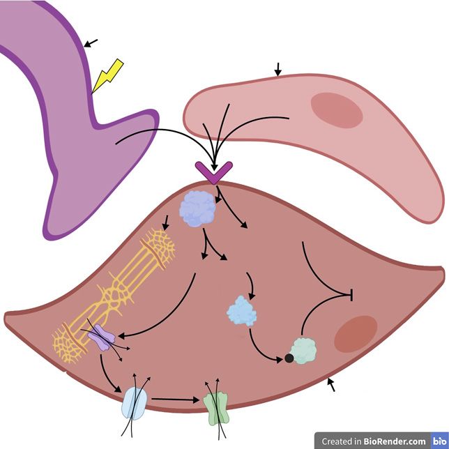
chogenic risk factors for ED include depression and anx-         dicted increase in ED prevalence between 1995 and 2025
iety [Yang et al., 2019], although these are beyond the          can be linked to an increasing ageing male population; the
scope of this review. Organic risk factors include vascular,     global proportion of men aged over 65 years in 1995 was
neurologic, and endocrine abnormalities [reviewed in             4.2% and will increase to 9.5% by 2025 [reviewed in Ayta
Ludwig and Phillips, 2014]. Interestingly, since the penile      et al., 1999]. However, the dramatic increase of ED prev-
vascular tissue that is responsible for erection is a compo-     alence is too rapid to be explained by ageing or genetic
nent of the global vascular system, ED of vascular origin        mutation alone. This is further supported by an excep-
is often an indicator of systemic endothelial dysfunction        tionally high prevalence of ED in younger men. For ex-
[Virag et al., 1981]. Thus, ED not only disrupts quality of      ample, a study using the IIEF-5 showed that Swiss men
life but can also be a strong indicator of cardiovascular        aged 18–25 years displayed a prevalence of 30% [Mialon
disease [Gandaglia et al., 2014].                                et al., 2012]. Another IIEF-based study demonstrated that
    ED is one of the most prevalent chronic conditions           ED prevalence in a population in India ranged from 9.9
and negatively impacts the quality of life of men and their      to 13% in 18–40-years-old men [Sathyanarayana Rao et
partners [Fisher et al., 2005]. The exact prevalence of ED       al., 2015]. Similarly, in Western Australia, ED prevalence
is difficult to estimate, because this relies on subjective      among men in their 20s and 30s was reported as 15.7 and
data-gathering and self-questionnaires. The Internation-         8.7%, respectively [Chew et al., 2008]. It was also found
al Index of Erectile Function (IIEF) and the Massachu-           that 1 in 4 Caucasian European men seeking medical help
setts Male Ageing Study (MMAS)-derived questionnaires            for new-onset ED were below 40 years of age [Capogros-
are the most commonly used for investigating ED in pop-          so et al., 2013]. These results are consistent with the in-
ulation studies. However, researchers use a number of            crease of younger men (40 years) while over-            be due to increased reporting alone; ED prevalence is in-
looking younger men [Feldman et al., 1994; Corona et al.,        creasing across all age groups, not just alongside an in-
2010; Weber et al., 2013]. As a result, it is challenging to     creasing ageing population.
understand the true prevalence of ED in different age                Thus, it is likely that environmental and lifestyle fac-
groups.                                                          tors are responsible for current global trends in ED prev-
    In addition, different geographical regions and age de-      alence. Indeed, several of these factors, which include
mographics yield varying results, creating further com-          smoking and diet, are implicated in the development of
plications in understanding the epidemiology of ED. For          ED [McVary et al., 2001; Bacon et al., 2006; Esposito et al.,
example, ED was reported at an overall prevalence of 23.2        2006; Francis et al., 2007; Ramírez et al., 2016]. However,
and 61% in Australian men from the ages of 35 and 45             the role of endocrine-disrupting chemicals (EDCs) in the
years, respectively [Weber et al., 2013; Martin et al., 2014],   aetiology of ED is unclear. The WHO defines an EDC as
and as high as 81.5% in Malaysian men over the age of 18         “an exogenous substance or mixture that alters function(s)
years [Nordin et al., 2019]. The landmark MMAS re-               of the endocrine system and consequently causes adverse
vealed a prevalence of mild to moderate ED in 52% of             health effects in an intact organism, or its progeny, or
men aged 40–70 years [Feldman et al., 1994], whereas the         (sub) populations” [Johansson and Svingen, 2020]. The
European Male Ageing Study (EMAS) found an average               term EDC in this review refers specifically to chemicals
ED prevalence of 30% in men at ages 40–79 years [Co-             which are known to alter hormonal pathways and cause
rona et al., 2010]. Although there are regional differences,     adverse health effects in humans. Although these adverse
it was estimated that ED affected 152 million men world-         health effects are not yet described to include ED, we
wide in 1995 and was predicted to increase to 322 million        present a logical connection between their impact on hor-
men globally by 2025 (using the lowest United Nations            monal pathways and the development and regulation of
population projections) [Ayta et al., 1999; McKinlay,            erectile tissues.
2000].                                                               Endocrine signalling, particularly that of androgens,
    Several studies have unequivocally demonstrated that         affects erectile function by driving penis development
ED prevalence increases with age [Feldman et al., 1994;          and also by regulating pathways in the adult involved in
Chew et al., 2008; Corona et al., 2010; Weber et al., 2013;      erection [Murakami, 1987; Foresta et al., 2004; Miyagawa
Martin et al., 2014; Nordin et al., 2019]. Thus, the pre-        et al., 2009]. Correct development of the erectile tissues in

188                  Sex Dev 2021;15:187–212                                          Cripps/Mattiske/Pask
                     DOI: 10.1159/000516600
the penis including the nerves, smooth muscle, vascula-
ture, and other structural features is essential for adult                                      Deep
erectile function. Although the role of androgens in erec-                           Sinusoidal dorsal vein Dorsal nerve
                                                                                     space
tile function is established, the role of other hormones in              Helicine artery                                Tunica
                                                                                                                        albuginea
this process is not well understood. However, endoge-
                                                                     Cavernous                                            Corpus
nous estrogen signalling has a recently discovered role in           artery                                               cavernosum
penis development [Cripps et al., 2019; Govers et al.,
2019] and may also regulate aspects of adult physiology            Circumflex
                                                                   vein
driving erection, including penile blood flow (discussed
below). Thus, endogenous estrogen signalling during de-            Subtunical
velopment and adulthood may contribute to erectile                 plexus
function. This is further supported by the presence of aro-
                                                                    Emissary vein
matase and estrogen receptors (ERs) throughout the rat
and human penis [Jesmin et al., 2002; Dietrich et al.,
                                                                                          Urethra    Corpus spongiosum
2004].
    ED is correlated with circulating testosterone and es-
trogen levels in men; a significantly higher level of circu-
                                                                Fig. 1. Transverse section of an adult human penis [Yafi et al.,
lating estrogen and a higher estrogen-testosterone ratio is     2016]. The corpus cavernosum (paired) and corpus spongiosum
found in men with ED [Chen et al., 2020]. Also, ageing is       constitute the 3 erectile tissues of the penis. The tunica albuginea
associated with reduced circulating testosterone, poten-        surrounds the corpora cavernosa. Blood flows into the corpus cav-
tially explaining ageing-induced ED [reviewed in Tsame-         ernosum via the cavernous artery, which branches into helicine
tis and Isidori, 2018]. This data suggests that alterations     arteries that supply the sinusoidal spaces. Blood drains from the
                                                                sinusoidal spaces into the subtunical plexus, which forms the em-
to hormonal balance, which could potentially occur via          issary vein that passes through the tunica albuginea. Emissary
EDCs, is likely to increase the risk of ED. Furthermore,        veins drain directly into the deep dorsal artery or into the circum-
the rapid increase in ED prevalence has occurred along-         flex veins which also drain into the deep dorsal artery. The dorsal
side our increasing exposure to EDCs via multiple sourc-        nerve is a sensory somatic nerve fibre responsible for reflexogenic
es, including plastics, plasticizers, and phytoestrogens        erections.
[reviewed in Diamanti-Kandarakis et al., 2009]. Due to
the lack of research investigating a link between EDCs
and ED, it is difficult to give an indication of how signifi-   lumbar sympathetic spinal cord nuclei, which in turn
cant a risk factor EDCs are for this condition. Thus, it is     transmit to the pelvic plexus [Reeves et al., 2016; de Groat,
crucial to understand the potential role of EDC exposure        2017]. These signals then travel through the cavernous
as a risk factor for ED. However, we first need a compre-       nerve, a branch of the pelvic plexus, which innervates the
hensive understanding of the physiological and develop-         erectile tissue of the penis [Colombel et al., 1999].
mental mechanisms which govern erectile function, with             Reflexogenic stimulus involves stimulation of the dor-
a focus on the role of endocrine signalling in this process,    sal nerve (Fig. 1), a sensory somatic nerve fibre in the pe-
before speculating on how EDCs may impact these pro-            nis, which relays messages to the spinal erection centres
cesses to cause ED.                                             via the pudendal nerve [de Groat, 2017]. In turn, efferent
                                                                nerves from the spine innervate the cavernous nerve as
                                                                described for the psychogenic response above. Individu-
   Physiology of Erectile Function                              als with spinal cord injury above the sacral pathways
                                                                maintain erectile responses, demonstrating the signifi-
   Neural Stimulation                                           cance of the reflexogenic response in erectile function
   Penile erection is an involuntary response elicited by a     [Courtois et al., 1993]. Taken together, psychogenic and
variety of stimuli and can arise via psychogenic and re-        reflexogenic stimulation induce erection (tumescence)
flexogenic mechanisms. Psychogenic stimulus occurs at           via stimulation of the cavernous nerve, which is com-
supraspinal centres via the senses, such as visual stimula-     posed of both parasympathetic and sympathetic nerve fi-
tion and smell, and imaginary factors, such as recall and       bres [Yilmaz et al., 2006].
sexual fantasies [de Groat, 2017]. These central stimuli           Parasympathetic stimulation of the cavernous nerve
send signals to the sacral parasympathetic or thoroco-          leads to increased blood flow within the penis, in turn

Impacts of EDCs on Erectile Dysfunction                         Sex Dev 2021;15:187–212                                            189
                                                                DOI: 10.1159/000516600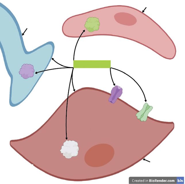
Endothelial cell

                                                         NANC nerve              NOS

                                                                              Androgens
                                                       NOS

Fig. 2. Androgen regulation of erectile tis-                                                   K+ channel
sue and molecular signalling involved in
                                                                                                                voltage-gated
erectile physiology. Androgen signalling                                                                        Ca2+channel
maintains non-adrenergic, non-choliner-
gic (NANC) nerve fibre and smooth mus-
cle levels in the erectile tissue. Androgens
also activate K+ channels in smooth mus-
cle, and androgen levels correlate with volt-
age-gated Ca2+ channel expression in the                               PDE5
smooth muscle of the erectile tissue. An-
drogens positively regulate phosphodies-                                                                      Smooth
terase 5 (PDE5) in the smooth muscle and                                                                      muscle cell
nitric oxide synthase (NOS) enzymes,
which are localised NANC nerves and en-
dothelial cells.

driving tumescence [Andersson and Wagner, 1995].                   Estrogen is also a known neuroprotective agent, which
Stimulation of the sympathetic nerves reduces blood flow        is demonstrated by a variety of mechanisms in several
to the penis, leading to the flaccid state (detumescence)       animal and clinical studies [Brann et al., 2007]. For ex-
[Andersson and Wagner, 1995]. Somatic nerves also have          ample, ERα protects rat neuronal cells in vitro via increas-
a role in erectile function via contraction of the bulbocav-    ing Bcl-XL mRNA (an anti-apoptotic transcript from
ernosus and ishiocavernosus muscles (described below).          Bcl-X) and downregulating BAD (considered a pro-apop-
    Androgen signalling has been implicated in the regula-      totic gene) [Gollapudi and Oblinger, 1999]. In addition,
tion of nerve structure required for erectile function. For     estrogen inhibits amyloid-beta-induced apoptosis and
example, castration in rats leads to a reduction in the         modulates apoptotic mechanisms such as maintaining
number of NOS-containing nerve fibres of the cavernosal         expression of Bcl2 (an anti-apoptotic gene) in rat hippo-
and dorsal nerves [Baba et al., 2000]; the dorsal nerve is      campal cells in vitro [Nilsen et al., 2006]. Future studies
not purely a sensory somatic nerve but is also composed         need to elucidate whether estrogen also exerts neuropro-
of autonomic NOS-containing nerve bundles [Burnett et           tection within the erectile tissue, although the expression
al., 1993; Carrier et al., 1995]. This is consistent with the   of ERs in the dorsal nerve of the rat glans penis suggests
findings that rat castration leads to an altered structure of   this may occur [Jesmin et al., 2002].
the dorsal nerve [Armagan et al., 2008] and a reduced
density of NANC nerve fibres innervating the erectile tis-         Anatomy, Vasculature, and Hemodynamics of
sue [Zvara et al., 1995; Schirar et al., 1997]. These studies      Erection
show that androgen signalling maintains the neural cir-            The erectile tissue within the penis comprises 3 cylin-
cuitry within the penis which is critical for erectile activ-   drical structures: the paired corpus cavernosa which are
ity (Fig. 2).                                                   dorsal to the urethra and the smaller corpus spongiosum

190                     Sex Dev 2021;15:187–212                                      Cripps/Mattiske/Pask
                        DOI: 10.1159/000516600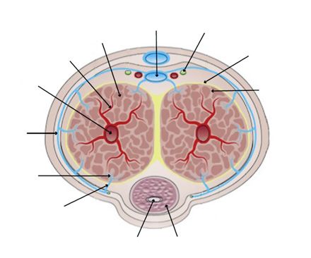
which encloses the urethra and forms the glans distally            Complete veno-occlusion occurs when the engorged
[Kuno et al., 2001]. The penile artery supplies the penis       corpora cavernosa are compressed at their base by con-
with blood and branches into the dorsal, bulbourethral,         traction of the ishiocavernosal muscles via somatic nerve
and the cavernous arteries [Keegan and Penson, 2013]. The       stimulation [Edey et al., 2011]. Similarly, the bulbospon-
cavernous arteries lie within the corpus cavernosa and          giosus muscle which surrounds the corpus cavernosum
branch into the helicine arteries, which supply the sinusoi-    and spongiosum contracts to force additional blood into
dal spaces [Smith and Axilrod, 2007; Keegan and Penson,         the penis during erection and compress the urethra to ex-
2013]. Sinusoidal spaces are blood-filled compartments          pel semen [Panchatsharam et al., 2020]. The corpus spon-
within the corpus cavernosum which are drained by ve-           giosum also contains sinusoidal spaces which expand
nules that coalesce into subtunical plexi which lie beneath     during erection and constrict the urethra to cause forceful
the tunica albuginea [Keegan and Penson, 2013]. The tu-         ejaculation [Clement and Giuliano, 2015; Panchatsharam
nica albuginea is a fibroelastic collagen-based structure       et al., 2020].
which surrounds the corpora cavernosa [Keegan and Pen-             Upon sympathetic stimulation, the penile smooth
son, 2013]; the collagen and fibroelastic fibres are arranged   muscle reverts to the contracted state, constricting the ar-
in an inner, circular layer and an outer, longitudinal layer    terioles and sinusoidal spaces which in turn decompress-
[Goldstein and Padma-Nathan, 1990]. These fibroelastic          es the penile veins [Andersson et al., 2000]. As a result,
properties allow for an increase in girth and elongation of     venous outflow increases which causes a reduction in in-
the penis during tumescence, while also providing enough        tracavernous pressure, inducing detumescence.
resilience for shrinkage to the flaccid state [Bitsch et al.,      Androgen signalling positively regulates smooth mus-
1990; Hsu et al., 1994; Iacono et al., 1995].                   cle content in the penis. Castration of rats, mice, rabbits,
    The subtunical plexi branch into emissary veins which       and dogs significantly reduces trabecular smooth muscle
penetrate the tunica albuginea [Keegan and Penson,              content accompanied by an increase in connective tissue
2013]. Superficial to the tunica albuginea, these veins         [Takahashi et al., 1991; Shabsigh, 1997; Traish et al., 1999;
drain into the deep dorsal vein or circumflex veins from        Palese et al., 2003; Shen et al., 2003]. Furthermore, andro-
the corpus spongiosum; the circumflex veins also ulti-          gens stimulate the differentiation of mouse pluripotent
mately drain into the deep dorsal vein (Fig. 1) [Quartey,       mesenchymal cells into smooth muscle cells in vitro
2006; Hsu et al., 2013].                                        [Singh et al., 2003]. The smooth muscle content within
    Upon sexual stimulation, parasympathetic neural sig-        the erectile tissue is correlated with the degree to which
nals cause the smooth muscle surrounding the cavernous          the corpus cavernosum can expand [Nehra et al., 1998].
and helicine arteries to relax, leading to dilation of these    Thus, the loss of smooth muscle induced by androgen
blood vessels and thus increased blood flow into the erec-      deprivation is likely to disrupt erectile function. Indeed,
tile tissue [Kuno et al., 2001]. In addition, trabecular        several animal studies have shown that a loss of androgen
smooth muscle within the corpus cavernosum relaxes so           signalling leads to attenuated erectile responses in vivo
that the sinusoidal spaces can expand following their en-       [Müller et al., 1988; Takahashi et al., 1991; Heaton and
gorgement of blood via the dilated arteries [Kuno et al.,       Varrin, 1994; Mills et al., 1994; Bivalacqua et al., 1998;
2001]. The expanding sinusoids then compress the sub-           Traish et al., 1999, 2003; Palese et al., 2003; Suzuki et al.,
tunical plexi against the unyielding tunica albuginea, oc-      2007; Traish, 2009]. This is consistent with the reduction
cluding venous outflow of the penis [Keegan and Penson,         of penile smooth muscle content in patients with ED
2013]. In addition, the pressure of the expanding sinu-         [Mersdorf et al., 1991; Claro et al., 2005] and those under-
soids causes the tunica albuginea to stretch and compress       going androgen deprivation [Tomada et al., 2013]. Inter-
the emissary veins, further restricting venous outflow          estingly, mice exposed to excess androgen levels also ex-
[Panchatsharam et al., 2020]. Also, subtunical venules          hibit smooth muscle loss in the corpus cavernosa in vivo
possess minimal geometric slack in the flaccid state (un-       [Hiremath et al., 2020]. Therefore, a balance of androgen
like the arteries and nerves), so when they elongate during     signalling maintains smooth muscle content (Fig. 2),
tumescence, they subsequently narrow which further re-          which in turn promotes erectile function.
stricts outflow from the corpus cavernosum [Udelson et             Androgen signalling also maintains the structural in-
al., 2001]. This overall process is known as veno-occlu-        tegrity of the tunica albuginea; castrated rats have re-
sion, whereby blood inflow increases and blood outflow          duced density of elastic fibres in the tunica albuginea
decreases, which in turn drastically increases the intra-       which are replaced by collagen [Shen et al., 2003]. A re-
cavernous pressure and results in tumescence.                   duction of elastic fibres may reduce the tunica albuginea’s

Impacts of EDCs on Erectile Dysfunction                         Sex Dev 2021;15:187–212                                   191
                                                                DOI: 10.1159/000516600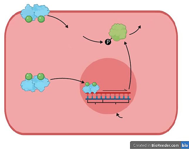
ability to expand, in turn disrupting veno-occlusion and       muscle contraction [Andersson, 2001]. However, the rise
causing ED [Akkus et al., 1997]. Indeed, rats with surgical    in Ca2+ concentration is short-lived and quickly drops to
injury to the tunica albuginea exhibit impaired erectile       baseline levels. Therefore, this process alone does not sus-
function following electric stimulation of the cavernous       tain chronic smooth muscle contraction required for the
nerve [Bivalacqua et al., 2000]. Taken together, andro-        flaccid state [Mills et al., 2003; Hill-Eubanks et al., 2011].
gens also promote erectile function by maintaining the             To achieve this, the protein RhoA activates Rho-ki-
fibroelastic properties of the tunica albuginea.               nase, which in turn deactivates MLCP by phosphoryla-
    Estrogen signalling within the vasculature of the erec-    tion. Since MLCP is deactivated and cannot dephosphor-
tile tissue may maintain the structural integrity of the en-   ylate MLC and thus drive smooth muscle relaxation, the
dothelium, a key signalling centre for the regulation of       MLCs can stay phosphorylated at basal Ca2+, increasing
vasodilation/vasorelaxation. Indeed, ERβ expression in         Ca2+ sensitivity of smooth muscle cells [Mills et al., 2003].
the male rat aorta is increased in the endothelium and         Ca2+ sensitivity refers to the dependence of MLC phos-
smooth muscle cells following vascular injury [Lindner et      phorylation on Ca2+ concentrations; sensitivity is high
al., 1998]. In addition, estrogen signalling inhibits TNFα-    when small increases in Ca2+ drive a greater degree of
and oxidized low-density lipoprotein (oxLDL)-induced           MLC phosphorylation (as in the flaccid state). In contrast,
apoptosis of human endothelial cells in vitro [Spyrido-        low sensitivity occurs when larger increases in Ca2+ con-
poulos et al., 1997; Florian and Magder, 2008]. Further-       centration are needed for a lesser degree of MLC phos-
more, estrogen-mediated activation of Notch1 protects          phorylation, which is when MLCP actively dephosphory-
human umbilical vein endothelial cells from TNFα-              lates MLC [Rembold, 1992].
induced apoptosis in vitro [Fortini et al., 2017]. Interest-       Inhibition of RhoA/Rho kinase-mediated calcium
ingly, siRNA-knockdown of ERβ, although not ERα,               sensitization induces erectile activity in the rat, demon-
eliminated the anti-apoptotic effect of estrogen [Fortini      strating the importance of this pathway in maintaining
et al., 2017].                                                 the flaccid state [Chitaley et al., 2001; Lasker et al., 2013].
    Estrogen also increases the expression of Bcl2 and         Interestingly, RhoA expression is 17-fold higher in the
Bcl-XL in human endothelial cells in vitro, potentially        rabbit corpus cavernosum compared to the ileum smooth
generating a protective effect on this tissue [Florian and     muscle, which is consistent with the chronic state of
Magder, 2008]. Thus, estrogen signalling has a role in         smooth muscle contraction in the corpus cavernosum
maintaining the structural integrity of the endothelium,       compared to other parts of the vascular system [Wang et
although this has not yet been demonstrated in the penile      al., 2002].
endothelium. However, the expression of ERs within the             Conversely, during tumescence, Ca2+ concentration in
vasculature of the rat penis raises this possibility [Jesmin   the smooth muscle cell drops so that MLCK cannot bind
et al., 2002].                                                 Cam-Ca2+ and induce contraction [Andersson, 2001].
                                                               However, reducing Ca2+ concentration is not sufficient to
   Calcium-Mediated Penile Smooth Muscle                       drive erection because the contractile machinery is sensi-
   Contraction/Relaxation and RhoA/Rho Kinase-                 tised to lower calcium concentrations through RhoA/
   Mediated Calcium Sensitisation                              Rho-kinase inactivation of MLCP. Thus, inhibition of the
   Smooth muscle contraction and relaxation is mediated        RhoA/Rho kinase pathway must also occur so that MLCP
by 2 critical proteins: myosin light chain kinase (MLCK)       can activate and dephosphorylate MLC, thereby decreas-
and myosin light chain phosphatase (MLCP). MLCK                ing Ca2+ sensitivity and driving smooth muscle relaxation
phosphorylates myosin light chains (MLCs), causing             [Mills et al., 2003]. In summary, detumescence and tu-
smooth muscle contraction [Kamm and Stull, 1985; An-           mescence depend on a simple switch mechanism on
dersson, 2001]. Conversely, MLCP dephosphorylates              whether MLC is phosphorylated (Fig. 3). However, the
MLCs, driving smooth muscle relaxation [Jin and Bur-           signalling pathways that regulate this switch by altering
nett, 2006]. In smooth muscle cells, cytosolic calcium ion     Ca2+ concentration and Ca2+ sensitivity in the smooth
(Ca2+) concentrations regulate MLCK and MLCP activity          muscle cells of the erectile tissue are extremely complex.
which facilitates contraction and relaxation, respectively.
Detumescence initiates with the increase in cytosolic Ca2+        Nitric Oxide (NO)-cGMP Mediated Tumescence
concentrations of smooth muscle cells; Ca2+ then binds to         Nitric oxide (NO) is a non-noradrenergic, non-cho-
calmodulin to form the Cam-Ca2+ complex which subse-           linergic (NANC) neurotransmitter and is essential for tu-
quently binds to and activates MLCK, leading to smooth         mescence, as evidenced by several animal and human

192                  Sex Dev 2021;15:187–212                                         Cripps/Mattiske/Pask
                     DOI: 10.1159/000516600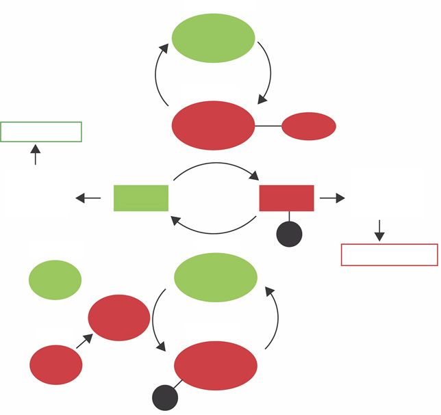
inactive

                                                                                 MLCK

                                                                                 active

                                                                                 MLCK          Cam-Ca
                                               Tumescence

Fig. 3. MLCK and MLCP mediate smooth
muscle contraction and relaxation, respec-
tively [Mas, 2010]. Ca2+ ions bind to          Smooth                                                     Smooth
                                               muscle               MLC                      MLC          muscle
calmodulin to form the Ca2+-calmodulin         relaxation                                                 contraction
complex (Cam-Ca) which then binds to
and activates MLCK. Active MLCK phos-                                                          P
phorylates MLC, facilitating smooth mus-                                         active
                                                    inactive                                               Detumescence
cle contraction. Conversely, active MLCP
dephosphorylates MLC, causing smooth                 RhoA                        MLCP
muscle relaxation and tumescence. Active
RhoA activates Rho kinase which deacti-                         Rho
vates MLCP by phosphorylation. Inactive                        kinase
                                                                                inactive
RhoA allows for the activation of MLCP.              active
Green refers to pathways driving tumes-
cence, red refers to that of detumescence.           RhoA                        MLCP
MLCK, myosin light chain kinase; MLCP,
myosin light chain phosphatase; MLC, my-                                  P
osin light chain; Cam-Ca, Ca2+-calmodulin
complex; P, phosphate group.

studies [Saenz de Tejada, 2002]. Upon parasympathetic              Hormonal signalling modulates cellular ion flux; tes-
stimulation, NO is released within the penis and activates     tosterone treatment of human corpus cavernosum
soluble guanylyl cyclase which enhances production of          smooth muscle cells in vitro activates K+ channels [Han
cyclic guanosine monophosphate (cGMP). In turn, cGMP           et al., 2008]. Also, castration of rats leads to reduced levels
activates protein kinase G (PKG) which reduces Ca2+            of voltage-gated Ca2+ channels in the corpus cavernosum
concentration through several mechanisms [Ghalayini,           smooth muscle, thus androgen signalling positively cor-
2004; Krassioukov and Elliott, 2017]. This includes phos-      relates with voltage-gated Ca2+ channel expression
phorylation of K+ channels, which leads to an efflux of K+     (Fig. 2) [Luo et al., 2009]. In addition, rapid estrogen sig-
and subsequent hyperpolarization of smooth muscle cells        nalling via membrane-bound ERs (discussed below) is
within the penis [Archer, 2002]. Hyperpolarization closes      known to modulate intracellular Ca2+ [Zhang et al., 2009;
voltage-dependent Ca2+ channels, thereby decreasing the        Rainville et al., 2015; Puglisi et al., 2019]. Therefore, an-
influx of Ca2+ into smooth muscle cells [Andersson and         drogens and estrogens may have a role in regulating the
Wagner, 1995]. In addition, PKG activates cation-ATPase        ion flux of smooth muscle cells which is a critical compo-
pumps in the plasma membrane of smooth muscle cells            nent of tumescence. Indeed, androgen treatment was re-
and the sarcoplasmic reticulum, leading to Ca2+ efflux out     ported to induce rapid relaxation of human cavernosal
of the cell and sequestration of Ca2+ in the sarcoplasmic      arteries and corpus cavernosum in vitro, although this
reticulum, respectively (Fig. 4) [Lucas et al., 2000]. Acti-   was not associated with increased levels of cGMP [Wald-
vated PKG can also inhibit the inositol triphosphate 3         kirch et al., 2008], suggesting androgens can regulate ion
(IP3) receptor, which blocks the influx of Ca2+ into the       channels via alternate mechanisms.
cytoplasm from the sarcoplasmic reticulum [Lucas et al.,           In addition to lowering cytosolic Ca2+ concentrations,
2000].                                                         the NO/cGMP/PKG pathway is thought to inhibit the
                                                               RhoA/Rho-kinase pathway, allowing for the activation of

Impacts of EDCs on Erectile Dysfunction                        Sex Dev 2021;15:187–212                                    193
                                                               DOI: 10.1159/000516600
NO
Diffusion Smooth muscle cell
Fig. 4. NO-cGMP mediated smooth mus-
sGC
cle relaxation. Extracellular nitric oxide
(NO) diffuses through the smooth muscle
SR
cell membrane and activates soluble gua-
nylyl cyclase (sGC), producing cGMP as a
PDEs cGMP RhoA/Rho-kinase
result. This activates protein kinase G
(PKG) which then activates K+ channels Ca2+
causing an efflux of K+ from the cell. This Ca2+
results in hyperpolarization (HP) which PKG
blocks Ca2+ channels so Ca2+ influx is re-
duced. In addition, PKG also activates cat-
ion ATPase pumps in the cell membrane
and sarcoplasmic reticulum (SR), driving K+ channel
HP
an efflux of Ca2+ out of the cell and seques-
tration of Ca2+ in the SR, respectively. PKG K+
Ca2+ channel
also suppresses the RhoA/Rho-kinase
Cation ATP-ase K+
pathway, thereby decreasing Ca2+ sensitiv- Ca2+
pump Ca2+
ity. NO-mediated reduction in cytosolic Ca2+
Ca2+
Ca2+ and increased Ca2+ sensitivity drives Ca2+ Ca2+
relaxation of the smooth muscle cell. The
phosphodiesterase proteins (PDEs) break
down cGMP.
MLCP which is essential for tumescence (Fig. 4) [Mills et The phosphodiesterase (PDE) protein family inhibits
al., 2003]. This is consistent with rodent studies which tumescence by breaking down secondary messenger mol-
disrupt the NO/cGMP/cGMP pathway: in vitro organ ecules such as cGMP and cAMP (discussed below) (Fig. 4)
bath experiments show that the corpus cavernosum of [Turko et al., 1999]. Interestingly, PDE5 (which breaks
PKG-null mice have diminished smooth muscle relax- down cGMP) mRNA is present in the human corpus cav-
ation [Hedlund et al., 2000]. Also, inhibition of guanylyl ernosum at levels 10- to 100-fold higher compared to oth-
cyclase in the corpus cavernosum of rats in vivo signifi- er non-reproductive tissues in males [Morelli et al., 2004].
cantly reduces the erectile response after electrostimula- Alongside higher levels of RhoA in the corpus caverno-
tion of the cavernous nerve [Martinez-Piñeiro et al., sum, this likely serves to maintain the penis in a chroni-
1993]. The disrupted erectile responses observed in these cally contracted state to maintain flaccidity.
studies are likely due to the RhoA/Rho-kinase pathway Androgen signalling is thought to upregulate PDE5 ex-
remaining active. Furthermore, PKG inhibits Ca2+ sensi- pression; castrated rabbits and rats display reduced PDE5
tization induced by RhoA in rat aortic smooth muscle expression and activity, which is restored by testosterone
cells in vitro by phosphorylation of RhoA [Sauzeau et al., replacement [Morelli et al., 2004; Zhang et al., 2005; Ar-
2000]. However, a direct effect of PKG on RhoA specifi- magan et al., 2006]. Also, transsexual individuals in a hy-
cally in penile smooth muscle has not yet been proven. pogonadal state also exhibit decreased PDE5 expression
NO/cGMP signalling is also considered the primary path- and activity in the corpus cavernosum [Morelli et al.,
way for increasing blood flow into the clitoris and vagina 2004]. In addition, treatment with a PDE5 inhibitor alone
during sexual arousal [Giuliano et al., 2002]. Thus, sexual has little effect on the erectile function of castrated ani-
function in the female and male genitalia arise from sim- mals, demonstrating that PDE5 expression relies on an-
ilar molecular pathways. drogen signalling [Traish et al., 2003; Zhang et al., 2005].
194 Sex Dev 2021;15:187–212 Cripps/Mattiske/Pask
DOI: 10.1159/000516600mER mER
Rapid, non-genomic signalling
NO
Fig. 5. Estrogen-mediated positive regula-
tion of eNOS expression/activation. In the PI3K/Akt eNOS
endothelial cell, when the estrogen recep- P
phosphorylation
tor (ER) binds to the estrogen ligand (en-
dogenous or exogenous estrogen or estro-
gen-mimicking EDCs; green circle), it di-
merises and translocates to the nucleus
where it binds to an estrogen-response ele-
ment (ERE) in the NOS3 promoter. This
induces transcription of NOS3 which leads ER ER transcription
to production of endothelial nitric oxide
synthase (eNOS). In addition, the associa-
tion of membrane-bound estrogen recep- ERE NOS3
tors (mERs) with estrogen initiates rapid,
non-genomic signalling. This involves ac- Nucleus
tivation of the phosphoinositide 3-kinase/
protein kinase B (PI3K/Akt) pathway, Endothelial cell
which in turn activates eNOS by phosphor-
ylation so that it produces NO. NO is the
same as shown in Figure 4.
However, androgens have no effect on PDE5 expres- rodents and humans [Gonzalez-Cadavid et al., 2000;
sion in cavernous smooth muscle cells in vitro, suggesting Dashwood et al., 2011], and the endothelial NOS (eNOS)
an indirect effect of androgens on PDE5 expression in isoform is expressed in the endothelium of the mouse and
vivo [Yang et al., 2009]. This is supported by the fact that human erectile tissues (Fig. 5, 6) [Burnett et al., 1996; Sef-
no androgen response element has been found in the rat tel et al., 1997]. Although the NOS enzymes are referred
Pde5a gene [Lin et al., 2013] and that genome-wide to as isoforms, they are encoded by separate genes and not
searches for genes regulated by androgens in human cells splice variants from a single gene [Huang et al., 1993].
did not yield PDE5A as a candidate [Bolton et al., 2007; Upon sexual stimulation of the parasympathetic sys-
Massie et al., 2007]. Rather than directly upregulating tem, NANC nerves within the penis depolarize via an in-
PDE5, androgens may provide the cellular context for flux of Ca2+ which then forms the Cam-Ca2+ complex,
PDE5 expression in the smooth muscle as these hor- activating nNOS [Bredt and Snyder, 1990]. As a result,
mones are critical for the development and maintenance nNOS produces NO which relaxes smooth muscles,
of vasculature within the erectile tissue (Fig. 2). Indeed, thereby dilating penile blood vessels and initiating the
castration of rats leads to the simultaneous reduction of erectile response. Despite this, nerve cell depolarization
cavernous smooth muscle and PDE5 expression [Liu et via Ca2+ influx is transitory and nNOS quickly deacti-
al., 2005; Yang et al., 2009]. vates, thus relaxing smooth muscles only briefly [Hurt et
al., 2012]. However, this initial increase in blood flow and
NO Production by Activation of Nitric Oxide Synthase shear stress on the endothelium activates phosphoinosit-
Isoforms ide 3-kinase (PI3K) which stimulates protein kinase B
NO in the penis is derived from 2 main sources: NANC (Akt), in turn activating eNOS by phosphorylation
parasympathetic nerves and the endothelium lining (Fig. 6) [Hurt et al., 2002; Musicki et al., 2005; Wen et al.,
blood vessels and sinusoids [Cartledge et al., 2001]. This 2011]. Phosphorylation activates NOS considerably lon-
is evident by the spatial expression of the nitric oxide syn- ger than by depolarization, and thus phosphorylated
thase (NOS) enzymes, of which there are several isoforms eNOS can continually produce NO to sustain smooth
that differ in tissue distribution. The neuronal NOS muscle relaxation (Fig. 6) [Hurt et al., 2012].
(nNOS) isoform is localised within penile nerve cells in
Impacts of EDCs on Erectile Dysfunction Sex Dev 2021;15:187–212 195
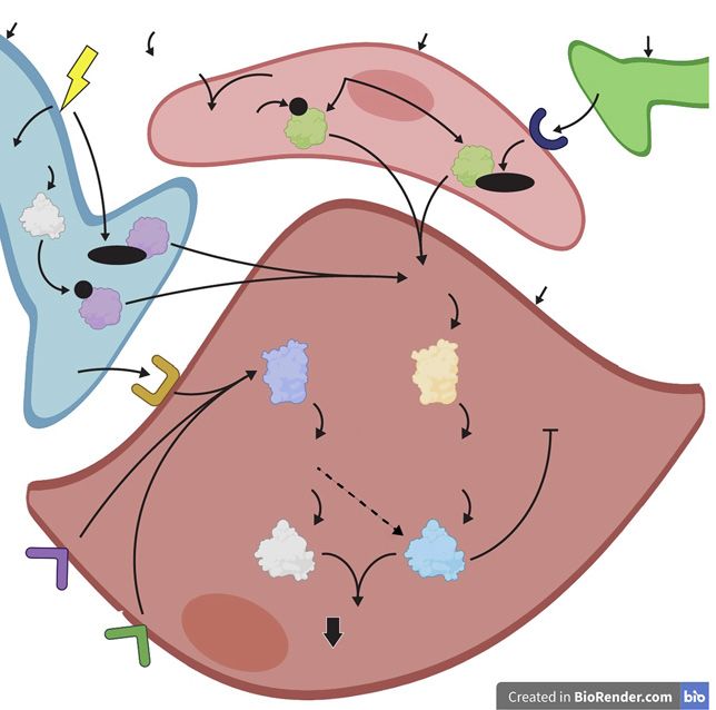
DOI: 10.1159/000516600NANC nerve SMC relaxation Endothelial cell Cholinergic nerve
Shear stress
Estrogen Acetylcholine
mAChR
P
PI3K/Akt eNOS
cAMP eNOS
Cam-Ca
PKA
nNOS
Cam-Ca
NO Smooth muscle cell
P
nNOS
VIP sAC sGC
VIP-R RhoA/Rho-kinase
cAMP cGMP
PKA PKG
PGE2
EP
Ca2+
PGI2 IP
Fig. 6. NO sources and other factors which drive smooth muscle dothelial cell. This leads to formation of Cam-Ca2+, which binds to
2+
relaxation. The NO-cGMP pathway reduces cytosolic Ca and in- and activates eNOS. Endogenous estrogen signalling also activates
hibits the RhoA/Rho-kinase pathway as depicted in Figure 4. eNOS by stimulating the PI3K/Akt pathway and upregulates ex-
When the NANC nerves are stimulated (lightning bolt), Ca2+ pression of eNOS (see Fig. 5). In addition to the NO-cGMP path-
binds to calmodulin to form the calmodulin-Ca2+ (Cam-Ca2+) way, vasoactive intestinal peptide (VIP) in the NANC nerves may
complex. This subsequently binds to and activates neuronal NOS bind to its receptor (VIP-R) on the smooth muscle cell to stimulate
(nNOS), driving NO production. Also, stimulation of NANC soluble adenylyl cyclase (sAC). This leads to production of cAMP
nerves drives production of cAMP in these cells. This activates in the smooth muscle cell, activating PKA to reduce cytosolic Ca2+
protein kinase A (PKA) which in turn activates nNOS by phos- concentration. cAMP may also mediate smooth muscle cell relax-
phorylation (P). The initial production of NO by the NANC nerves ation via activation of PKG. The prostanoids prostaglandin E2
leads to smooth muscle cell (SMC) relaxation, in turn leading to (PGE2) and prostacyclin (PGI2) can also drive cAMP production
shear stress on the endothelial cells. This triggers the PI3K/Akt via association with the EP and IP receptors on the smooth muscle
pathway, which then activates eNOS by phosphorylation. Acetyl- cell, respectively. NANC is the same as shown in Figure 2sGC, PKG
choline released from cholinergic nerves binds to the muscarinic and NO are the same as shown in Figure 4. PI3K/Akt and eNOS are
acetylcholine receptor (mAChR), which increases Ca2+ in the en- the same as shown in Figure 5.
In addition, sexual stimulation increases production [Hurt et al., 2012]. Administration of mice with the non-
of cyclic adenosine monophosphate (cAMP) (discussed specific NOS inhibitor (i.e., inhibits all NOS isoforms)
further below), which activates protein kinase A (PKA). L-nitroarginine methyl ester (L-NAME) abolishes or sig-
In turn, PKA phosphorylates nNOS so it also continually nificantly attenuates erection, revealing the critical nature
produces NO (Fig. 6) [Hurt et al., 2012]. These findings of the NO-cGMP pathway for tumescence [Burnett et al.,
demonstrate that while nNOS initiates NO-mediated 1996; Mizusawa et al., 2001; Cashen et al., 2002].
erection upon parasympathetic stimulation, both nNOS Androgens positively regulate the NOS enzymes; nu-
and eNOS sustain erection via their phosphorylated state merous animal studies have demonstrated that androgen
196 Sex Dev 2021;15:187–212 Cripps/Mattiske/Pask
DOI: 10.1159/000516600signalling is associated with increased NOS activity/ex- tion of estrogen into ovariectomized rats increases nNOS
pression in the erectile tissue and nerves innervating the mRNA in the hypothalamus and hippocampus [Ceccatel-
penis [Lugg et al., 1995; Penson et al., 1996; Reilly et al., li et al., 1996; Grohe et al., 2004]. The stimulation of neu-
1997; Schirar et al., 1997; Marin et al., 1999; Park et al., ronal NO production by estrogen may also explain the
1999; Seo et al., 1999; Armagan et al., 2006]. However, neuroprotective properties of estrogen as NO is a known
androgen deprivation was found to increase eNOS mRNA neuroprotective agent [Chiueh, 1999; Wen et al., 2004].
and have no effect on nNOS mRNA levels in rat erectile Thus, estrogen signalling may positively regulate nNOS
tissue [Shen et al., 2000a]. Furthermore, androgen with- in nerves innervating the erectile tissue. However, to the
drawal was reported to increase eNOS levels in the hu- best of our knowledge this remains to be proven.
man corpus cavernosum [Tomada et al., 2013]. The con-
flicting data suggests that a precisely controlled balance Disruptions of NO-cGMP Pathway and Compensatory
of androgen signalling is required for maintaining nor- Mechanisms
mal levels of NOS expression and activity (Fig. 2). Also, The NO-cGMP pathway has a profound impact on tu-
cGMP can stimulate expression of the human PDE5A mescence, and compensatory mechanisms exist if it is
gene [Lin et al., 2001, 2006]. Thus, androgens may indi- disrupted. For example, mice with a mutation for nNOS
rectly upregulate PDE5 via positive regulation of the NOS display normal mating behaviour and erectile function;
enzymes which in turn drives cGMP production. eNOS is upregulated in these mice which may compen-
Estrogen signalling may also promote smooth muscle sate for disrupted NO production [Burnett et al., 1996].
relaxation by stimulating NOS expression and activity in Although eNOS is defined by its localisation to the endo-
the erectile tissue. Indeed, in humans and animals, ERs thelium, it may also localize to neural cells within the pe-
upregulate eNOS via an estrogen-response element in the nis, potentially substituting the function of nNOS [Cash-
eNOS promoter (Fig. 5) [MacRitchie et al., 1997; Yang et en et al., 2002]. This remains to be proven, although eNOS
al., 2000; McNeill Anne et al., 2002; Min, 2007]. Interest- is localised in the dendritic spines of primary culture cor-
ingly, in human endothelial cell cultures, activated mem- tical and hippocampal neurons from rats at embryonic
brane-bound ERs rapidly stimulate the PI3K/Akt path- day 18 [Caviedes et al., 2017].
way via a non-genomic mechanism, which in turn acti- Mice with mutations for eNOS also display normal
vates eNOS by phosphorylation (Fig. 5, 6) [Haynes et al., erectile function and retain about 60% of the NOS activ-
2000, 2003]. This is consistent with the significantly high- ity in the penis compared to that of WT mice [Burnett et
er basal release of endothelium-derived NO in the male al., 2002]. This shows that other NOS isoforms synthesise
mouse aorta compared to that of the male estrogen recep- NO in mice lacking eNOS, compensating for erectile
tor knockout (ERKO) mouse, suggesting that ER levels function [Burnett et al., 2002]. In addition, although
are related to basal NO production in endothelium nNOS is defined by its neuronal localization, its expres-
[Rubanyi et al., 1997]. sion in endothelial cells within the penis may also com-
Furthermore, estrogen-deficient post-menopausal pensate for a loss of eNOS [Cashen et al., 2002]. This is
women have reduced levels of ERα, eNOS, and phosphor- reinforced by the co-expression of nNOS with eNOS in
ylated eNOS in endothelial cells of the antecubital vein the human umbilical vein endothelial cells in vitro [Ba-
compared to premenopausal women [Gavin et al., 2009]. chetti et al., 2004].
Postmenopausal women also display reduced endotheli- Overall, the activity of NOS isoforms can compensate
al-dependent dilation of the brachial artery, suggesting for each other if one is mutated, thereby allowing for tu-
that a loss of estrogen leads to a reduction in NO bioavail- mescence despite disruption of the NO-cGMP pathway.
ability [Gavin et al., 2009]. Taken together, estrogen sig- Further compensation may arise by potential overlap of
nalling in the endothelium can upregulate and activate eNOS and nNOS localisation in the erectile tissue.
eNOS via genomic and non-genomic mechanisms, re-
spectively. Additional Pro-Erectile Signalling Pathways
Estrogen may also promote tumescence by positively While parasympathetic signalling mediated by the
regulating nNOS activity/expression. The treatment of NO-cGMP pathway is primarily responsible for tumes-
human nNOS-expressing neuroblastoma cell lines with cence, other signalling pathways modulate erectile func-
estrogen was reported to cause a rapid increase in NO tion through stimulation of cGMP and cAMP produc-
production via activation of eNOS and nNOS in vitro tion. These factors may also compensate for deficiencies
[Wen et al., 2004; Xia and Krukoff, 2004]. Also, the injec- in NO-signalling, potentially explaining normal erectile
Impacts of EDCs on Erectile Dysfunction Sex Dev 2021;15:187–212 197
DOI: 10.1159/000516600function in NOS mutant mice from the studies men- en to the relaxant effects of vasoactive intestinal peptide
tioned above. (VIP) in this process. Nerves innervating the erectile tis-
sue in humans and rabbits contain VIP, and thus it may
cAMP/PKA Pathway function as a neurotransmitter in the penis to promote
The second messenger cAMP is produced by adenylyl tumescence [Polak et al., 1981; Willis et al., 1983]. This is
cyclase and activates PKA [Sassone-Corsi, 2012]. In addi- supported by the presence of VIP receptors in smooth
tion to cGMP signalling, cAMP/PKA signalling is thought muscle cells of the rat corpus cavernosum in vitro [Gui-
to mediate smooth muscle relaxation in the penis. Indeed, done et al., 2002]. Furthermore, administration of a VIP-
several studies have identified cAMP signalling in the antagonist to the rabbit corpus cavernosum reduces re-
corpus cavernosum smooth muscle [Lin et al., 2005]. In laxation in vitro following electric stimuli [Kim et al.,
addition, forskolin (adenylyl cyclase activator) adminis- 1995] and also to the rat penis in vivo following cavernous
tration relaxes the human corpus cavernosum in vitro; nerve stimulation [Suh et al., 1995]. In addition, tumes-
the magnitude of relaxation correlates with the level of cence is associated with increased VIP concentration in
cAMP accumulation induced by forskolin in human cor- the cavernous blood in humans, and administration of
poral smooth muscle cells in vitro [Palmer et al., 1994]. VIP drives erection [Ottesen et al., 1984]. The mechanism
The mechanism by which cAMP/PKA signalling relaxes of VIP-mediated tumescence most likely involves elevat-
penile smooth muscle cells likely involves the activation ing cAMP levels in smooth muscle cells of the erectile tis-
of K+ channels on the smooth muscle cell membrane, hy- sue; VIP dose-dependently induces production of cAMP
perpolarizing the smooth muscle cell and thereby de- in rat corpus cavernosum smooth muscle cells [Guidone
creasing cytosolic Ca2+ levels. This is illustrated by the et al., 2002].
ablation of PGE1 (a relaxing factor discussed below) in- However, VIP is not the primary relaxant agent in tu-
duced activation of K+ channels in human corporal mescence: injection of VIP into the rat penis enhances erec-
smooth muscle cells in vitro by a PKA inhibitor [Lee et tion in vivo but does not induce a full erection [Suh et al.,
al., 1999]. 1995]. Also, electrical field stimulation-induced relaxation
In contrast, the treatment of rats with an adenylyl cy- of the human corpus cavernosum in vitro is not diminished
clase inhibitor does not affect the erectile response in vivo by VIP inactivation [Pickard et al., 1993]. For a rigid erec-
following electrostimulation of the cavernous nerve tion sufficient for penetration, it is likely that VIP functions
[Martinez-Piñeiro et al., 1993]. There is also little evi- alongside other pro-erectile pathways. Indeed, VIP and
dence to suggest that the cAMP/PKA pathway reduces NOS are colocalised in nerves innervating the erectile tissue
Ca2+ sensitivity to the contractile machinery in penile in animals and humans, suggesting that NO and VIP com-
smooth muscle through inhibition of the RhoA/Rho-ki- plement each other to facilitate erectile activity (Fig. 6)
nase pathway, a critical component for tumescence. [Ehmke et al., 1995; Andersson, 2001]. This is supported by
Therefore, it is likely that the NO/cGMP/PKG pathway is injection of a combination of VIP and SIN-1 (NO-releasing
the key driver for tumescence while cAMP/PKA signal- compound) into the rabbit corpus cavernosum which aug-
ling has a relatively minor role by reducing cytosolic Ca2+ ments the erectile response in vivo compared to those in-
concentration (Fig. 6). jected with SIN-1 or VIP alone, revealing an additive effect
Importantly, these pathways are not mutually exclu- of the NO and VIP pathways [Sazova et al., 2002]. Overall,
sive; crosstalk exists between cAMP and cGMP signal- VIP is indisputably a smooth muscle relaxant and most
ling. This is partially discussed above with cAMP/PKA- likely complements NO-cGMP signalling via the cAMP
mediated phosphorylation of nNOS. In addition, both pathway to promote tumescence (Fig. 6).
cAMP and cGMP can activate PKG in cavernosal smooth VIP signalling appears to be independent of androgen
muscle cell cultures from young (16 weeks) and old (28 signalling; men with chemical castration display no sig-
months) rats [Lin et al., 2002]. Therefore, while activation nificant change in VIP levels in the corpus cavernosum
of the cAMP pathway may have minor direct effects on compared to non-castrated individuals [Cormio et al.,
tumescence, it may also indirectly contribute to it by re- 2005]. Also, castrated rats display no significant change
inforcing the cGMP/PKG-signalling pathway (Fig. 6). of VIP mRNA levels in the corpus cavernosum [Shen et
al., 2000b]. However, the erectile function of castrated
Vasoactive Intestinal Peptide rats display greater responsiveness to VIP, suggesting that
Before NO-cGMP signalling was established as the key androgens negatively regulate the VIP/cAMP pathway
pathway for tumescence, considerable attention was giv- [Zhang et al., 2011].
198 Sex Dev 2021;15:187–212 Cripps/Mattiske/Pask
DOI: 10.1159/000516600Adrenergic nerve
Endothelial cell
Ang-II
ET-1
TXA2
NA
Receptor (α1, ETA, AT1, TP)
SR PLC
RhoA/Rho-kinase
IP3 DAG
MLCP
PKC
IP3R
Ca2+ CPI-17
P
Ca2+Ca2+
Ca2+ Ca2+ Ca2+
DP Smooth muscle cell
TRPC3
Ca2+ channel
Fig. 7. Smooth muscle contraction pathways. Upon stimulation of channels, leading to an influx of extracellular Ca2+. Increased cy-
adrenergic nerves (lightning bolt), noradrenaline (NA) is released tosolic Ca2+ in the smooth muscle cell causes depolarization (DP),
and binds to the α1-adrenoreceptor (α1). Endothelin-1 (ET-1), an- activating Ca2+ channels in the smooth muscle cell membrane
giotensin-II (Ang-II), and the prostanoid thromboxane A2 (TXA2) which leads to a further influx of Ca2+. DAG leads to activation of
released from the endothelial cell bind to their receptors ETA, AT1, protein kinase C (PKC), which activates CPI-17 by phosphoryla-
and TP, respectively, on the smooth muscle cell. Association of tion. This then inhibits MLCP. Association of NA, ETA, Ang-II,
these ligands with their receptors leads to activation of phospholi- and TXA2 with their receptors may also drive the RhoA/Rho-ki-
pase C (PLC), which then produces inositol triphosphate 3 (IP3) nase pathway to inhibit MLCP. Thus, these signalling factors drive
and diacylglycerol (DAG). IP3 associates with the IP3 receptor smooth muscle cell contraction by increasing cytosolic Ca2+ and
(IP3R) on the sarcoplasmic reticulum (SR), which acts as a channel increasing Ca2+ sensitivity. MLCP is the same as shown in Figure
to release Ca2+ from the SR. The activated IP3Rs couple with mem- 3.
brane-bound transient receptor potential canonical 3 (TRPC3)
Prostanoids (Involved in Tumescence and prostanoids exert dual functions on erectile function as
Detumescence) some contract or relax penile smooth muscle: administra-
Prostanoid metabolites are produced via several syn- tion of a TP receptor agonist and PGF2α to isolated hu-
thases from arachidonic acid in the endothelium [Minhas man corpus cavernosum preparations results in contrac-
et al., 2000]. The prostanoid metabolites identified in the tion [Hedlund and Andersson, 1985b]. On the other
human and animal corpus cavernosum are the prosta- hand, treatment with PGE2 and PGE1 leads to relaxation
glandins (PGI2, PGF2α, PGE2, PGD2) and thromboxane [Hedlund and Andersson, 1985b].
A2 (TXA2) [Khan et al., 1999; Andersson, 2011]; these Prostanoid-induced relaxation is supported by studies
mediate their functions via the IP, FP, EP, DP, and TP which show that injection of PGE1 leads to relaxation of
receptors, respectively (Fig. 6, 7) [Andersson, 2011]. The the monkey [Bosch et al., 1989] and rat corpus caverno-
Impacts of EDCs on Erectile Dysfunction Sex Dev 2021;15:187–212 199
DOI: 10.1159/000516600sum in vivo [Chen et al., 1992]. In addition, the EP recep- Ca2+ concentration and contraction in vitro [Grann et al.,
tors are known to mediate PGE1- and PGE2-induced re- 2016]. The authors also found that treatment of the Rho-
laxation of the human corpus cavernosum in vitro [An- kinase inhibitors Y27632 and glycyl-H1152P dose-de-
gulo et al., 2002]. In fact, the documented relaxant effects pendently attenuated U46619-induced contraction, pro-
of PGE1 has led to its use as a treatment for ED and results viding further evidence that TXA2 mediates contraction
in greater satisfaction in sexual performance [Linet and via activating the RhoA/Rho-kinase pathway. DP recep-
Neff, 1994; Urciuoli et al., 2004]. Prostanoids may con- tors (for PGF2α) can also increase Ca2+ concentration and
tribute to tumescence by stimulating cAMP production; inhibit production of cAMP, potentially explaining its
Gs-protein coupled EP and IP receptors (for PGE2 and contractile properties in the penis [Ricciotti and FitzGer-
PGI2) are known to stimulate adenylyl cyclase (Fig. 6) ald, 2011].
[Ricciotti and FitzGerald, 2011]. This is supported by Contradictory findings have been reported on the ef-
PGE1 administration in combination with an inhibitor of fects of PGI2 on penile smooth muscle: PGI2 treatment to
a cAMP-specific PDE which leads to relaxation and in- isolated human corpus cavernosum was reported to cause
creased cAMP levels in primary culture human cavernosal contraction [Hedlund and Andersson, 1985b], while a
smooth muscle cells [Bivalacqua et al., 1999]. Further- separate study found that treatment of iloprost (PGI2 an-
more, in equine penile arteries, treatment of a PKA in- alogue) to isolated rat corpus cavernosum leads to relax-
hibitor decreases the relaxant effects of PGE1, demonstrat- ation [Bassiouni et al., 2019]. Another study found that
ing that this prostaglandin relaxes penile blood vessels via PGI2 relaxed 4 of 6 isolated human corpus cavernosum
the cAMP/PKA pathway [Ruiz Rubio et al., 2004]. samples but had no effect on the remaining 2 [Kirkeby et
Interestingly, treatment of rats with PGE1 dose-de- al., 1993]. Thus, it is unclear from these results as to
pendently increases NO production and increases n/ whether PGI2 has a contractile, relaxant, or neutral effect
eNOS expression in the rat corpus cavernosum in vivo, on smooth muscle cells in the corpus cavernosum.
revealing that PGE1 may also relax erectile tissue through This is unexpected as PGI2 is an established vasodila-
the NO-cGMP pathway [Escrig et al., 1999]. This contra- tor in blood vessels. Lue [2011] suggests that this discrep-
dicts the finding that inhibition of NOS did not affect ancy arises from varying distribution of IP receptors (for
PGE1-mediated relaxation of equine penile arteries in vi- PGI2) within the penis. Indeed, it is unlikely the IP recep-
tro [Ruiz Rubio et al., 2004]. However, the same authors tor is present in trabecular smooth muscle because PGI2
demonstrated that the combined inhibition of PKA and fails to relax trabecular smooth muscle in human corpus
PKG reduced PGE1-mediated relaxation, suggesting cavernosum in vitro [Angulo et al., 2002]. However, PGI2
PGE1 primarily influences cAMP signalling and poten- is a potent vasodilator in human penile arteries in vitro,
tially the cGMP pathway. It should be noted that to the which is confirmed by the presence of IP receptors in this
best of our knowledge, PGE1 has not been identified as a tissue [Angulo et al., 2002]. Thus, the specific distribution
naturally occurring prostaglandin in the penis. Thus, the of prostanoid receptors in the vascular bed of the penis
relaxant effects of PGE1 described above do not necessar- can coordinate the effects of prostanoids on smooth mus-
ily reflect that of the native prostaglandins. cle relaxation.
In contrast to relaxant prostanoids, the TP receptor Taken together, prostanoid signalling relaxes and con-
(for TXA2) can activate phospholipase C (PLC), an en- tracts penile smooth muscle, thus contributing to tumes-
zyme which hydrolyses phosphatidylinositol 4,5-biphos- cence and detumescence, respectively (Fig. 6, 7).
phate (PIP2) into diacylglycerol (DAG) and IP3, intracel- Interestingly, in addition to the role that prostanoids
lular messengers which increase cytosolic Ca2+ concen- have in erectile physiology, the mechanism by which an-
tration (discussed further below) (Fig. 7) [Feletou, 2010]. drogens masculinize mouse embryos involves the arachi-
In addition, the TP receptor can activate RhoGEF, which donic acid cascade which leads to prostaglandins [Gupta
in turn activates RhoA [Feletou, 2010]. Thus, TXA2 and Goldman, 1986]. Thus, androgen-mediated pros-
through its receptor may drive smooth muscle contrac- tanoid signalling may also drive development of the erec-
tion in the penis by elevating cytosolic Ca2+ and promot- tile tissue, although more research is required to elucidate
ing the RhoA/Rho-kinase pathway (Fig. 7). Indeed, TP this.
receptors are identified as contractile factors of human
penile arteries and trabecular smooth muscle in vitro Acetylcholine
[Angulo et al., 2002]. Also, treatment of rat cavernous ar- In addition to NANC nerves which release NO, the
teries with the TXA2 analogue U46619 led to increased mammal penis is also innervated by cholinergic nerves
200 Sex Dev 2021;15:187–212 Cripps/Mattiske/Pask
DOI: 10.1159/000516600You can also read