Sonographic Approach of the Lumbar Portion of the Psoas Muscle Abordaje ecográfico de la porción lumbar del músculo psoas
←
→
Page content transcription
If your browser does not render page correctly, please read the page content below
Published online: 28.06.2019
THIEME
46 Practice forum | Foro practico
Sonographic Approach of the Lumbar Portion of the
Psoas Muscle
Abordaje ecográfico de la porción lumbar del músculo psoas
Jaime Ríos Serra1 Ana de Groot Ferrando2
1 Clínica Serra, San Vicente del Raspeig, Alicante, Spain Address for correspondence Jaime Rios, Clínica Serra, San Vicente del
2 Campos Fisioterapia, Alicante, Spain Raspeig, Alicante, España (e-mail: jaimerios_19@hotmail.com).
Rev Fisioter Invasiva 2019;2:46–47.
Introduction
muscles. This can expand our sonographic assessment of
The deep part of the psoas major muscle originates from the this muscle.
transverse processes of lumbar vertebrae L1 to L5, whereas
the superficial part originates from the lateral surfaces of the
Case in Images
T12 vertebral body. The muscle descends, crossing the ante-
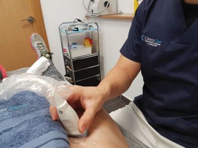
romedial portion of the vertebral bodies, and, at the level of Patient Position
the pelvis, it joins the iliacus muscle, forming the iliopsoas The patient is placed in supine position, lying with the knee
muscle and inserting onto the lesser trochanter of the femur. extended and the arms on either side of the body to ensure a
This is the only muscle that inserts at this site, and it is good approach from the anterior aspect of the hip to the
innervated by the branches from the ventral rami of L1 to L4, abdominal region. The probe is placed transversely over the
which correspond to the crural nerve. muscle fibers. (►Fig. 1)
The iliopsoas is involved in dynamic movements such as
walking and is important for the maintenance of static Optimization, Probe Position and Sonographic Image
standing. Its primary functions include producing hip flexion For an appropriate examination of the iliopsoas muscle, a low
(with a fixed trunk) and lumbar extension by increasing the frequency range is established (6–10 MHz), which should be
lordosis (when the muscle contracts with fixed legs, the determined according to the volume of the body mass of the
anterior pelvic tilt increases).1–4 Besides, this muscle has a subject. To visualize the iliopsoas, the position of the probe
stabilizing role for the hip and lumbar spine.5–11 The function should be transverse to the fibers (►Fig. 1). In this manner,
of the same cannot be recognized as being separate from the we must first locate the anterosuperior iliac spine as a
iliacus muscle, as demonstrated via electromyography.12 reference and visualize the difference between the psoas
The aim of this study was to demonstrate a new approach and iliacus muscles13 (►Fig. 2). Based on this reference
for locating the muscle to enable a sonographic assessment image, we should continue the examination, taking the
of the lumbar origin of the psoas muscle, beginning with a probe upwards, trying to clear the ‘dirty’ shadow generated
basic examination in the groin region. Moreover, we sought by the bowel loops, until the psoas is visualized in the
to establish an accessible and safe sonographic approach to transverse section of L4, which is at the height of the
enable us to assess a portion of the psoas muscle, which has umbilicus, and, in this manner, the muscle can be bilaterally
been poorly studied to date. Hence, this may help establish a compared along its muscle belly (►Fig. 3)
possible evolution of lesions affecting this muscle. From this position of reference, a scan is performed in a
This sonographic description may be relevant within the proximal direction, during which we will ask the patient to
field of physiotherapy as it is an approach that is seldom flex the hip with the knee extended, to visualize the entire
used. Additionally, it provides useful information regarding muscle belly, until reaching D12- L1.
the quality of the contraction of the psoas muscle along its
path besides detecting changes that affect the size of the
Discussion
same and possible alterations in its cortical insertions.
Ultimately, it allows clinicians to detect any asymmetries, With this approach, we hope to broaden the range of
as comparisons can be performed with the other psoas sonographic applications for the assessment of this highly
DOI https://doi.org/ Copyright © 2019 by Thieme Revinter
10.1055/s-0039-1688507. Publicações Ltda, Rio de Janeiro, Brazil
ISSN 2386-4591.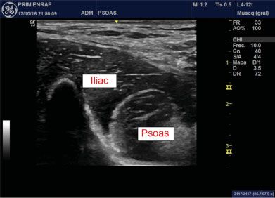
Sonographic Approach of the Lumbar Portion of the Psoas Muscle                               Serra, Ferrando           47

                                                                      relevant muscle in the lumbar area and hips. With a good
                                                                      ultrasound machine, a proper technique and a resonant
                                                                      patient, this assessment may be included within our sono-
                                                                      graphic examinations of the psoas, with absolute certainty of
                                                                      extracting further useful information.

                                                                           Conflicts of Interest
                                                                           The authors have no conflicts of interest to declare.

                                                                      References
                                                                       1 Sahrmann SA. Diagnosis and treatment of movement impairment
                                                                           syndromes. St. Louis: Mosby; 2002
                                                                       2 Neumann DA. Kinesiology of the hip: a focus on muscular actions.
                                                                           J Orthop Sports Phys Ther 2010;40(02):82–94. Doi: 10.2519/
                                                                           jospt.2010.3025
Fig. 1 Placement of the probe.                                         3   Neumann DA, Garceau LR. A proposed novel function of the psoas
                                                                           minor revealed through cadaver dissection. Clin Anat 2015;28
                                                                           (02):243–252. Doi: 10.1002/ca.22467
                                                                       4   Yoshio M, Murakami G, Sato T, Sato S, Noriyasu S. The function of
                                                                           the psoas major muscle: passive kinetics and morphological
                                                                           studies using donated cadavers. J Orthop Sci 2002;7(02):199–207
                                                                       5   Levangie PK, Norkin CC. Joint structure and function: A compre-
                                                                           hensive analysis (3rd ed.). Philadelphia: F. A. Davis; 2001
                                                                       6   Muscolino JE. Kinesiology: the skeletal system and muscle func-
                                                                           tion. Elsevier Health Sciences; 2014
                                                                       7   Mcginnis PM. Biomechanics of sport and exercise (2nd ed.).
                                                                           Champaign: Human Kinetics; 2005
                                                                       8   Basmajian JV, DeLuca CJ. Muscles alive: Their functions revealed by
                                                                           electromyography (5th ed.). Baltimore: Williams & Wilkins; 1985
                                                                       9   Blankenbaker DG, Tuite MJ, Keene JS, del Rio AM. Labral injuries
                                                                           due to iliopsoas impingement: can they be diagnosed on MR
                                                                           arthrography? AJR Am J Roentgenol 2012;199(04):894–900
                                                                      10   Blankenbaker DG, Tuite MJ. The painful hip: new concepts.
                                                                           Skeletal Radiol 2006;35(06):352–370 Review
                                                                      11   Balius R, Pedret C, Blasi M, et al. Sonographic evaluation of the
Fig. 2 Sonographic visualization of the psoas fibers and the iliacus        distal iliopsoas tendon using a new approach. J Ultrasound Med
muscle.                                                                    2014;33(11):2021–2030. Doi: 10.7863/ultra.33.11.2021
                                                                      12   Lewis CL, Sahrmann SA, Moran DW. Anterior hip joint force
                                                                           increases with hip extension, decreased gluteal force, or
                                                                           decreased iliopsoas force. J Biomech 2007;40(16):3725–3731
                                                                      13   Levangie PK. The association between static pelvic asymmetry
                                                                           and low back pain. Spine 1999;24(12):1234–1242

Fig. 3 Sonographic image of the L4 vertebral body and the psoas
muscle belly.

                                                                                  Journal of Invasive Techniques in Physical Therapy   Vol. 2   No. 1/2019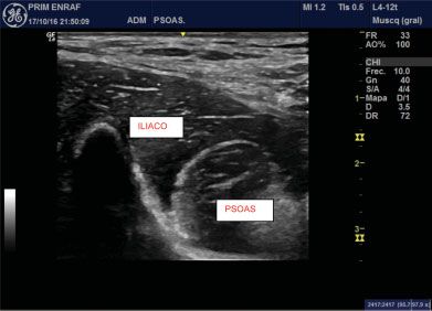
Published online: 28.06.2019
                                                                                                                                              THIEME
           46    Practice forum | Foro practico

                 Abordaje ecográfico de la porción lumbar del músculo
                 psoas
                 Sonographic Approach of the Lumbar Portion of the Psoas Muscle
                 Jaime Ríos Serra1         Ana de Groot Ferrando2

                 1 Clínica Serra, San Vicente del Raspeig, Alicante, España        Address for correspondence Jaime Rios, Clínica Serra, San Vicente del
                 2 Campos Fisioterapia, Alicante, España                           Raspeig, Alicante, España (e-mail: jaimerios_19@hotmail.com).

                  Rev Fisioter Invasiva 2019;2:46–47.

                 Introducción                                                      tamaño del mismo y posibles alteraciones en sus
                                                                                   inserciones corticales. En definitiva, permite detectar
                 El músculo psoas tiene su origen más profundo en las              cualquier tipo de asimetría al poderlo comparar con el
                 transversas de L1 a L5, y el más superficial en la parte           otro psoas. Es una manera de ampliar nuestro estudio
                 lateral del cuerpo vertebral de D12. Se dirige hacia distal       ecográfico de este músculo.
                 pasando por la zona anteromedial de los cuerpos
                 vertebrales, uniéndose en la pelvis con el músculo ilíaco,
                                                                                   Caso en imágenes
                 formando de este modo el psoas-ilíaco para insertarse en el
                 trocánter menor del fémur, siendo este el único músculo           Posición del paciente
                 que se inserta ahí. La inervación de ese músculo                  Se coloca al paciente en decúbito supino con rodilla
                 corresponde a las ramas anteriores de L1 a L4,                    extendida y los brazos pegados al cuerpo, para poder tener
                 correspondientes al nervio crural.                                un buen acceso desde la cara anterior de la cadera hasta la
                     El psoas ilíaco tiene una gran importancia en los             zona abdominal, con la sonda colocada de forma transveral a
                 movimientos dinámicos como caminar, y en el                       las fibras del músculo. (fig. 1)
                 mantenimiento de la bipedestación en estático. Entre sus
                 funciones más destacadas está la de producir la flexión de         Optimización, posición del transductor e imagen
                 cadera (con tronco fijo), y la extensión lumbar aumentado la       ecográfica
                 lordosis (al contraerse con las piernas fijas aumenta la           Para la adecuada exploración del músculo psoas ilíaco, se
                 inclinación pélvica anterior).1–4 Además, es estabilizador de     establecerá un rango de baja frecuencia (6-10 Mhz), que
                 la cadera y la columna lumbar.5–11 Su función no se reconocería   vendrá determinado en función del volumen de masa
                 por separado del iliaco, como ya se demostró mediante             corporal del sujeto. La posición de la sonda para visualizar
                 electromiografía.12                                               el psoas iliaco debe ser transversal a las fibras (►Fig. 1). De
                     El objetivo de este trabajo es mostrar un nuevo abordaje de   ese modo, localizaremos la espina ilíaca anterosuperior
                 localización del músculo que permita evaluar ecográficamente       como referencia y visualizaremos la diferencia entre el
                 el origen lumbar del músculo psoas, partiendo de su               psoas y el ilíaco13 (►Fig.2). A partir de esa imagen de
                 exploración básica a nivel inguinal. Además, se pretende a        referencia, continuamos la exploración ascendiendo con la
                 través del presente trabajo, establecer un abordaje ecográfico     sonda, intentando salvar la sombra sucia generada por las
                 accesible y seguro, que nos permita evaluar una porción del       asas intestinales, hasta que lleguemos a visualizar el psoas en
                 músculo psoas muy poco estudiada; de manera que nos sirva         la transversa de L4, la cual quedará a la altura del ombligo y
                 de posible evolución lesional.                                    así podremos comparar bilateralmente el músculo en su
                     Esa descripción ecográfica, creemos que es relevante           vientre muscular (►Fig. 3)
                 dentro nuestro ámbito de la fisioterapia, debido a que es un           Desde esa posición de referencia, se realizará un barrido
                 abordaje que no se utiliza, y que nos da una información          hacia proximal, en el cual le pediremos al paciente una
                 útil en cuanto a la calidad de contracción del músculo            flexión de cadera con la rodilla extendida para visualizar
                 psoas en todo su trayecto; aumento o disminución del              todo el vientre muscular hasta llegar a D12- L1.

                                                    DOI https://doi.org/           Copyright © 2019 by Thieme Revinter
                                                    10.1055/s-0039-1688507.        Publicações Ltda, Rio de Janeiro, Brazil
                                                    ISSN 2386-4591.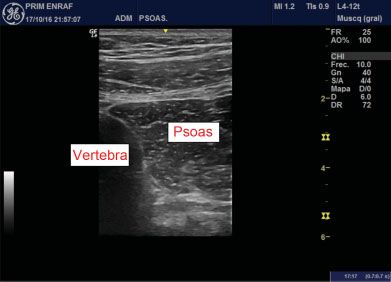
Abordaje ecográfico de la porción lumbar del músculo psoas                           Serra, Ferrando           47

                                                                      Discusión

                                                                      Con este abordaje pretendemos ampliar el abanico de
                                                                      valoración ecográfica de este músculo de gran relevancia
                                                                      para las lumbares y cadera. Con un buen equipo ecográfico,
                                                                      una buena técnica y un paciente que sea resonante, podemos
                                                                      incluir esa valoración dentro de nuestras exploraciones
                                                                      ecográficas del psoas, con la total seguridad de extraer
                                                                      mucha más información útil.

                                                                           Conflictos de interés
                                                                           Los autores no tienen conflictos de intereses que declarar.

                                                                      Bibliografía
Fig. 1 La posición del paciente y colocación de la sonda.              1 Sahrmann SA. Diagnosis and treatment of movement impairment
                                                                           syndromes. St. Louis: Mosby; 2002
                                                                       2 Neumann DA. Kinesiology of the hip: a focus on muscular actions.
                                                                           J Orthop Sports Phys Ther 2010;40(02):82–94. Doi: 10.2519/
                                                                           jospt.2010.3025
                                                                       3   Neumann DA, Garceau LR. A proposed novel function of the psoas
                                                                           minor revealed through cadaver dissection. Clin Anat 2015;28
                                                                           (02):243–252. Doi: 10.1002/ca.22467
                                                                       4   Yoshio M, Murakami G, Sato T, Sato S, Noriyasu S. The function of
                                                                           the psoas major muscle: passive kinetics and morphological
                                                                           studies using donated cadavers. J Orthop Sci 2002;7(02):199–207
                                                                       5   Levangie PK, Norkin CC. Joint structure and function: A
                                                                           comprehensive analysis (3rd ed.). Philadelphia: F. A. Davis; 2001
                                                                       6   Muscolino JE. Kinesiology: the skeletal system and muscle
                                                                           function. Elsevier Health Sciences; 2014
                                                                       7   Mcginnis PM. Biomechanics of sport and exercise (2nd ed.).
                                                                           Champaign: Human Kinetics; 2005
                                                                       8   Basmajian JV, DeLuca CJ. Muscles alive: Their functions revealed by
                                                                           electromyography (5th ed.). Baltimore: Williams & Wilkins; 1985
                                                                       9   Blankenbaker DG, Tuite MJ, Keene JS, del Rio AM. Labral injuries
                                                                           due to iliopsoas impingement: can they be diagnosed on MR
Fig. 2 Visualización ecográfica de las fibras del psoas y del músculo        arthrography? AJR Am J Roentgenol 2012;199(04):894–900
iliaco.                                                               10   Blankenbaker DG, Tuite MJ. The painful hip: new concepts.
                                                                           Skeletal Radiol 2006;35(06):352–370 Review
                                                                      11   Balius R, Pedret C, Blasi M, et al. Sonographic evaluation of the
                                                                           distal iliopsoas tendon using a new approach. J Ultrasound Med
                                                                           2014;33(11):2021–2030. Doi: 10.7863/ultra.33.11.2021
                                                                      12   Lewis CL, Sahrmann SA, Moran DW. Anterior hip joint force
                                                                           increases with hip extension, decreased gluteal force, or
                                                                           decreased iliopsoas force. J Biomech 2007;40(16):3725–3731
                                                                      13   Levangie PK. The association between static pelvic asymmetry
                                                                           and low back pain. Spine 1999;24(12):1234–1242

Fig. 3 Imagen ecográfica del cuerpo vertebral de L4 y el cuerpo
muscular del Psoas.

                                                                                                     Revista Fisioterapia Invasiva   Vol. 2   No. 1/2019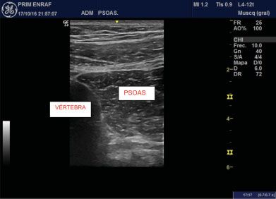
You can also read