Cycle Immunoglobulins in the mouse uterus during the oestrous
←
→
Page content transcription
If your browser does not render page correctly, please read the page content below
Immunoglobulins                 in the      mouse          uterus     during            the oestrous
                                                   cycle
        Flore Rachman, V. Casimiri, A. Psychoyos and O. Bernard
 Unité de Recherche d'Hépatologie Infantile, INSERM U 56, & Clinique de Pédiatrie, Université
Paris-Sud, Hôpital d'Enfants, and *Laboratoire de Physiologie de la Reproduction, ER 203, CNRS,
                   Hôpital de Bicêtre (Bât. INSERM), F-94270 Bicêtre, France

     Summary. The distribution of IgA, IgG and IgM was studied by an immunoperoxidase
     technique on sections of mouse uteri at each stage of the oestrous cycle. Staining for
     IgG and IgA was highest at pro-oestrus, declined at oestrus and was very low during the
     other stages. At pro-oestrus IgG was found throughout the stroma, in the uterine
     lumen, and in 10% of glandular lumina; very few IgG-containing plasma cells were
     present. At pro-oestrus, IgA was found in the uterine lumen, and in most of the uterine
     glands, both in the lumen and in the epithelium; little IgA was present in the stroma.
     IgA-plasma cells were detected at each stage of the cycle and were particularly
     numerous at pro-oestrus and oestrus. These results suggest that IgA is secreted locally
     from plasma cells into the uterine gland through the glandular epithelium, but that IgG
     enters the stroma from the local capillaries. The obvious increase in IgG and IgA
     secretion at pro-oestrus, when plasma oestradiol levels are highest, supports the
     hypothesis that, during the oestrous cycle, the humoral immune response is regulated in
     the uterus by ovarian hormones.

                                              Introduction

Several investigations have now established that the uterus forms part of a mucosal immune system
which is common to the gut, bronchi, mammary gland and liver. Lymphocyte migration towards
the mouse uterus and the presence of immunoglobulins in rat uterine fluid have been demonstrated
and are probably hormone dependent (Wira & Sandoe, 1977; McDermott, Clark & Bienenstock,
1980; Wira, Hyde, Sandoe, Sullivan & Spencer, 1980). Less is known, however, about the tissue
distribution of immunoglobulins and immunoglobulin-containing cells in the uterus. The data
obtained in humans are conflicting (Tourville, Ogra, Lippes & Tornasi, 1970; Kelly & Fox, 1979).
IgA-plasma cells seem to be present throughout the oestrous cycle in rats (Wira et al., 1980).
    Our previous observations suggest that during the preimplantation period of pregnancy in mice,
the IgA produced locally by plasma cells is secreted into the uterine lumen via uterine glands,
whereas IgG infiltrates the stroma but does not seem to be secreted locally (Bernard, Rachman &
Bennett, 1981). This report presents the results of studies of immunoglobulin distribution in the
mouse uterus at the various stages of the oestrous cycle.

                                       Materials and Methods

Six-week-old random-bred female mice of the OF1 strain were         kept in cages containing 4 females
each, next to cages containing male mice of the same strain, under a schedule of 14 h light and                  10 h

                                     0022-4251/83/050017-06S02-00/0
                            ©   1983 Journals ofReproduction & Fertility   Ltd
                                                                   Downloaded from Bioscientifica.com at 03/06/2020 05:20:41AM
                                                                                                                 via free access
darkness. These conditions were maintained for 2 weeks before the study. Mice whose vaginal
smears showed regular oestrous cycles of 5 days were selected at a given stage of the cycle. They
were killed by cervical dislocation and the vagina and uterine horns were removed. To confirm the
stage in the oestrous cycle, the histology of the corresponding vaginal epithelium was studied after
fixation in Bouin's alcoholic fluid, embedding in paraffin wax and staining with haematoxylin and
eosin. Only mice in which the vaginal epithelial histology and vaginal smears were clearly
characteristic of a particular stage of the cycle (Allen, 1922) were further examined for uterine
immunoglobulins. Uterine horns were treated as previously described (Bernard, Ripoche &
Bennett, 1977; Bernard et al., 1981); briefly, each horn was ligated at both ends, fixed in 4%
paraformaldehyde and embedded in polyethylene glycol 1000; 5 µ sagittal and transverse sections
were incubated first with rabbit anti-mouse IgA, IgG or IgM (Litton Bionetics, Kensington,
Maryland), then with peroxidase-labelled sheep anti-rabbit immunoglobulin (adsorbed with mouse
immunoglobulin) (Institut Pasteur Production, Dr M. Lavergne), and stained with diaminobenzi-
dine and H202. Uterine horns from 3-5 mice were studied for each stage of the cycle.
Controls. The specificity of the various antisera and conjugates was checked as previously
described (Bernard et al., 1981). Endogenous peroxidase activity was only present in eosinophils.
These cells are known to be abundant in the endometrium at pro-oestrus and oestrus in the rat
(Psychoyos, 1973) and were clearly distinguishable from cells staining specifically for
immunoglobulins.
Quantitation of immunoglobulin-containing glands. To determine the proportion of glandular
lumina containing IgA or IgG, all the glands visible in each sagittal section of the whole uterine
horn were examined and the glandular lumina with and without IgA or IgG were counted
separately. The number of glandular lumina in each section ranged from 36 to 322 (mean 104).
Statistical analysis was performed using Student's t test.
Results
Distribution of immunoglobulins
The distribution of plasma cells and immunoglobulins in the various parts of the uterus is shown
in Table 1 and PI. 1, Figs 1-6.
Table 1. Distribution of IgG, IgA and IgM in the various structures of the mouse uterus at different
stages of the oestrous cycle
Plasma cells Luminal Uterine
Stroma in stroma epithelium lumen
Stage of -
oestrous cycle IgA IgG IgM IgA
-
IgG IgM IgA
-
IgG IgM IgA
-
IgG IgM
Dioestrus Occas. ± Occas. + Occas.
Pro-oestrus Rare
—
++
—
++
—
+ ++
Oestrus + -
++ Very + ±
-
rare
Metoestrus I ± + Occas. ± Rare
Metoestrus II —
Occas. -
+ Occas. —
Rare Occas.
— — —
+ -t-, strong staining; +, medium staining; ±, faint staining; —, no staining; Occas., occasional.
Dioestrus. Very little immunoglobulin was detectable in the uterus except for a few IgA-
containing plasma cells in the stroma and some IgA visible in a few glandular and uterine lumina.
Pro-oestrus. The amount of immunoglobulins found in the uterus was highest at this stage of the
cycle. IgG was present throughout the stroma (PI. 1, Fig. 5) but no IgG-containing plasma cells
Downloaded from Bioscientifica.com at 03/06/2020 05:20:41AM
via free accessPLATE 1
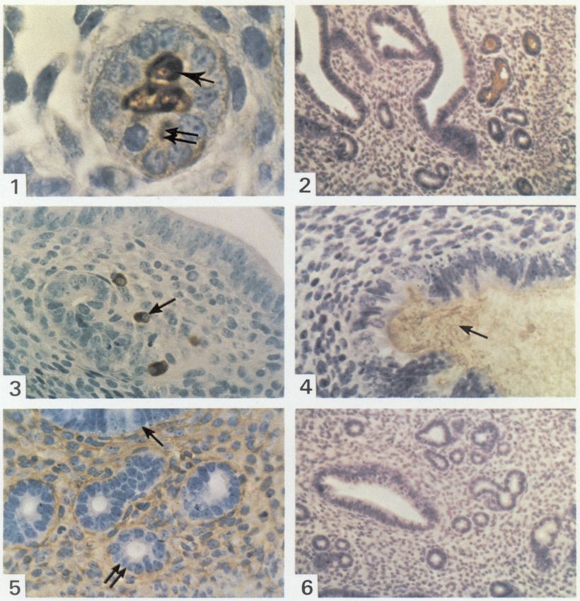
Immunoglobulins in the mouse endometrium during the oestrous cycle (indirect immunoperox-
idase localization followed by haematoxylin staining).
Fig. 1. At pro-oestrus, IgA is present in the gland lumen (single arrow) and in epithelial cells
(double arrow), 633.
Fig. 2. At pro-oestrus, about half of the glandular lumina contain IgA. 100.
Fig. 3. At dioestrus, some IgA-plasma cells (arrowed) are seen around empty glands, 250.
Fig. 4. At pro-oestrus, IgA is present in the uterine lumen (arrowed), 250.
Fig. 5. At pro-oestrus, IgG invades the stroma but is not seen crossing the uterine (single arrow)
or glandular (double arrow) epithelium, 250.
Fig. 6. At pro-oestrus, there is no IgM visible in the plasma cells, glands or stroma, 100.
(Facing p. 18)
Downloaded from Bioscientifica.com at 03/06/2020 05:20:41AM
via free accesswere detected. The largest amount of IgG was observed in uteri from mice at the late pro-oestrous
stage, as shown by vaginal epithelial histology, and staining was especially strong in the stroma
next to the luminal epithelium. No IgG was visible in the glandular or luminal epithelia but it was
clearly detectable in the uterine lumen and in a few glandular lumina. IgA-containing plasma cells
were very conspicuous ; most of these cells surrounded the glands. IgA was usually not detectable in
the stroma, except occasionally as faint staining, but nearly half the glandular lumina (PI. 1, Fig. 2)
and a few cells in the glandular epithelium contained IgA (PI. 1, Fig. 1). IgA was also present in the
uterine lumen (PI. 1, Fig. 4), but not in the luminal epithelium. The number of IgA-plasma cells in
the stroma seemed largest, and staining of IgA in the uterine lumen strongest, in the late pro-
oestrous stage. IgM and IgM-containing plasma cells were never detected (PI. 1, Fig. 6), except in a
few blood capillaries.
Oestrus. There was a general decrease in immunoglobulin levels, compared to pro-oestrus. Some
staining of IgG was still present in the stroma and about one third of the glandular lumina
contained IgA.
Metoestrus I. IgG was virtually undetectable, and IgA was less evident both in plasma cells and
glands.
Metoestrus II. Staining for immunoglobulin was the faintest of all the stages of the oestrous
cycle.
No IgG, IgA or IgM was visible in the epithelial cells of the uterine lumen at any stage of the
cycle.
Quantitation of immunoglobulin-containing glandular lumina
During the oestrous cycle, the mean percentage of IgA-containing glands rose significantly
from dioestrus to pro-oestrus (P < 001); at oestrus, it decreased slightly but not significantly and
then significantly at metoestrus I (P < 005), thereafter declining further at metoestrus II (Text-fig.
I). The same trend was observed for IgG, although the percentage of IgG-containing glands was
much lower.
Text-fig. 1. The number of glandular lumina in mouse uteri containing IgA, IgG or IgM at
various stages of the oestrous cycle (D, dioestrus; P, pro-oestrus; O, oestrus; MI, metoestrus I;
Mil, metoestrus II). *Significantly different from value at dioestrus (P < 0-01 for IgA; <
005 for IgG). fSignificantly different from values at metoestrus I and II (P < 0-05).
Downloaded from Bioscientifica.com at 03/06/2020 05:20:41AM
via free accessDiscussion
These results show that, in mice, IgG and IgA are present in the endometrium and their
distribution and amounts appear to vary with the stage of the oestrous cycle and the corresponding
hormone fluctuations. These findings (1) further support the concept of hormonal regulation of IgA
and IgG secretion in the rodent uterus, and (2) suggest that, during both the oestrous cycle and the
first days of pregnancy, the mechanisms responsible for the presence of IgA and IgG are different.
The regulation of uterine immunoglobulin secretion by ovarian hormones is suggested by several
data; our results are consistent with those showing that IgA and IgG levels in the rat uterine fluid
vary throughout the oestrous cycle, the highest levels being observed at pro-oestrus, when plasma
oestradiol and progesterone concentrations are highest, and the lowest at dioestrus (Wira &
Sandoe, 1977, 1980). Also, oestradiol injection into ovariectomized rats induced accumulation
of IgA and IgG into the uterine fluid (Wira & Sandoe, 1977, 1980). It is therefore likely that the
immunohistological changes affecting IgG and IgA observed here in mouse endometrium during
the oestrous cycle are also hormone-dependent.
The mechanisms of immunoglobulin secretion in the uterus are probably different for each
class of immunoglobulin. Our results for the mouse uterus show that IgM does not seem to be
involved in local secretory immunity during the oestrous cycle, or in early pregnancy; the limited
amount detected at the time of implantation was probably due to passive diffusion because of the
major increase in vascular permeability at this stage (Psychoyos, 1960). Such increased
permeability is probably responsible for the presence of IgG in the uterine stroma at pro-oestrus
and oestrus (present study) and at the time of implantation (Bernard et al., 1981). Very few IgG-
containing plasma cells were visualized in the stroma during the oestrous cycle, and oestradiol is
known to increase the blood flow as well as an oedematous tendency favouring the passive diffusion
of macromolecules (Spaziani & Szego, 1958). The mechanisms by which IgG reaches the uterine
lumen at pro-oestrus remain unclear. As it was observed during the implantation period, despite the
striking contrast between the IgG-filled stroma and the absence of luminal epithelial staining, some
IgG may cross the glandular epithelium to reach the uterine lumen since it was detected in about
10% of glandular lumina at pro-oestrus. Alternatively, it might cross the luminal epithelium in a
form not detectable by light microscopy (Parr, 1980). Previous data concerning IgA secretion in the
rodent uterus have shown that, in mice, IgA-B lymphocytes migrate to the endometrium from the
mesenteric lymph nodes, that this migration rate is highest at pro-oestrus (McDermott et al., 1980)
and that, in ovariectomized rats, IgA lymphocytes also concentrate in the uterus after oestradiol
injection but this effect is blocked by progesterone (Wira et al., 1980); it is also known that, in rats,
the secretory component is detectable in the uterine fluid, mainly at pro-oestrus but also in
ovariectomized animals after oestradiol injection as either free or IgA-linked secretory component
(Sullivan & Wira, 1981). Our results provide further information on the steps between lymphocyte
migration and presence of IgA in the uterine fluid. They indicate that, in mice, IgA-plasma cells
infiltrate the stroma during the oestrous cycle and the early stage of pregnancy (Bernard et al., 1981 )
mostly when oestradiol plasma levels are highest. These results also show that IgA is most probably
transferred from the plasma cells into the glands, since it was detected in the cells of the glandular
epithelium and in the lumina of about 40% of glands at pro-oestrus. This is consistent with the
hypothesis that IgA binds to secretory component on the basal surface of the glandular epithelial
cell, crossing the cell to be released in the gland lumen and then in the uterine lumen; the very faint
staining for free IgA in the stroma at pro-oestrus may also reflect some IgA diffusion from the
capillaries, as suggested previously (Bernard et al., 1981). Our results further indicate that IgA
secretion in the uterine glands is continuous, because at each stage of the cycle plasma cells were
detected, and IgA was visualized in the gland lumina and in a few glandular epithelial cells. As
shown by the increased number of IgA-containing glands at pro-oestrus, enhanced IgA secretion
may result from an increase in the number of IgA-containing plasma cells at that time, leading to
local release of IgA and an augmented transfer of secretory IgA through glandular epithelial cells.
Downloaded from Bioscientifica.com at 03/06/2020 05:20:41AM
via free accessWhether secretory component synthesis is also enhanced in rodents at pro-oestrus under the
influence of oestradiol has not yet been established but seems to be indicated by the findings of
Sullivan & Wira (1981).
We thank Mme M. Grelier and Mme R-M. Dreyfus for help with the preparation and revision
of the manuscript. This work was supported by CNRS No. 03.3673 and UER Kremlin-Bicêtre No.
867.
References
Allen, E. (1922) The estrous cycle in the mouse. Am. J. Spaziani, E. & Szego, CM. (1958) The influence of
Anat. 30, 297-371. estradiol and cortisol on uterine histamine of the
Bernard, O., Ripoche, M.A. & Bennett, D. (1977) ovariectomized rat. Endocrinology 63, 669-678.
Distribution of maternal immunoglobulins in the SuUivan, D.A. & Wira, CE. (1981) Estradiol regulation of
mouse uterus and embryo in the days after implanta¬ secretory component in the female reproductive
tion. J. exp. Med. 145, 58-75. tract. J. Steroid Biochem. 15, 439^144.
Bernard, O., Rachman, F. & Bennett, D. (1981) Immu¬ Tourville, D.R., Ogra, S.S., Lippes, J. & Tornasi, T.B.
noglobulins in the mouse uterus before implantation. (1970) The human female reproductive tract: im-
J. Reprod. Pert. 63, 237-240. munohistological localization of yA, yG, yM, secre¬
Kelly, J.K. & Fox, H. (1979) The local immunological tory "piece" and lactoferrin. Am. J. Obstet. Gynec.
defence system of the human endometrium. /. 108, 1102-1108.
Reprod. Immunol. 1, 39-45. Wira, C.R. & Sandoe, C.P. (1977) Sex steroid hormone
McDermott, M.R., Clark, D.A. & Bienenstock, J. (1980) regulation of IgA and IgG in rat uterine secretions.
Evidence for a common mucosal immunologie Nature, Lond. 268, 534-535.
system. II. Influence of the estrus cycle on Wira, C.R. & Sandoe, C.P. (1980) Hormonal regulation
immunoblast migration into genital and intestinal of immunoglobulins: influence of estradiol on
tissues. J. Immunol. 124, 2536-2538. immunoglobulins A and G in the rat uterus.
Parr, M.B. (1980) Endocytosis in the uterine epithelium Endocrinology 106, 1020-1026.
during early pregnancy. Prog. Reprod. Biol. 7, 81-91. Wira, C.R., Hyde, E., Sandoe, C.P., SuUivan, D. &
Psychoyos, A. (1960) Nouvelle contribution à l'étude de Spencer, S. (1980) Cellular aspects of the rat uterine
la nidation de l'oeuf chez la rate. C.r. hebd. Séance. IgA response to estradiol and progesterone. J. Steroid
Acad. Sci. Paris D 251, 3073-3075. Biochem. 12, 451-459.
Psychoyos, A. (1973) Endocrine control of egg implanta¬
tion. Handbook of Physiology: Endocrinology II, Vol.
2, pp. 187-215. Eds R. O. Greep & E. V. Astwood.
Am. Physiol. Soc, Washington D.C. Received 10 November 1982
Downloaded from Bioscientifica.com at 03/06/2020 05:20:41AM
via free accessYou can also read