Btk-dependent Rac activation and actin rearrangement following Fc RI aggregation promotes enhanced chemotactic responses of mast cells
←
→
Page content transcription
If your browser does not render page correctly, please read the page content below
2576 Research Article
Btk-dependent Rac activation and actin
rearrangement following FcRI aggregation promotes
enhanced chemotactic responses of mast cells
Hye Sun Kuehn1, Madeleine Rådinger1, Jared M. Brown1, Khaled Ali2, Bart Vanhaesebroeck2,
Michael A. Beaven3, Dean D. Metcalfe1 and Alasdair M. Gilfillan1,*
1
Laboratory of Allergic Diseases, National Institute of Allergy and Infectious Diseases, National Institutes of Health, 10 Center Drive MSC 1881,
Bethesda, MD 20892-1881, USA
2
Centre for Cell Signalling, Institute of Cancer, Barts and the London School of Medicine and Dentistry, John Vane Science Centre,
Charterhouse Square, London, EC1M 6BQ, UK
3
Laboratory of Molecular Immunology, National Heart, Lung, and Blood Institute, National Institutes of Health, Bethesda, MD 20892, USA
*Author for correspondence (agilfillan@niaid.nih.gov)
Accepted 28 April 2010
Journal of Cell Science 123, 2576-2585
© 2010. Published by The Company of Biologists Ltd
doi:10.1242/jcs.071043
Summary
Mast cells infiltrate the sites of inflammation associated with chronic atopic disease and during helminth and bacterial infection. This
process requires receptor-mediated cell chemotaxis across a concentration gradient of their chemotactic ligands. In vivo, mast cells are
likely to be exposed to several such agents, which can cooperate in a synergistic manner to regulate mast cell homing. Here, we report
that chemotaxis of mouse bone-marrow-derived mast cells (BMMCs) in response to the chemoattractants stem-cell factor (SCF) and
Journal of Cell Science
prostaglandin (PG)E2, is substantially enhanced following antigen-dependent ligation of the high-affinity receptor for IgE (FcRI).
These responses were associated with enhanced activation of phosphoinositide 3-kinase (PI3K), and downstream activation of the
tyrosine protein kinase Btk, with subsequent enhanced phospholipase (PL)C-mediated Ca2+ mobilization, Rac activation and F-actin
rearrangement. Antigen-induced chemotaxis, and the ability of antigen to amplify responses mediated by SCF, adenosine and PGE2
were suppressed following inhibition of PI3K, and were impaired in BMMCs derived from Btk–/– mice. There were corresponding
decreases in the PLC-mediated Ca2+ signal, Rac activation and F-actin rearrangement, which, as they are essential for BMMC
chemotaxis, accounts for the impaired migration of Btk-deficient cells. Taken together, these data demonstrate that, by regulating
signaling pathways that control F-actin rearrangement, Btk is crucial for the ability of antigen to amplify mast-cell chemotactic
responses.
Key words: Mast cells, FcRI, Btk, Rac activation, Actin rearrangement, Chemotaxis, SCF, PGE2
Introduction mast-cell homing, in addition to its role in release of the mast-cell
Mast cells are important players in host defence mechanisms mediator. As demonstrated in several cell types, chemotaxis is a
associated with innate and adaptive immune responses (Galli et al., Ca2+-dependent process that requires cytoskeletal rearrangement
2005; Marshall, 2004; Mekori, 2004). However, in afflicted attributable to initial polymerization and then depolymerization of
individuals, inflammatory mediators released from activated mast actin filaments (Nishida et al., 2005; Shimizu et al., 2009). In mast
cells can also induce atopic allergic inflammatory responses (Brown cells, sensitization with IgE alone enhances cortical F-actin-ring
et al., 2008; Metcalfe et al., 1997). Mast cells infiltrate the sites of formation, as a consequence of F-actin polymerization, and
inflammation associated with chronic atopic disease, and helminth subsequent IgE-FcRI crosslinking with antigen induces
and bacterial infection (Echtenacher et al., 1996; Madden et al., disassembly of this structure (Allen et al., 2009; Nishida et al.,
1991). These processes rely on the ability of mast cells to migrate 2005). The events that regulate SCF-mediated mast-cell chemotaxis
to target tissues in response to the appropriate chemotactic stimuli. might in part be regulated by phosphoinositide 3-kinase (PI3K)-
Mast cells express surface receptors for a number of endogenous mediated pathways (Kim et al., 2008; Samayawardhena et al.,
ligands that are known to be potent mast-cell chemoattractants 2007)
under experimental conditions. Of these, the most widely Inflamed tissues in allergic individuals accumulate inflammatory
investigated is stem cell factor (SCF), the natural ligand for the mast-cell-activating agents including PGE2 (Harris et al., 2002;
growth-factor receptor KIT (Meininger et al., 1992; Nilsson et al., Vancheri et al., 2004) and adenosine (Spicuzza et al., 2006). In
1994). Others include several agonists of G-protein-coupled addition, chronically inflamed tissues undergo fibrotic changes
receptors (GPCRs), such as prostaglandin E2 (PGE2), which that might lead to increased production of SCF by fibroblasts
positively regulates mast-cell responses through the EP3 receptor (Hogaboam et al., 1998). Therefore, it is likely that mast cells
(Weller et al., 2007). There is also evidence to suggest that IgE- within these tissues are exposed to co-activating factors, which
sensitized mast cells migrate toward antigen, potentially through might thus contribute to tissue targeting of mast cells in a
the release of chemoattractants from mast cells themselves (Ishizuka pathological environment through synergistic interactions with
et al., 2001; Kitaura et al., 2005). This evokes a potential antigen. We therefore investigated whether such synergy can occur
contributory role for the high-affinity receptor for IgE (FcRI) in and, if so, the molecular mechanisms regulating these responses in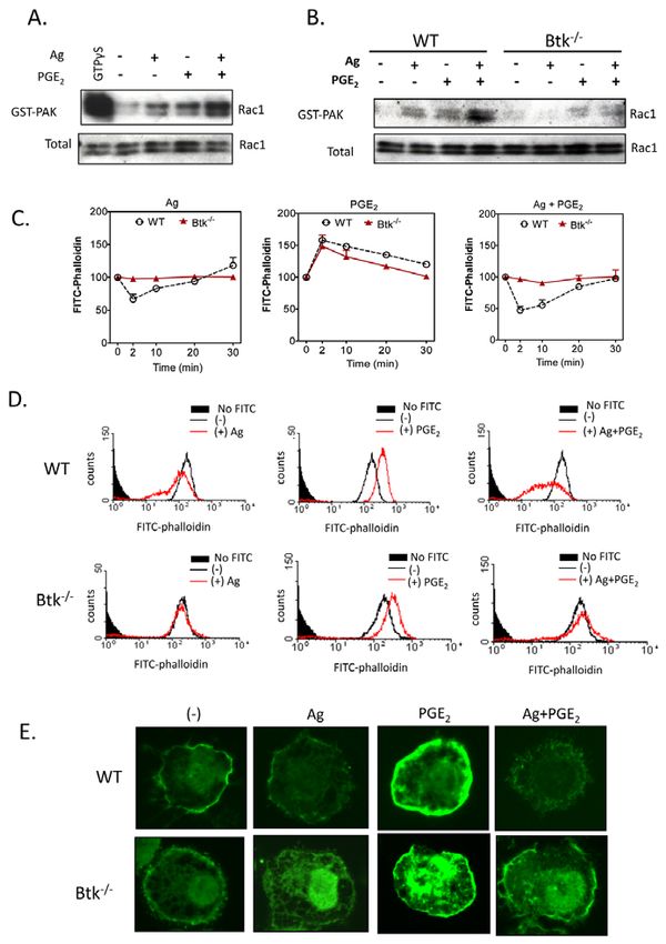
Btk in chemotactic responses 2577
mast cells derived from the bone marrow (BMMCs) of wild-type alone (100 nM) was more rapid than that produced by SCF
and gene-deficient mice. Signaling events induced by antigen were (compare Fig. 1C with 1A). In combination with antigen, the
found to synergistically interact with those elicited by SCF and the response to PGE2 was substantially enhanced, and comparable
GPCR agonists, adenosine and PGE2, to dramatically enhance with that observed for the combination of antigen and SCF.
chemotaxis in mouse BMMCs. Such synergy was associated with However, as the response of PGE2 alone was more robust than that
enhanced increases in Ca2+ mobilization and Rac-dependent produced by SCF alone, the degree of enhancement of the SCF
cytoskeletal reorganization through actin polymerization and response by antigen was of greater relative magnitude, at least at
depolymerization. We finally demonstrate that, by regulating such the 1 hour time point. There are no reports of the effect of adenosine
synergistic signaling events, Btk is an integral component of the on mast-cell chemotaxis but, unlike SCF and PGE2, we found that
integrated signal-transduction pathway that permits antigen to adenosine by itself had little or no ability to induce chemotaxis
amplify the chemotactic responses of mast cells. (Fig. 1D). However, the combination of adenosine and antigen
challenge induced a chemotactic response approximately fourfold
Results greater than that induced by antigen alone. A similar pattern has
Mast-cell migration toward SCF and GPCR agonists is been noted for mast-cell degranulation where adenosine potentiates
synergistically enhanced by antigen antigen-induced degranulation without inducing degranulation by
To investigate potential synergistic interactions between the itself (Ali et al., 1990). Nevertheless, the chemotactic response to
signaling events elicited through KIT and GPCRs in the context of the adenosine-antigen combination was lower than that observed
FcRI ligation, we first examined the ability of antigen to enhance with the combinations of SCF or PGE2 with antigen.
the chemotactic response elicited by SCF and GPCR agonists in To determine whether or not the ability of antigen to enhance
BMMCs. As expected, optimal concentrations of SCF (10 ng/ml) GPCR-agonist-induced chemotaxis was mediated through Gi-
and, to a much lesser extent antigen (10 ng/ml), produced significant dependent signaling, we examined the ability of the Gi inhibitor,
chemotaxis towards each stimulant over the course of 4 hours (Fig. pertussis toxin, to block this response. As predicted, the chemotactic
1A). However, the chemotactic response was enhanced responses to SCF, antigen or the combination of the two, were
approximately threefold when SCF and antigen were added unaffected by pertussis toxin pre-treatment (Fig. 1E, left panel). By
Journal of Cell Science
simultaneously, and this was apparent at all time points examined contrast, such treatment completely blocked the chemotactic
(Fig. 1B). GPCR agonists were examined in a similar manner, and, response to PGE2 as well as the synergistic enhancement of
on the basis of their efficacy in enhancing antigen-mediated response to antigen caused by PGE2 and adenosine (Fig. 1E, middle
degranulation in BMMCs (Kuehn et al., 2008a; Kuehn and Gilfillan, and right panels). These results demonstrate that PGE2 and
2007; Zhong et al., 2003), PGE2 and adenosine were selected as adenosine mediate their effects on chemotaxis exclusively through
examples for study. From Fig. 1C, it can be seen that the Gi and that the responses to antigen or SCF are not due to the
chemotactic response induced by an optimal concentration of PGE2 release of agonists of Gi-coupled receptors, such as adenosine,
Fig. 1. FcRI aggregation synergistically enhances
chemotactic responses elicited by KIT and GPCRs.
(A-D)BMMCs were sensitized overnight with IgE in
cytokine-free medium. In these, and other, studies described,
3⫻105 cells were added to the upper chambers. The
chemotaxis assay was performed as described in the
Materials and Methods. (E)BMMCs were preincubated with
pertussis toxin (1g/ml) for 4 hours then washed and used
for the assay. The indicated agonists [used at optimal
concentrations (see supplementary material Fig. S1), antigen,
Ag (10 ng/ml), SCF (10 ng/ml), adenosine (1M), PGE2
(100 nM)] were added in the lower chamber, and the
migrated BMMCs were counted after 4 hours. Results are
means ± s.e. of three separate experiments. *P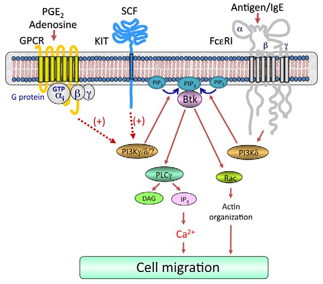
2578 Journal of Cell Science 123 (15)
which can then act in an autocrine or paracrine manner to induce To verify that the responses were due to directed chemotaxis
chemotaxis. rather than chemokinesis (random migration), we examined the
migration of sensitized BMMCs, with or without a concentration
Synergistic migration of BMMCs occurs by a direct gradient of stimuli across the membranes. Without a gradient, the
chemotaxis response migration of BMMCs towards antigen, SCF and PGE2 was largely
To exclude the possibility that mast-cell products, other than those abrogated, as was the synergy between antigen and other stimulants
acting through Gi, contribute to the responses described above, (Fig. 2C). These data demonstrate that the migration of sensitized
supernatants from BMMCs that had been previously stimulated BMMCs towards the various stimulants was primarily due to
with the various stimulants, or combinations thereof, were applied chemotaxis and not chemokinesis. As the data show that SCF and
to the lower wells of the chemotactic chambers. Sensitized (with GPCR-mediated responses were similarly potentiated by antigen,
IgE) or non-sensitized BMMCs were then placed in the upper the detailed mechanistic studies were investigated with PGE2.
chambers. No differences were observed in the migration of Similar responses were, however, observed when cells were
sensitized cells towards the chambers containing the supernatants challenged with SCF or adenosine in the absence of antigen (data
from previously stimulated cells (Fig. 2A) compared with cells not shown).
that were stimulated in the conventional manner (compare data to
Fig. 1). Virtually no migration of non-sensitized BMMCs occurred Synergy in chemotaxis is associated with synergistic
when the lower chambers contained supernatants from cultures F-actin cytoskeletal rearrangement and an enhanced Ca2+
stimulated with antigen, even though normal migration was still signal
apparent when the lower chambers contained supernatants from We next explored whether the synergy in chemotactic responses
cells stimulated with SCF or PGE2 (Fig. 2A). Furthermore, the was accompanied by a similar enhancement of actin
synergy between antigen and the other stimulants was substantially polymerization and depolymerization, as monitored by FITC-
decreased for non-sensitized cells. To further establish that the labeled phalloidin, which interacts with F-actin but not with G-
observed responses were not dependent on the generation and actin (monomeric, globular G-actin). As shown in Fig. 3A, F-actin
release of a chemotactic protein following antigen challenge, we staining with FITC-phalloidin was reduced by antigen at early
Journal of Cell Science
examined whether such chemotaxis was prevented by incubation time points (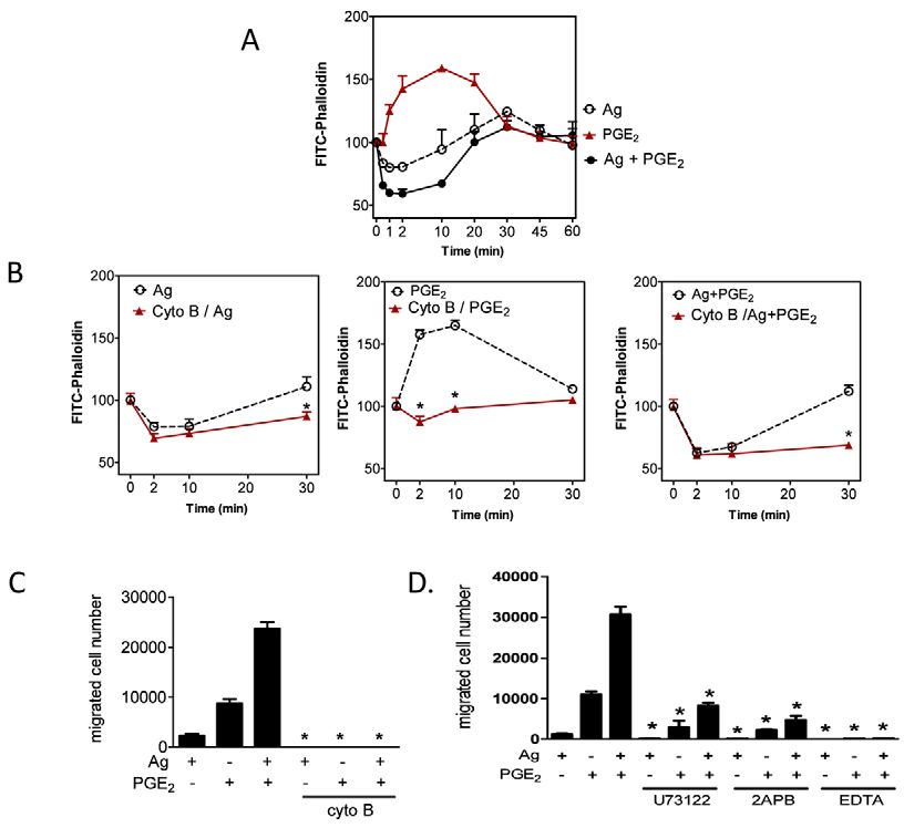
Btk in chemotactic responses 2579
Fig. 3. The role of cytoskeletal reorganization and Ca2+ in
mast-cell migration. (A,B)Sensitized BMMCs were
preincubated with or without cytochalasin B (10M) for 20
minutes. After preincubation with cytochalasin B, cells were
stimulated with indicated agonists [Ag (10 ng/ml), PGE2 (100
nM)] for the indicated time, fixed, then permeabilized. Actin
rearrangement was measured by using FITC-labeled phalloidin
staining of the cells, followed by flow cytometry. Data are mean
values of fluorescence intensities of phalloidin staining.
(C)Sensitized BMMCs were preincubated with or without
Cytochalasin B (10M) for 20-30 minutes, and cell migration
was measured. (D)Sensitized BMMCs were preincubated with
U73122 (1M), 2APB (50M), or EDTA (5 mM) in upper
chambers and placed in 600l HEPES buffer containing 0.5%
BSA and indicated inhibitors for 30 minutes, and then upper
chambers were placed in the lower chambers containing the
indicated agonists. Results are means ± s.e. of three separate
experiments. *P2580 Journal of Cell Science 123 (15)
PI3K might have a role in the responses mediated by GPCRs,
PI3K is the major isoform regulating the responses to antigen
alone, or in combination with other stimulants.
Antigen-mediated and synergistic chemotactic responses
require Btk
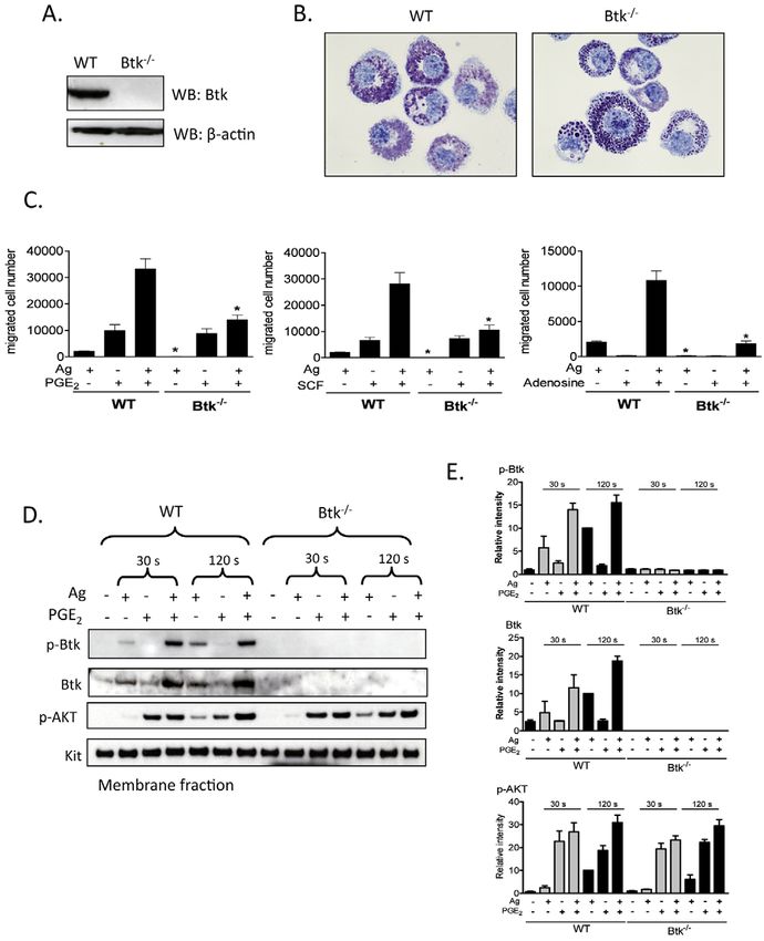
Btk is one of several downstream regulators of PI3K-dependent
responses in hematopoietic cells, including B-cells (de Gorter et
al., 2007) and mast cells (Kuehn et al., 2008b). We used BMMCs
from Btk–/– mice to investigate whether Btk regulates chemotactic
responses downstream of PI3K. Lack of Btk in these cells was
confirmed by western blot analysis (Fig. 5A). Toluidine Blue
staining demonstrated that Btk–/– BMMC morphology was similar
to that of WT BMMCs (Fig. 5B). Btk–/– BMMCs are also known
to express KIT and FcRI to the same extent as WT BMMCs (Hata
et al., 1998).
The chemotactic response to antigen was virtually absent in the
Btk–/– BMMCs and yet these cells responded fully to SCF and
PGE2 (Fig. 5C). As in normal BMMCs (Fig. 1), adenosine was an
ineffective chemotactic stimulant for Btk-deficient cells (Fig. 5C)
Nevertheless, the synergistic chemotactic responses induced upon
interactions of antigen with SCF, PGE2 and adenosine, were largely
abrogated in the Btk–/– BMMCs. These results suggest that antigen-
induced migration and the synergistic responses elicited by antigen
Journal of Cell Science
are mediated by Btk downstream of PI3K, but the chemotactic
responses induced by SCF or PGE2 alone are regulated by a PI3K-
regulated, but Btk-independent, pathway.
Activation of Btk requires coordinated translocation to the cell
membrane and tyrosine phosphorylation at positions Y223 and
Y551. We thus next examined the ability of PGE2 to enhance the
antigen-mediated phosphorylation of Btk in the membrane fractions
of BMMCs derived from WT and Btk–/– mice, using an antibody
that recognized Btk phosphorylated at Y551. To verify PI3K
activation in these experiments, we also determined AKT
phosphorylation in the membrane fractions.
Consistent with previous results (Kuehn et al., 2008a), and the
ability of wortmannin to block BMMC chemotaxis (Fig. 4A),
antigen-mediated phosphorylation of AKT was enhanced by PGE2
Fig. 4. The role of PI3K in synergistic chemotactic responses. and this enhancement was most prominent at the 2 minute time
(A)Sensitized BMMCs were preincubated with or without wortmannin (100 point (Fig. 5D,E). This response was still observed in the Btk–/–
nM), in the upper chamber and placed in 600l HEPES buffer containing BMMCs, which verified that activation of PI3K was upstream of
0.5% BSA and wortmannin for 30 minutes. The upper chambers were then
Btk. Unlike antigen, which induced Btk phosphorylation in the
replaced in the lower chambers containing the indicated agonists.
membrane fraction of WT BMMCs, PGE2 had minimal effect on
(B)Sensitized BMMCs were preincubated with indicated inhibitors for 20
minutes and then stimulated with the indicated agonists for 5 minutes. Btk phosphorylation in the absence of antigen at both time points
Following electrophoresis and membrane transfer, proteins were probed using examined (30 seconds and 120 seconds) (Fig. 5D,E). This was
anti-phospho-AKT (Ser473-P). To normalize protein loading, membranes consistent with the finding that PGE2-mediated chemotaxis is not
were stripped and probed for -actin, or alternatively identically loaded dependent on Btk as demonstrated in Btk–/– BMMCs, Nevertheless,
samples were probed for -actin. The data shown are from three separate in combination with antigen, PGE2 produced a marked synergistic
experiments, each repeated at least three times, with identical results, on enhancement of membrane-associated Btk phosphorylation (Fig.
separate cell preparations. (C)Sensitized BMMCs were preincubated with or 5D,E). This was associated with a net increase in Btk protein level
without AS 252424 (3M) or IC 87114 (3M) in the upper chambers and within the membrane fraction. Hence the increase in membrane-
placed in 600l HEPES buffer containing 0.5% BSA and indicated inhibitors
associated phosphorylated Btk could be attributed, at least in part,
for 30 minutes, and then upper chambers were replaced in the lower chamber
to increased Btk translocation rather than increased
containing the indicated agonists. Results in A and C are means ± s.e. of three
separate experiments. *PBtk in chemotactic responses 2581
Fig. 5. The role of Btk in synergistic
chemotactic responses. (A)To confirm
knock out of Btk, cell lysates were
prepared from WT, Btk–/– BMMCs and
proteins were probed using an anti-Btk
antibody. (B)4-week-old BMMCs from
WT and Btk–/– mice were stained with
Toluidine Blue and images were obtained
with an original magnification of 100⫻.
(C)Sensitized WT and Btk–/– BMMCs
were washed and placed in the upper
chambers. Indicated agonists [Ag (10
ng/ml), SCF (10 ng/ml), PGE2 (100 nM),
adenosine (1M)] were added to the lower
chamber. Migrated cells were counted after
incubation for 4 hours. (D)Sensitized WT
and Btk–/– BMMCs were stimulated with
Journal of Cell Science
indicated agonists [antigen, Ag (10 ng/ml),
PGE2 (100 nM)] for 30 seconds or 2
minutes, and then membrane fractions
were prepared to analyse the activation
status. Following electrophoresis and
membrane transfer, proteins were probed
using the following antibodies: anti-
phospho-Btk (Tyr551-P), anti-Btk, anti-
phosphorylated AKT (Ser473-P). To
normalize protein loading, membranes
were probed for KIT. (E)Data were
generated by scanning the blots in three
independent experiments, and then
normalizing to the response at 2 minutes
obtained with Ag in WT BMMCs. Results
are means ± s.e. of three separate
experiments. *P2582 Journal of Cell Science 123 (15)
Fig. 6. The role of Btk in PGE2-enhanced, antigen-mediated
Ca2+ signaling. (A)Sensitized WT and Btk–/– BMMCs were
stimulated with the indicated agonists [antigen, Ag (10 ng/ml),
PGE2 (100 nM)] for 30 seconds or 2 minutes, and then membrane
fractions were prepared. Following electrophoresis and membrane
transfer, proteins were probed using anti-phospho-PLC (Tyr783-P).
To normalize protein loading, membranes were probed for KIT.
(B)The data were generated by scanning the blots from three
independent experiments, and then normalizing to the response at 2
minutes obtained with Ag in WT BMMCs. The data are presented
as the means + s.e. of three separate experiments. (C)WT and Btk–/–
BMMCs were stimulated with antigen (Ag, 10 ng/ml), PGE2 (100
nM) or Ag and PGE2. After 30 seconds, the samples were processed
and the Ins(1,4,5)P3 levels determined. The data are presented as
means + s.e. of four separate experiments conducted in duplicate.
(D)WT Btk–/– BMMCs were loaded with Fura-2 AM, and then
changes in intracellular Ca2+ levels were determined after challenge
with Ag (10 ng/ml) or PGE2 (100 nM) or Ag and PGE2. The Ca2+
Journal of Cell Science
data are from three representative experiments conducted on
separate cell preparations. Results are means ± s.e. of three separate
experiments. *PBtk in chemotactic responses 2583
Fig. 8. Proposed integrated signaling pathway responsible for the
synergistic chemotactic responses elicited by antigen in the presence of
SCF or GPCR agonists. For clarity, the pathways by which GPCRs and KIT
regulate chemotaxis in the absence of FcRI aggregation have not been
included. Dotted lines indicate an amplification pathway and does not imply
Journal of Cell Science
direct regulation. Concurrent ligation of KIT or GPCRs with FcRI
aggregation leads to enhanced activation of PI3Ks, which would result in
elevated production of phosphoinositide-3,4,5-trisphosphate [PtdIns(3,4,5)P3
or PIP3] from phosphoinositide-4,5-bisphosphate [PtdIns(4,5)P2 or PIP2]. This
would allow the synergistic translocation and activation of Btk (Fig. 5D). This
in turn induces an enhancement in the Ca2+ signal and Rac-dependent F-actin
rearrangement, which in combination lead to the observed synergy in mast-cell
chemotaxis.
Fig. 7. The role of Btk in PGE2-enhanced, antigen-mediated cytoskeletal indeed accompanied by a similar synergistic increase in the amount
reorganization. (A,B)Rac activation was determined using a PAK-1 PBD of membrane-associated phosphorylated Btk (Fig. 5D,E). This was,
assay kit. BMMCs were stimulated with indicated agonists [antigen, Ag (10
however, probably due to an increase in total membrane-associated
ng/ml) or PGE2 (100 nM)] for 2 minutes and lysates were mixed with GST-
PBD bound to glutathione-agarose, and incubated for 4 hours at 4°C. For a
Btk rather than enhanced phosphorylation. However, the
positive control, cell lysates were incubated with 100M GTPS. Precipitates mechanism for this increase is unclear. Although it might be argued
were washed and suspended in sample buffer. Proteins were separated and that this reflects an enhanced PI3K signal, leading to binding of
blotted with an anti-Rac1 antibody. To establish equal Rac content in the the PH domain of Btk to the PtdIns(3,4,5)P3 formed at the cell
reactions, cell lysates from each sample were probed by immunoblot analysis membrane, the lack of Btk translocation observed with PGE2 alone
prior to reaction with GST-PBD. The western blots represent data from four despite robust AKT phosphorylation would suggest that additional
separate experiments. (C-E)Cells were stimulated with indicated agonist Ag signals are required to stabilize this interaction.
(10 ng/ml), PGE2 (100 nM) for indicated time (C) or 2 minutes (D,E), and then The lack of antigen-regulated chemotactic responses in Btk–/–
fixed and permeabilized. Actin polymerization was measured using FITC- BMMCs might be directly attributed to the impaired Ca2+ signal
labeled phalloidin staining of the cells, followed by flow cytometry (C,D) and
and F-actin rearrangement in response to antigen in the presence
imaging (E). Data are from four representative experiments conducted on
separate cell preparations.
or absence of PGE2 (Figs 6,7). In several cell types, increased
cytosolic Ca2+ levels and cytoskeletal reorganization associated
with actin polymerization and depolymerization are crucial events
either inhibitor alone (Fig. 4C) or in combination (data not shown). for cell migration (Allen et al., 2009; Jung et al., 2009; Shimizu et
The fact that this response was completely blocked by wortmannin al., 2009). The antigen-and PGE2-induced Ca2+ signal in mast cells
suggests that additional PI3K isoforms also contribute to the ability is initially dependent on the activation of PLC and PLC,
of the antigen to enhance PGE2- mediated chemotaxis. respectively, with resulting generation of Ins(1,4,5)P3, which
Chemotaxis was also severely impaired in Btk-deficient BMMCs liberates Ca2+ from intracellular stores (Kuehn et al., 2008a; Ma
(Fig. 5). Our previous studies provided evidence that Btk activation and Beaven, 2009). PLC activation, Ins(1,4,5)P3 generation and
is PI3K dependent (Kuehn et al., 2008b) and that both are crucial the resulting Ca2+ signal were substantially enhanced in BMMCs
for degranulation, as well as production of cytokines and reactive when stimulated concurrently with antigen and PGE2 (Kuehn et
oxygen species with respect to antigen and SCF (Hata et al., 1998; al., 2008a) (Fig. 6), as was Rac activation leading to F-actin
Iwaki et al., 2005; Kuehn et al., 2008b). In assessing whether this rearrangement (Fig. 7). The requirement for these signals was also
was true for chemotaxis, we observed that the Btk-dependent demonstrated by the fact that inhibitors of PLC- and Ins(1,4,5)P3-
synergy in the chemotactic response to PGE2 plus antigen was dependent Ca2+ mobilization (Fig. 3D) and prevention of2584 Journal of Cell Science 123 (15)
cytoskeletal reorganization with cytochalasin B (Fig. 3B,C) ablated from EMD Biosciences. EDTA, actinomycin D were purchased from Sigma. AS
252424 and IC 87114 were Serono (Geneva, Switzerland).
the chemotactic responses to antigen and PGE2 individually, or in
combination. In addition, all these events were impaired in Btk–/– Fractionation of cells and immunoblotting
BMMCs (Figs 6,7). Irrespective of the PI3K isoform used by the To prepare membrane fractions, BMMCs (2⫻106 cells/sample) were sensitized and
washed as above, then stimulated with antigen (10 ng/ml) and/or PGE2 (100 nM) at
individual receptors, these latter observations provide a direct
37°C for the indicated times. For experiments in which the effects of inhibitors were
insight as to how the PI3K-Btk axis might regulate antigen- examined, BMMCs were preincubated with the indicated inhibitors for 20 min
mediated chemotaxis and its ability to enhance the responses before the addition of the indicated agonists. The reactions were terminated by
elicited by GPCR agonists. We have previously demonstrated that washing with ice-cold PBS followed by the addition of 200 l ice-cold lysis buffer
[50 mM Tris-HCl, pH 7.4, 2 mM EDTA, 2 mM DTT, 1 mM sodium orthovanadate,
the ability of SCF to enhance the antigen-dependent PLC1- 50 mM sodium pyrophosphate, 50 mM sodium fluoride, protease inhibitor cocktail
mediated Ca2+ signal required for degranulation is also regulated (Roche) and Sigma phosphatase inhibitor cocktails 1 and 2 (Sigma)]. The cells were
by Btk (Iwaki et al., 2005). Thus, the synergistic enhancement of lysed by sonication. Sonicates were then centrifuged at 700 g for 10 minutes to
chemotaxis produced by SCF and antigen is likely to occur by a remove intact cells and nuclei. The recovered supernatants were further centrifuged
at 20,000 g for 30 minutes to recover the membrane fractions from the resulting
similar mechanism as that required by PGE2 in conjunction with pellet. Proteins in the pellet fractions were solubilized with lysis buffer containing
antigen. 1% Triton X-100, 1% NP40, and 0.1% SDS for 30 minutes on ice followed by
As outlined in Fig. 8, our data as a whole demonstrate that centrifugation at 15,000 g for 15 minutes. The recovered supernatants were saved as
membrane fractions. Proteins from the membrane fractions were separated by
simultaneous challenge of mast cells with antigen, specific GPCR electrophoresis on 4-12% NuPAGE Bis-Tris gels (Invitrogen) and the proteins were
ligands and SCF result in activation of receptor-proximal signaling probed for immunoreactive proteins utilizing the following antibodies (Abs): anti-
pathways leading to integrated activation of PI3Ks with a resulting phospho-AKT (Ser473-P) and anti-KIT (Cell Signaling); anti-phospho-PLC1
synergistic activation of membrane-associated Btk (Fig. 5D). This (Tyr783-P) (Biosource) and anti-phospho-Btk (Tyr551-P) and anti-Btk (BD
Biosciences). Protein loading of the membrane fractions was normalized by stripping
in turn induces an enhancement in the Ca2+ signal and Rac- and probing for KIT; or alternative by probing identically loaded samples.
dependent F-actin rearrangement, which in combination lead to the For immunoblot analyses, BMMC lysates were prepared as described (Tkaczyk
observed synergy in mast-cell chemotaxis. We thus conclude that et al., 2002) and proteins separated by electrophoresis on 4-12% NuPAGE BisTris
gels (Invitrogen). Following membrane transfer, proteins were probed using the
the signal-transduction events elicited by FcRI act co-ordinately following antibodies: anti--actin mAb (clone AC-15) (Sigma); anti-phospho-AKT
with those initiated by GPCRs and the SCF receptor KIT to amplify (Ser473-P) (Cell Signaling). To normalize protein loading, membranes were stripped
Journal of Cell Science
mast-cell chemotaxis and that the PI3K-Btk axis is required for and probed for -actin, or alternatively identically loaded samples were probed for
these responses. This model could therefore provide a paradigm -actin. To quantify changes in protein phosphorylation, the ECL films were scanned
using a Quantity One scanner (Bio-Rad).
for how signal-transduction cascades initiated by several classes of
receptors might integrate to promote migration of mast cells, or Rac activation assay
indeed other inflammatory cells, into inflammatory tissues or Affinity precipitation with GST-PBD (p21 Binding Domain) was performed using
PAK-1 PBD (Rac effector protein, p21 activated kinase-1) assay kit (Upstate)
infected sites in disease states in a physiological setting. according to the manufacturer’s instructions, BMMC lysates were mixed with 10 l
GST-PBD bound to glutathione-agarose, and incubated for 4 hours at 4°C. For a
Materials and Methods positive control, cell lysates were incubated for 15 minutes at 30°C with 100 M
Cell isolation and sensitization GTPS in the presence of 1 mM EDTA. The reaction was stopped by the addition
Mouse bone-marrow-derived mast cells (BMMCs) were obtained by flushing bone of 60 mM MgCl2. GTPS-loaded lysates were incubated with GST-PBD for 30
marrow cells from the femurs of C57BL/6 mice (The Jackson Laboratory), then minutes at 4°C. Finally, precipitates were washed three times with MLB and
culturing the cells for 4-6 weeks in RPMI 1640 containing IL-3 (30 ng/ml) (Peprotech) suspended in Laemmli sample buffer. Proteins were separated on 4-12% NuPAGE
as described (Kuehn et al., 2008a). The Btk–/– mice, kindly provided by Anne B. BisTris gels, transferred onto nitrocellulose membrane, and blotted with anti-Rac1
Satterthwaite (University of Texas Southwestern Medical Center, Dallas, TX), and antibody. To establish equal Rac content in the reactions, cell lysates from each
the wild-type (WT) mice used in this study have been described (Iwaki et al., 2005). sample were probed by immunoblot analysis before reaction with GST-PBD.
Mice were backcrossed with C57BL/6 (The Jackson Laboratory) over six generations
and age-matched 8- to 10-week-old littermate mice were used as controls. The Intracellular Ca2+ determination
genotype of these mice was confirmed by RT-PCR of tail biopsies (data not shown). Ca2+ flux was measured in sensitized and activated WT or Btk–/– BMMCs following
BMMCs were sensitized overnight with an optimal concentration (100 ng/ml) of loading of the cells with Fura-2 AM ester (Molecular Probes) as described (Tkaczyk
mouse IgE anti-DNP (clone, SPE-7) (Sigma) in cytokine-free medium. The cells et al., 2003). Cells were loaded with Fura-2 AM (2 M) for 30 minutes at 37°C,
were then rinsed three times with HEPES buffer (10 mM HEPES, pH 7.4, 137 mM rinsed, and resuspended in HEPES buffer containing 0.04% BSA and sulfinpyrazone
NaCl, 2.7 mM KCl, 0.4 mM Na2HPO4•7H2O, 5.6 mM glucose, 1.8 mM CaCl2•2H2O, (0.3 mM) (Sigma), and then placed in a 96-well black culture plate (2⫻104 cells/well)
and 1.3 mM MgSO4•7H2O) containing 0.04% BSA (Sigma) to remove excess IgE. (CulturPlat-96 F, PerkinElmer Life Sciences). Fluorescence was measured at two
Cells were then resuspended in this buffer at the appropriate cell density for a excitation wavelengths (340 and 380 nm) and an emission wavelength of 510 nm.
specific assay. The ratio of the fluorescence readings was calculated following subtraction of the
fluorescence of the cells that had not been loaded with Fura-2 AM.
Chemotaxis assay
Chemotaxis assays were performed using Transwell® permeable support with 5.0 Ins(1,4,5)P3 assay
M pore polycarbonate membranes on 6.5 mm inserts (Costar) placed within 24- Sensitized WT or Btk–/– BMMCs (2⫻106) were stimulated with antigen (10 ng/ml)
well polystyrene plates. Sensitized BMMCs from WT or Btk–/– (3⫻105 cells/100 l and/or PGE2 (100 nM) in the same buffer (400 l). After 30 seconds, the reaction
in HEPES buffer containing 0.5% BSA) were placed in the upper Transwell support was stopped by adding 80 l ice-cold 100% trichloroacetic acid. Ins(1,4,5)P3 was
chamber. The upper chambers were then preincubated within each well of the plate then extracted from the trichloroacetic acid precipitates using 1,1,2-
(functionally, the lower chamber) containing 600 l HEPES buffer containing 0.5% trichlorofluorethane-trioctylamine. Cellular Ins(1,4,5)P3 content was determined
BSA for 30 minutes. The upper chambers were then placed in the lower chambers using a commercially available kit (Amersham Biosciences) according to the
containing the agonists, DNP-human serum albumin [DNP-HSA (antigen, Ag)] manufacturer’s instructions. The results are expressed as picomoles Ins(1,4,5)P3 per
(Sigma), SCF, PGE2 and adenosine, as indicated. After incubation for 4 hours at 2⫻106 cells.
37°C, cells migrating to the lower chambers were collected and counted by
microscopy. For the inhibitor studies, BMMCs were preincubated with pertussis Measurement of F-actin (polymeric, filamentous actin) content by flow
toxin (1 g/ml) (Sigma) for 4 hours, washed, then chemotaxis assessed as above. In cytometry
other studies, cells were pre-incubated with or without wortmannin (100 nM), AS Sensitized BMMCs (1⫻106 cells/sample) were washed, then challenged with DNP-
252424 (3 M), IC 87114 (3 M), U73122 (1 M), 2APB (50 M), EDTA (5 mM), HSA (Ag, 10 ng/ml) and/or PGE2 (100 nM) for 2 minutes as indicated. Next, cells
actinomycin D (5 g/ml) or cytochalasin B (10 M) in the upper chambers and were fixed at RT by the addition of 1 ml of 4% paraformaldehyde for 15 minutes.
placed in 600 l HEPES buffer containing 0.5% BSA and indicated inhibitors for 30 The cells were then permeabilized with 0.1% Saponin-PBS for 5 minutes and stained
minutes. The upper chambers were replaced in the lower chamber containing with 2 g/ml FITC-labelled phalloidin (Sigma) in 1% BSA, 0.1% Saponin-PBS for
indicated agonists. Wortmannin, U73122, 2APB, cytochalasin B were purchased 1 hour in the dark at room temperature. After washing three times with PBS, cellularBtk in chemotactic responses 2585
F-actin content was determined using FACScan flow cytometer by gating on 10,000 Jung, I. D., Lee, H. S., Lee, H. Y. and Choi, O. H. (2009). FcRI-mediated mast cell
living cells. migration: signaling pathways and dependence on cytosolic free Ca2+ concentration.
Cell Signal. 21, 1698-1705.
Fluorescence microscopy Kim, M. S., Kuehn, H. S., Metcalfe, D. D. and Gilfillan, A. M. (2008). Activation and
For imaging, sensitized BMMCs (50,000/sample) were stimulated with Ag (10 function of the mTORC1 pathway in mast cells. J. Immunol. 180, 4586-4595.
ng/ml) and/or PGE2 (100 nM) for 2 minutes. After stimulation, cells were immediately Kirshenbaum, A. S. and Metcalfe, D. D. (2006). Growth of human mast cells from bone
attached to glass slides using a cytospin centrifuge (450 r.p.m., 3 minutes, RT). Cells marrow and peripheral blood-derived CD34+ pluripotent progenitor cells. Methods
were then fixed and permeabilized and stained with FITC-labeled phalloidin as Mol. Biol. 315, 105-112.
described above. After washing the slides carefully with PBS, images were obtained Kitaura, J., Kinoshita, T., Matsumoto, M., Chung, S., Kawakami, Y., Leitges, M., Wu,
D., Lowell, C. A. and Kawakami, T. (2005). IgE- and IgE+Ag-mediated mast cell
using fluorescence microscopy.
migration in an autocrine/paracrine fashion. Blood 105, 3222-3229.
Kuehn, H. S. and Gilfillan, A. M. (2007). G protein-coupled receptors and the modification
Toluidine Blue staining
of FcvarepsilonRI-mediated mast cell activation. Immunol. Lett. 113, 59-69.
Four-week-old WT or Btk–/– BMMCs (50,000 cells/sample) were stained with Kuehn, H. S., Beaven, M. A., Ma, H. T., Kim, M. S., Metcalfe, D. D. and Gilfillan, A.
Toluidine Blue as described (Kirshenbaum and Metcalfe, 2006). Images were M. (2008a). Synergistic activation of phospholipases C and C: a novel mechanism
obtained using confocal microscopy (100⫻). for PI3K-independent enhancement of FcRI-induced mast cell mediator release. Cell.
Signal. 20, 625-636.
Statistical analysis Kuehn, H. S., Swindle, E. J., Kim, M. S., Beaven, M. A., Metcalfe, D. D. and Gilfillan,
Data are represented as the mean ± s.e. The statistical analyses were performed by A. M. (2008b). The phosphoinositide 3-kinase-dependent activation of Btk is required
unpaired Student’s t-test. Differences were considered significant when PYou can also read