Anti-inflammatory effect of amalgam on periapical lesion cells in culture
←
→
Page content transcription
If your browser does not render page correctly, please read the page content below

Vojnosanit Pregl 2021; 78(3): 289–295.           VOJNOSANITETSKI PREGLED                                                   Page 289

ORIGINAL ARTICLES                                                                                                   UDC: 616.31
                                                                                          https://doi.org/10.2298/VSP190225043E

Anti-inflammatory effect of amalgam on periapical lesion cells in
culture
        Anti-inflamacijski efekat amalgama na ćelije iz periapeksne lezije u kulturi

                          Mile Eraković*, Miloš Duka*†, Marina Bekić‡, Marijana Milanović†,
                                 Sergej Tomić‡, Dragana Vučevi憇, Miodrag Čoli憇§

                     Military Medical Academy, *Clinic for Stomatology, Belgrade, Serbia; University of
                      Defence, †Faculty of Medicine of the Military Medical Academy, Belgrade, Serbia;
                   ‡Institute for the Application of Nuclear Energy, Belgrade-Zemun, Serbia; University of

                   East Sarajevo, §Faculty of Medicine, Foča, Republic of Srpska, Bosnia and Herzegovina

Abstract                                                            Apstrakt

Background/Aim. Amalgam has been used for years in                  Uvod/Cilj. Amalgam se godinama koristi u stomatologiji, ali
dentistry, but the controversy on its adverse effects, both on      i dalje postoje kontroverze o njegovim neželjenim efektima
local oral/dental tissues and systemic health, still exists.        na lokalno oralno/dentalno tkivo i sistemsko zdravlje. Kada
When used for retrograde filling in apical surgery, amalgam         se koristi za retrogradno punjenje u apikalnoj hirurgiji, amal-
comes in close contact with the periapical tissue, and it is        gam dolazi u blizak kontakt sa periapeksnim tkivom, što je
sometimes responsible for the induction of periapical lesion        ponekad povezano sa indukcijom periapeksne lezije (PL) ili
(PL) or its exacerbation. Therefore, the aim of the study was       njenom egzacerbacijom. Zato je cilj ovog rada bio da se ispita
to examine the effect of amalgam on cytotoxicity and pro-           efekat amalgama na citotoksičnost i produkciju pro-
duction of pro-inflammatory cytokine by cells isolated from         inflamacijskih citokina od strane ćelija izolovanih iz PL.
PL. Methods. Conditioned medium from freshly prepared               Metode. Od sveže napravljenog amalgama pripremljen je
amalgam (ACM) was performed according to the ISO                    kondicionirani medijum (ACM) inkubiranjem legure na 37°C
10993-12 by incubating the alloy in RPMI medium (0.2                u RPMI medijumu u toku 3 dana (0.2 g/mL) kako je pred-
g/mL) for 3 days at 37°C. Cells were isolated from 20 hu-           loženo standardom ISO 10993-12. Ćelije su izolovane iz 20
man PLs after apicoectomy by collagenase/DNA-ase diges-             humanih PL nakon apikoektomije, digestijom tkiva pomoću
tion and cultured with different dilutions of ACM. Cytotox-         kolagenaze/DNA-aze, a zatim su korišćene za kulturu u pri-
icity was determined by MTT assay (n = 7 cultures) and              sustvu različitih razblaženja ACM. Citotoksičnost je ispitivana
apoptosis/necrosis assays (n = 8 cultures), whereas cytokine        pomoću MTT testa (n = 7 kultura) i detekcijom apop-
production was measured by a Flow Cytomix Microbeads                toze/nekroze (n = 8), dok je nivo produkovanih citokina me-
Assay (n = 8 cultures). Results. Undiluted (100%) and 75%           ren simultano pomoću eseja sa mikrokuglicama uz pomoć
ACM was cytotoxic due to induction of apoptosis of PL               protočne citometrije (n = 8). Rezultati. Nerazblažen ACM
cells. Non-cytotoxic concentrations of ACM (50% and                 (100%) i onaj od 75% pokazali su citotoksični efekat, in-
25%) inhibited the production of pro-inflammatory cyto-             dukujući apoptozu PL ćelija. Necitotoksične koncentracije
kines (TNF-α, IL-1β, IL-6, and IL-8), concentration-                ACM (50% i 25%) inhibirale su produkciju pro-inflamacijskih
dependently. Conclusion. For the first time, our results            citokina (TNF-α, IL-1β, IL-6 i IL-8) na dozno-zavisan način.
showed an unexpected anti-inflammatory property of amal-            Zaključak. Naši rezultati po prvi put pokazuju neočekivano
gam on PL cells, which could be beneficial for PL healing           antiinflamacijsko svojstvo amalgama na PL ćelije, što može
after apicoectomy.                                                  biti korisno za zarastanje lezije nakon apikoektomije.

Key words:                                                          Ključne reči:
dental amalgam; periapical tissue; cytokines;                       amalgam, stomatološki; periapeksno tkivo;
cytotoxicity, immunologic; inflammation;                            citokini; citotoksičnost, imunološka; zapaljenje;
apicoectomy.                                                        apikoektomija.

Correspondence to: Miodrag Čolić, Institute for the Application of Nuclear Energy, Banatska 31b, 11 080 Belgrade-Zemun, Serbia.
E-mail: mjcolic@eunet.rs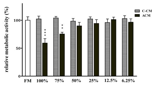
Page 290                                              VOJNOSANITETSKI PREGLED                                                Vol. 78, No 3

      Introduction                                                        study was approved by the Ethics Committee of the MMA,
                                                                          followed by informed consent from patients. The exclusion
      Dental amalgam is one of the most versatile restorative             criteria included the following: patients with malignant,
materials that has been used in dentistry for about 170 years,            autoimmune, and other chronic inflammatory diseases, as well
particularly as the first choice for restoring posterior teeth.           as those on immunosuppressive/immunomodulatory therapy.
However, it has myriads of uses, including root-end filling in            The patients included had not been treated with antibiotics for
periapical surgery 1–3. This procedure prevents the invasion of           one month prior to the PLs excision. PLs were diagnosed by
irritants from infected root canals into the periapical tissues.          clinical and radiographic criteria. No distinction was made
The advantage of using amalgam for retrograde filling for such            between age, sex, tooth type, size, and clinical presentation of
a long period of time is its self-sealing capacity, easy                  PLs. After extraction, PLs were immediately placed in a
manipulation, radio-opacity, and insolubility in tissue fluids 2.         medium consisting of RPMI-1640 (Sigma, Munich, Germany)
The preferred amalgam is a high copper-zinc-free amalgam,                 and antibiotics/antimycotics and transported to the laboratory.
composed of silver 40%–70%, tin 12%–30%, and copper
12%–24%. However, it has many disadvantages, such as the                       Isolation of cells from PLs
production of corrosive by-products 4, 5, cytotoxicity of
mercury and other dissolved metal ions, moisture sensitivity,                   The cells from PLs were isolated by a procedure that has
and staining of hard and soft tissues 1, 6, 7. There is a possibility     been previously introduced by our research group 22. Briefly,
of releasing non-resorbable scattered particles during amalgam            periapical tissue was placed in a Petri dish containing 1 mL
manipulation, which may be difficult to retrieve 2. Moreover,             RPMI-1640 medium and cut into 2–3 mm diameter pieces
amalgam does not properly seal the root end three-                        using a scalpel. The tissue was then digested for 20 min with
dimensionally, has poor marginal adaptation, and does not                 0.05% collagenase type IV (Sigma) and 0.02% DNA-ase
prevent the leakage of microorganisms and their products in               (Sigma) dissolved in RPMI-1640 medium in a cell incubator at
the peri-radicular tissue 2. However, despite these disadvan-             37 °C. After that, the tissue was pressed through a stainless-
tages and evidence of a decrease in its use, amalgam’s cost,              steel mesh using a syringe plunger, filtered, and resuspended
durability, and ease of manipulation have persuaded many                  in RPMI-1640 medium containing 1 mM EDTA. The released
dentists to continue to use it, and amalgam remains a standard            cells were pooled, washed twice by centrifugation in the RPMI
to which other materials are compared 2, 8.                               medium at room temperature (400 g for 10 min), and counted.
      The major concern for using amalgam in dentistry is its             The viability of cells, determined by Trypan Blue dye, was
cytotoxic effect, which has been documented in many human                 93% ± 3%. The cells were used for in vitro experiments. Eight
and animal cells as well as in established cell lines in vitro 6, 9–11.   periapical lesions were used to study cytokine production and
In the past few decades, however, potential systemic and local            apoptosis/necrosis. Twelve PLs containing either a larger
toxic effects have been described in vivo 2, 3, 12, 13. Patients may      number of cells (higher than 2.0 × 10⁶ cells; n = 4 PLs) or
suffer from hypersensitivity reactions to mercury or other                pooled PLs from the same donors (n = 8 PLs from 3 patients)
amalgam components. Other reactions to amalgam with a                     were used for the MTT assay. The total number of individual
variety of clinical symptoms, collectively termed “amalgam                cultures for this assay was 7.
disease,“ have been reported, including adverse
immunological effects and autoimmune phenomena 12, 14, 15.                     Preparation of conditioned medium
      Clinical and histopathological studies show that
amalgam, implanted subcutaneously or in the bone, is well                       Amalgam, consisting of the encapsulated alloy
tolerated 16, 17. This is in contrast with some studies showing           (Extracap) and mercury, was purchased from Galenika,
the capability of amalgam particles to cause periapical                   Belgrade, Serbia. One-gram (g) powder of the alloy contained
lesions 18 and to cause a cytotoxic effect on periodontal                 silver (500 mg), tin (299 mg), and cooper (201 mg). The alloy
ligament cells and periodontal fibroblasts 19–21. However, there          mass was 0.360 g, and the mercury mass was 0.400 g.
is no study investigating the effect of amalgam on human                  Amalgam specimens were prepared by triturating amalgam
periapical lesion (PL) cells in vitro, which was the main goal            alloy powder with pure mercury in an amalgamator, and after
of our study. This knowledge is important since the alloy                 the mixture, disc-form specimens, diameter around 10 mm,
communicates with the periapical tissue for a long period of              thickness about 1–2 mm, were prepared. The freshly prepared
time. Our results showed for the first time an unexpected anti-           amalgam discs were used for the preparation of amalgam
inflammatory effect of amalgam on PL cells which could be                 conditioned medium (ACM) by placing the amalgam disc in a
beneficial for PL healing.                                                glass tube containing RPMI-1640 medium with an addition of
                                                                          antibiotics/antimycotics. The mass of amalgam to the volume
                                                                          of RPMI medium was 0.2 g/mL according to ISO 10993-5 and
      Methods
                                                                          ISO 10993-12. The conditioning lasted for 3 days. Control
      Periapical lesion samples                                           CM was prepared by incubating control inert material,
                                                                          polystyrene, under the same conditions. ACM and control (C)-
     Human PLs (n = 20) were extracted during apicoectomy                 CM were supplemented with 10% FCS. There was no need for
at the Department of Oral Surgery, Clinic for Stomatology,                pH adjustment, which remained 7.4. Such prepared CM were
Military Medical Academy (MMA), Belgrade, Serbia. The                     further used for PL cell culture experiments.

                                                                                   Eraković M, et al. Vojnosanit Pregl 2021; 78(3): 289–295.
Vol. 78, No 3                                      VOJNOSANITETSKI PREGLED                                                 Page 291

     Cell cultures                                                   binding buffer, followed by incubation with Annexin-V–
                                                                     FITC and PI. The labeled cells were analyzed on a flow
      The cells isolated from PLs were cultivated in 96-wells,       cytometer (Partec, Cube 6). Annexin-V-FITC+ cells were
with round-bottomed plates (ICN, Costa Mesa, CA) (1×10⁵              recognized as primary apoptotic cells (early phase of
cells/well, 200 µL) in the complete culture medium consisted         apoptosis), PI+ cells were primary necrotic cells, whereas
of RPMI-1640 medium supplemented with 10% fetal calf                 double-positive cells were apoptotic/secondary necrotic cells
serum (FCS) (Sigma) and standard culture solutions of                (late phase of apoptosis).
antibiotics 22. The cultures were treated with different dilutions
of ACM or C-CM. Undiluted CM was considered 100% CM.
                                                                          Cytokine assays
After 24 h, the cell supernatants were collected, centrifuged,
and frozen at -70 °C until the levels of cytokines were                   The concentrations of interleukin (IL)-1β, IL-6, IL-8,
determined. The cells were used for apoptosis/necrosis assay.        and TNF-α in culture supernatants were detected by a
                                                                     FlowCytomix Microbeads Assay. This is a bead-based
     MTT assay                                                       ELISA-like assay optimized for flow cytometry, allowing the
                                                                     simultaneous detection of several cytokines in a volume of
      PL cells were cultivated in 96-well plates (1×10⁵/well;        samples (50 µL). The inflammation kit, containing
triplicates) in either fresh complete RPMI medium, different         microbeads coupled with antibodies to pro-inflammatory
dilutions of ACM or C-CM. After a 24-hour incubation                 cytokines, was purchased from Biolegend. The levels of
period, the plates were centrifuged, and the medium was              cytokines were determined by constructing standard curves
carefully removed. The solution of 3-[4,5-dimethyl-2-                based on the known concentration of these cytokines.
thiazolyl]-2,5-diphenyl tetrazolium bromide (MTT) (Sigma)
(100 μL/well, final concentration 100 μg/mL), was added.                  Statistical analysis
Wells with an MTT solution without cells served as blank
controls. The plates were incubated with MTT for 3 hours in                The Studentʼs t-test was used for comparison of parametric
an incubator at 37 °C. Dissolution of formazan was done by           variables between two groups. The Friedmanʼs test (paired one-
incubating the MTT-treated cultures with 0.1N HCl/10%                way ANOVA) was used for comparison between groups for
SDS (sodium dodecyl sulphate) (100 μL/well) overnight.               non-parametric variables with Dunn's multiple comparison post-
The next day, the optical density (OD) of the developed              test. The values of p < 0.05 were considered to be statistically
colour was read atw 570/650 nm (ELISA reader, Behring II).           significant. Software SPSS version 23.0 (IBM, Armonk, New
The results were expressed as the relative metabolic activity        York, USA) was used to analyze the data.
compared to the metabolic activity of control cultures.
      The relative metabolic activity was calculated as                   Results
follows: metabolic activity (%) = (OD of cultures with
ACM/OD of cultures with control fresh medium) × 100.                      The first aim of this study was to examine the
                                                                     cytotoxicity of ACM on PL cells in culture. By using the
     Apoptosis/necrosis assay                                        MTT test (Figure 1), we showed that only concentrated
                                                                     (100%) and 75% ACM significantly reduced the viability of
      Apoptosis/necrosis was detected by Annexin-V–                  PL cells (p < 0.001 and p < 0.01, respectively). The
fluorescein isothiocyanate (FITC) and Propidium iodide (PI)          cytotoxicity was due to the induction of apoptosis (Figures
staining kit (R&D), following the manufacturer’s protocol.           2A and 2B). Figure 2B shows that ACM increased the
Briefly, cultivated PL cells were collected, washed with             proportion of late apoptotic/secondary necrotic cells.

                 Fig. 1 ‒ Cytotoxicity effect of amalgam on periapical lesion cells (PL) in culture. PL cells,
                 prepared as described in Materials and methods, were cultured with different dilutions of
                amalgam conditioned medium (ACM) for 24 hours. The viability of PL cells was determined
                  by the MTT test, as described. Values are given as mean ± SD (n = 7 cultures) of relative
                      metabolic activity of cells. **p < 0.01; ***p < 0.001 compared to control cultures.
                               FM – fresh medium; C-CM – control-conditioning medium.

Eraković M, et al. Vojnosanit Pregl 2021; 78(3): 289–295.
Page 292 VOJNOSANITETSKI PREGLED Vol. 78, No 3
Fig. 2 ‒ Effect of amalgam on apoptosis of periapical lesion (PL) cells in culture. PL
cells, prepared as described in Materials and methods, were cultured with different
dilutions of amalgam conditioned medium (ACM) for 24 hours. The apoptosis of PL
cells was determined by the Annexin V-FITC/ PI assay, as described.
A) Values are given as mean ± SD (n = 8 cultures) of apoptotic cells
(*p < 0.05; **p < 0.01 compared to control cultures).
B) Representative histograms showing that ACM accelerate apoptosis of PL cells,
manifested by an increase of late apoptotic/secondary necrotic cells.
CM ‒ control medium.
The second aim was to investigate the effect of ACM and 25% concentrations of ACM suppressed the production
on the production of pro-inflammatory cytokines (IL-1β, of all four cytokines dose-dependently (Figure 3), whereas
TNFα, IL-6, and IL-8) by PL cells. We used non-cytotoxic the 12.5% concentration did not show any modulatory effect
concentrations (50%, 25%, and 12.5%) of ACM. The 50% (data not shown).
Fig. 3 ‒ Effect of amalgam on the levels of pro-inflammatory cytokines in the culture of
periapical lesion (PL) cells. PL cells, prepared as described in Materials and methods, were
cultured with different dilutions of amalgam conditioned medium (ACM) for 24 hours. The
levels of pro-inflammatory cytokines in culture supernatants were determined by Flow Cytomix
Microbeads Assay. Values are given as mean ± SD (n= 8 cultures) levels of cytokines (*p < 0.05;
**p < 0.01; ***p < 0.001 compared to control cultures or compared to 50% ACM, indicated by
corresponding bars). CM ‒ control medium.
Eraković M, et al. Vojnosanit Pregl 2021; 78(3): 289–295.Vol. 78, No 3 VOJNOSANITETSKI PREGLED Page 293
Discussion are believed to exist due to the high reactivity of mercury
species toward thiol-groups and other functional groups,
The first aim of this study was to examine the notably in proteins 27. It has been shown that both organic
cytotoxicity in vitro of a copper-zinc-free amalgam, which is and inorganic mercury induce apoptosis of different cells,
the oldest root-end filling material in apical surgery. including human lymphocytes 27, 28.
Apicotomy is a common procedure for removing periapical The second part of this study was related to the effect of
lesions (granuloma or cysts) when the conventional ACM on the production of pro-inflammatory cytokines by
endodontic treatment is not efficacious. Amalgam is still PL cells. We tested non-cytotoxic concentrations of ACM
used for this purpose because of its self-sealing capacity, because toxic concentrations would not be relevant for a
radio-opacity, insolubility in tissue fluids, and low price. proper conclusion, partly due to the spontaneous release of
However, since amalgam does not properly seal the root-end cytokines from dead cells. We observed an unexpected result
three-dimensionally, has poor marginal adaptation, and does where ACM at non-cytotoxic concentrations significantly
not prevent the leakage of microorganisms in the peri- inhibited the secretion of pro-inflammatory cytokines (IL-1β,
radicular tissue successfully 1–3, 23, we hypothesized that TNFα, IL-6, and IL-8). Therefore, our hypothesis was
amalgam, due to its cytotoxic effect, could aggravate rejected.
periapical inflammation. Therefore, cells isolated from PLs, The anti-inflammatory effect of ACM is contrary to the
which are dominantly composed of infiltrating inflammatory data published on the proinflammatory effect of amalgam
cells 22, 24, were the most suitable target to test this particles which could induce the PL development if released
hypothesis, and this was our original approach. into the periapical tissue during endodontic surgery 18.
Before starting with crucial experiments, it was Similarly, amalgam has been found to cause an inflammatory
necessary to determine the cytotoxicity of amalgam by using response in the dental pulp, which is transitory and
this culture model. Up to now, many different tests have significantly decreased in due time 29. These differences
been used for assessing amalgam cytotoxicity, but MTT, (pro-inflammatory versus anti-inflammatory properties of
based on the evaluation of cellular metabolic activity, is the amalgam) can be explained by the difference in setting
most acceptable as a first screening assay 6. It is known that experiments. Namely, cytotoxic effects of amalgam on
amalgam causes cytotoxicity either in direct contact with periodontal tissue in vivo can provoke an inflammatory
examined cells or indirectly by metallic ions released from reaction due to direct contact, where, in the vicinity of the
the alloy 2, 6, 9. We decided to study the effect of amalgam alloy, relatively high concentrations of cytotoxic metallic
indirectly by analyzing the effect of ACM in which its ions can be released. This effect dominates over anti-
leachable products are present and which are considered inflammatory effects seen at non-cytotoxic concentrations of
dominant cytotoxic factors 9, 25. The study was conducted leachable amalgam components.
exactly as recommended by the ISO 10993-5 standard. We No one has ever published a study related to amalgam
showed that only high concentrations of ACM (concentrated nor examined the changes of multiple pro-inflammatory and
and 75%) were cytotoxic for PL cells due to apoptosis other cytokines. The most relevant paper is the one published
induction, suggesting that amalgam is generally cytotoxic by Schedle et al. 30, who investigated the effects of dental
alloy as similarly shown on other target cells. A relatively amalgam on cytokine production by human peripheral blood
high proportion of apoptotic cells were also observed in mononuclear cells (PBMC) from healthy donors. To induce
control PL cell cultures, and the most sensitive cells were cytokine production, they stimulated PBMC in culture with
granulocytes, followed by macrophages, whereas lymphoid lipopolysaccharide, phytohemagglutinin, or staphylococcal
cells were more resistant (data not shown). These enterotoxin A in the presence of fresh amalgam, aged
observations are in line with the already known facts about amalgam, or ACM prepared from fresh amalgam. They
the high apoptotic rate of extravasated neutrophils as showed that freshly prepared amalgam, as well as ACM,
terminally differentiated cells 26. reduced the production of interferon-γ (IFN-γ) and IL-10 but
We did not examine the concentrations of released ions increased the levels of TNF-α. Both fresh amalgam and
from amalgam because this has been extensively investigated ACM had no effects on the levels of IL-2, IL-6, or
and published 5, 6. In fact, all metal ions can be released in granulocyte-macrophage colony-stimulating factor. Amal-
CM from amalgam, such as mercury, silver, copper, and thin. gam aged for 6 weeks did not modulate the concentrations of
Out of them, cooper is the most cytotoxic, but it can be any of the above cytokines. To investigate which heavy
hypothesized that other ions act synergistically in inducing metal cations released from amalgam caused the observed
cytotoxicity 1, 2, 5, 6, 9. This hypothesis was based on previous immunomodulatory effects, Cu2+, Hg2+, and Sn2+, which
publications which thoroughly investigated the release and were detected in ACM, were added as salts to PBMC
cytotoxicity of metal ions from amalgams of different cultures. Cu2+ and Hg2+ decreased the IFN-γ and IL-10
composition. In this context, Kaga et al. 9 have demonstrated levels. However, Hg2+ increased TNF-α concentrations,
that pure copper showed the highest cytotoxicity among the whereas Sn2+ had no modulatory effect.
metals tested in zinc-free amalgams. Silver and mercury It is evident that our results, showing a decrease in
showed reduced cytotoxicity, while tin was non-cytotoxic. In TNF-α production, are opposite. The difference could be due
contrast, zinc-containing amalgams are more cytotoxic due to the following reasons, respectively: different concen-
to the easy release of Zn ions. The toxic effects of mercury trations of ACM (concentrated vs. diluted ACM); different
Eraković M, et al. Vojnosanit Pregl 2021; 78(3): 289–295.Page 294 VOJNOSANITETSKI PREGLED Vol. 78, No 3
cells (stimulated PBMC vs. non-stimulated PL cells); Th-17 cells, by producing interferon-γ (IFN-γ) and IL-17,
different mass/volume ratio for ACM preparation (1.92 g/mL respectively, are involved in the progression of PLs and
vs. 0.2 g/mL); different incubation time for cell cultures (48 bone destruction, whereas T-helper 2 (Th2) cytokines, such
h vs. 24 h). Some other studies investigated the effect of as interleukin 4 (IL-4), IL-5, IL-10, and IL-33, are involved
mercury. In this context, Soleo et al. 31 showed an increase in in the humoral immune response and attenuation of the
the number of CD4+ cells in peripheral blood of subjects tissue damage 22, 35, 36. Therefore, further experiments
exposed to mercury from dental amalgam together with a investigating the effect of amalgam on this panel of
decrease of serum IL-8 levels. Podzimek et al. 32 examined cytokines could make a much better conclusion.
cytokine production (IL-1β, IL-4, IL-6, TNF-α, and IFN-γ)
by human lymphocytes in cultures treated with mercury and Conclusion
found increased production of TNF-α and IFN-γ. Ilday et
al. 33 observed reduced clinical periodontal findings in By using inflammatory cells isolated from human PL,
patients after overhang amalgam restoration removal, but we showed, for the first time, a potent anti-inflammatory
these findings did not correlate with the changes in the levels effect of non-cytotoxic concentrations of ACM. This finding
of IL-6, IL-8, and TNF-α in the gingival crevicular fluid. is in contrast with the previous findings, which state that
This is in contrast with another study, published previously, particular amalgam particles released during retrograde
which showed that removal of dental amalgam restorations filling can cause chronic apical periodontitis. Our results
was associated with decreased concentrations of Th1-type suggest that, in contrast to the high release of toxic ions from
pro-inflammatory cytokines in serum, supporting the amalgam, slow release of leachable components from this
hypothesis that amalgam could be responsible for stimulating amalgam, by down-modulating the production of pro-
the Th1-type response in vivo 34. inflammatory cytokines, may control аn excessive
It is known that cytokines play a key role in the inflammation and promote PL healing.
pathogenesis of PLs 35. Pro-inflammatory cytokines, such
as IL-1, IL-6, IL-8, and TNF-α, orchestrate the recruitment Acknowledgement
and activation of innate immune cells, presumably
neutrophil granulocytes and monocytes in the early This work was supported by the Ministry of Education,
inflammatory phase and T and B cells in the later Science and Technological Development, Republic of Serbia
inflammatory phase, respectively. In this context, cytokines (grant No: OI 175102) and grants from the Ministry of
of T cells are the main controllers of the Defence, Republic of Serbia (MFVMA/7/17-19 and
immune/inflammatory reactions. T-helper 1 (Th1) cells and MFVMA/9/16-18).
R E F E R E N C E S
1. Mahler DB. The high-copper dental amalgam alloys. J Dent Res 12. Enwonwu C. Potential health hazard of use of mercury in den-
1997; 76(1): 537‒41. tistry: critical review of the literature. Environ Res 1987; 42(1):
2. Bhagat K, Goel M, Bhagat N. Root End Filling Materials and Re- 257‒74.
cent Advances: A Review. EC Dent Sci 2017; 12: 46‒57. 13. Moszczyński P. Mercury compounds and the immune system: a
3. Mjor IA, Jokstad A, Qvist V. Longevity of posterior restora- review. Int J Occup Med Environ Health 1997; 10(3): 247‒58.
tions. Int Dent J 1990; 40(1): 11‒7. 14. Enestrom S, Hultman P. Does amalgam affect the immune sys-
4. Hohenfeldt PR, Aurelio JA, Gerstein H. Electrochemical corrosion tem? A controversial issue. Int Arch Allergy Immunol 1995;
in the failure of apical amalgam. Report of two cases. 106(3): 180‒203.
Oral Surg Oral Med Oral Pathol Oral Radiol Endod 1985; 15. Meurman JH, Porko C, Murtomaa H. Patients complaining about
60(6): 658‒60. amalgam‐related symptoms suffer more often from illnesses
5. Pleva J. Corrosion and mercury release from dental amalgam. J and chronic craniofacial pain than their controls. Eur J Oral
Orthomol Med 1989; 4(3): 141‒58. Sci 1990; 98(2): 167‒72.
6. Kaga M, Seale N, Hanawa T, Ferracane J, Waite D, Okabe T. Cyto- 16. Flanders DH, James GA, Burch B, Dockum N. Comparative his-
toxicity of amalgams, alloys, and their elements and phases. topathologic study of zinc-free amalgram and Cavit in connec-
Dent Mater 1991; 7(1): 68‒72. tive tissue of the rat. J Endod 1975; 1(2): 56‒9.
7. Bodrumlu E. Biocompatibility of retrograde root filling materi- 17. Torabinejad M, Hong CU, Pitt Ford TR, Kaiyawasam SP. Tissue
als: a review. Aust Endod J 2008; 34(1): 30‒5. reaction to implanted super-EBA and mineral trioxide aggre-
8. Osborne JW, Norman RD, Gale EN. A 14-year clinical assess- gate in the mandible of guinea pigs: a preliminary report. J En-
ment of 12 amalgam alloys. Quintessence Int 1991; 22(11): dod 1995; 21(11): 569‒71.
857‒64. 18. Nair PN. On the causes of persistent apical periodontitis: a re-
9. Kaga M, Seale N, Hanawa T, Ferracane J, Okabe T. Cytotoxicity view. Int Endod J 2006; 39(4): 249‒81.
of amalgams. J Dent Res 1988; 67(9): 1221‒4. 19. Keiser K, Johnson CC, Tipton DA. Cytotoxicity of mineral triox-
10. Leirskar J. On the mechanism of cytotoxicity of silver and ide aggregate using human periodontal ligament fibroblasts. J
copper amalgams in a cell culture system. Eur J Oral Sci 1974; Endod 2000; 26(5): 288‒91.
82(1): 74‒81. 20. Tai KW, Chang YC. Cytotoxicity evaluation of perforation re-
11. Meryon S. The effect of zinc on the biocompatibility of dental pair materials on human periodontal ligament cells in vitro. J
amalgams in vitro. Biomaterials 1984; 5(5): 293‒7. Endod 2000; 26(7): 395‒7.
Eraković M, et al. Vojnosanit Pregl 2021; 78(3): 289–295.Vol. 78, No 3 VOJNOSANITETSKI PREGLED Page 295
21. Lin CP, Chen YJ, Lee YL, Wang JS, Chang MC, Lan WH, et al. Ef- 30. Schedle A, Rausch‐Fan XH, Samorapoompichit P, Franz A, Leutme-
fects of root-end filling materials and eugenol on mitochondrial zer F, Spittler A, et al. Effects of dental amalgam and heavy
dehydrogenase activity and cytotoxicity to human periodontal metal cations on cytokine production by peripheral blood
ligament fibroblasts. J Biomed Mater Res 2004; 71(2): 429‒40. mononuclear cells in vitro. J Biomed Mater Res 1998; 42(1):
22. Colic M, Gazivoda D, Vucevic D, Vasilijic S, Rudolf R, Lukic A. 76‒84.
Proinflammatory and immunoregulatory mechanisms in peri- 31. Soleo L, Colosio C, Alinovi R, Guarneri D, Russo A, Lovreglio P, et
apical lesions. Mol Immunol 2009; 47(1): 101‒13. al. Immunologic effects of exposure to low levels of inorganic
23. Letzel H, van 't Hof MA, Vrijhoef MM, Marshall GW Jr, Marshall mercury. Med Lav 2002; 93(3): 225‒32.
SJ. A controlled clinical study of amalgam restorations: survival, 32. Podzimek S, Tomka M, Nemeth T, Himmlova L, Matucha P, Pro-
failures, and causes of failure. Dent Mater 1989; 5(2): 115‒21. chazkova J. Influence of metals on cytokines production in
24. Čolić M, Lukić A, Vučević D, Milosavljević P, Majstorović I, Marja- connection with successful implantation therapy in dentistry.
nović M, et al. Correlation between phenotypic characteristics Neuro Endocrinol Lett 2010; 31(5): 657‒62.
of mononuclear cells isolated from human periapical lesions 33. Ilday NO, Celik N, Dilsiz A, Alp HH, Aydin T, Seven N, et al.
and their in vitro production of Th1 and Th2 cytokines. Arch The effects of overhang amalgam restoration on levels of cy-
Oral Biol 2006; 51(12): 1120‒30. tokines, gingival crevicular fluid volume and some periodontal
25. Ferracane JL, Mafiana P, Cooper C, Okabe T. Time-dependent parameters. Am J Dent 2016; 29(5): 266‒70.
dissolution of amalgams into saline solution. J Dent Res 1987; 34. Bjorkman L, Brokstad KA, Moen K, Jonsson R. Minor changes in
66(8): 1331‒5. serum levels of cytokines after removal of amalgam restora-
26. Greenlee-Wacker MC. Clearance of apoptotic neutrophils and res- tions. Toxicol Lett 2012; 211(2): 120‒5.
olution of inflammation. Immunol Rev 2016; 273(1): 357‒70. 35. Braz-Silva PH, Bergamini ML, Mardegan AP, De Rosa CS, Hasseus
27. Schwenk M, Klein R, Templeton DM. Immunological effects of B, Jonasson P. Inflammatory profile of chronic apical periodon-
mercury (IUPAC Technical Report). Pure Appl Chem 2009; titis: a literature review. Acta Odontol Scand 2019; 77(3):
81(1): 153‒67. 173‒80.
28. Shenker BJ, Berthold P, Decker S, Mayro J, Rooney C, Vitale L, et al. 36. Naufel AO, Aguiar MCF, Madeira FM, Abreu LG. Treg and
Immunotoxic Effects of Mercuric Compounds on Human Th17 cells in inflammatory periapical disease: a systematic re-
Lymphocytes and Monocytes. II. Alterations in Cell Viability. view. Braz Oral Res 2017; 31: e103.
Immunopharmacol Immunotoxicol 1992; 14(3): 555‒77.
29. Baek SH, Plenk Jr H, Kim S. Periapical tissue responses and Received on February 25, 2019.
cementum regeneration with amalgam, SuperEBA, and MTA Accepted on March 19, 2019.
as root-end filling materials. J Endod 2005; 31(6): 444‒9. Online First April, 2019.
Eraković M, et al. Vojnosanit Pregl 2021; 78(3): 289–295.You can also read