Comparative Effect of Aspirin, Meloxicam and Terminalia catappa Leaf Extract on Serum Levels of Some Inflammatory Markers in Alloxan Induced ...
←
→
Page content transcription
If your browser does not render page correctly, please read the page content below

Asian Journal of Research in Biochemistry

                            4(1): 1-10, 2019 Article no.AJRB.47442

       Comparative Effect of Aspirin, Meloxicam and
  Terminalia catappa Leaf Extract on Serum Levels of
      Some Inflammatory Markers in Alloxan Induced
                                       Diabetic Rats
                                   Ezekiel E. Ben1, Asuquo E. Asuquo1* and Daniel U. Owu2
              1
             Department of Physiology, Faculty of Basic Medical Sciences, University of Uyo, Nigeria.
         2
          Department of Physiology, Faculty of Basic Medical Sciences, University of Calabar, Nigeria.

                                                                                             Authors’ contributions

        This work was carried out in collaboration among all authors. Author EEB designed the study,
       performed the statistical analysis, wrote the protocol, and wrote the first draft of the manuscript.
       Authors AEA and DUO managed the analyses of the study. Author AEA managed the literature
                                          searches. All authors read and approved the final manuscript.

                                                                                                  Article Information

                                                                                     DOI: 10.9734/AJRB/2019/v4i130058
                                                                                                                 Editor(s):
      (1) Dr. Khadiga Mohamed Abu-Zied, Professor, Department of Photochemistry, National Research Centre, Cairo, Egypt.
                                                                                                               Reviewers:
                                                    (1) Sharafudeen D. Abubakar, Ahmadu Bello University, Zaria, Nigeria.
                                                               (2) Md. Riaj Mahamud, University of Oklahoma HSC, USA.
                                            Complete Peer review History: http://www.sdiarticle3.com/review-history/47442

                                                                                        Received 24 October 2018
                                                                                       Accepted 13 February 2019
  Original Research Article
                                                                                         Published 04 March 2019

ABSTRACT

Background: The association between diabetes mellitus and inflammation is established but the
use of non-steroidal anti-inflammatory drugs is not without some health risk.
Aim: The study was aimed at comparing the levels of some inflammatory biomarkers in diabetic rats
treated with aqueous leaf extract of Terminalia catappa, non steroidal anti-inflammatory drugs
(NSAIDs) and exogenous insulin.
Materials and Methods: Thirty six (36) Wistar rats were assigned to 6 groups of 6 animals each.
Group 1 and 2 served as normal and diabetic controls and received orally 5ml/kg body weight of
distilled water. Group 3 was diabetic treated orally with 130mg/kg body weight of aqueous leaf
extract of Terminalia catappa. And groups 4, 5 and 6 were administered orally with aspirin
(30mg/kg), meloxicam (2mg/kg) and 0.75U/kg body weight of insulin subcutaneously. Diabetes was
induced with intraperitoneal injection of 150mg/kg body weight of alloxan solution and diabetes
confirmed after 72 hours with blood glucose levels ≥200mg/dl. The experiment lasted for 14 days
_____________________________________________________________________________________________________

*Corresponding author: E-mail: asuquoetim59@yahoo.com;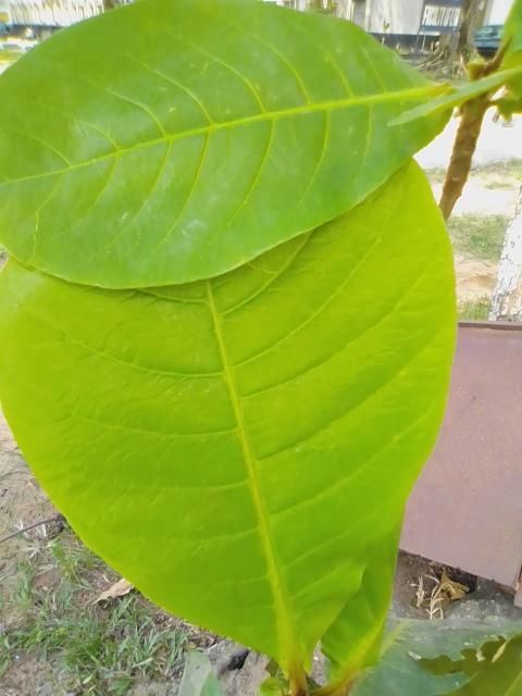
Ben et al.; AJRB, 4(1): 1-10, 2019; Article no.AJRB.47442

and blood was collected by cardiac puncture for serum analysis of C-reactive protein, Interleukin-6
and Fibrinogen by ELISA method.
Results: The results showed significant (P
Ben et al.; AJRB, 4(1): 1-10, 2019; Article no.AJRB.47442

Health Organization encouraged the option of              The animals were housed in a well ventilated
exploring the efficacy of medicinal plants in the         cage in the animal house and they were allowed
treatment of diseases [18]. Among many                    to acclimatize for two weeks and maintained in a
ethnopharmacological plants, Terminalia catappa           24 hours dark and light cycle. The animals were
(Combretaceae) is useful in treatment of different        fed with standard pellets (from Guinea Feeds,
ailments [19]. The plant is also called Almond            Plc Nigeria) and have access to water ad libitum.
tree or Tropical Almond and found commonly in
the Tropics [20]. Extracts from the leaves and            2.3 Induction of Diabetes
bark of this plant have been reported as
antioxidant [21], anti-diabetic [22] and anti-            Diabetes was induced by intraperitoneal injection
inflammatory      [23].   The    current    adjunct       of alloxan monohydrate at a dose of 150mg/Kg
therapeutic target for diabetes is on inflammation        body weight [24,25,26]. The animals were
and scientific exploration on anti-inflammatory           assessed for development of diabetes after 72
potentials of this plant becomes necessary to             hours [27] by obtaining blood sample from the tip
investigate the possible link to its anti-diabetic        of the tail. The blood sample was dropped into
property and compare it to the actions of non-            glucose strip to measure the glucose level using
steroidal anti-inflammatory drugs. This research          a glucometer (One Touch Ultra, Life Scan Inc,
aims at comparing the effect of Terminalia                U.S.A). Blood glucose of ≥200mg/dl was
catappa leaf extract and non steroidal anti-              considered diabetic (normal range of blood
inflammatory drugs (NSAIDs) on some                       glucose in rat is 80–120mg/dl) and were used for
inflammatory biomarkers in diabetic rats.                 the experiments [25,27].

2. MATERIALS AND METHODS                                  2.4 Experimental Design
2.1 Preparation of Plant Extract                          The experimental animals were randomly
                                                          distributed into Six (6) groups of six (n=6) rats
Fresh leaves of Terminalia catappa were                   per group as follows:
collected at the premises of the University of
Calabar and the area was free of pesticides and           Group 1: Control group administered with only
other contaminants. The photographs of the leaf           distilled water orally at a dose of 5ml/kg body
as taken directly are shown in plates 1 and 2 in          weight.
the result section of this article. The leaves were       Group 2: Diabetic group administered with only
authenticated by a botanist (Mrs E. G. Udoma) at          distilled water orally at a dose of 5ml/Kg body
the Department of Botany and Ecological                   weight.
studies, University of Calabar, Nigeria and it is         Group 3: Diabetic group treated with aqueous
documented in the herbarium with voucher                  leaf extract of Terminalia catappa at a dose of
number UUPH 22(a). The leaves were then                   130mg/Kg body weight by oral administration.
washed with clean water to remove debris. The             Group 4: Diabetic group treated orally with
water was blotted out and kept overnight at room          30mg/kg body weight of aspirin.
temperature to dry up. The clean leaves were              Group 5: Diabetic group treated orally with
pulverized and 5000g of the pulverized leaves             2mg/kg body weight of meloxicam.
were soaked in 5 litres of deionised water for 18         Group 6: Diabetic group treated with exogenous
hours. The mixture was filtered using muslin              Insulin at a dose of 0.75U/Kg body weight by
cloth and evaporated to dryness using                     subcutaneous administration.
thermostatic water bath at 45oC until a semi solid
paste of 204.18g of the extract was obtained              2.5 Acute Toxicity Test (LD50)
after evaporation representing a percentage yield
of 4.08%. The extract was stored in refrigerator          The toxicity test of the Terminalia catappa
for later use.                                            aqueous leaf extract was carried out by Lorke’s
                                                          method [28] on mice weighing 15 and 25g. The
2.2 Preparation of Experimental Animal                    animals were randomly divided into seven (7)
                                                          groups of six mice per group. The extract was
Healthy adult male albino Wistar rats weighting           respectively administered to groups 1 to 7 at a
between 150-200g were used for the study. The             dose of 500mg//Kg, 800mg/Kg, 1000mg/Kg,
animals were procured from the animal house,              1200mg/Kg,        1400mg/Kg,       1500mg/Kg,
Department of Physiology, Faculty of Basic                1600mg/Kg body weight intraperitoneally. The
Medical Sciences, University of Calabar, Nigeria.         animals were observed for physical signs of

                                                      3
Ben et al.; AJRB, 4(1): 1-10, 2019; Article no.AJRB.47442
toxicity such as gasping, writhing, palpitation and specific antibody. The absorbance or optical
death after 24 hours. The median leathal dose density (OD) was measured
(LD50) was calculated as square root of spectrophotometrically at a wavelength of 450nm
maximum (most tolerable) dose producing 0% and readings are obtained using Microtiter Plate
mortality (x) and minimum (least tolerable) dose Reader and the Optical Density value is
producing 100% mortality (y) using the formula: proportional to the concentration of Interleukin-6.
LD50 = √xy.
2.9 Rat Fibrinogen Assay
2.6 Ethical Approval
Whole blood was collected into plain sample
The experimental protocol received full approval bottle and used for determination of fibrinogen.
from Faculty Animal Research Ethics Committee The fibrinogen level was determined by Enzyme
(FAREC-FBMS), Faculty of Basic Medical Linked Immunosorbent Assay (ELISA) method
Sciences University of Calabar, Nigeria with [31]. Standards or samples were added to the
approval number 021PY30417. appropriate microelisa strip plate wells and
combined to the fibrinogen antibody. The
2.7 Rat C-reactive Protein Assay absorbance or optical density (OD) was
measured by spectrophotometry at wavelength
Serum C-reactive protein level was analysed by of 450nm and the result read on a microplate
ELISA method. Commercial rat C-reactive immediately. The Optical Density value is
protein analysis kit using Sandwich-ELISA proportional to the concentration of fibrinogen.
(Enzyme Linked Immunosorbent Assay) method The assay was performed at room temperature
[29]. Standards or samples were added to the (18-25°C).
appropriate microelisa strip plate wells and
combined to the specific antibody. The 2.10 Statistical Analysis
absorbance or optical density (OD) is measured
spectrophotometrically at a wavelength of 450nm The data obtained from the result was subjected
and readings are obtained using Microtiter Plate to statistical testing using one way ANOVA
Reader and the Optical Density value is followed by Tukey test using Graph Pad Prisms
proportional to the concentration of C-reactive software 6.0. Data was expressed as mean +
protein. standard error of mean (SEM). Results with
values of P< 0.05 were considered significant.
2.8 Rat Interleukin-6 Assay
3. RESULTS
Serum from the blood of the experimental
animals was used to analyse the Interleukin-6. 3.1 Photograph of the Terminalia catappa
Interleukin-6 assay was carried out with Leaf
commercially prepared rat Interleukin-6 analysis
kit using Sandwich-ELISA (Enzyme Linked Plate 1 shows the leaves of Terminalia cattapa
Immunosorbent Assay) method [30]. Standards clustered at the end of the twigs and plate 2
or samples are added to the appropriate shows a smooth coriaceous single green
microelisa strip plate wells and combined to the Terminalia catappa leaf.
Plate 1 Plate 2
Source: Direct photograph taken by Ben and colleagues, 7/2/2019
4Ben et al.; AJRB, 4(1): 1-10, 2019; Article no.AJRB.47442
3.2 Toxicity Test (Ld50) which was significant when compared to diabetic
group. The mean value in the insulin treated
The result of the acute toxicity test showed that diabetic group also showed significant (PBen et al.; AJRB, 4(1): 1-10, 2019; Article no.AJRB.47442
180
160
*
140
Interleukin-6 level (ng/ml)
120
100 a a a
a
80
60
40
20
0
Control Diabetic Diabetic + Diabetic Diabetic + Diabetic +
Extract +Aspirin Meloxicam Insulin
Groups
Fig. 2. Serum level of Interleukin-6 in experimental group compared with control and
diabetic control groups
Values are mean ± SEM. * =Significant change compared with control group (P< 0.05)
a=Significant change compared with diabetic control group (P< 0.05)
400
350
Fibrinogen level (mg/ml)
*
300
250
200
150
100
50
0
Control Diabetic Diabetic + Diabetic Diabetic + Diabetic +
Extract +Aspirin Meloxicam Insulin
Groups
Fig. 3. Serum level of fibrinogen in experimental group compared with control and diabetic
control groups
Values are mean ± SEM. * =Significant change compared with control group (P< 0.05)
a=Significant change compared with diabetic control group (P< 0.05)
and insulin in their respective diabetic treated similar pattern of being increased significantly in
groups. These results reveal that the reduction in the diabetic group and was lowered significantly
C-reactive protein and Interleukin-6 by the by the extract, aspirin, meloxicam and insulin,
extract was higher than that of aspirin and with insulin having higher effect compared to the
meloxicam. Moreover, fibrinogen level follows a other agents. Both aspirin and meloxicam are
6Ben et al.; AJRB, 4(1): 1-10, 2019; Article no.AJRB.47442
non-steroidal anti-inflammatory drugs used in the effect of insulin as shown in this study on
treatment of inflammation besides other functions fibrinogen may suggest the usefulness of insulin
as analgesia and anti-pyretic. These anti- in thrombosis more than the non steroidal anti-
inflammatory drugs inactivate cyclooxygenase inflammatory drugs.
(COX) enzymes with meloxicam being more
COX-2 specific blocker than COX-1 [35]. In view of these findings, NSAIDs such as aspirin
Blocking COX enzymes leads to suppressed or meloxicam adjunct to insulin therapy in
production of prostaglandins and thromboxanes diabetes may be advocated. But reports on
[36] as the mechanism of reducing inflammation. various side effects of these drugs are
documented. Gastrointestinal toxicity and
The level of reduction of these biomarkers varies increased risk of cardiovascular events such as
between the two anti-inflammatory drugs. It was heart attack and stroke are associated with many
observed that C-reactive protein was more non steroidal anti-inflammatory drugs [47].
reduced by aspirin than meloxicam while the Meloxicam is specifically contraindicated in
level of reduction of Interleukin-6 and fibrinogen persons with hypertension and diabetes despite
were similar between the two drugs. The its modification for gastrointestinal tolerability
irreversible effect of aspirin on COX-1 enzyme thereby ruling out its use in diabetes condition.
[37] makes the drug different from other anti- Moreover, recent clinical findings have revealed
inflammatory drugs in addition to regular the development of NSAIDs induced enteropathy
pathways such as the formation of nitric oxide [48,49]. It is reported that the permeability of the
free radicals in the body as independent small intestinal mucosa in a single dose of
mechanism in reducing inflammation [38], NSAID is within 12 hours and this is by
uncoupling of oxidative phosphorylation in uncoupling of the mitochondrial phosphorylation
mitochondria [39] and signal modulation through which breaks the integrity of the mucosa junction
NF-kB [40]. The present study cannot explain if normally protected by COX1 and COX 2 [50,51].
the specificity in inactivation of the COX However, this enteropathy is asymptomatic and
isoforms; COX-1 and COX-2 may play a role in physicians continue to treat with this drug
the effectiveness of aspirin in the reduction of the because the side effect is vague [52].
acute phase inflammatory marker, C-reactive
protein compared to meloxicam despite the The reduction of these biomarkers of
similarity in the reduction of pro-inflammatory inflammation by the aqueous leaf extract
cytokine; Interleukin-6. suggests that the extract is in agreement with
current position that natural treatments can help
It was observed that insulin reduced C-reactive to reduce inflammation in the blood [53]. But the
protein and Interleukin-6 and fibrinogen with higher level of reduction observed may suggest
comparatively higher effect on fibrinogen than that the extract activates the pathways used by
the other agents. Following the established these agents and other pathways not known to
association between hyperglycaemia and express its anti-inflammatory actions. However
inflammation, the anti-inflammatory effect of this assertion requires a more empirical
insulin will first involve lowering of blood glucose investigation to ascertain the signalling
and this might show existing positive link pathway(s) involved.
between hyperglycaemia, fibrinogen level and
risk of cardiovascular disease in diabetes. It is 5. CONCLUSION
reported that insulin suppresses important
inflammatory mediators namely: Intercellular The serum level of C-reactive protein,
adhesion molecule-1(I-CAM-1), Monocyte Interleukin-6 and fibrinogen were significantly
Chemoattractant Protein-1 (MCP-1) expression, reduced by the aqueous leaf extract of
necrosis factor NF-kB binding [41,42] in human Terminalia catappa compared to aspirin and
aortic endothelial cells in vitro via nitric oxide meloxicam. And the effect of aspirin was more
signalling pathway [43,44,45]. It is suggested that marked on C-reactive protein than meloxicam
Toll like receptors (TLRs) which plays an while the two affected Interleukin-6 and
important role in tissue inflammation and damage fibrinogen similarly. Therefore aqueous leaf
such as cardiac ischemia and atherosclerosis extract of Terminalia catappa may be a potent
[46] is also suppressed by insulin. Although the anti-inflammatory agent than non steroidal anti-
reduction of C-reactive protein and Interleukin-6 inflammatory drugs and could complement the
by insulin in this study is not as much as the non function of insulin in diabetes treatment.
steroidal anti-inflammatory drugs, the significant
7Ben et al.; AJRB, 4(1): 1-10, 2019; Article no.AJRB.47442
COMPETING INTERESTS and inflammation to hinder the progression
of vascular complications of diabetes.
Authors have declared that no competing Front. Physiol. 2019;9:1-18.
interests exist. 12. Zhou R, Tardivel A, Thorens B, Choi I,
Tschopp J. Thioredoxin interacting protein
REFERENCES links oxidative stress to inflammasome
activation. Nat Immunol. 2010;11:136-140.
1. Pollack R, Donath M, LeRoith D, Leibowitz 13. Isoda K, Young J, Zirlik A, MacFarlane L,
G. Anti-inflammatory Agents in the Tsuboi N, Gerdes N. Metformin inhibits
Treatment of Diabetes and Its Vascular proinflammatory responses and nuclear
Complications. Diabetes Care. 2016; factor-kappaB in human vascular wall
39(Suppl. 2):244–252. cells. Arterioscler Thromb Vasc Biol.
2. Pradhan A, Manson J, Rifai N, Buring J, 2006;26:611–617.
Ridker P. C-reactive protein, interleukin 6, 14. Lee H, Kim J, Kim H, Shong M, Ku B, Jo E.
and risk of developing type 2 diabetes Upregulated NLRP3 inflammasome
mellitus. JAMA. 2001;286:327–334. activation in patients with type 2 diabetes.
3. Han T, Sattar N, Williams K, Gonzalez- Diabetes. 2013;62:194–204.
Villalpando C, Lean M, Haffner S. 15. Foretz M, Guigas B, Bertrand L, Pollak M,
Prospective study of C-reactive protein in Viollet B. Metformin: From mechanisms of
relation to the development of diabetes action to therapies. Cell Metab. 2014;20:
and metabolic syndrome in the Mexico City 953–966.
Diabetes Study. Diabetes Care. 2002;25: 16. Hansen T, Thiel S, Wouters P,
2016–2021. Christiansen J, Van den Berghe G.
4. Sjoholm A, Nystrom T. Inflammation and Intensive insulin therapy exerts anti
the etiology of type 2 diabetes. Diabetes inflammatory effects in critically ill patients
Metab Res Rev. 2006;22:4–10. and counteracts the adverse effect of low
5. Gulhar R, Jialal I. Physiology, Acute phase mannose-binding lectin levels. J Clin
reactant. Bethesda: StatPearls Publishing Endocrinol Metab. 2003;88:1082–1088.
LLC; 2018. 17. Fowler M. Microvascular and
6. Hotamisligil G, Shargill N, Spiegelman B. macrovascuar complications of diabetes.
Adipose expression of tumor necrosis Clinical Diabetes. 2011;29(3):116-122.
factoralpha: direct role in obesity-linked 18. WHO expert committee on the use of
insulin resistance. Science. 1993;259:87– essential drugs. Criteria for selection of
91. essential drugs. The use of essential
7. Duncan B, Schmidt M, Pankow J, drugs, WHO Tech Rep Ser, WHO Geneva,
th
Ballantyne C, Couper D, Vigo A, 8 edition, 825, 1992.
Hoogeveen R, Folsom A, Heiss G. Low- 19. Koffi N, Yvette F, Noel Z. Effect of
Grade systemic Inflammation and the Aqueous extract of Terminalia catappa
development of Type 2 Diabetes. leaves on the glycaemia of rabbits. JAPS.
Diabetes. 2003;52:1799-1804. 2011;1(8):59-64.
th
8. Esposito K, Nappo F, Marfella R, Giugliano 20. Nadkarni KM. Indian Materia Medica. 4
G, Giugliano F, Ciotola M, Quagliaro L, ed. Vol 1:1205: Popular Prakashn
Ceriello A, Giugliano D. Inflammatory Boombay; 1976.
cytokine concentrations are acutely 21. Masuda T, Yonemori Y, Oyama Y, Takeda
increased by hyperglycemia in humans: T, Tanaka T. Evaluation of the antioxidant
role of oxidative stress. Circulation. activity of environmental Plants: activity of
2002;106:2067–2072. the leaf extracts from seashore plants. J
9. Bergman M. Pathophysiology of Agric Food Chem. 1999;47:1749-54.
prediabetes and treatment implications for 22. Ahmed S, Swamy V, Dhanapal G,
the prevention of type 2 diabetes mellitus. Chandrashekara V. Anti-diabetic Activity of
Endocrine. 2013;43:504–513. Terminalia catappa Linn. Leaf Extracts in
10. Alam U, Asghar O, Azmi S, Malik R. Alloxan-Induced Diabetic Rats. IJPT. 2005;
General aspects of diabetes mellitus. 4:36-39.
Handb. Clin. Neurol. 2014;126:211–222. 23. Pawar SP, Pal SC, Kasture VS. Anti-
11. Teodoro J, Nunes S, Rolo A, Reis F, inflammator y activity of Terminalia
Palmeira C. Therapeutic options targeting catappa Linn; Pharmacy Focus in the new
oxidative stress, mitochondrial dysfunction millennium abstracts. P110,1997.
8Ben et al.; AJRB, 4(1): 1-10, 2019; Article no.AJRB.47442
24. Katsumata K, Katsumata Y, Ozawa T, 37. Toth L, Muszbek L, Komoromi I.
Katsumata J. Potentiating effect of Mechanism of the irreversible inhibition of
combined usage of three sulfonylures human cyclooxygenase-1 by aspirin as
drugs on the occurrence of alloxan predicted by QM/MM calculations. J Mol
diabetes in rats. Horm Metab Res. Graph. 2013;40:90-109.
1993;25:125-126. 38. Paul-Cark M, Van Cao T, Moradi-Bidhendi
25. Kulkarni S. Commonly used drugs, their N, Cooper D, Gilrow D. 15-epilipoxin A4-
doses and nature of action in laboratory mediated induction of nitic oxide explains
rd
animals. 3 ed. Vallabh Prakashan Delhi: how aspirin inhibits acute inflammation. J
Hand book of Experimental Pharmacology. Exp Med. 2004;200(1):69-78.
2005;190-195. 39. Somasundaram S, Sigthorsson G,
26. Etuk E. Animals models for studying Simpson R, Watts J, Jacob M, Tavares I,
diabetes mellitus. Int J Agric Biol. Rafi S, Roseth A. Uncouping of Intestinal
2010;1:130-4. mitochondrial oxidative phosphorylation
27. Borgohain R, Lahon K, Das S, Gohain K. and inhibition of cyclooxygenase are
Evaluation of mechanism of anti-diabetic required for the development of NSAID-
activity of Terminalia chebula on alloxan enteropathy in the rat. Ailment Pharmacol.
and Adrenaline induced Diabetic albino Ther. 2000;14(5):639-650.
rats. IJPBS. 2012;3(3):256-266. 40. McCarty M, Block K. Pre-administration of
28. Lorke D. A new approach to practical acute high-dose salicylates, suppressors of NF-
toxicity testing. Arch Toxicol. 1993;54:275- kappaB activation, may increase the
287. chemosensitivity of many cancers: An
29. Kilpatrick J, Volanakis JE. Molecular example of proapoptotic signal modulation
genetics, structure and function of C therapy. Integr. Cancer Ther. 2006;5(3):
reactive protein. Immun. Res. 1991;10:43- 252-268.
53. 41. Kopp E, Ghosh S. Inhibition of NF-_B by
30. Swardfager W, Lanctot K, Rothenburg L, sodium salicylate and aspirin. Science.
Wong A, Cappell J, Hermann N. A meta 1994;265:956–959.
analysis of cytokines in alzheimers 42. Grilli M, Pizzi M, Memo M, Spano P.
disease. Biological Psychiatry. 2010; Neuroprotection by aspirin and sodium
68(10):930-941. salicylate through blockade of NF-_B
31. Handley DA, Hughes TE. Pharmacological activation. Science. 1996;274:1383–1385.
approaches and strategies for therapeutic 43. Aljada A, Ghanim H, Saadeh R, Dandona
modulation of fibrinogen. 1997;Throm. P. Insulin inhibits NFkappaB and MCP-1
Res. 87:1 expression in human aortic endothelial
32. Ben E, Asuquo A, Owu D. Serum levels of cels. J Clin Endocrinol Metab. 2001;86:
some Inflammatory Markers in Alloxan 450-453.
induced Diabetic rats Treated with 44. Aljada A, Saadeh R, Assian E, Ghanim H,
Terminalia catappa leaf extract and Dandona P. Insulin inhibits the expression
Exogenous Insulin; 2019. AJRIMPS; (In of intercellular adhesion molecule-1 by
press) human aortic endothelial cell stimulation of
33. Ford E. Body mass index, diabetes, and C- nitric oxide. J. Clin Endocrinol. Metab.
reactive protein among U.S. Adults. 2000;85:2572-2575.
Diabetes Care. 1999;22:1971–1977. 45. Bazzoni F, Beutler B. The tumor necrosis
34. Wu T, Dorn J, Donahue R, Sempos C, factor ligand and receptor families. N Engl
Trevisan M. Associations of serum C- J Med. 1996;334:1717-1725.
reactive protein with fasting insulin, 46. Chao W. Toll-like receptor signalling: A
glucose, and glycosylated hemoglobin. Am critical modulator of cell survival and
J Epidemiol. 2002;155:65–71. ischemic injury in the heart. Am J Physiol
35. Noble S, Balfour J. Meloxicam. Drugs. Heart Circ Physiol. 2009;296:H1-12.
1996;51(3):424-30. 47. Fanelli A, Ghisi D, Aprile P, Lapi F.
36. Patrono C, Ciabattoni G, Pinca E, Pugliese Cardiovascular and cerebrovascular risk
F, Castrucci G, De Salvo A, Satta M, with nonsteroidal anti-inflammatory drugs
Peskar B. 1980. Low dose aspirin and and cyclooxygenase 2 inhibitors: Latest
inhibition of thromboxane B2 production in evidence and clinical implications. Ther
healthy subjects. Thromb. Res. 1980;17: Adv Drug Saf. 2017;8(6):173-182.
317–327.
9Ben et al.; AJRB, 4(1): 1-10, 2019; Article no.AJRB.47442
48. Higuchi K, Umegaki E, Watanabe T, Yoda comparing rofecoxib with placebo and
Y, Morita E, Murano E, Tokioka S. Present indomethacin. Gut. 2000;47:527-32.
status and strategy of NSAIDs-induced 51. Maiden L, Thjodleifsson B, Seigal A,
small bowel injury. J Gastroenterol. Bjarnason II, Scott D, Birgisson S,
2009;44:879-88. Bjarnason I. Long-term effects of Non-
49. Endo H, Hosono K, Inamori M, Nozaki Y, steroidal anti-inflammatory drugs and
Yoneda K, Fujita K, Takahashi H. cyclooxygenase-2 selective agents on the
Characteristics of small bowel injury in small bowel: A cross-sectional capsule
symptomatic chronic low-dose aspirin enteroscopy study. Clin Gastroenterol
users: The experience of two medical Hepatol. 2007;5:1040-5.
centers in capsule endoscopy. J 52. Chaitanya A, Mohit T, Aucar A, Greenberg
Gastroenterol. 2009;44:544–549. E. Meloxicam-induced enteropathy of the
50. Sigthorsson G, Crane R, Simon T, Hover small bowel. CMAJ. 2011;183(5):577-580.
M, Quan H, Bolognese J, Bjarnason I. 53. Rao M, Gopal S. C-Reactive Protein - A
COX-2 inhibition with rofecoxib does not critical Review. Int. J. Curr. Microbiol. App.
increase intestinal permeability in healthy Sci. 2015;4(12):55-61.
subjects: A double blind crossover study
_________________________________________________________________________________
© 2019 Ben et al.; This is an Open Access article distributed under the terms of the Creative Commons Attribution License
(http://creativecommons.org/licenses/by/4.0), which permits unrestricted use, distribution, and reproduction in any medium,
provided the original work is properly cited.
Peer-review history:
The peer review history for this paper can be accessed here:
http://www.sdiarticle3.com/review-history/47442
10You can also read


