A Painful Coincidence? - CLINICAL CARE CONUNDRUM - MDedge
←
→
Page content transcription
If your browser does not render page correctly, please read the page content below
CLINICAL CARE CONUNDRUM

                                                              A Painful Coincidence?
                 Saikrishna Patibandla, MD1, Moises Auron, MD2, Andrew PJ Olson, MD3, Samantha Chamberlain, MD4,
                                                    Sima S Pendharkar, MD, MS1*

       This icon represents the patient’s case. Each paragraph that follows represents the discussant’s thoughts.

1
 Department of Medicine, The Brooklyn Hospital Center, Academic Affiliate of The Icahn School of Medicine at Mount Sinai, Clinical Affiliate of The
Mount Sinai Hospital, Brooklyn, New York; 2Department of Hospital Medicine, Cleveland Clinic, Cleveland, Ohio; 3Departments of Medicine and
Pediatrics, University of Minnesota Medical School, Minneapolis, Minnesota; 4Department of Emergency Medicine, Ascension St John’s Hospital,
Detroit, Michigan.

      An 81-year-old woman with a remote history of left
      proximal femoral fracture (status post–open reduction
and internal fixation) acutely developed severe pain in her
left lateral thigh while at her home. A few days prior to her
left thigh pain, the patient had routine blood work done.
Her lab results (prior to the onset of her symptoms) revealed
that her hemoglobin decreased from 10 g/dL, noted 9
months earlier, to 6.6 g/dL. Her primary care physician, who
was planning to see the patient for her next regularly sched-
uled follow-up, was made aware of the patient’s decline in
hemoglobin prior to the planned visit. The primary care phy-
sician called the patient to inform her about her concerning
lab findings and coincidentally became aware of the acute,
new-onset left thigh pain. The primary care physician re-
quested that the patient be taken by her daughter to the
emergency department (ED) for further evaluation.

The acute decrease in hemoglobin carries a broad differential
                                                                               FIG 1. Radiograph of the pelvis showing internal fixation of the left hip with
and may or may not be related to the subsequent development                    an intramedullary nail and compression screw, no evidence of acute fracture,
of thigh pain. The presentation of an acute onset of pain in the               moderate degenerative changes involving the joint, and no soft tissue injury.
thigh within the context of this patient’s age and gender suggests
a femur fracture; this can be osteoporosis-related or a patho-
logic fracture associated with malignancy. Several malignancies                2 with diabetic retinopathy and peripheral neuropathy, oste-
are plausible, including multiple myeloma (given the anemia) or                oporosis, nonalcoholic fatty liver disease (NAFLD), and inter-
breast cancer. The proximal part of long bones is the most com-                nal hemorrhoids. Her medications included apixaban, me-
mon site of pathologic fractures, and the femur accounts for half              toprolol succinate, metformin, losartan, sitagliptin, calcium,
of these cases. Plain radiographs would be appropriate initial im-             vitamin D, alendronate, and fish oil. She had mild tenderness
aging and may be followed by either a computed tomography                      to palpation of her thigh, but her exam was otherwise nor-
(CT) scan or magnetic resonance imaging (MRI).                                 mal. Radiography of the left hip and pelvis showed no acute
                                                                               fracture (Figure 1). An upper and lower endoscopy 3 years
       In the ED, she denied any recent trauma, hemoptysis,                    prior to her presentation revealed internal hemorrhoids.
     Irecent dark or bloody stools, vaginal bleeding, abdomi-
nal pain, or history of gastric ulcers. She had not experienced                The patient is taking apixaban, a direct factor Xa inhibitor. The
any similar episodes of thigh pain in the past. She had a his-                 absence of other obvious sources of bleeding suggests that
tory of atrial fibrillation, hypertension, diabetes mellitus type              the cause of anemia and pain is most likely bleeding into the
                                                                               anterior thigh compartment, exacerbated by the underlying
*Corresponding Author: Sima S Pendharkar, MD; Email: pendharkars0@gmail.       anticoagulation. Since there was no trauma preceding this ep-
com; Telephone: 919-360-2987; Twitter: @SimaPendharkar.                        isode, the differential diagnosis must be expanded to include
Published online first May 19, 2021.                                           other, less common sources of bleeding, including a vascular
Received: November 5, 2019; Revised: July 31, 2020; Accepted: August 4, 2020   anomaly such as a pseudoaneurysm or arteriovenous malfor-
© 2021 Society of Hospital Medicine DOI 10.12788/jhm.3514                      mation. While the radiographs were normal, a CT scan or MRI

An Official Publication of the Society of Hospital Medicine                              Journal of Hospital Medicine® Vol 16 | No 6 | June 2021          371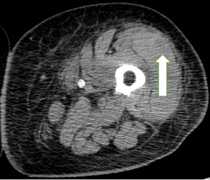
Patibandla et al | Diabetic Myonecrosis–Related Hematoma
contribute to malabsorption (eg, iron, vitamin
B12). The lack of abnormalities with respect to
the liver and kidneys makes anasarca second-
ary to hepatic and renal dysfunction less likely.
The iron deficiency anemia prompted fur-
ther evaluation for a gastrointestinal
source of bleeding. Esophagogastroduode-
noscopy showed a single, clean, 3-cm healing
ulcer in the antrum, mild gastritis, and a super-
ficial erosion in the duodenal bulb, all of which
were biopsied. Because of inadequate bowel
preparation, most of the colon was not opti-
mally visualized and evaluation revealed only
internal and external hemorrhoids in the rec-
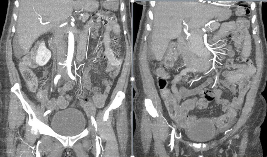
FIG 2. Computed tomography scan images of the abdomen and pelvis with intravenous contrast
tum. On hospital day 4, the patient’s hemoglo-
showing no extravascular extravasation of contrast from major intra-abdominal vasculature. Mild bin decreased from 9.6 g/dL to 7.3 g/dL. She
abdominal and pelvic ascites, a small right pleural effusion with compressive atelectasis, and had dark stools and also complained of left
generalized anasarca are present.
hip pain and swelling of the left knee and
thigh. Another unit of packed red blood
may allow for identification of a fracture, other bone lesion, cells was given. A push enteroscopy and repeat colonosco-
and/or hematoma. py showed no bleeding from the antral ulcer or from the
internal and external hemorrhoids.
A complete blood count revealed a hemoglobin of
6.6 g/dL (normal, 11.5-14.1 g/dL) with a mean corpus- The patient has an antral ulcer, which most likely was a source
cular volume of 62 fL (normal, 79-96 fL). A CT scan of the of chronic blood loss and the underlying iron deficiency. How-
abdomen and pelvis with intravenous contrast (Figure 2) ever, the presence of healing and lack of signs of bleeding as
was obtained to evaluate for intra-abdominal hemorrhage demonstrated by negative repeat endoscopic studies sug-
and retroperitoneal hematoma; it showed mild abdominal gests that the ulcer has little active contribution to the current
and pelvic ascites, a small right pleural effusion with com- anemia episode. A capsule enteroscopy could be performed,
pressive atelectasis, and generalized anasarca, but no evi- but most likely would be low yield. The presence of left thigh
dence of bleeding. She was administered 2 units of packed and knee swelling associated with worsening thigh pain rais-
red blood cells. Apixaban was held and 40 mg intravenous es the suspicion of a hemorrhagic process within the anterior
pantoprazole twice daily was started. Her iron level was thigh compartment, perhaps associated with an occult femoral
12 µg/dL (normal, 50-170 µg/dL); total iron-binding capacity fracture. A CT scan of the thigh would be valuable to identify a
(TIBC) was 431 µg/dL (normal, 179-378 µg/dL); and ferritin fracture or bone lesion as well as the presence of a hematoma.
level was 19 ng/mL (normal, 10-204 ng/mL). Her basic met- There are no widely available tests to evaluate apixaban anti-
abolic panel, liver enzymes, international normalized ratio, coagulant activity; the anticoagulant effect would be expected
partial thromboplastin time, and folate were normal. Serum to dissipate completely 36 to 48 hours after discontinuation in
vitamin B12 level was 277 pg/mL (normal, 213-816 pg/mL), the context of normal renal function.
and the reticulocyte count was 1.7%.
On hospital day 5, the patient’s left leg pain worsened.
The studies reveal microcytic anemia associated with iron de- A physical exam showed edema of her entire left low-
ficiency, as demonstrated by an elevated TIBC and very low er extremity with ecchymoses in several areas, including
ferritin. She also has a low-normal vitamin B12 level, which the left knee and lower thigh. A duplex ultrasound was
can contribute to poor red blood cell production; assessing negative for deep venous thrombosis, and X-ray of her left
methylmalonic acid levels would help to confirm whether true knee was normal. Her repeat hemoglobin was 8.8 g/dL. A
vitamin B12 deficiency is present. Anasarca can be secondary repeat CT scan of the abdomen and pelvis again revealed
to severe hypoalbuminemia due to either protein-losing pro- no retroperitoneal bleeding. Orthopedic surgery was con-
cesses (eg, nephrotic syndrome, protein-losing enteropathy) sulted on hospital day 7 and had low suspicion for compart-
or cirrhosis with poor synthetic function (given her history of ment syndrome. Physical exam at that time showed mild
NAFLD); it can also be secondary to severe heart failure or swelling of the left thigh, moderate swelling of the left
end-stage renal disease. The CT scan with contrast ruled out knee joint and pretibial area, two areas of ecchymosis on
inferior vena cava thrombosis as a cause of ascites and did not the left thigh, and diffuse ecchymosis of the left knee; all
reveal an obvious intra-abdominal malignancy as the cause compartments were soft, and motor and nervous system
of her anemia. Intestinal edema associated with anasarca can functions were normal. A CT scan of the left lower extrem-
372 Journal of Hospital Medicine® Vol 16 | No 6 | June 2021 An Official Publication of the Society of Hospital Medicine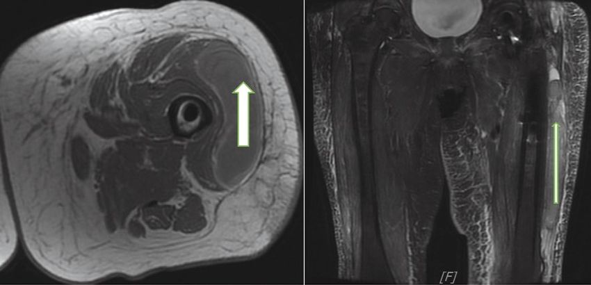
Diabetic Myonecrosis–Related Hematoma | Patibandla et al

ity (Figure 3) revealed findings suspicious for hemorrhagic
myositis with diffuse left thigh swelling with skin thickening
and edema. There was no evidence of abscess, gas collec-
tion, foreign body, acute osteomyelitis, fracture, or disloca-
tion. The patient’s hemoglobin remained stable.

Myopathies can be hereditary or acquired. Hereditary myop-
athies include congenital myopathies, muscular dystrophies,
channelopathies, primary metabolic myopathies, and mito-
chondrial myopathies. Acquired myopathies include infectious
myopathies, inflammatory myopathies, endocrine myopathies,
secondary metabolic myopathies, and drug-induced and tox-
ic myopathies. The findings of hemorrhagic myositis and skin
edema are very intriguing, especially given their localized fea-
tures. An overt femur fracture was previously ruled out, and
an anterior thigh compartment syndrome was considered less
likely after orthopedic surgery consultation. There is no de-
scription of the patient taking medications that could cause
                                                                   FIG 3. Computed tomography scan image of the left thigh with emphasis on
myopathy (such as statins), and there are also no clinical fea-    the bean-shaped encapsulated collection in the lateral muscle tissue of the left
tures suggestive of primary inflammatory myopathy, such as         thigh (white arrow) that raised suspicion for hemorrhagic myositis and diffuse
dermatomyositis. Increased suspicion of a focal inflammatory       cellulitis/edema.

process such as localized scleroderma with regional inflamma-
tory myopathy or another focal myopathy must be considered.        cated by hemorrhagic transformation. Diabetic myonecrosis is
The next diagnostic steps would include measuring the cre-         relatively uncommon and a diagnosis made by combining his-
atine kinase level, as well as obtaining an MRI of the leg to      tory, examination, and laboratory findings and excluding other
assess the nature and extent of the myopathy.                      alternative conditions.
                                                                      A clear schema for approaching the patient with acute, non-
     Multidisciplinary involvement, including hematology,          traumatic myopathies is important in avoiding diagnostic er-
     rheumatology, and surgery, aided in narrowing the dif-        ror. One effective schema is to divide myopathy into infectious
ferential diagnosis. On hospital day 10, an MRI of the left        and noninfectious categories. Causes of infectious myopathy
thigh was performed for suspicion of diabetic myonecrosis          include bacterial infections (eg, pyomyositis), inflammatory
(Figure 4). The MRI revealed a 10 cm × 3.6 cm × 22 cm in-          damage to muscles associated with viruses (eg, influenza), as
tramuscular hematoma in the belly of the vastus lateralis          well as rarer causes. Bacterial processes tend to be relative-
muscle with associated soft tissue swelling, overlying sub-        ly focal and affect a specific muscle group or anatomic com-
cutaneous edema, and skin thickening that was suggestive           partment, while viral causes are often more diffuse and occur
of hemorrhagic diabetic myonecrosis with some atypical             in the context of a systemic viral syndrome. Bacterial causes
features. A rheumatology consult was requested to evalu-           range in severity, and life-threatening conditions, such as nec-
ate for possible vasculitis in the left lower extremity, and       rotizing soft tissue infection, must be considered. In this case,
vasculitis was not considered likely. The diagnosis of dia-        bacterial causes were less likely given the patient’s lack of fever,
betic myonecrosis with associated intramuscular hemor-             leukocytosis, and systemic signs of infection.1,2 However, these
rhage secondary to apixaban was made after careful recon-          findings are not uniformly sensitive, and clinicians should not
sideration of the clinical presentation, imaging and               exclude potentially life- or limb-threatening infections without
laboratory data, and overall picture. Based on the clinical        thorough evaluation. For example, pyomyositis may present
findings, imaging results, and exclusion of alternative caus-      without fever in the subacute stage, without leukocytosis if
ative pathologies of thigh swelling, no biopsy was per-            the patient is immunocompromised, and without overt pus if
formed, as it was not considered necessary to make the             the infection is not in the suppurative stage.3 Viral causes were
diagnosis of diabetic myonecrosis. The patient was dis-            made less likely in this patient given the lack of a current or
charged on hospital day 11 and was doing well. She fol-            recent systemic viral syndrome.
lowed up with her primary care doctor and has regained                Once infectious etiologies are deemed unlikely, noninfec-
normal function of her leg.                                        tious etiologies for nontraumatic myopathies should be con-
                                                                   sidered. Some causes of noninfectious myopathy present with
DISCUSSION                                                         the muscle symptoms as a predominant feature, while others
Diabetic myonecrosis, or diabetic muscle infarction, is an un-     present in the context of another illness such as cancer, met-
common nontraumatic myopathy that occurs in patients with          abolic disorders, or other systemic disorders. Many noninfec-
diabetes who develop acute, focal muscle pain without recent       tious causes of myopathy associated with systemic illnesses
trauma. In this case, the muscle infarction was further compli-    have diffuse or relatively diffuse symptoms, with pain and/or

An Official Publication of the Society of Hospital Medicine                   Journal of Hospital Medicine® Vol 16 | No 6 | June 2021          373
Patibandla et al | Diabetic Myonecrosis–Related Hematoma
of the combination of bleeding risk from
apixaban and the underlying mechanisms of
diabetic myonecrosis.
The treatment of diabetic myonecrosis is
mainly supportive, with an emphasis on rest,
nonsteroidal anti-inflammatory agents, anti-
platelet agents, and strict glycemic control.10
There is conflicting information about the
value of limb immobilization versus active
physical therapy as appropriate treatment
modalities.11 Patients who present with clin-
ical concern for sepsis or compartment syn-
drome require consultation for consideration
FIG 4. Magnetic resonance image of the left thigh that shows a large hematoma (thick arrow in image on of acute surgical intervention.10 The short-
the left and thin arrow in image on the right) encapsulated in the muscle belly of the vastus lateralis muscle.
term prognosis is promising with supportive
therapy, but the condition may recur.12 The
weakness in multiple muscle groups, often in a bilateral distri- recurrence rate may be as high as 40%, with a 2-year mortality
bution. Such examples include dermatomyositis and polymy- of 10%.13 Ultimately, patients need to be followed closely in the
ositis as well as myositis associated with other rheumatologic outpatient setting to reduce the risk of recurrence.
conditions. Nontraumatic rhabdomyolysis is diffuse and can In this patient, the simultaneous occurrence of focal pain and
occur in association with medications and/or genetic condi- acute blood loss anemia led to a diagnosis of diabetic myone-
tions. crosis that was complicated by hemorrhagic conversion, a truly
Angervall and Stener4 first described diabetic myonecrosis painful coincidence. The patient underwent a thorough evalu-
in 1965 as tumoriform focal muscular degeneration due to di- ation for acute blood loss before the diagnosis was ultimately
abetic microangiopathy. The most commonly affected muscle made. Clinicians should consider diabetic myonecrosis in pa-
groups in diabetic myonecrosis are the anterior thigh, calf, and tients with diabetes who present with acute muscle pain but no
posterior thigh, followed by muscles in the upper extremities.5 evidence of infection.
Patients with diabetic myonecrosis have an overall mean age
at presentation of 44.6 years; affected patients with type 1 di- KEY TEACHING POINTS
abetes mellitus present at a mean age nearly 20 years young- • Diabetic myonecrosis is an underrecognized entity and
er than those with type 2 diabetes mellitus (35.9 years vs 52.2 should be included in the differential diagnosis for patients
years, respectively).6 Patients tend to have a long (often >15 with diabetes who present with acute muscle pain and no
years) history of diabetes with microvascular complications history of trauma.
such as retinopathy (reported in 71%), nephropathy (reported • Imaging with CT and/or MRI of the affected region is the
in 57%), and/or neuropathy (reported in 55%).7 mainstay of diagnosis; treatment is predicated on severity
The mainstay of the diagnosis of diabetic myonecrosis is a and risk factors and can range from conservative therapy to
thorough history and physical examination and imaging. Rou- operative intervention.
tine laboratory evaluation is relatively unhelpful in diagnosing • Although the prognosis is good in these patients, careful
diabetic myonecrosis, but appropriate imaging can provide outpatient follow-up is necessary to oversee their recovery
valuable supportive information. A CT scan and MRI are both to help reduce the risk of recurrence.
helpful in excluding other etiologies as well as identifying fea-
tures consistent with diabetic myonecrosis. A CT scan can help
exclude a localized abscess, tumor, or bone destruction and, Acknowledgment
in affected patients, may show increased subcutaneous atten- The authors thank Dr Vijay Singh for his radiology input on image selection for
uation and increased muscle size with decreased attenuation this manuscript.
secondary to edema. However, a CT scan may not give opti-
2 Disclosures: The authors have nothing to disclose.
mal assessment of muscle tissue, and therefore MRI may need
to be considered. MRI T2 images have a sensitivity nearing References
90% for detecting myonecrosis.1 The diagnostic value of MRI 1. Ivanov M, Asif B, Jaffe R. Don’t move a muscle: a case of diabetic
often obviates the need for muscle biopsy. myonecrosis. Am J Med. 2018;131(11):e445-e448. https://doi.org/10.1016/
j.amjmed.2018.07.002
Spontaneous infarction with hemorrhagic features seen on 2. Morcuende JA, Dobbs MB, Crawford H, Buckwalter JA. Diabetic muscle
imaging can be explained by a combination of damage from infarction. Iowa Orthop J. 2000;20:65-74.
atherosclerotic or microvascular disease, an activated coagu- 3. Crum-Cianflone NF. Bacterial, fungal, parasitic, and viral myositis. Clin
Microbiol Rev. 2008;21(3):473-494. https://doi.org/10.1128/CMR.00001-08
lation cascade, and an impaired fibrinolytic pathway.8 Hemor-
4. Angervall L, Stener B. Tumoriform focal muscular degeneration in two
rhagic conversion in diabetic myonecrosis appears to be un- diabetic patients. Diabetologia. 1965;1(1):39-42. https://doi.org/10.1007/
common.9 In our case, we suspect that it developed because BF01338714
374 Journal of Hospital Medicine® Vol 16 | No 6 | June 2021 An Official Publication of the Society of Hospital MedicineDiabetic Myonecrosis–Related Hematoma | Patibandla et al 5. Lawrence L, Tovar-Camargo O, Lansang MC, Makin V. Diabetic myonecrosis: Skeletal Radiol. 2016;45(8):1069-1078. https://doi.org/10.1007/s00256-016- a diagnostic and treatment challenge in longstanding diabetes. Case Rep 2389-4 Endocrinol. 2018;2018:1723695. https://doi.org/10.1155/2018/1723695 10. Khanna HK, Stevens AC. Diabetic myonecrosis: a rare complication of diabe- 6. Horton WB, Taylor JS, Ragland TJ, Subauste AR. Diabetic muscle infarc- tes mellitus mimicking deep vein thrombosis. Am J Case Rep. 2017;18:38-41. tion: a systematic review. BMJ Open Diabetes Res Care. 2015;3(1):e000082. https://doi.org/10.12659/ajcr.900903 https://doi.org/10.1136/bmjdrc-2015-000082 11. Bunch TJ, Birskovich LM, Eiken PW. Diabetic myonecrosis in a previous- 7. Bhasin R, Ghobrial I. Diabetic myonecrosis: a diagnostic challenge in pa- ly healthy woman and review of a 25-year Mayo Clinic experience. Endocr tients with long-standing diabetes. J Community Hosp Intern Med Perspect. Pract. 2002;8(5):343-346. https://doi.org/10.4158/EP.8.5.343 2013;3(1). https://doi.org/10.3402/jchimp.v3i1.20494 12. Mukherjee S, Aggarwal A, Rastogi A, et al. Spontaneous diabetic myone- 8. Bjornskov EK, Carry MR, Katz FH, Lefkowitz J, Ringel SP. Diabetic muscle crosis: report of four cases from a tertiary care institute. Endocrinol Diabetes infarction: a new perspective on pathogenesis and management. Neuromuscul Metab Case Rep. 2015;2015:150003. https://doi.org/10.1530/EDM-15-0003 Disord. 1995;5(1):39-45. 13. Kapur S, McKendry RJ. Treatment and outcomes of diabetic muscle in- 9. Cunningham J, Sharma R, Kirzner A, et al. Acute myonecrosis on MRI: etiol- farction. J Clin Rheumatol. 2005;11(1):8-12. https://doi.org/10.1097/ ogies in an oncological cohort and assessment of interobserver variability. 01.rhu.0000152142.33358.f1
You can also read