Langer axillary arch in breast surgery: a narrative review - Annals of Breast Surgery
←
→
Page content transcription
If your browser does not render page correctly, please read the page content below
Review Article
                                                                                                                                      Page 1 of 8

Langer axillary arch in breast surgery: a narrative review
Pier Carlo Rassu

Department of General Surgery, “San Giacomo” Hospital, Novi Ligure (AL), Italy
Correspondence to: Pier Carlo Rassu, MD. Department of General Surgery, “San Giacomo” Hospital, Via Edilio Raggio, 12, 15067 Novi Ligure (AL),
Italy. Email: piercarlo.rassu@gmail.com.

                Abstract: The Langer’s axillary arch (LAA), an anomalous accessory muscle causing potential lymphedema
                and compression of vessels or nerves, has been long identified and described form cadaveric studies even
                if the presence of this muscular variation during axillary access in breast surgery plays a key-role in the
                correct identification of anatomical landmarks both in sentinel lymph node biopsy (SLNB) and in axillary
                lymph node dissection (ALND). As a consequence, the capability of pre-operatively and intra-operatively
                techniques to detect the LAA may prevent the surgeon to mistakes in axillary clearance. With the aim to
                point out about the feasibility of pre-operative techniques to identify the LAA in patients and to analyse the
                surgical implications from the LAA during axillary access, this narrative review screened the literature reports
                on clinical significance of LAA in breast surgery. Remarks on SLNB and lymphadenectomy where the
                positioning, the detectability with different tracers, and the malignancy of lymph nodes (LNs) might become
                crucial in relation to the LAA were detailed. Then, features taking part of a Decision Support System (DSS)
                to determine how to handle the surgical approach when LAA is encountered during breast surgery were
                investigated. Finally, open issues on the opportunity to resect or preserve such accessory muscle outlined the
                need motivate more research on clinical features of LAA in breast surgery.

                Keywords: Breast surgery; Langer’s axillary arch (LAA); sentinel lymph node biopsy; axillary dissection; echography

                Received: 13 September 2020; Accepted: 09 December 2020.
                doi: 10.21037/abs-20-115
                View this article at: http://dx.doi.org/10.21037/abs-20-115

Introduction                                                                  (4,5). Variants of LAA with possible structure and position
                                                                              in relation to muscles, nerves, and axillary vessels have been
The axillary surgery plays a fundamental role in the                          further described (6-9). LAA is usually asymptomatic, but
treatment of breast cancer, melanoma of the trunk and                         it could be implicated in the syndrome of costoclavicular
the upper limb, in the histological diagnosis of lymphoma                     compression and of hyper abduction, thoracic outlet and
and in all those cases where it is necessary to remove for                    shoulder instability, as like as in the entrapment of the
diagnostic purposes both lymph nodes (LNs), and axillary                      median nerve; further it can present with upper limb deep
masses of unclear nature.                                                     vein thrombosis or venous congestive symptoms (10,11).
   The axillary surgery for breast cancer has further                            Several data about morphological terminology and
stimulated the interest in Langer’s axillary arch (LAA)                       frequency of LAA can be obtained from the study by Jelev
that was described as an extensive sheet of skin-associated                   et al. (6) along with the clinical significance of axillary arch
musculature between the superficial fascia and the                            which presents some peculiar features to be taken into
subcutaneous fat (1). Initially reported as muscle variation                  account during procedures of axillary surgery. Despite this,
in the axillary fossa by Bugnone in 1783 (2), the “axillary                   a recent meta-analysis (12) outlined that the majority of
arch” was identified by Ramsay in 1795 (3). However, it                       studies have been conducted on cadavers. Consequently, the
was Langer in 1846 who explained the axillary muscle more                     possibility to calculate the total prevalence of the atypical
accurately, being aware of tension lines in the skin which                    muscles in the general population comes from cadaveric
were recognized as potentially related to surgical incisions                  studies (13), more than from surgical axillary procedures (12).

© Annals of Breast Surgery. All rights reserved.                                     Ann Breast Surg 2021 | http://dx.doi.org/10.21037/abs-20-115
Page 2 of 8                                                                                                                    Annals of Breast Surgery, 2021

                          Records identified using specific terms ‘breast surgery’,
                         ‘Langer’s axillary arch’, ‘lymphadenectomy’, ‘dissection’,
                                ‘sentinel lymph node’, and ‘preoperatively’
                                                      n=42

                                Records screened for relevance to subject
                                                 n=42

                                                                                       Excluded articles based on abstract not relevant
                                                                                                       specific reasons:
                                                                                                    Not related to surgery
                                                                                                Not related to breast cancer
                                                                                                             n=16

                                   Full text articles access for eligibility
                                                     n=26
                                                                                      Excluded articles after review of full-text articles
                                                                                      No relevance in context of axillary diagnosis for
                                                                                                       breast cancer
                                                                                                             n=0
                                      Articles included in final review
                                                    n=26

Figure 1 Diagram, modified from PRISMA chart, detailing identification of studies included in narrative review. PRISMA, Preferred
Reporting Items for Systematic Review and Meta-Analysis.

The occurrence of LAA is at 7–8%, varying frequencies                                 ‘dissection’, ‘sentinel lymph node’, and ‘preoperatively’.
from 0.25–43.8% depending on the population studied.                                  Following acquisition of the full texts, other potentially
Probably on account of most of the axillary surgery applied                           eligible articles that could have been missing in the
to women, the reported occurrence of the LAA is major                                 electronic databases’ search were screened with a reference
on female than male patients, bilateral or unilateral as well                         search. The Preferred Reporting Items for Systematic
(3,6,8,14).                                                                           Review and Meta-Analysis (PRISMA) guidelines (19) was
   Although intraoperative recognition of muscle variations                           applied in scrutinising references of relevant articles for
in a given anatomical region does not necessarily reflect its                         pertinent studies. Articles which did not identify the LAA
surgical significance (9), the failure to report or to identify                       in breast surgery were excluded. The search was carried out
the accessory muscles of the axilla undoubtedly affects                               in August 2020 with no prior time limit set for inclusion
their clinical significance so that even experienced breast                           of data as well as inclusion of every original language of
or thoracic surgeons are expected to deliver a detailed                               publication.
knowledge of the axilla (8,9,15-18).
   Therefore, this review seeks to explore the literature
                                                                                      Study endpoints
on LAA in breast surgery along with features of the
Decision Support System (DSS) used by surgeons (15) to                                The primary endpoint was finding the capability of pre-
identify whether the LAA might be preserved or resected.                              operatively and intra-operatively techniques to detect the
We present the following article in accordance with the                               LAA in breast surgery. The secondary endpoint included
Narrative Review reporting checklist (available at http://                            surgeon DSS while encountering LAA during axillary access
dx.doi.org/10.21037/abs-20-115).                                                      and examining features associated with the arch.

Methods                                                                               Results

Search strategy                                                                       Literature search

The major electronic database (PubMed, Web of Science,                                There was a total of 42 potential citations from the initial
and Scopus) were thoroughly searched for studies on ‘breast                           database search (Figure 1). Amongst these, articles discussed
surgery’, ‘Langer’s axillary arch’, ‘lymphadenectomy’,                                only axillary dissection in cadavers and anatomical studies

© Annals of Breast Surgery. All rights reserved.                                               Ann Breast Surg 2021 | http://dx.doi.org/10.21037/abs-20-115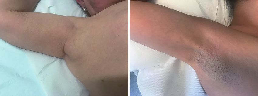
Annals of Breast Surgery, 2021                                                                                                         Page 3 of 8

 Table 1 Number of detected LAAs and related treatment during SLNB and ALND in 17 studies with breast surgeries selected in this review
                                                                                           Number of
 Ref. Study (year)                                 Procedure                                         Treatment of LAA
                                                                                             LAAs

 (8)   Kutiyanawala et al. (1998)                  ALND                                         6       Resection

 (14) Suzuma et al. (2003)                         SLNB with indigo carmine                     1       Resection during secondary ALND

 (15) Rassu (2020)                                 SLNB with TcLC and ICG and ALND              10      8 preserved, 2 resected

 (16) Ridgway et al. (2011)                        SLNB with technetium and blue dye            1       Resection during secondary ALND

 (17) Upasna et al. (2015)                         ALND                                         2       Resection

 (18) Daniels & Querci della Rovere (2000) ALND                                                 1       Resection

 (20) Karanlik et al. (2013)                       ALND                                         9       7 preserved, 2 resected

 (21) Ando et al. (2010)                           SLNB with indigo carmine                     59      Not reported

 (22) Keshtgar et al. (1999)                       SLNB with radioisotope colloid               2       Resection during secondary ALND

 (23) Abudhaise et al. (2019)                      SLNB                                         8       Not reported

 (24) Sang et al. (2019)                           SLNB with methylene blue and ICG            132      Resection during secondary ALND

 (25) Chêne et al. (2007)                          SLNB with radioisotope colloid               5       Resection during secondary ALND

 (26) Smart et al. (2005)                          SLNB with technetium antimony                1       Resection
                                                   sulphide and patent blue dye

 (27) Ku et al. (2008)                             ALND                                         1       Resection

 (28) Petrasek et al. (1997)                       ALND                                         1       Preservation

 (29) Lamichhane et al. (2017)                     ALND                                         1       Preservation

 (30) Al Maksoud et al. (2015)                     ALND                                         1       Preservation
 LAA, Langer’s axillary arch; SLNB, sentinel lymph node biopsy; ALND, axillary lymph node dissection; TcLC, 99mTc‐labeled nanocolloid;
 ICG, indocyanine green.

on LAA along with studies examining LAA in other surgery                      information for intraoperative navigation of SLNB, even if
than the ones for breast cancer were excluded. In total,                      it appears to be difficult as this muscular variation crosses
26 articles discussing relevant issues pertaining to LAA in                   in front of the great axillary vessels and the first intercostal
breast surgery were included in this review. LAA occurrence                   brachial nerve. Since LAA may hide the LNs of the first
during breast surgery either in sentinel lymph node biopsy                    level of Berg (Figure 2), it can be confused with enlarged
(SLNB) or in axillary lymph node dissection (ALND) along                      LNs or soft tissues tumours (8,9,20,21). Intermittent
with LAA treatment is described in 17 studies (Table 1).                      compression of the axillary vein or hyperabduction
Other four papers evaluated the preoperative investigation                    syndrome could be indicative for LAA presence (10,11,18).
of the axillary arch, whilst the remaining five articles                         Sometimes, the arch could be easily visible at clinic
described implications of LAA in breast surgical procedures                   judgment (Figure 3). Imaging analysis, as like as
like SLNB and ALND. Due to the few studies concerning                         mammography, echography, computed tomography (CT)
breast surgery and management of LAA in clinical setting, a                   and magnetic resonance imaging (MRI) have demonstrated
narrative review was performed.
                                                                              to detect the presence of the axillary arch only in few
                                                                              clinical reports (22,31-35). Ko et al. (32) detailed on the
Discussion                                                                    capability of mammography to get imagines about a band-
                                                                              like structure overlapped with pectoralis muscle which they
Preoperatively diagnosis of LAA
                                                                              revealed to be the axillary arch. Keshtgar et al. reported
Preoperative diagnosis of the axillary arch supplies pieces of                that the LAA is fully stretched during imaging, so that

© Annals of Breast Surgery. All rights reserved.                                       Ann Breast Surg 2021 | http://dx.doi.org/10.21037/abs-20-115
Page 4 of 8 Annals of Breast Surgery, 2021
A B
Figure 2 Displacement of the LAA muscular structure in the axilla. (A) The LAA appears as a ventral extension of the anterior margin of the
latissimus dorsi; (B) the LAA is displaced laterally to allow the complete view of the surgical field. LAA, Langer’s axillary arch.
Figure 3 Occurrence of LAA at clinical examination in two patients (own data). LAA, Langer’s axillary arch.
the echography scanning was misleading in showing the images more with fast spin-echo T2-weighted images
location of the SLN (22). Indeed, echography examination than on spin-echo T1-weighted images thanks to the
could be more easily provided after surgery as an attempt signal intensity contrast between LAA and fat. For a
to assess if a mass due to LAA could be resolved in preoperative diagnosis with MRI, Suzuma et al. described
patients where LAA is preserved (Figure 4). Anyway, at the anatomical site of LAA takes origin at the anterior
ultrasonography inspection, the muscular arch seemed to edge of the latissimus dorsi in the middle of the posterior
appear as a grey oval structure (15). fold of the axilla and tapering to a narrow tendon which
Multidetector row CT (MDCT) and MRI may allow was inserted into the posterior aspect of the trilaminar
at obtaining LAA recognition preoperatively (14,21,31). tendon of the pectoralis major (14).
Rajakulasingam & Saifuddin reported MRI images with Hong et al. presented the submission to MDCT of a
two slips of muscle arising from the anterior margin of the case with a palpable, non-tender mass in left axilla with
left latissimus dorsi muscle (33). The thicker slip crosses complains of intermittent ulnar numbness in left arm to
antero-inferior to the axillary neurovascular bundle forearm, aggravated with hyperabduction and relieved with
without any compression or displacement. Guy et al. adduction of the arm. The MDCT was able to discriminate
characterized at first the prevalence, anatomic relations, between right and left axilla where a nodular elongated
possible LN concealment, and potential neurovascular soft tissue structure was detected (35). With the same
impingement of the axillary arch muscle in an extensive imaging technique, Ando et al. reported not significant
review of shoulder MRI data (34). They reported that the different numbers of MDCT-LNs and failure rates of SLN
LAA was detected or excluded best on oblique coronal identification between patients with or without the LAA (21).
© Annals of Breast Surgery. All rights reserved. Ann Breast Surg 2021 | http://dx.doi.org/10.21037/abs-20-115Annals of Breast Surgery, 2021 Page 5 of 8
A B
A
LD
Figure 4 Tentative detection of LAA with echography after axillary surgery. (A) Right LAA in a patient submitted to SLNB with TcLC and
to secondary ALND (own data); (B) left LAA in a patient submitted to SLNB with ICG (own data). A and *** = arch. LAA, Langer’s axillary
arch; SLNB, sentinel lymph node biopsy; TcLC, 99mTc‐labeled nanocolloid; ALND, axillary lymph node dissection; ICG, indocyanine
green; LD, latissimus dorsi.
LAA in SLNB counts are commonly maximal at the inferior edge of the
axillary hairline just posterior to the pectoralis muscle in
The presence of LAA might affect the SLNB. Keshtgar
usual SLNB (16). In their experience, the maximal counts
et al. reported that the SLN localization was extremely
were more cranial and lateral than the common location
easy when the node was lying over the arch, whilst if the
of the sentinel nodes when LAA occurred. Further, they
SLN was underneath LAA the node detection was more
found no difficulty in finding LNs under an arch when
difficult (22). Ando et al. detailed the anatomical localization
radioisotope was used, even if an arch could conceal the
of LNs with respect to the common position of a SLN in
nodes when blue dye was applied. In his paper investigating
a group of 56 patients with a diagnosis of axillary arch and
SLNs identification (21). In some cases, SLNs were located SLNB with 99mTc-labeled nanocolloid (TcLC) and
in more lateral and superficial axillary positions, in others indocyanine green (ICG) as tracers, Rassu (15) outlined
they were allocated in more cranial positions or in more that in the patient group with only SLNB, a mean number
dorsal positions or below anomalous muscles of the axilla. of 2.42 (±1.76) and 2.42 (±1.83) LNs per patient were
Examining eight patients over 3 years, Abudhaise et al. harvested with TcLC and ICG, respectively. Similarly,
found sentinel LNs laterally to the LAA in the sub-pectoral the metastatic LNs were not significantly different:
region and associated with stretching of the efferent 0.08 (±0.34) with TcLC and 0.14 (±0.55) with ICG (15)
lymphatic vessels (23). so to corroborate the ability of radioisotope colloids
Few pieces of information are currently available in “technetium-based” to provide accuracy in collecting LNs
literature (Table 1) on the most useful tracers to be used even in presence of anomalous muscular structure in the
during axillary access to avoid mistakes due to LAA. axilla (16,22,25).
Inadequate clearance of Level 1 nodes due to them being The ICG tracer was evaluated along with methylene
partially covered by the arch (Figure 2A) could lead blue only in another study to identify LNs when masked
relatively inexperienced surgeons to a level above the by LAA (24). Literature reports evidence that SLNB
axillary vein, with an increased risk of lymphedema of the using the fluorescent dye ICG allows at detecting LNs
arm postoperatively. Besides, as the LAA could act as a non-invasively with high accuracy and sensitivity (36).
potential site of metastatic LNs (24) accurate identification Indeed, Rassu outlined the meaningful advantage recorded
by means of intraoperative localization with standardised applying the ICG tracer to map lymphatic vessels in
techniques and performing tracers is strongly advised order to minimize the confounding factor given by the
(15,16,24). anatomical limits of the operative field in case of Langer’s
Ridgway et al. reported that percutaneous radioisotope anatomical variation (15).
© Annals of Breast Surgery. All rights reserved. Ann Breast Surg 2021 | http://dx.doi.org/10.21037/abs-20-115Page 6 of 8 Annals of Breast Surgery, 2021
LAA in lymphadenectomy because of the recurrence risk to undissected axillopectoral
muscles harbouring positive nodes is larger in magnitude
The surgeon awareness of the anatomical variations that
(14,24). With a procedure of LAA division, as illustrated in
may appear in a specific axillary region is the leading factor
Figure 2B, Ku et al. obtained a complete dissection of the
for their intraoperative recognition along with their main
medial and lateral axillary group with no evidence of axillary
anatomical features (9). During lymphadenectomy in LAA’s
recurrence, lymphedema or any limitation of motion of the
patients, Rassu outlined the axillary location that contains
right arm after 2-year follow-up (27).
the vast majority of the LNs as almost fully covered by
Another reason for muscle resection is based on the
the arch (15): thus, amplifies the recurrence risk from
association of LAA with neurovascular compression leading
undissected axillopectoral muscles harbouring positive
to postoperative upper limb lymphedema and thoracic
nodes. Indeed, a very recent work confirmed that LAA’s
outlet syndrome with compression particularly noted during
LN has a relatively high metastasis rate (24), but the same
abduction or external rotation of the arm (18,20,25).
research did not provide any comparison between LN
On the other hand, especially whether the LAA does
status of patients with and without LAA as it was provided
not reveal to be problematic for the patient in order to
previously by Smart et al. (26) and more recently by
permit later reconstructive surgery (1,8,14,18,20,26)
Rassu (15) who observed no metastatic LNs in LAA patients
the preservation of the neurovascular elements should
even in secondary ALND.
continuously be taken into consideration by the
In their systematic review of medical literature published
surgeon (15). Gentle dissection of tissues with a reluctance
between 1996 and 1999, Babu & Khashaba concluded that
to divide any horizontally lying veins should allow this
LAA should be recognized in axillary dissection to avoid
anomaly to be identified and prevent inadvertent damage
confusion in staging of LNs and to prevent injury to axillary
(8,38). This suggested to preserve the arch in certain LAA
vessels and brachial plexus (37). The LAA might restrict
patients in line with the “SLNB era” advocated by Veronesi
axillary access during ALND as it is tautened by abduction
et al. (39). In two studies (15,20), the LAA was kept
and elevation of the arm: thus, allowing free mobility and
(Table 1). Indeed, in a mean 23 months follow-up carried
relaxation of the muscle is therefore advocated (26).
out by Petrasek et al. there were no known complications
related to the axillary arch, such as lymphedema, brachial
Conclusions plexus injury, axillary vein injury or thrombosis (28). The
same uneventful procedure was provided by Lamichhane
LAA and features of the decision supporting system in
et al. (29) and Al Maksoud et al. (30) who were able to
breast surgery
dissect nodes beneath the arch without the need to divide it.
The presence of the LAA muscular structure on the lateral Thus, breast surgeon’s awareness of the possible
side of the surgical field may be mistakenly confused with complications of an axillary arch muscle, the understanding
the latissimus dorsi muscle (Figure 2A). In this situation, the of any functional deficit from it, along with their clinical
band that adheres to the medial edge of the muscle could judgment targeted for each patient may continue to
not be recognized and the dissection might be continued stimulate the debate on the preservation or the cutting
along in a wrong plan too cranial than the axillary vein. of axillary arches. Karanlik et al. and Rassu asserted their
As a consequence, the cords of the brachial plexus would clinical judgment in a DSS to determine how to identify
be exposed to the risk to be injured with impairment of LAA, to question whether the axillary arch could be
breast reconstruction (1,8,14,18,20,26) even after a radical perceived as potentially problematic for the patient, and
mastectomy (27). to handle results to the final decision (15,20). Such DSS
On the other hand, the LAA may complicate procedures approach, based on multiple variables, could motivate
as the SLNB and the lymphadenectomy giving risks as like more research on clinical features of LAA in breast surgery
as suboptimal staging and limited regional control of disease where also patient age and status should be taken into
(1,16,17,20-22,24,26). account.
Accordingly, in order to get an optimal axillary clearance,
research commonly suggests the cutting of this anomalous
Acknowledgments
axillary muscle (Table 1). In detail, authors who support
such a decision consider performing the LAA resection Funding: None.
© Annals of Breast Surgery. All rights reserved. Ann Breast Surg 2021 | http://dx.doi.org/10.21037/abs-20-115Annals of Breast Surgery, 2021 Page 7 of 8
Footnote variants during axillary dissection. Br J Surg 1998;
85:393-4.
Reporting Checklist: The author has completed the Narrative
9. Natsis K, Vlasis K, Totlis T, et al. Abnormal muscles that
Review reporting checklist. Available at http://dx.doi.
may affect axillary lymphadenectomy: surgical anatomy.
org/10.21037/abs-20-115
Breast Cancer Res Treat 2010;120:77-82.
10. Magee C, Claire Jones MB, McIntosh S, et al. Upper limb
Conflicts of Interest: The author has completed the ICMJE
deep vein thrombosis due to Langer’s axillary arch. J Vasc
uniform disclosure form (available at http://dx.doi.
Surg 2012;55:234-6.
org/10.21037/abs-20-115). The author has no conflicts of
11. Clarys JP, Barbaix E, Van Rompaey H, et al. The muscular
interest to declare.
arch of the axilla revisited: its possible role in the thoracic
outlet and shoulder instability syndromes. Man Ther
Ethical Statement: The author is accountable for all 1996;1:133-9.
aspects of the work in ensuring that questions related 12. Taterra D, Henry BM, Zarzecki MP, et al. Prevalence and
to the accuracy or integrity of any part of the work are anatomy of the axillary arch and its implications in surgical
appropriately investigated and resolved. practice: a meta-analysis. Surgeon 2019;17:43-51.
13. Douvetzemis S, Natsis K, Piagkou M, et al. Accessory
Open Access Statement: This is an Open Access article muscles of the anterior thoracic wall and axilla. Cadaveric,
distributed in accordance with the Creative Commons surgical and radiological incidence and clinical significance
Attribution-NonCommercial-NoDerivs 4.0 International during breast and axillary surgery. Folia Morphol (Warsz)
License (CC BY-NC-ND 4.0), which permits the non- 2019;78:606-16.
commercial replication and distribution of the article with 14. Suzuma T, Sakurai T, Yoshimura G, et al. Magnetic
the strict proviso that no changes or edits are made and the resonance axillography for preoperative diagnosis of the
original work is properly cited (including links to both the axillopectoral muscle (Langer’s axillary arch): a case report.
formal publication through the relevant DOI and the license). Breast Cancer 2003;10:281-3.
See: https://creativecommons.org/licenses/by-nc-nd/4.0/. 15. Rassu PC. A single-center study on 12 year-experience
in lymphadenectomy and in sentinel lymph-node biopsy
References with 99mTc-labeled nanocolloid and indocyanine green as
tracers: relationships with detection and management of
1. Besana-Ciani I, Greenall MJ. Langer's axillary arch: the Langer’s axillary arch. Breast J 2020;26:1056-60.
anatomy, embryological features and surgical implications. 16. Ridgway PF, Collins AM, McCready DR. The surgical
Surgeon 2005;3:325-7. importance of an axillary arch in sentinel node biopsy.
2. Pitzorno H. Contributo alla morfologia dell’arco ascellare Surg Radiol Anat 2011;33:147-9.
muscolare di Langer. Arch Ital Anat Embryol 1911; 17. Upasna, Kumar A, Singh B, et al. Muscular variations
10:129-44. during axillary dissection: A clinical study in fifty patients.
3. Ramsay A. An account of unusual conformation of some Niger J Surg 2015;21:60-2.
muscles and vessels. Edinb Med Surg J 1812;8:281-3. 18. Daniels IR, Querci della Rovere G. The axillary arch of
4. Langer C. Zur Anatomie Des Musculus Latissimus Dorsi. Langer--the most common muscular variation in the axilla.
Osterreichische Med Wocheschrift 1846;15:454-8. Breast Cancer Res Treat 2000;59:77-80.
5. Carmichael SW. The tangled web of Langer’s lines. Clin 19. Moher D, Liberati A, Tetzlaff J, et al. Preferred reporting
Anat 2014;27:162-8. items for systematic reviews and meta-analysis: the
6. Jelev L, Georgiev GP, Surchev L. Axillary arch in human: PRISMA statement. Int J Surg 2010;8:336-41.
common morphology and variety. Definition of “clinical” 20. Karanlik H, Fathalizadeh A, Ilhan B, et al. Axillary arch
axillary arch and its classification. Ann Anat 2007; may affect axillary lymphadenectomy. Breast Care (Basel)
189:473-81. 2013;8:424-7.
7. Bertone VH, Ottone NE, Lo Tartaro M, et al. The 21. Ando J, Kitamura T, Kuroki Y, et al. Preoperative
morphology and clinical importance of the axillary arch. diagnosis of the axillary arch with multidetector row
Folia Morphol (Warsz) 2008;67:261-6. computed tomography and the axillary arch in association
8. Kutiyanawala MA, Stotter A, Windle R. Anatomical with anatomical problems of sentinel lymph node biopsy.
© Annals of Breast Surgery. All rights reserved. Ann Breast Surg 2021 | http://dx.doi.org/10.21037/abs-20-115Page 8 of 8 Annals of Breast Surgery, 2021
Breast Cancer 2010;17:3-8. frequent but rarely discussed anatomical variant in the
22. Keshtgar MRS, Saunders C, Ell PJ, et al. Langer’s axillary radiologic literature. Pediatr Radiol 2018;48:433-6.
arch in association with sentinel lymph node. Breast 32. Ko K, Han BK, Shin JH, et al. The axillopectoral muscle
1999;8:152-3. seen on mammography. Clin Radiol. 2006;61:625-9.
23. Abudhaise H, Merh R, Devalia H. The anatomical 33. Rajakulasingam R, Saifuddin A. Fullness in the left
relationship between the xillary arch of Langer and axilla-answer: Langer's axillary arch. Skeletal Radiol
sentinel lymph node in breast cancer surgery. Ann R Coll 2020;49:1677-9.
Surg Engl 2019;101:533. 34. Guy MS, Sandhu SK, Gowdy JM, et al. MRI of the axillary
24. Sang Y, Kong X, Li X, et al. Langer’s axillary arch lymph arch muscle: prevalence, anatomic relations, and potential
node metastatis in breast cancer patients: A prospective consequences. AJR Am J Roentgenol 2011;196:W52-7.
clinical study. Surg Oncol 2019;29:48-52. 35. Hong HJ, Choi NJ, Han DH, et al. Axillary arch: detailed
25. Chêne G, Le Bouëdec G, Dauplat G. Arch and sentinel: ultrasonographic images with multiplanar CT correlation.
surgical technique of sentinel node biopsy with the J Med Ultrason (2001) 2015;42:121-5.
axillopectoral muscle. Gynecol Obstet Fertil 2007;35:25-9. 36. Troyan SL, Kianzad V, Gibbs-Strauss SL, et al. The
26. Smart PJ, Shayan R, Mann GB. Axillopectoral muscle: an FLARE intraoperative near-infrared fluorescence
important anomaly in axillary surgery. Surgical Practice imaging system: a first-in-human clinical trial in breast
2005;9:147-9. cancer sentinel lymph node mapping. Ann Surg Oncol
27. Ku SK, Sang AH, Sairhee K. et al. The axillary arch of 2009;16:2943-52.
Langer (axillopectoral muscle): a case report. J Breast 37. Babu ED, Khashaba A. Axillary arch and its implications in
Cancer 2008;11:106-8. axillary dissection--a review. Int J Clin Pract 2000;
28. Petrasek AJ, Semple JL, McCready DR. The surgical and 54:524-5.
oncologic significance of the axillary arch during axillary 38. Ivanovic N, Granic M, Randjelovic T, et al. Fragmentation
lymphadenectomy. Can J Surg 1997;40:44-7. of axillary fibrofatty tissue during dissection facilitates
29. Lamichhane D, Agrawal SK, Mukhopadhyay S, et al. preservation of the intercostobrachial nerve and the lateral
Axillary arch: clinical significance in breast cancer patients. thoracic vein. Breast 2008;17:293-5.
Int J Case Rep Images 2017;8:758-61. 39. Veronesi U, Paganelli G, Viale G, et al. A randomized
30. Al Maksoud AM, Barsoum AK, Moneer MM. Langer’s comparison of sentinel node biopsy with routine axillary
arch: a rare anomaly affects axillary lymphadenectomy. J dissection in breast cancer. N Engl J Med 2003;
Surg Case Rep 2015;2015:rjv159. 349:546-53.
31. Koberlein GC, Hoffmann C. Langer's axillary arch: a
doi: 10.21037/abs-20-115
Cite this article as: Rassu PC. Langer axillary arch in breast
surgery: a narrative review. Ann Breast Surg 2021.
© Annals of Breast Surgery. All rights reserved. Ann Breast Surg 2021 | http://dx.doi.org/10.21037/abs-20-115You can also read