Future of Endoscopic Ear Surgery - BINASSS
←
→
Page content transcription
If your browser does not render page correctly, please read the page content below
F u t u re o f E n d o s c o p i c E a r
  S u r ge ry
  Joao Flavio Nogueira, MDa,*, Raquel de Sousa Lobo Ferreira Querido, MDb,
  Janaina Gonçalves da Silva Leite, MD, MSc, Ticiana Cabral da Costa, MDd

    KEYWORDS
     Endoscopic ear surgery  Transcanal endoscopic ear surgery  Ear surgery
     Minimally invasive ear surgery  Otoendoscopy

  INTRODUCTION

  Endoscopic ear surgery (EES) has gained popularity in recent years, becoming stan-
  dard practice in otology centers around the world as an adjunct to conventional micro-
  scopic surgery and as a sole tool for limited disease.
     Reports on EES started to appear in the literature in 1990 thanks to the work of
  Thomassin and colleagues,1,2 Tarabich and colleagues,3 and Poe and colleagues.4
  Thomassin used the endoscope as an adjuvant tool to work on the epitympanic
  recess and posterior sinus. Later, McKennan5 described the technique of endoscopic
  “second look,” establishing the effectiveness of the endoscope in detecting residual
  or recurrent disease. Magnan and colleagues6 used the endoscope in the cerebello-
  pontine angle and they were the first to use the expression “looking around the
  corner,” now widely cited by endoscopic ear surgeons as one of the advantages of
  the endoscope over the microscope. In 1997, Tarabichi3 was the first to describe a
  fully endoscopic technique for removal of middle ear cholesteatoma.
     In 2004 we started seeing a gradual introduction of transcanal endoscopic tech-
  niques to treat middle ear diseases. Endoscopes were primarily used for the visualiza-
  tion of hidden areas, such as the posterior epitympanum during classic microscopic
  tympanoplasties.7 Gradually, it was also used during operations, such as myringo-
  plasty, tympanoplasty, ossiculoplasty, and cholesteatoma resection, to replace the
  microscope as the main tool in middle ear surgery.8,9
     Now we like the term “transcanal endoscopic ear surgery” (TEES) to describe
  fully endoscopic minimally invasive procedures. Such surgeries offers several
  benefits when compared with conventional binocular retroauricular microscopic

    a
      Medicine Faculty, State University of Ceará, Dr. Silas Munguba Av., 1700, Fortaleza 60741-000,
    Brazil; b Columbia University, Department of Otolaryngology–Head and Neck Surgery, P & S 11-
    452, 630 W. 168th Street, New York, NY 10032, USA; c Federal University of Ceará, Costa
    Mendes St., 1608, Fortaleza 60416-200, Brazil; d Cepto – Otos, Carolina Sucupira St., 1151,
    Fortaleza 60140-120, Brazil
    * Corresponding author.
    E-mail address: joaoflavioce@hotmail.com

    Otolaryngol Clin N Am 54 (2021) 221–231
    https://doi.org/10.1016/j.otc.2020.09.023                                                      oto.theclinics.com
    0030-6665/21/ª 2020 Elsevier Inc. All rights reserved.

  Descargado para Irene Ramírez (iramirez@binasss.sa.cr) en National Library of Health and Social Security de ClinicalKey.es por
Elsevier en enero 08, 2021. Para uso personal exclusivamente. No se permiten otros usos sin autorización. Copyright ©2021. Elsevier
                                                Inc. Todos los derechos reservados.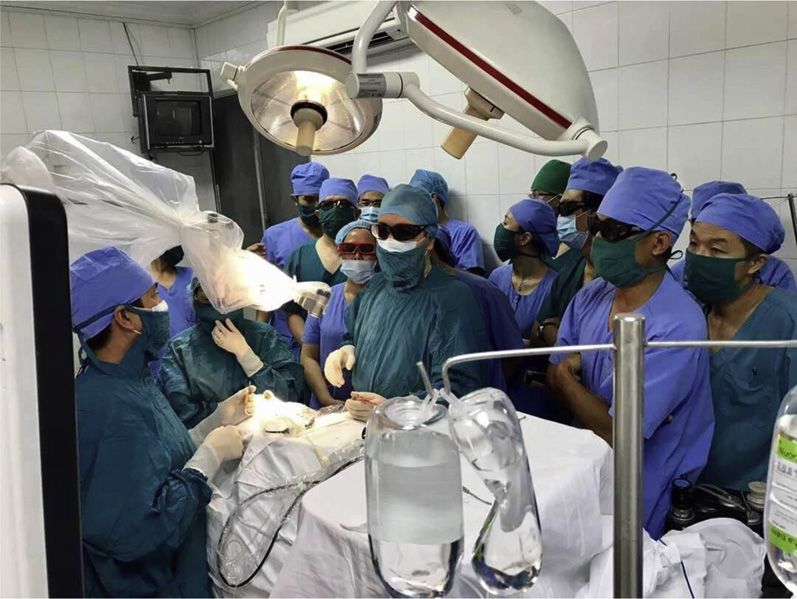
222        Nogueira et al

           approaches, including a wide visualization of the surgical field,10 enhanced optics
           with higher amplification, ocular access to hidden areas of the middle ear, avoid-
           ance of unnecessary incisions and soft tissue dissections, and a decreased oper-
           ative time and less postoperative morbidity. However, the main disadvantage of
           EES is that the surgery has to be performed with one single hand, which is certainly
           restrictive for an ear surgeon who has been trained operating with two hands under
           otologic microscopic views for years and this certainly requires a learning period
           and perseverance.
              Endoscopic instrumentation, techniques, and knowledge have improved during the
           last few years, and we believe that, in the future, endoscopic surgical techniques will
           gain even more importance in otologic surgery.

           ENDOSCOPIC EAR SURGERY EQUIPMENT

           Fundamental tools needed for endoscopic middle ear surgery incorporates11 a light
           source, rigid endoscopes (0 and 30 , and in some cases 45 ),12 and an HD3-CCD
           camera and video screen.13 Choices of different lights at different prices are easily
           accessible, such as halogen, light-emanating diode (LED), and xenon lights. There
           is no conclusive information advocating any of the sources of light and presently
           the choice depends on the surgeon’s inclinations and accessibility.14 It is important
           to mention that any of these light sources should never be at fully 100% capability.
           The middle ear cavity is small and to have a great view the surgeon needs no more
           than 40% of light power. This also reduces the probability of thermal injuries in middle
           ear structures.
              Endoscopic camera systems are accessible through an assortment of sellers. The
           fundamental necessity for a camera system is a 3-CCD camera. The 3-CCD cameras
           allow for high-definition and clear video picture quality by depending on individual
           CCDs for red, green, and blue light. Single CCD cameras tend to “red out” and
           become saturated when used in a small area that contains bleeding. Currently, oto-
           logic and sinus surgery light sources are indistinguishable and are commonly acces-
           sible in otolaryngology operating theaters. Although not essential, nowadays there are
           3-CCD high-definition (1080p) and 4K cameras. These produce a high-quality image
           (Figs. 1–3).
              The diameters for the rigid endoscopes used in ear surgery are usually 2.7, 3, and
           4 mm. If the outer canal is wide enough, EES or TEES is completed using a 4-mm

           Fig. 1. 3-CCD HD camera.

    Descargado para Irene Ramírez (iramirez@binasss.sa.cr) en National Library of Health and Social Security de ClinicalKey.es por
  Elsevier en enero 08, 2021. Para uso personal exclusivamente. No se permiten otros usos sin autorización. Copyright ©2021. Elsevier
                                                  Inc. Todos los derechos reservados.
Future of Endoscopic Ear Surgery                      223

  Fig. 2. Examples of the most used endoscopes in current EES.

  diameter scope. TEES endoscopic shaft lengths are ordinarily 11, 14, and 18 cm in
  length. There is no evidence to conclude that there is an ideal endoscopic measure-
  ment and the choice depends on the surgeon’s inclination, accessibility, and patient
  anatomy.14

  HOLDER

  Endoscope/camera holders have been designed to facilitate and allow two-handed
  EES. An ideal endoscope holder should be easy to set up, easily controlled, provide

  Fig. 3. Example of a 4K camera.

  Descargado para Irene Ramírez (iramirez@binasss.sa.cr) en National Library of Health and Social Security de ClinicalKey.es por
Elsevier en enero 08, 2021. Para uso personal exclusivamente. No se permiten otros usos sin autorización. Copyright ©2021. Elsevier
                                                Inc. Todos los derechos reservados.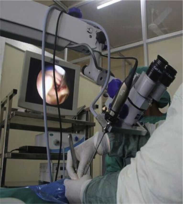
224        Nogueira et al

           a variety of angled views, and allow the surgeon to operate with two hands, thus facil-
           itating tympanomeatal flap elevation, delicate dissections of diseases from the ossic-
           ular chain, and allowing constant suctioning when needed for a blood-free operative
           site.15
              Although endoscope holders have been designed to facilitate two-handed proced-
           ures, there are still some certain technical difficulties: (1) because of the narrowness of
           the external ear canal, in addition to the endoscope, there must be sufficient room for
           two surgical instruments; (2) during hemorrhage, the manipulation of the endoscope in
           and out of the operative field may be time consuming; (3) if the patient moves abruptly
           during the operation, an endoscope placed in the fixed position could cause injury to
           the outer and/or middle ear canal; and (4) because of the heat generated and thermal
           trauma risk of the stationary endoscope in the middle ear certain questions are raised
           regarding its long-term safety (Fig. 4).16

           THREE-DIMENSIONAL ENDOSCOPIC EAR SURGERY

           EES started to gain popularity with the release of high-definition camera systems with
           image quality a thousand times more detailed than with a standard camera. Consid-
           ering the microscopic size of middle ear structures and the presence of important neu-
           rovascular structures in a small space the use of low-definition systems could
           negatively affect the performance and outcomes of EES. Since the studies by Presutti7
           and Marchioni and colleagues9,17,18 who used the new high-definition system to study
           the endoscopic anatomy of the attic and retrotympanum, EES started to become a
           widely accepted technique for the management of middle ear disease.
              Despite these technical improvements, the lack of depth perception provided by the
           two-dimensional (2D) vision and the need to operate with only one hand are the most
           cited limitation in EES. Even though an experienced endoscopic surgeon could over-
           come the loss of the sense of depth using other visual cues and anatomic knowledge,

           Fig. 4. Example of a holder model for two-handed EES.

    Descargado para Irene Ramírez (iramirez@binasss.sa.cr) en National Library of Health and Social Security de ClinicalKey.es por
  Elsevier en enero 08, 2021. Para uso personal exclusivamente. No se permiten otros usos sin autorización. Copyright ©2021. Elsevier
                                                  Inc. Todos los derechos reservados.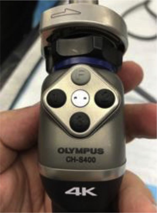
Future of Endoscopic Ear Surgery 225
a system that provides a three-dimensional (3D) image could be beneficial for begin-
ners and more experienced surgeons.
The use of a 3D high-definition system has already been reported in some experi-
mental studies in the laparoscopic and gynecologic fields.19,20 However, the diameter
(more than 4 mm) of the endoscope limited their use in ear surgery because the diam-
eter of the external auditory canal is an important limiting factor.
With the release of a 4-mm high-definition 3D endoscope, this technology is now
suitable for use in ear surgery (Figs. 5 and 6).
In their study, Bernardeschi and colleagues21 used 4-mm 0 and 30 angled 3D en-
doscopes. These endoscopes incorporated the HD camera with a resolution of
1920 1080 pixels with two integrated video chips. The frame rate was 50/60 Hz,
working length was 175 mm, and weight was 295 g. The light source was a POWER
LED 300 cold light source. It was possible to change from 3D to 2D high-resolution
images with the click of a button, which is an attractive feature for those surgeons
who are not comfortable with 3D vision.21
However this new system showed some limitations: (1) no diameters less than 4 mm
are available, and this could be an issue when dealing with a narrow external auditory
canal or in pediatric cases; (2) only two angled endoscopes (0 and 30 ) are currently
available; and (3) the 3D endoscope has digital zoom capability compared with the 2D
system, which has optical zoom, and this could induce some loss of resolution when
zooming with the 3D system.
THREE-DIMENSIONAL EXOSCOPES
The microscope has long been considered the best tool for microsurgical visualiza-
tion.22 However, it is limited by positioning at extreme angles, and otolaryngologists
are often forced into awkward positions with little freedom of motion for extended pe-
riods of time, which can explain the high prevalence of neck and back pain with long-
term detrimental effects.23
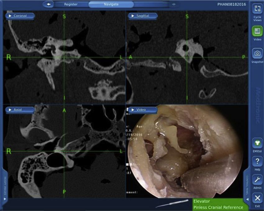
A new surgical visualization system called the 3D exoscope system is a viable alter-
native to surgical microscopes for otologic and neurotologic surgery.24,25 The 3D exo-
scope is meant to be exterior to the body surface like a microscope and to have dual
image sensors for 3D visualization. The images obtained from the 3D exoscope are
visualized on a monitor, and a surgeon observes 3D stereoscopic images wearing
3D glasses (Figs. 7 and 8).
Fig. 5. Example of a 3D endoscope.
Descargado para Irene Ramírez (iramirez@binasss.sa.cr) en National Library of Health and Social Security de ClinicalKey.es por
Elsevier en enero 08, 2021. Para uso personal exclusivamente. No se permiten otros usos sin autorización. Copyright ©2021. Elsevier
Inc. Todos los derechos reservados.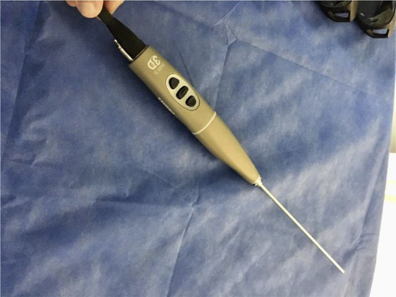
226        Nogueira et al

           Fig. 6. Example of 3D endoscope.

             An exoscope allows for excellent visualization at extreme angles because the sur-
           geons are able to independently position themselves and they no longer depend on
           the eyepiece but operate using the monitors. Better ergonomics also extends to the
           assistant and the entire surgical team, which also allows better instrument exchange
           and communication between team members (including residents, anesthesiologists,
           and nurses), allowing an active participation in procedures and education and thus
           increasing patient’s safety.26
             The main drawbacks of exoscopes are: (1) limited depth perception, which is over-
           come using newer monitors, such as 3D or 3D 4K monitors; (2) low lighting, especially
           when visualizing through narrow surgical fields; and (3) lower image quality when
           compared with the microscope, which can be improved with continued device
           development.

           4K TECHNOLOGY AND NARROW-BAND IMAGING IN OTOLOGY

           Some of the greatest advantages of using an endoscope includes the higher magnifi-
           cation and wide angled views, allowing the surgeon to access and remove disease
           from delicate areas in a safer manner.

           Fig. 7. Example of 3D exoscope.

    Descargado para Irene Ramírez (iramirez@binasss.sa.cr) en National Library of Health and Social Security de ClinicalKey.es por
  Elsevier en enero 08, 2021. Para uso personal exclusivamente. No se permiten otros usos sin autorización. Copyright ©2021. Elsevier
                                                  Inc. Todos los derechos reservados.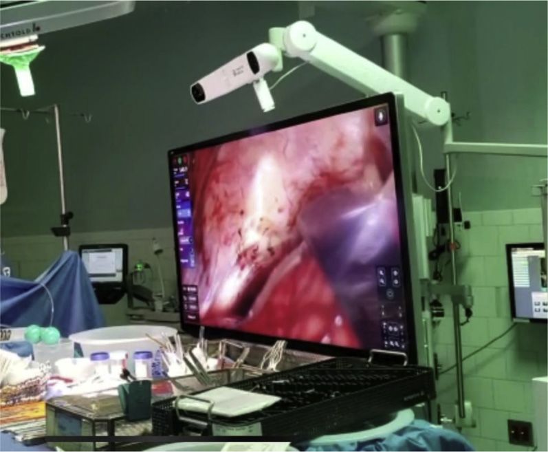
Future of Endoscopic Ear Surgery                      227

  Fig. 8. Exoscope in action during live surgery.

     Advances in optical technology have led to the development of high-definition visu-
  alization, such as 4K technology, which increases the magnification even further. The
  increased resolution improves image sharpness and provides a much more detailed
  view of all anatomic and pathologic structures (when compared with standard sys-
  tems currently in use), which is particularly important when working around delicate
  and critical structures, improving the safety and efficacy of the surgical procedure.
     Narrow band imaging (NBI), which has been implemented in oncologic surgery in all
  fields,27 uses a filter that allows different wavelengths of narrow band light (415 and
  540 nm wavelengths), in a sequential red-green-blue illumination, to pick up hypervas-
  cularity,27 which could indicate areas of dysplasia. Recently there has been interest in
  using this technology in noncancer cases.28
     In otologic surgery there is a difference in how structures and disease in the middle
  ear appear under the narrow band filter compared with ordinary white light. Because
  of the avascular nature of tympanosclerosis and cholesteatoma, they appear whiter
  than usual on NBI, whereas hypervascular structures, such as granulation tissue,
  are highlighted by the filter.28
     The association of 4K magnification with NBI technology could improve the identi-
  fication and dissection of middle ear pathology, such as cholesteatoma during middle
  ear surgery. The 4K magnification would allow a better visualization of the sinus
  tympani, facial recess, and stapes foot plate while dissecting the disease. However,
  NBI not only allows assessment of the extent of disease, but also ensures small resid-
  ual disease is not left behind at the time of operation.
     Further studies using the same NBI technology combined with 4K resolution and
  rigid scope are necessary to validate this extremely interesting potential.

  NEURONAVIGATION

  Advances in microsurgical techniques have led to an expansion of minimally invasive
  otologic/neurotologic procedures. Simultaneously, the complexity and proximity of
  critical anatomic structures, particularly on the skull base, sometimes requires the
  intraoperative use of navigation techniques.
     Surgical approaches through the temporal bone require some form of temporal
  bone drilling to create an adequate access toward the surgical target. Skull base sur-
  geons use anatomic landmarks as means of orientation during temporal bone drilling,
  to optimize access creation while minimizing bone removal and evading critical

  Descargado para Irene Ramírez (iramirez@binasss.sa.cr) en National Library of Health and Social Security de ClinicalKey.es por
Elsevier en enero 08, 2021. Para uso personal exclusivamente. No se permiten otros usos sin autorización. Copyright ©2021. Elsevier
                                                Inc. Todos los derechos reservados.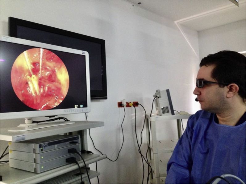
228        Nogueira et al

           structures, such as the facial nerve and sigmoid sinus. However, these landmarks are
           subject to high interindividual variability29 and is eroded by tumor, inflammation, or
           previous surgery.
              To reduce the risks during the procedure, surgical navigation systems have been
           increasingly used in otologic surgery.30,31 These image-guidance systems are devel-
           oped to help surgeons to identify critical anatomic landmarks intraoperatively; howev-
           er, it cannot substitute a thorough knowledge of the surgical anatomy. Initially
           developed for neurosurgical procedures, these systems use computerized tracking
           devices to monitor the position of the endoscopes or instruments relative to the pa-
           tient’s anatomic landmarks. The system displays the location of the tip of a tracked
           drill, and other instruments, in real-time, on a navigation map of the patient’s preoper-
           ative anatomy image (computed tomography or MRI).32,33
              However, the cost and lack of portability make the current image-guidance systems
           unavailable in many institutions and hard to transport to different hospitals.32,34
              In their study, Nogueira and colleagues35 used a third-generation camera and a pre-
           defined black and white pattern for optical tracking. The camera was attached in a
           holder at a distance from 60 cm to 100 cm from the patient’s marker, and it was linked
           to an up-to-date laptop through a standard firewire port. This port provides a power
           supply and a fast hub for information exchange, because the orientation functions
           needed to be performed repeatedly at real-time rates.35
              The laptop-based image-guidance system achieved an accuracy rate of 1.16 mm,
           showing its effectiveness and possible use in real patients (Figs. 9 and 10).

           NEW SURGICAL APPROACHES

           In recent years, technical improvements and growing expertise in the handling of the
           endoscope allowed introducing an exclusive endoscopic approach to the middle ear,
           lateral skull base, middle cranial fossa, and posterior fossa/cerebellopontine angle
           pathologies.
              Although the endoscope has been commonly used in transcanal middle ear sur-
           geries, which have proved to be highly successful, EES has also proven to be a
           feasible option in the approach of cochlear schwannoma involving internal auditory
           canal.

           Fig. 9. Neuronavigation in EES.

    Descargado para Irene Ramírez (iramirez@binasss.sa.cr) en National Library of Health and Social Security de ClinicalKey.es por
  Elsevier en enero 08, 2021. Para uso personal exclusivamente. No se permiten otros usos sin autorización. Copyright ©2021. Elsevier
                                                  Inc. Todos los derechos reservados.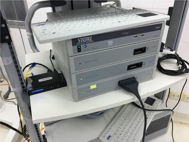
Future of Endoscopic Ear Surgery                      229

  Fig. 10. Neuronavigation in EES.

    There are no proven indications against the advisability of EES. Any otologic surgery
  that is conducted with a microscope can also use an endoscope. Therefore, these are
  some of the current indications for EES.
  1. External ear: Cholesteatoma, exostosis repair, canalplasty, debridement, and
     biopsy.
  2. Middle ear: Myringotomy, myringoplasty, medial graft tympanoplasty, lateral graft
     tympanoplasty, the retraction of the tympanic membrane, acquired cholesteatoma,
     congenital cholesteatoma, neoplasms of middle ear (eg, glomus tympanicum), os-
     siculoplasty, and stapes surgery.
  3. Inner ear/skullbase: Intracochlear schwannoma, small symptomatic neoplasms of
     internal auditory canal fundus of facial nerve, petrous apex cyst, and the repair of
     perilymph fistulas (congenital or traumatic).
  4. Middle cranial fossa: The repair of superior canal dehiscence.
  5. Posterior fossa/cerebellopontine angle: Establishing enduring schwannoma in in-
     ternal auditory canal fundus, and localization and sealing of externalized air cells
     during the decompression of internal auditory canal to reduce the risk of cerebro-
     spinal fluid leaks.

  DISCLOSURE

  The Authors have nothing to disclose.

  REFERENCES

    1. Thomassin JM, Duchon-Doris JM, Emram B, et al. Endoscopic ear surgery. Initial
       evaluation. Ann Otolaryngol Chir Cervicofa 1990;107:564–70.
    2. Thomassin JM, Korchia D, Doris JM. Endoscopic-guided otosurgery in the pre-
       vention of residual cholesteatomas. Laryngoscope 1993;103:939–43.

  Descargado para Irene Ramírez (iramirez@binasss.sa.cr) en National Library of Health and Social Security de ClinicalKey.es por
Elsevier en enero 08, 2021. Para uso personal exclusivamente. No se permiten otros usos sin autorización. Copyright ©2021. Elsevier
                                                Inc. Todos los derechos reservados.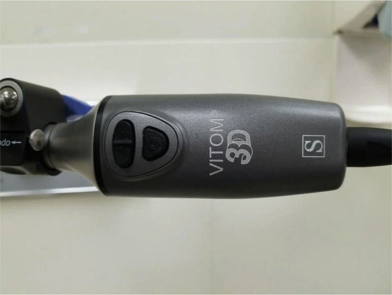
230        Nogueira et al

            3. Tarabichi M. Endoscopic management of acquired cholesteatoma. Am J Otol
               1997;18:544–9.
            4. Poe D, Rebeiz E, Pankratov M, et al. Transtympanic endoscopy of the middle ear.
               Laryngoscope 1992;102:993–6.
            5. McKennan KX. Endoscopic “second look” mastoidoscopy to rule out residual ep-
               itympanic/mastoid cholesteatoma. Laryngoscope 1993;103:810–4.
            6. Magnan J, Chays A, Lepetre C, et al. Surgical perspectives of endoscopy of the
               cerebellopontine angle. Am J Otol 1994;15:366–70.
            7. Presutti L, Marchioni D, Mattioli F, et al. Endoscopic management of acquired
               cholesteatoma: our experience. J Otolaryngol Head Neck Surg 2008;37(4):
               481–7.
            8. Tarabichi M. Endoscopic management of limited attic cholesteatoma. Laryngo-
               scope 2004;114(7):1157–62.
            9. Marchioni D, Alicandri-Ciufelli M, Molteni G, et al. Endoscopic tympanoplasty in
               patients with attic retraction pockets. Laryngoscope 2010;120(9):1847–55.
           10. Marchioni D, Alicandri-Ciufelli M, Piccinini A, et al. Inferior retrotympanum revis-
               ited: an endoscopic anatomic study. Laryngoscope 2010;120(9):1880–6.
           11. Kozin ED, Gulati S, Kaplan AB, et al. Systematic review of outcomes following
               observational and operative endoscopic middle ear surgery. Laryngoscope
               2015;125(5):1205–14.
           12. Bottrill I, Perrault D, Poe D. In vitro and in vivo determination of the thermal effect
               of middle ear endoscopy. Laryngoscope 1994;106:213–6.
           13. Kozin ED, Lehmann A, Carter M, et al. Thermal effects of endoscopy in a human
               temporal bone model: implications for endoscopic ear surgery. Laryngoscope
               2014;124(8):E332–9.
           14. Kozin ED, Daniel JL. Basic principles of endoscopic ear surgery. Oper Tech Oto-
               laryngol Head Neck Surg 2017;28:2–10.
           15. Khan MM, Parab SR. Endoscopic cartilage tympanoplasty: a two-handed tech-
               nique using an endoscope holder. Laryngoscope 2016;126:1893–8.
           16. Ito T, Kubota T, Takagi A, et al. Safety of heat generated by endoscope light sour-
               ces in simulated transcanal endoscopic ear surgery. Auris Nasus Larynx 2016;
               43:501–6.
           17. Marchioni D, Mattioli F, Alicandri-Ciufelli M, et al. Endoscopic approach to tensor
               fold in patients with attic cholesteatoma. Acta Otolaryngol (Stockh) 2009;129:
               946–54.
           18. Marchioni D, Mattioli F, Alicandri-Ciufelli M, et al. Transcanal endoscopic
               approach to the sinus tympani: a clinical report. Otol Neurotol Otol Neurotol
               2009;30:758–65.
           19. Storz P, Buess GF, Kunert W, et al. 3D HD versus 2D HD: surgical task efficiency
               in standardised phantom tasks. Surg Endosc 2012;26:1454–60.
           20. Spille J, Wenners A, von Hehn U, et al. 2D versus 3D in laparoscopic surgery by
               beginners and experts: a randomized controlled trial on a Pelvitrainer in objec-
               tively graded surgical steps. J Surg Educ 2017. https://doi.org/10.1016/j.jsurg.
               2017.01.011.
           21. Bernardeschi D, Lahlou G, Seta D, et al. 3D endoscopic ear surgery: a clinical
               pilot study. Eur Arch Otorhinolaryngol 2017. https://doi.org/10.1007/s00405-
               017-4839-6.
           22. Uluc K, Kujoth GC, Baskaya MK. Operating microscopes: past, present, and
               future. Neurosurg Focus 2009;27:E4.
           23. Carlucci C, Fasanella L, Ricci Maccarini A. Exolaryngoscopy: a new technique for
               laryngeal surgery. Acta Otorhinolaryngol Ital 2012;32:326–8.

    Descargado para Irene Ramírez (iramirez@binasss.sa.cr) en National Library of Health and Social Security de ClinicalKey.es por
  Elsevier en enero 08, 2021. Para uso personal exclusivamente. No se permiten otros usos sin autorización. Copyright ©2021. Elsevier
                                                  Inc. Todos los derechos reservados.
Future of Endoscopic Ear Surgery 231
24. Rossini Z, Cardia A, Milani D, et al. VITOM 3D: preliminary experience in cranial
surgery. World Neurosurg 2017;107:663–8.
25. Sack J, Steinberg JA, Rennert RC, et al. Initial experience using a high-definition
3-dimensional exoscope system for microneurosurgery. Oper Neurosurg 2018;
14:395–401.
26. Mamelak AN, Drazin D, Shirzadi A, et al. Infratentorial supracerebellar resection
of a pineal tumor using a high definition video exoscope (VITOM). J Clin Neurosci
2012;19:306–9.
27. Ochsner M, Klein A. The utility of narrow band imaging in the treatment of laryn-
geal papillomatosis in awake patients. J Voice 2015;29(3):349–51.
28. Plaat B, Zwakenberg MA, Zwol JGV, et al. Narrow-band imaging in transoral laser
surgery for early glottic can-cer in relation to clinical outcome. Head Neck 2017;
39(7):1343–8.
29. Gharabaghi A, Rosahl SK, Feigl GC, et al. Image-guided lateral suboccipital
approach: part 1-individualized landmarks for surgical planning. Neurosurgery
2008;62:18–22.
30. Miller RS, Hashisaki GT, Kesser BW. Image-guided localization of the internal
auditory canal via the middle cranial fossa approach. Otolaryngol Head Neck
Surg 2006;134:778–82.
31. Woerdeman PA, Willems PW, Noordmans HJ, et al. Auditory feedback during
frameless image-guided surgery in a phantom model and initial clinical experi-
ence. J Neurosurg 2009;110:257–62.
32. Stamm AC, Pignatari S, Sebusiani S, et al. Image-guided endoscopic sinus and
skull base surgery. Rev Bras Otorinolaringol 2002;68:502–9.
33. Metson R, Cosenza M, Gliklish RE, et al. The role of image-guidance systems for
head and neck surgery. Arch Otolaryngol Head Neck Surg 1999;125:1100–4.
34. Chu ST. Endoscopic sinus surgery under navigation system: analysis report of 79
cases. J Chin Med Assoc 2006;69:529–33.
35. Nogueira JF Jr, Stamm AC, Lyra M. Novel compact laptop-based image-
guidance system: preliminary study. Laryngoscope 2009;119:576–9.
Descargado para Irene Ramírez (iramirez@binasss.sa.cr) en National Library of Health and Social Security de ClinicalKey.es por
Elsevier en enero 08, 2021. Para uso personal exclusivamente. No se permiten otros usos sin autorización. Copyright ©2021. Elsevier
Inc. Todos los derechos reservados.You can also read