Middle ear polyps: results of traction avulsion after a lateral approach to the ear canal in 62 cats (2004-2014) - Dermato clinica
←
→
Page content transcription
If your browser does not render page correctly, please read the page content below
660356
research-article2016
JFM0010.1177/1098612X16660356Journal of Feline Medicine and SurgeryJanssens et al
Original Article
Journal of Feline Medicine and Surgery
Middle ear polyps: results of traction 1–6
© The Author(s) 2016
Reprints and permissions:
avulsion after a lateral approach to sagepub.co.uk/journalsPermissions.nav
DOI: 10.1177/1098612X16660356
the ear canal in 62 cats (2004–2014) jfms.com
This paper was handled and processed
by the European Editorial Office (ISFM)
for publication in JFMS
Sara DS Janssens1, Annika N Haagsman1 and Gert Ter Haar2
Abstract
Objectives The objective of this study was to report the surgical outcome and complication rate of deep traction
avulsion (TA) of feline aural inflammatory polyps after a lateral approach (LA) to the ear canal.
Methods This was a retrospective analysis of data retrieved from an electronic database of 62 cats treated with TA
after an LA (TALA) for removal of ear canal polyps. Long-term outcome was assessed via a telephone questionnaire
survey with the owners.
Results Domestic shorthair cats (48%) and Maine Coons (37%) were over-represented. The most common
presenting clinical signs were otorrhoea, ear scratching and head shaking. Video-otoscopic examination confirmed
a polypous mass in the ear canal in all patients. All 62 cats underwent TALA with a mean surgical time of 33 mins
for experienced surgeons (n = 4) and 48 mins (n = 12) for less experienced surgeons. The recurrence rate of
polyp regrowth for experienced surgeons was 14.3% vs 35% for the less experienced surgeons. Postoperative
complications included Horner’s syndrome (11.5%) and facial nerve paralysis (3%). Otitis interna was not observed.
Conclusions and relevance A lateral approach to the ear canal in combination with deep TA of an aural inflammatory
polyp is an effective first-line technique that results in a low recurrence and complication rate.
Accepted: 16 June 2016
Introduction least invasive technique, yet has the highest documented
Middle ear polyps in cats are relatively common benign recurrence rate (up to 50%).1,17 Recently, however, better
masses that arise from the mucosal lining of the middle results have been reported using a per-endoscopic tran-
ear, Eustachian tube or the nasopharynx.1-5 Whereas stympanic traction technique, which resulted in polyp
many cats will not demonstrate any specific clinical recurrence in only 13.5% of the 37 cats treated.
signs associated with middle ear polyps, extension of the Complications are minimal with these techniques.21 A
polyps beyond the boundaries of the middle ear leads to ventral bulla osteotomy (VBO) is the most invasive tech-
signs of otitis externa, otitis interna or nasopharyngitis. nique with the highest associated complication risks but
Diagnosis of polyps is relatively straightforward when with the lowest recurrence rates (0–8%).1,16,17,20,22 In one
polyps have protruded into the ear canal or into the relatively large study with 19 cats treated with VBO,
nasopharynx where they can easily be demonstrated reported postoperative complications included Horner’s
using otoscopy and nasopharyngoscopy. Advanced syndrome (n = 11), otitis interna (n = 2) and facial nerve
imaging in the form of CT or MRI is advised for cats that
have disease confined to the middle ear cavity, and have
1Department of Clinical Sciences of Companion Animals, Utrecht
concomitant nasal disease or otitis interna.6-15
University, Utrecht, The Netherlands
The treatment of middle ear polyps that have pro- 2Department of Clinical Sciences and Services, Royal Veterinary
truded beyond the middle ear cavity is surgical in all College, London, UK
cases. Described techniques for polyp removal from the
ear canal include traction avulsion (TA), ventral bulla Corresponding author:
Sara DS Janssens DVM, Department of Clinical Sciences of
osteotomy, lateral ear resection and total ear canal abla-
Companion Animals, Utrecht University, Yalelaan 108, Utrecht
tion with lateral bulla osteotomy, although the latter two 3584 CM, The Netherlands
techniques are not commonly advised.1-3,16-20 TA is the Email: s.d.s.janssens@uu.nl
Downloaded from jfm.sagepub.com at COLORADO STATE UNIV LIBRARIES on August 26, 2016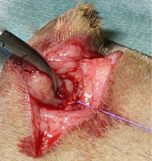
2 Journal of Feline Medicine and Surgery
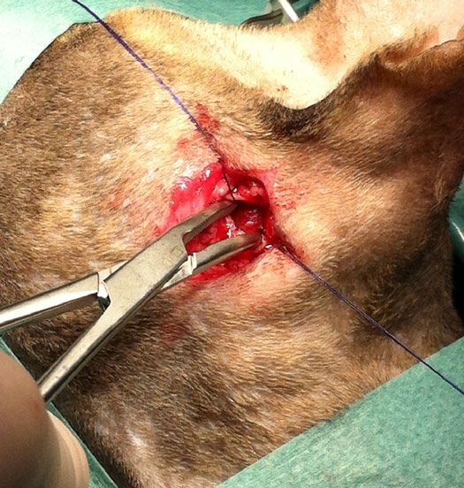
Figure 1 The forceps is gently rotated while grasping the Figure 2 Traction is applied until the polyp avulses
aural polyp to make sure no other tissue has been grasped
After aseptic preparation of the surgical site, an inci-
paralysis (n = 5), and the recurrence rate was 3–5%.20 sion was made in the skin in a dorsoventral direction
Venker-van Haagen introduced TA after a lateral approach over the palpable vertical ear canal, starting just cranio-
(LA) to the ear canal (TALA) as a simple, quick and effec- ventral to the tragus over approximately 2.5 cm.5,23 The
tive method bypassing complications seen with VBO and subcutaneous tissues were dissected with small scissors
generally leading to a low recurrence rate.5 Whereas this to free the cartilage of the vertical ear canal to the level of
technique has been used for decades, postoperative com- the junction between the auricular and annular carti-
plications, long-term outcome and recurrence rates using lages, reflecting the parotid gland. A vertical stab incision
this technique have so far not been reported in the veteri- was made ventrally to dorsally in the auricular cartilage
nary literature. Therefore, the aim of the present study just above this junction with a Bard Parker scalpel handle
was to report epidemiological data, clinical signs, postop- with a no. 11 blade over 7–10 mm.5,23 Stay sutures were
erative complications and long-term surgical outcomes of placed on both sides of the incision in the ear canal carti-
TALA for removal of middle ear polyps. lage with fine monofilament suture material to increase
visualisation and avoid damaging the cartilage. Small
Materials and methods closed curved haemostatic forceps were then introduced
Animals and data collection into the ear canal, meticulously following the direction of
Data from all cats treated surgically by a lateral approach the horizontal ear canal until the polyp was encountered.
at our department between December 2004 and June The forceps were then opened and advanced deeper over
2014 were collected retrospectively from an electronic the polyp until it could be grasped as close as possible to
database. Information collected included age, breed, sex, the osseous meatus (Figure 1). When a firm grip had been
clinical signs, findings during initial physical examina- achieved, the forceps were gently rotated to make sure no
tion, otoscopic findings, diagnostic imaging results, other tissue than the polyp itself had been grasped and
surgeons performing the procedure, results of histopa- traction was applied until the polyp avulsed from its ori-
thology and follow-up examinations. Patients were gin (Figure 2). With complete removal of a classical mid-
excluded from this study if digital files were incomplete dle ear inflammatory polyp, a small stalk at the base of
or patients were lost to follow-up. the polyp could usually be identified. The middle ear
cavity was flushed with warm saline and with a small
Surgery curette the osseous meatus and most lateral aspect of the
Preoperative protocol for all cats consisted of a thorough tympanic cavity was ‘palpated’ to check for additional
otoscopic examination to determine the presence of a visi- inflammatory tissue, which was removed with this
ble polyp on the affected site. curette when encountered.23 The stay sutures were
Downloaded from jfm.sagepub.com at COLORADO STATE UNIV LIBRARIES on August 26, 2016Janssens et al 3
Table 1 Preoperative clinical signs in 62 cats with a study. Domestic shorthair cats and Maine Coons were
middle ear polyp over-represented in our population, 48% (30/62) and
37% (23/62), respectively. Other breeds that were pre-
Clinical signs sented more than once included the Norwegian Forest
Head tilt 20 (32) Cat (2/62; 3%) and the Oriental Shorthair (2/62; 3%).
Ataxia 16 (26) The mean age of affected cats was 3.9 years (range 0.5–14
Anorexia 3 (5) years) with a median of 2 years. There were 27 (44%)
Otorrhoea 55 (89) female cats, 81% of which were neutered, and 35 (56%)
Ear scratching 40 (65) male cats of which 80% were neutered.
Head shaking 42 (68)
Deafness 10 (16) Clinical signs and imaging results
Horner’s syndrome 1 (2) Presenting clinical signs related to the presence of an ear
polyp are listed in Table 1. The most common clinical
Data are n (%)
signs were otorrhoea, ear scratching and head shaking.
Signs such as ataxia, anorexia, Horner’s syndrome and
removed and the cartilage of the ear canal was closed deafness were rarely seen. Diagnostic imaging in the form
with 4-0 monofilament suture material in an interrupted of a CT scan was performed in 16/62 cats included in this
pattern; three or four sutures were usually sufficient. The study. Eight cats underwent CT as part of the initial work-
subcutis was closed in a continuous pattern with 4-0 up prior to surgery and 10 when presented with signs of
absorbable monofilament material and the skin was recurrence (including two that had CT previously, at ini-
closed in a subdermal suture pattern using the same tial work-up). Only the first CT scan (16 cats) results were
material. reviewed for this study. A soft tissue opacity within the
Postoperative management consisted of amoxicillin- tympanic bulla was seen in all cases, with additional osse-
clavulanic acid (Synulox; Pfizer Animal Health) or enro- ous changes/thickening of the bulla wall in 9/16.
floxacin (Baytril; Bayer Animal Health) for 7 days for Extension of soft tissue opacity from the middle ear
cats without clinical signs of otitis interna and for 14–21 through the external bony meatus into the ear canal was
days for those presenting with signs of otitis interna, and seen in 12/16 cases. In one cat the soft tissue opacity
meloxicam (Metacam cat; Boehringer Ingelheim) for 5 extended to the level of the external bony meatus and in
days. No corticosteroids were administrated. three cats this specific information could not be retrieved.
In six cats, in addition to the middle ear and ear canal
abnormalities reported above, a protrusion of polypous
Questionnaire
tissue towards the nasopharynx was diagnosed, based on
Each owner was contacted by telephone at least 6 months
the presence of a soft tissue opacity at the level of the
after intervention (follow-up 6 months to 10 years) and
Eustachian tube opening in the nasopharynx (9.7%).
asked the following questions: (1) recovery time after
surgery (estimated number of weeks for clinical signs to
disappear completely: 1–2, 2–3, 3–4, 5–10, >10 weeks); Outcome
(2) presence of postsurgical residual signs (head tilt, No complications were encountered during surgery in
ataxia, anorexia, otorrhoea, ear scratching, head shaking, all cases. Postoperative complications included Horner’s
facial nerve paralysis and Horner’s syndrome); (3) clini- syndrome in seven cases and facial nerve paralysis in
cal signs fitting with recurrence of polyp growth after two. Vestibular signs were not observed. All patients
initial full recovery; (4) overall owner satisfaction as were discharged from the hospital.
evaluated on a 1–4 analogue scale (1 = displeased, 4 = Forty-seven (75.8%) cat owners indicated that their cats
very satisfied). Medical terminology was explained dur- had completely recovered following surgery and were not
ing the conversation with the owner. exhibiting any residual signs. Clinical signs had disap-
peared within 1–3 weeks in 91% of these animals, with a
Statistical analysis mean of 2.6 weeks. In six of the 47 (12.7%) cats that made
Collected data were statistically analysed using SPSS an initial full recovery after the surgery, clinical signs com-
software (V.20.0; IBM). A Kaplan–Meier was performed patible with possible recurrence of polyp growth devel-
to evaluate the disease free interval. Cox regression oped 12, 21, 23, 34, 41 and 101 months postoperatively,
analysis was preformed to evaluate significant differences respectively. For these late recurrences the median dis-
(P4 Journal of Feline Medicine and Surgery
recurrences) and confirmed the presence of an inflam-
matory polyp in all.
Discussion
The aim of this study was to evaluate the efficacy, long-
term outcome of and complications associated with
TALA of aural inflammatory polyps in cats.
The current study demonstrates that TALA of an
aural polyp is a time-efficient surgical technique, associ-
ated with an uneventful postoperative recovery, minor
postoperative complications and a low recurrence rate of
14% for experienced surgeons. Although the surgery is
relatively easy to perform, a learning curve is associated
with the technique as both surgical time and recurrence
rates depend on the experience of the surgeon.
No apparent breed or sex predilection has been
reported for aural polyps, but it has been noted by many
Figure 3 Kaplan–Meier curve of disease-free interval in months authors that polyp formation is more commonly seen in
specific cat breeds like the Norwegian Forest, Sphynx,
otorrhoea, head shaking and ear scratching persisted Maine Coon, Persian, Ragdoll and Abyssinian.1-3,18,24,25
postoperatively beyond 4 weeks. Recurrence of (or resid- Similar to what has been reported before, domestic
ual) polyp growth was confirmed at re-examination in shorthair cats were over-represented in our study.1,21,25-28
our clinic using CT scan and/or video-otoscopy in 8/15 In contrast to previous publications, however, the Maine
(53%) cases. Of the other seven cats with persisting clini- Coon was also over-represented (37%). Although this
cal signs after surgery two owners declined further confirms previous anecdotal evidence, further research
work-up and five cats were lost to follow-up. is needed to see if this reflects a bias in our study popula-
Auxiliary surgery in the cats with confirmed regrowth tion or truly reflects a breed predisposition.
of polyps consisted of polyp removal via TA under The mean age at presentation, male–female distribu-
video-otoscopic guidance (3/12), VBO (7/12) or a sec- tion and the most common clinical findings (ie, otor-
ond lateral approach as described above (2/12). rhoea, head shaking and ear scratching) were similar to
An experienced ear, nose and throat surgeon had per- those that have been previously reported.1,2,5,20,21,24,26
formed the polyp removal in 40 cats included in this study. Clinical signs associated with otitis interna (ie, vestibular
In the remainder of the cases (n = 22), the procedure had ataxia, anorexia and deafness) were less commonly seen
been carried out by a less experienced surgeon (resident). in our study compared with previous reports.4 Only 16
Mean surgical time was 33 mins for experienced surgeon cats presented with ataxia in our study, which resolved
and 48 mins for the less experienced surgeon. in all cats after surgical removal of the polyp and con-
There were no significant differences in recurrence secutive antibiotic treatment. Prognosis of animals pre-
rates between the experienced surgeon, (average recur- senting with vestibular signs did not appear to be worse
rence rate was 14.3% vs 35% for the less experienced sur- than that of those presenting without. Recurrence rates
geon). The cats with confirmed recurrences (n = 13) were similar for both groups.
included seven domestic shorthairs, five Maine Coons Advanced diagnostic imaging was not performed in
and one Birman. Recurrence was seen in six females most animals included in this study and diagnosis was
(6/27; 22%) vs seven male cats (7/35; 20%). There were confirmed by (video-)otoscopy in all cases with subse-
no significant differences in recurrence rates between quent histopathological evaluation in 60% of these. In
males and females, and neutered and intact cats. most of the cases where recurrence was suspected,
A Kaplan–Meier analysis was performed to evaluate advanced diagnostic imaging was performed. Even
the time between surgery and recurrence of polyp though polyps can be visualised on radiographs in
growth (disease-free interval) (Figure 3). For this pur- many cases,18,24,25,29 for proper (surgical) treatment
pose, a time frame between surgery (t = 0) and the time planning, CT of the entire skull is advised. CT also
of recurrence of polyp growth, or the time at which the offers a higher true-positive diagnostic rate for the
case was censored was used. Cases were censored when detection of otitis media, otitis interna and provides
recurrence of polyp growth did not occur (48 cats). At 1 information on concomitant changes in the nasal cavity,
year 97% and at 2 years 88% of the cats had survived nasopharynx and frontal sinuses.30,31 Findings on diag-
without recurrence of a polyp. Histopathological exami- nostic imaging in our population were similar to
nation was performed in 39 cases (including 10 of the 12 changes associated with ear canal and middle ear
Downloaded from jfm.sagepub.com at COLORADO STATE UNIV LIBRARIES on August 26, 2016Janssens et al 5
polyps reported before. Eight patients received a CT TALA is associated with minor complications of
before the initial surgery and involvement of the mid- transient nature. Peri- and postoperative bleeding, and
dle ear was demonstrated in all. Only three of these vestibular signs upon recovery were not noted in any of
eight cats developed a recurrence of the polyp, despite the cats. Horner’s syndrome was seen in seven cats
obvious changes in the middle ear. Whereas definite (11.5%). Two cats developed postoperative facial nerve
conclusions cannot be drawn based on our data, it does paralysis, one of which had resolved at the time of
suggest that despite middle ear involvement on CT follow-up. The other cat was lost to follow-up. An inex-
imaging TALA is usually curative. Further studies spe- perienced resident performed the surgeries in the latter
cifically designed to evaluate the use of CT as a predic- two cats and facial nerve damage could have resulted
tor of outcome after TALA are required in a larger from inadvertent ventral dissection beyond the level of
population. Diagnostic imaging such as CT, based on the transition between vertical and horizontal ear canal.
our results, is advised for cats that require more elabo- Owing to the retrospective nature of this study, it is
rate surgical procedures such as ventral bulla osteot- possible that some minor complications (ie, Horner’s
omy or present with recurrence after polyp removal. syndrome) were under-reported. Major complications
In this study histopathological confirmation of the including otitis interna and/or facial nerve paralysis
diagnosis was, unfortunately, only performed in 63% of most likely would have been reported. The number of
the cases. Although this would have ideally been per- complications appears comparable with previous
formed in all cases, especially in the older animals,3,24-26 reports on TA techniques, including the PTT study
the absence of recurrence of signs after removal of the where three cats (8%) developed Horner’s syndrome,25
mass in the ear canal indicates the presumptive diagnosis but is much higher after VBO. Horner’s syndrome is
was correct. However, this clearly presents a limitation observed in 25–80% of the cases,26,32 and vestibular
inherent to the retrospective study design. As a general signs after VBO range from 4–42%.16,17 Facial nerve
rule, histopathology should be performed for accurate paralysis was seen in 16% of 18 procedures,24 but when
diagnosis in all cases. Histopathology was performed in accompanied by total ear canal ablation the incidence
83% of the animals that presented with recurrence, which can increase up to 31%.33 In contrast to vestibular symp-
confirmed the diagnosis of inflammatory polyp in all. toms and/or facial nerve paralysis, Horner’s syndrome
TA and VBO have been the most commonly used is considered only a mild complication. The condition
techniques for treatment of aural inflammatory polyps. does not affect quality of life and often resolves in 2–4
VBO is associated with a low recurrence rate (0–8%) but weeks.26
is more invasive and is associated with a higher inci- Recurrence of polyp growth was seen in 13 cats of the
dence of iatrogenic complications (Horner’s syndrome, 62 included in this study. The choice between a lateral
facial nerve paralysis, otitis interna).20 In this study approach and a VBO for revisional surgery was made
TALA was used as a first-choice treatment for all cases based on the size of the polyp and the owners prefer-
with ear canal polyps visible upon otoscopy, regardless ences. Very small polyps that barely extend beyond the
of imaging results, indicating middle ear involvement or external bony meatus are difficult to grasp using the lat-
clinical signs indicative of otitis interna. This technique eral approach. For polyps properly protruding beyond
resulted in a much lower recurrence rate of 14.3% com- the external bony meatus, a lateral approach can be
pared with other described TA techniques (around 50% repeated. For very small polyps, a VBO is advised, espe-
recurrence) and a similar recurrence rate as per- cially when dealing with possibly multiple small polyps
endoscopic transtympanic TA (PTT). Whereas recur- confined to the middle ear cavity.
rence rates are higher than reported for VBO, TALA is
easy to perform, efficient and not associated with major Conclusions
complications (ie, bleeding, facial nerve paralysis). Inflammatory polyps in the ear canal and middle ear are
Another major advantage of this lateral approach is the frequently encountered in cats. Whereas diagnosis
short anaesthetic and surgical time compared with heavily relies on visualisation of a polypous growth in
previously described surgical techniques. The average the ear canal during otoscopy, advanced imaging such
surgical time amounted to only 33 mins for experienced as CT can be a useful aid. First-line treatment for removal
ear, nose and throat surgeons, and 48 mins for less of polyps can consist of a lateral approach to the ear
experienced surgeons. The study on PTT reported a min- canal in combination with TA of the polyp. This study
imal surgery duration of 45 mins and a maximum of 2.5 demonstrates this is an effective, fast and inexpensive
h.21 Although the latter is a minimally invasive technique technique that results in a low recurrence and complica-
associated with minor complications, the present study tion rate. The technique can be performed by first-line
proved to have similar results in outcome and complica- practitioners but is associated with a higher recurrence
tion rates with a significantly shorter surgical time and rate if not performed by a surgeon familiar with the
without the need for endoscopic equipment. technique.
Downloaded from jfm.sagepub.com at COLORADO STATE UNIV LIBRARIES on August 26, 20166 Journal of Feline Medicine and Surgery
Acknowledgements We would like to acknowledge 16 Faulkner J and Budsberg S. Results of ventral bulla oste-
Sharon Rook for her assistance with study data retrieval. otomy for treatment of middle ear polyps in cats. J Am
Further, we would like to acknowledge Dr Jan van den Broek Anim Hosp Assoc 1990; 26: 496–499.
for his assistance in the statistical analysis. 17 Kapatkin A, Matthiesen D, Noone K, et al. Results of sur-
gery and long-term follow-up in 31 cats with nasopharyn-
Conflicts of interest The authors declared no potential con- geal polyps. J Am Anim Hosp Assoc 1990; 26: 387–392.
flicts of interest with respect to the research, authorship, and/ 18 Muilenburg RK and Fry TR. Feline nasopharyngeal pol-
or publication of this article. yps. Vet Clin North Am Small Anim Pract 2002; 32: 839–849.
19 Schmidt J and Kapatkin A. Nasopharyngeal and ear canal
Funding The authors received no financial support for the polyps in the cat. Feline Pract 1990; 18: 16–19.
research, authorship, and/or publication of this article. 20 Trevor PB and Martin RA. Tympanic bulla osteotomy for
treatment of middle-ear disease in cats: 19 cases (1984–
References 1991). J Am Vet Med Assoc 1993; 202: 123–128.
1 Anderson DM, Robinson RK and White RA. Management of 21 Greci V, Vernia E and Mortellaro CM. Per-endoscopic
inflammatory polyps in 37 cats. Vet Rec 2000; 147: 684–687. trans-tympanic traction for the management of feline
2 Fan TM and de Lorimier LP. Inflammatory polyps and aural inflammatory polyps: a case review of 37 cats.
aural neoplasia. Vet Clin North Am Small Anim Pract 2004; J Feline Med Surg 2014; 16: 645–650.
34: 489–509. 22 Bradley R, Noone K, Saunders GK, et al. Nasopharyngeal
3 Pope ER. Feline inflammatory polyps. Semin Vet Med Surg and middle ear polypoid masses in five cats. Vet Surg
(Small Anim) 1995; 10: 87–93. 1985; 14: 141–144.
4 Haar Ter G. Inner ear dysfunction related to ear disease in 23 Haar Ter G. Basic principles of surgery of the external
dogs and cats. Eur J Companion Anim Pract 2006; 16: 127–136. ear (pinna and ear canal). In: Kirpensteijn J and Klein WR
5 Venker-van Haagen A. The ear. In: Venker-van Haagen A (eds). The cutting edge: basic operating skills for the veteri-
(eds). Ear, nose, throat, and tracheobronchial diseases in nary surgeon. London, UK: Roman House Publishers, 2006,
dogs and cats. Hannover: Schlütersche, 2005, pp 1–50. pp 272–283.
6 Allgoewer I, Lucas S and Schmitz SA. Magnetic resonance 24 Davidson J. Otopharyngeal polyps. In: Bojrab M (eds).
imaging of the normal and diseased feline middle ear. Vet Current techniques in small animal surgery. 4th ed. Phila-
Radiol Ultrasound 2000; 41: 413–418. delphia: Lea & Febiger, 1998, pp 147–150.
7 Barthez PY, Koblik PD, Hornof WJ, et al. Apparent wall 25 Harvey CE and Goldschmidt MH. Inflammatory polypoid
thickening in fluid filled versus air filled tympanic bulla growths in the ear canal of cats. J Small Anim Pract 1978; 19:
in computed tomography. Vet Radiol Ultrasound 1996; 37: 669–677.
95–98. 26 Anders BB, Hoelzler MG, Scavelli TD, et al. Analysis of
8 Dayrell-Hart BL. MR imaging of the vestibular apparatus auditory and neurologic effects associated with ventral
of the dog. In: Proceedings 15th ACVIM Forum, 1997, May bulla osteotomy for removal of inflammatory polyps or
22–25, Orlando, FL: American College of Veterinary Inter- nasopharyngeal masses in cats. J Am Vet Med Assoc 2008;
nal Medicine, 1997, pp 606–607. 233: 580–585.
9 Dvir E, Kirberger RM and Terblanche AG. Magnetic reso- 27 Lane J, Orr C, Lucke V, et al. Nasopharyngeal polyps aris-
nance imaging of otitis media in a dog. Vet Radiol Ultra- ing in the middle ear of the cat. J Small Anim Pract 1981; 22:
sound 2000; 41: 46–49. 511–522.
10 Garosi LS, Lamb CR and Targett MP. MRI findings in a 28 Veir JK, Lappin MR, Foley JE, et al. Feline inflammatory
dog with otitis media and suspected otitis interna. Vet Rec polyps: historical, clinical, and PCR findings for feline
2000; 146: 501–502. calici virus and feline herpes virus-1 in 28 cases. J Feline
11 Garosi LS, Dennis R, Penderis J, et al. Results of mag- Med Surg 2002; 4: 195–199.
netic resonance imaging in dogs with vestibular disor- 29 Kirpensteijn J. Aural neoplasms. Semin Vet Med Small Anim
ders: 85 cases (1996–1999). J Am Vet Med Assoc 2001; 218: 1993; 8: 17–23.
385–391. 30 Bischoff MG and Kneller SK. Diagnostic imaging of the
12 Garosi LS, Dennis R and Schwarz T. Review of diagnostic canine and feline ear. Vet Clin North Am Small Anim Pract
imaging of ear diseases in the dog and cat. Vet Radiol Ultra- 2004; 34: 437–458.
sound 2003; 44: 137–146. 31 Detweiler DA, Johnson LR, Kass PH, et al. Computed tomo-
13 Hoskinson JJ. Imaging techniques in the diagnosis of middle graphic evidence of bulla effusion in cats with sinonasal
ear disease. Semin Vet Med Surg (Small Anim) 1993; 8: 10–16. disease: 2001–2004. J Vet Intern Med 2006; 20: 1080–1084.
14 Russo M, Covelli EM, Meomartino L, et al. Computed 32 Hardie EM. Surgical diseases of the middle ear. In: Mon-
tomographic anatomy of the canine inner and middle ear. net E (eds). Small animal soft tissue surgery. Iowa: John
Vet Radiol Ultrasound 2002; 43: 22–26. Wiley & Sons, 2012, pp 149–156.
15 Seitz SE, Losonsky JM and Marretta SM. Computed tomo- 33 Sharp N. Chronic otitis externa and otitis media treated
graphic appearance of inflammatory polyps in three cats. by total ear canal ablation and ventral bulla osteotomy in
Vet Radiol Ultrasound 1996; 37: 99–104. thirteen dogs. Vet Surg 1990; 19: 162–166.
Downloaded from jfm.sagepub.com at COLORADO STATE UNIV LIBRARIES on August 26, 2016You can also read