Clinical application of COVID-19 Reporting and Data System in computed tomography of bilateral pneumonia diagnostic: a literature review ...
←
→
Page content transcription
If your browser does not render page correctly, please read the page content below
JOURNAL AVAILABLE AT RADIOLOGYUPDATE.ORG

Clinical application of COVID-19 Reporting
and Data System in computed tomography of
bilateral pneumonia diagnostic: a literature
review
Ugnė Kulnickaitė1, Laura Dobrovaitė1, Kamilė Grigaitė1

Faculty of Medicine, Lithuanian University of Health Sciences, Kaunas, Lithuania
1

    ABSTRACT
    Background. The 2019 coronavirus disease pandemic (COVID-19) has spread at an astonishing speed across the world,
    causing major morbidity and mortality. Computed tomography (CT) examination plays an important role in crisis
    areas in the diagnosis of COVID-19. COVID-19 Reporting and Data System (CO-RADS) has a five-point scale of suspi-
    cion for COVID-19 pneumonia in chest CT picture which standardizes the evaluation scheme and simplifies reporting.
    Aim. To summarise and present the role of COVID-19 Reporting and Data System in computed tomography of bilat-
    eral pneumonia diagnostic.
    Materials and methods. Recently published studies were reviewed to evaluate COVID-19 Reporting and Data System
    scale as effective tool to detect COVID-19 pneumonia on chest CT scans. Databases from the subscription list of Lithu-
    anian University of Health Sciences were selected: Medline (PubMed), SpringerLink and ScienceDirect.
    Results. Chest CT features, as bilateral involvement, subpleural or peripherally distributed GGO, consolidation, retic-
    ulation, crazy paving pattern, air bronchogram signs, intralobular septal thickening, pulmonary vascular enlargement,
    are considered to be characteristic manifestations of COVID-19 infection. Studies show that Dutch Radiological So-
    ciety presented CO-RADS scale may reach CT scans sensitivity and specificity for detecting COVID-19 pulmonary
    involvement up to 88 % and 98 %, respectively.
    Conclusion. Chest CT scan has a high sensitivity for COVID-19 diagnosis and could reduce false negative results ob-
    tained by RT-PCR tests. Furthermore, a standardized reporting system could increase clarification, minimize reporting
    variability and help radiologists recognize the results they observe, especially, for less experienced specialists.

    Keywords: COVID-19, pneumonia, CT scan, CO-RADS, SARS-Cov-2.

CLINICAL APPLICATION OF COVID-19                                  cilitate development and distribution of COV-
REPORTING AND DATA SYSTEM IN COM-                                 ID-19-related knowledge and tools around the
PUTED TOMOGRAPHY OF BILATERAL                                     nation. Standardized CT scoring systems, such
PNEUMONIA DIAGNOSTIC: A LITERA-                                   as the COVID-19 Reporting and Data System
TURE REVIEW                                                       (CO-RADS), have been proposed to improve
                                                                  communication between radiologists and other
                                                                  health care providers through converting radi-
1. INTRODUCTION
                                                                  ologic findings into standardized scores. CO-
The coronavirus disease pandemic of 2019                          RADS rates the likelihood of COVID-19 pul-
(COVID-19) has spread across the globe at an                      monary involvement on a scale of 1 to 5 (very
unprecedented pace, causing substantial mor-                      low to very high). A technically insufficient ex-
bidity and mortality. Immediate triage of COV-                    amination (CO-RADS category 0) and RT-PCR–
ID-19 infection suspected patients using chest                    proven severe acute respiratory syndrome coro-
computer tomography (CT) may be helpful when                      navirus 2 (SARS-CoV-2) infection at the time of
results from definitive viral testing are pending                 examination (CO-RADS category 6) are two ad-
(1). The Dutch Radiological Society (Neder-                       ditional categories (2). Futhermore, this scoring
landse Vereniging voor Radiologie) launched a                     system also reports the extent of parenchymal
COVID-19 network in early March 2020 to fa-                       involvement by assigning a CT severity score to

     44
RADIOLOGY UPDATE VOL. 5 (8) ISSN 2424-5755
patients highly suspected of having COVID-19 4.1 CO-RADS 0
(1). COVID-19 has CT findings that partly over- CO-RADS 0 defines technically insufficient
lap with those of other diseases, mostly viral scans, such as those with respiratory motion in-
infections, but also has distinct characteristics terference or scans that are invalid with a very
that are less common in other settings (2). Such poor quality (2).
standardized scoring systems allow for quick 4.2 CO-RADS 1
and consistent clinical decision-making, which CO-RADS 1 represents a very low presumption
is particularly important during these difficult of COVID-19, includes findings such as a nor-
times. CO-RADS is a CT-based method that is mal scan or ones that reveal apparent evidence
used in COVID-19 to determine the suspicion of of non-infectious pathology that encompasses a
pulmonary involvement (1). The actual interpre- variety of other findings, including emphysema,
tation of whether a patient has COVID-19 needs
lung tumors, fibrosis, or perifissural nodules (2).
to include information, such as laboratory test
This correlates with the Radiological Society of
results, clinical observations, and the type and
North America (RSNA) consensus statement on
duration of symptoms. Positive RT-PCR results
reporting chest CT findings for Negative Pneu-
are still the gold standard for diagnosing COV-
monia category (6) that indicates no features in
ID-19 at the moment (2).
the parenchyma of the lungs that may be caused
2. MATERIALS AND METHODS by infection, in particular, no peripheral or nod-
ular ground-glass opacities (GGO) and consoli-
Recently published studies were reviewed to dation could be seen (7).
evaluate COVID-19 Reporting and Data Sys- 4.3 CO-RADS 2
tem scale as effective tool to detect COVID-19
CO-RADS 2 shows a low level of suspicion of
pneumonia on chest CT scans. Databases from
pulmonary damage caused by COVID-19 in-
the subscription list of Lithuanian University
fection, though, it has imaging results that are
of Health Sciences were selected: Medline (Pu-
characteristic of infective etiology but not COV-
bMed), SpringerLink and ScienceDirect.
ID-19-compatible. These findings may cover
3. AIM bronchitis, infectious bronchiolitis, broncho-
pneumonia, lobar pneumonia, and pulmonary
This article aims to summarise and present the abscess that could be seen in a chest CT as a
role of COVID-19 Reporting and Data System tree-in-bud sign, a centrilobular nodular pat-
in computed tomography of bilateral pneumonia tern, lobar or segmental consolidation, and lung
diagnostic. cavitation (2). Atypical Appearance category
of RSNA consensus statement has CT findings
4. RESULTS (3) that are close to CO-RADS 2, however, it
excludes smooth interlobular septal thickening
Chest CT has become a significant imaging mo-
with pleural effusion and appoints as CO-RADS
dality in the assessment and monitoring of COV-
1 if it manifesting as interstitial pulmonary oede-
ID-19 pneumonia patients (3), with sensitivity of
ma or CO-RADS 3 if ground-glass opacities are
97 % (4), indicated the need for a standardized
noticed, giving it a better interpretation of pul-
assessment model that would ease the analysis
monary damage assessment.
and reporting of imaging examinations, serve
as a basis for reliable referral generation, and 4.4 CO-RADS 3
increase patient care quality (5). Towards this CO-RADS 3 applies to CT findings that are
aim, the Dutch Radiological Society introduced equivocal with COVID-19 pulmonary involve-
CO-RADS demonstrating a sufficient diagnostic ment but can also be found in other forms
accuracy for predicting COVID-19 pulmonary of viral pneumonia or non-infectious caus-
presence (2). CO-RADS is classified into 6 cate- es. Findings include perihilar ground-glass,
gories and are the following: homogenous extensive ground-glass with or
45JOURNAL AVAILABLE AT RADIOLOGYUPDATE.ORG without sparing of some secondary pulmonary based on CT findings that are typical for COV- lobules, or ground-glass together with smooth ID-19 but exhibiting some overlap with other interlobular septal thickening with or without (viral) pneumonias. Findings are similar to those pleural effusion if there are no other common for CO-RADS 5 but they are not located in con- CT findings. Category 3 also includes small tact with the visceral pleura, nor are they located ground-glass opacities that are not centrilobu- strictly unilaterally in a predominant peribron- lar (otherwise they would be CO-RADS 2) or chovascular distribution or superimposed on not located close to the visceral pleura (other- severe diffuse preexisting pulmonary abnormal- wise they would be CO-RADS 4). (2,8) ities. (2,8) 4.5 CO-RADS 4 4.6 CO-RADS 5 CO-RADS 4 implies a high level of suspicion for Based on standard CT results, CO-RADS 5 indi- pulmonary lession resulting from COVID-19 cates a very high degree of suspicion for COV- 46
RADIOLOGY UPDATE VOL. 5 (8) ISSN 2424-5755
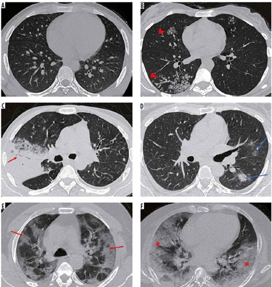
ID-19 pulmonary involvement. (9) The findings arrows) in both lungs. This was later identified as
associated with this category can be broken down COVID-19 pneumonia. (F) A patient with acute
into two groups: Mandatory features, which respiratory distress syndrome due to COVID-19
must be present in all cases, and confirmatory pneumonia had extensive, peripheral confluent
patterns of features. At least one confirmatory bilateral GGOs with septal thickening. (Jain A,
pattern must be present. (10) Mandatory features Patankar S, Kale S, Bairy A. Imaging of coronavi-
are ground-glass opacities with or without con- rus disease (COVID-19): a pictorial review. Pol-
solidations in lung regions close to visceral pleu- ish Journal of Radiology 2021; 86(1): 4-18).
ral surfaces, including the fissures, and a multi-
focal bilateral distribution. Subpleural sparing 5. DISCUSSION
can be present. (2) There are three confirmato-
COVID-19 has now posed a major threat to
ry patterns that emerge at various times during
public wellbeing, the social health sector and
the disease's progression. At an early stage, this
remains a global challenge (11). In this case,
pattern presents multiple ground-glass areas,
Coronavirus, which is highly contagious, must
which can be rounded or half-rounded in shape
be diagnosed quickly and accurately in order
and have unsharp demarcation, or multiple and
to begin adequate therapy, restrict further virus
sharply limited ground-glass areas outlining the
transmission, and effectively remove the virus
limits of multiple adjacent secondary pulmonary
from circulation (12). The standard norm for
lobules. Later in the disease's progression, visible
COVID-19 detection is RT-PCR of viral nucleic
intra-lobular interstitial thickening combined
acid, although, current reports have acknowl-
with ground-glass opacities forms a "crazy pav-
edged the relevance of chest CT scans analysis in
ing" pattern. Later, the pattern changes to one
COVID-19 patients with false negative RT-PCR
compatible with organizing pneumonia, which
findings, especially, when there is a clinical sus-
includes the reversed halo sign, ground-glass
picion of infection (13-15). Chest CT features, as
consolidation associated with extensive sub-
bilateral involvement, subpleural or peripheral-
pleural consolidations and an air bronchogram,
ly distributed GGO, consolidation, reticulation,
curvilinear subpleural bands, and ground-glass
crazy paving pattern, air bronchogram signs, in-
bands with or without consolidation, but with an tralobular septal thickening, pulmonary vascu-
arching pattern with pleural contact. (10) lar enlargement (16-18, 19-25), are considered to
4.7 CO-RADS 6 be characteristic manifestations for COVID-19
CO-RADS 6 was introduced to classify COV- infection (26) and with a high sensitivity (4,27)
ID-19 that had been confirmed by a positive RT- may let suspect this infection both in sympto-
PCR test for virus-specific nucleic acid. (9) matic and in some cases asymptomatic patients
Figure.CO-RADS is depicted in representative (28). However, some lately published meta-anal-
axial high-resolution computed tomography yses, evaluating accuracy of CT scans detecting
(HRCT) chest images. (A) CO-RADS 1 – normal COVID-19 pulmonary involvement, points out
HRCT chest. (B) CO-RADS 2 – centrilobular a risk of a low specificity and false-positive find-
nodules in the right lung with a tree-in-bud con- ings also (27, 29). Dutch Radiological Society
figuration (red arrowheads) – bronchiolitis, later presented CO-RADS reporting model declares
diagnosed as active pulmonary tuberculosis. (C) high predictability of COVID-19 in patients
CO-RADS 3 – In a patient with Klebsiella pneu- with moderate to severe symptoms (2), leading
monia, dense consolidation with air broncho- to the importance to evaluate this assessment
gram (red arrow) and surrounding ground-glass tool and see whether a standardised model will
opacity (GGO). (D) CO-RADS 4 - left lung provide more specificity and sensitivity into the
peribronchovascular GGOs (blue arrows). (E) practice. By using chest CT images of Japanese
CO-RADS 5 – peripheral and subpleural pre- data to detect COVID-19 pneumonia, Fujioka
dominant multifocal GGOs with interlobular et al. reported that CO-RADS retains signifi-
septal thickening – “crazy paving” pattern (red cant efficiency and great interobserver consen-
47JOURNAL AVAILABLE AT RADIOLOGYUPDATE.ORG sus with average sensitivity of 87.8%, specificity pre-test disease probability (26) and combining of 66.4%, and an AUC of 0.859 (8). Other ret- it with a predictive assessment and reporting rospective study in Italy demonstrated high di- system could further improve the accuracy of di- agnostic accuracy and moderate interrater con- agnosing the COVID-19 pneumonia (35). sensus among readers of varying skill levels with In conclusion, chest CT scan has a high sensi- even higher than previous report specificity of tivity for COVID-19 diagnosis and could reduce 81%, but lower sensitivity of 61% and an AUC false negative results obtained with RT-PCR of 0.72 (30). Dutch themselves measured the tests. Furthermore, a standardized reporting real-life performance of radiologist emergency system could increase clarification, minimize department chest CT interpretation for tracing reporting variability and help radiologists recog- COVID-19, using CO-RADS and revealed a nize the results they observe, especially, for less high precision of diagnosing with AUC of 0.87, experienced specialists. especially when symptoms last longer than 48 hours (31). Another retrospective analysis of the practical use of CO-RADS in the emergen- cy department of patients with possible COV- ID-19 infection also showed a great sensitivity and specificity of CO-RADS at 83.8% and 78.6%, respectively, with an AUC of 0.890 and provided a better risk classification by 65.8% in patients affected and by 82.1% in patients not affected by COVID-19 (26). A study made by Özel et al. in Turkey to assess CO-RADS model agrees with Dutch Radiology society findings by discovering this reporting scheme being incredibly effective in detecting COVID-19 pneumonia, especially, CO-RADS 5, which suggests a very high prob- ability for infection and statistically significant correlation with RT-PCR results (32). A com- parison analysis of CO-RADS scale and sever- ity scoring system (CT-SS), suggested by Yang at al. (33), of predicting severe COVID-19 dis- ease revealed both providing great performance with an AUC of 0.97 and 0.89, respectively, and CO-RADS scale giving even a better capability, with specificity of 98% at cut-off point > 4.5, sen- sitivity of 88% (34). Showing a great standard- ised reporting model sensitivity and specificity by previous studies Lessmann et al. introduced an artificial intelligence (AI) system to rate the probability of COVID-19 pulmonary involve- ment on CT scans by using CO-RADS and CT severity scores, providing high patients with COVID-19 identification results with an AUC of 0.95 in an internal cohort and an AUC of 0.88 in an external cohort (1). Some analysis indi- cate that CT imagining could be addressed for COVID-19 screening, thorough assessment, and follow-up, especially in epidemic areas with high 48
RADIOLOGY UPDATE VOL. 5 (8) ISSN 2424-5755

REFERENCES                                                             rRT-PCR or CT? Eur J Radiol. 2020;126:108961.
                                                                       16. Martínez Chamorro E, Díez Tascón A, Ibáñez Sanz L, Ossaba
1. Lessmann N, Sánchez CI, Beenen L, Boulogne LH, Brink M,             Vélez S, Borruel Nacenta S. Radiologic diagnosis of patients with
Calli E, Charbonnier JP, Dofferhoff T, van Everdingen WM,              COVID-19. Radiologia. 2021;63(1):56-73.
Gerke PK, et al. Automated Assessment of COVID-19 Reporting            17. Bao C, Liu X, Zhang H, Li Y, Liu J. Coronavirus Disease 2019
and Data System and Chest CT Severity Scores in Patients Sus-          (COVID-19) CT Findings: A Systematic Review and Meta-anal-
pected of Having COVID-19 Using Artificial Intelligence. Radi-         ysis. J Am Coll Radiol. 2020; 17(6): 701-709.
ology. 2021; 298(1): 18-28.                                            18. Song F, Shi N, Shan F, Zhang Z, Shen J, Lu H, Ling Y, Jiang Y,
2. Prokop M, van Everdingen W, van Rees Vellinga T, Quarles van        Shi Y. Emerging 2019 Novel Coronavirus (2019-nCoV) Pneumo-
Ufford H, Stöger L, Beenen L, Geurts B, Gietema H, Krdzalic J,         nia. Radiology. 2020; 295(1): 210-217.
Schaefer-Prokop C, et al. CO-RADS: A Categorical CT Assess-            19. Awulachew E, Diriba K, Anja A, Getu E, Belayneh F. Comput-
ment Scheme for Patients Suspected of Having COVID-19-Defi-            ed Tomography (CT) Imaging Features of Patients with COV-
nition and Evaluation. Radiology. 2020;296(2):97-104.                  ID-19: Systematic Review and Meta-Analysis. Radiol Res Pract.
3. Lee EYP, Ng MY, Khong PL. COVID-19 pneumonia: what has              2020;2020:1023506.
CT taught us? Lancet Infect Dis. 2020; 20(4): 384-385.                 20. Wan S, Li M, Ye Z, Yang C, Cai Q, Duan S, Song B. CT Man-
4. Ai T, Yang Z, Hou H, Zhan C, Chen C, Lv W, Tao Q, Sun Z, Xia        ifestations and Clinical Characteristics of 1115 Patients with
L. Correlation of Chest CT and RT-PCR Testing for Coronavirus          Coronavirus Disease 2019 (COVID-19): A Systematic Review
Disease 2019 (COVID-19) in China: A Report of 1014 Cases. Ra-          and Meta-analysis. Acad Radiol. 2020;27(7):910-921.
diology. 2020;296(2):32-40.                                            21. Zheng Y, Wang L, Ben S. Meta-analysis of chest CT fea-
5. Salehi S, Abedi A, Balakrishnan S, Gholamrezanezhad A.              tures of patients with COVID-19 pneumonia. J Med Virol.
Coronavirus disease 2019 (COVID-19) imaging reporting                  2021;93(1):241-249.
and data system (COVID-RADS) and common lexicon: a                     22. Ojha V, Mani A, Pandey NN, Sharma S, Kumar S. CT
proposal based on the imaging data of 37 studies. Eur Radiol.          in coronavirus disease 2019 (COVID-19): a systematic re-
2020;30(9):4930-4942.                                                  view of chest CT findings in 4410 adult patients. Eur Radiol.
6. Simpson S, Kay FU, Abbara S, Bhalla S, Chung JH, Chung              2020;30(11):6129-6138.
M, Henry TS, Kanne JP, Kligerman S, Ko JP, et al. Radiological         23. Salehi S, Abedi A, Balakrishnan S, Gholamrezanezhad A.
Society of North America Expert Consensus Statement on Re-             Coronavirus Disease 2019 (COVID-19): A Systematic Review
porting Chest CT Findings Related to COVID-19. Endorsed by             of Imaging Findings in 919 Patients. AJR Am J Roentgenol.
the Society of Thoracic Radiology, the American College of Ra-         2020;215(1):87-93.
diology, and RSNA - Secondary Publication. J Thorac Imaging.
                                                                       24. Sun Z, Zhang N, Li Y, Xu X. A systematic review of chest
2020;35(4):219-227.
                                                                       imaging findings in COVID-19. Quant Imaging Med Surg.
7. Bernheim A, Mei X, Huang M, Yang Y, Fayad ZA, Zhang N,              2020;10(5):1058-1079.
Diao K, Lin B, Zhu X, Li K, et al. Chest CT Findings in Coronavi-
                                                                       25. Zhu J, Zhong Z, Li H, Ji P, Pang J, Li B, Zhang J. CT imaging
rus Disease-19 (COVID-19): Relationship to Duration of Infec-
                                                                       features of 4121 patients with COVID-19: A meta-analysis. J Med
tion. Radiology. 2020;295(3):200463.
                                                                       Virol. 2020;92(7):891-902.
8. Fujioka T, Takahashi M, Mori M, Tsuchiya J, Yamaga E, Horii
                                                                       26. Turcato G, Zaboli A, Panebianco L, Scheurer C, Venturini A,
T, Yamada H, Kimura M, Kimura K, Kitazume Y, et al. Evaluation
                                                                       Tezza G, Canelles MF, Ausserhofer D, Pfeifer N, Wieser A. Clin-
of the Usefulness of CO-RADS for Chest CT in Patients Suspect-
                                                                       ical application of the COVID-19 Reporting and Data System
ed of Having COVID-19. Diagnostics (Basel). 2020;10(9):608.
                                                                       (CO-RADS) in patients with suspected SARS-CoV-2 infection:
9. Liu H, Ren H, Wu Z, Xu H, Zhang S, Li J, Hou L, Chi R, Zheng        observational study in an emergency department. Clin Radiol.
H, Chen Y, et al. CT radiomics facilitates more accurate diagnosis     2021;76(1):74.e23-74.e29.
of COVID-19 pneumonia: compared with CO-RADS. J Transl
                                                                       27. Xu B, Xing Y, Peng J, Zheng Z, Tang W, Sun Y, Xu C,
Med. 2021; 19(1): 29.
                                                                       Peng F. Chest CT for detecting COVID-19: a systematic re-
10. Penha D, Pinto EG, Matos F, Hochhegger B, Monaghan C,              view and meta-analysis of diagnostic accuracy. Eur Radiol.
Taborda-Barata L, Irion K, Marchiori E. CO-RADS: Coronavirus           2020;30(10):5720-5727.
Classification Review. J Clin Imaging Sci. 2021; 11:9.
                                                                       28. Shao JM, Ayuso SA, Deerenberg EB, Elhage SA, Augenstein
11. Wang MY, Zhao R, Gao LJ, Gao XF, Wang DP, Cao JM. SARS-            VA, Heniford BT. A systematic review of CT chest in COVID-19
CoV-2: Structure, Biology, and Structure-Based Therapeutics            diagnosis and its potential application in a surgical setting.
Development. Front Cell Infect Microbiol. 2020;10:587269.              Colorectal Dis. 2020;22(9):993-1001.
12. Liu X, Liu C, Liu G, Luo W, Xia N. COVID-19: Pro-                  29. Kim H, Hong H, Yoon SH. Diagnostic Performance of
gress in diagnostics, therapy and vaccination. Theranostics.           CT and Reverse Transcriptase Polymerase Chain Reaction
2020;10(17):7821-7835.                                                 for Coronavirus Disease 2019: A Meta-Analysis. Radiology.
13. Xie X, Zhong Z, Zhao W, Zheng C, Wang F, Liu J. Chest              2020;296(3):145-155.
CT for Typical Coronavirus Disease 2019 (COVID-19) Pneu-               30. Bellini D, Panvini N, Rengo M, Vicini S, Lichtner M, Tieghi
monia: Relationship to Negative RT-PCR Testing. Radiology.             T, Ippoliti D, Giulio F, Orlando E, Iozzino M, et al. Diagnostic
2020;296(2):41-45.                                                     accuracy and interobserver variability of CO-RADS in patients
14. Fang Y, Zhang H, Xie J, Lin M, Ying L, Pang P, Ji W. Sensitivity   with suspected coronavirus disease-2019: a multireader valida-
of Chest CT for COVID-19: Comparison to RT-PCR. Radiology.             tion study. Eur Radiol. 2021; 31(4): 1932-1940.
2020;296(2):115-117.                                                   31. Schalekamp S, Bleeker-Rovers CP, Beenen LFM, Quarles van
15. Long C, Xu H, Shen Q, Zhang X, Fan B, Wang C, Zeng B, Li           Ufford HME, Gietema HA, Stöger JL, Harris V, Reijers MHE,
Z, Li X, Li H. Diagnosis of the Coronavirus disease (COVID-19):        Rahamat-Langendoen J, Korevaar DA, et al. Chest CT in the

                                                                                                                                  49
JOURNAL AVAILABLE AT RADIOLOGYUPDATE.ORG Emergency Department for Diagnosis of COVID-19 Pneumo- nia: Dutch Experience. Radiology. 2021;298(2):98-106. 32. Özel M, Aslan A, Araç S. Use of the COVID-19 Reporting and Data System (CO-RADS) classification and chest computed tomography involvement score (CT-IS) in COVID-19 pneumo- nia. Radiol Med. 2021; 12: 1–9. 33. Yang R, Li X, Liu H, Zhen Y, Zhang X, Xiong Q, Luo Y, Gao C, Zeng W. Chest CT Severity Score: An Imaging Tool for As- sessing Severe COVID-19. Radiol Cardiothorac Imaging. 2020; 2(2): e200047. 34. Zayed, N.E., Bessar, M.A. & Lutfy, S. CO-RADS versus CT- SS scores in predicting severe COVID-19 patients: retrospective comparative study. Egypt J Bronchol. 2021; 15(1): 13. 35. Hermans JJR, Groen J, Zwets E, Boxma-De Klerk BM, Van Werkhoven JM, Ong DSY, Hanselaar WEJJ, Waals-Prinzen L, Brown V. Chest CT for triage during COVID-19 on the emer- gency department: myth or truth? Emerg Radiol. 2020; 27(6): 641-651. 36. Jain A, Patankar S, Kale S, Bairy A. Imaging of coronavirus disease (COVID-19): a pictorial review. Polish Journal of Radiol- ogy 2021; 86(1): 4-18. 50
You can also read