Risk factors for cerebral vasospasm in patients with aneurysmal subarachnoid hemorrhage
←
→
Page content transcription
If your browser does not render page correctly, please read the page content below
Open Medicine 2020; 15: 598–604
Research Article
Valentina Opancina*, Snezana Lukic, Slobodan Jankovic, Radisa Vojinovic, Milan Mijailovic
Risk factors for cerebral vasospasm in patients
with aneurysmal subarachnoid hemorrhage
https://doi.org/10.1515/med-2020-0169 intensity is probably directly related to the occurrence of
received May 6, 2020; accepted June 5, 2020 vasospasm and its adverse consequences.
Abstract Keywords: cerebral vasospasm, endovascular coiling,
Introduction ‒ Aneurysmal subarachnoid hemorrhage is aneurysm, international normalized ratio, white blood
a type of spontaneous hemorrhagic stroke, which is caused cells
by a ruptured cerebral aneurysm. Cerebral vasospasm
(CVS) is the most grievous complication of subarachnoid
hemorrhage (SAH). The aim of this study was to examine
the risk factors that influence the onset of CVS that 1 Introduction
develops after endovascular coil embolization of a ruptured
aneurysm. Aneurysmal subarachnoid hemorrhage (ASAH) is a type
Materials and methods ‒ The study was designed as a of spontaneous hemorrhagic stroke, which is caused by a
cross-sectional study. The patients included in the study ruptured cerebral aneurysm in about 80% of patients with
were 18 or more years of age, admitted within a period of subarachnoid hemorrhage (SAH) [1,2]. Nearly one-third of
24 h of symptom onset, diagnosed and treated at a ASAH survivors develop delayed cerebral ischemia, which
university medical center in Serbia during a 5-year is caused by the narrowing of cerebral blood vessels and
period. decreased cerebral blood flow [3,4]. Segmental or diffuse
Results ‒ Our study showed that the maximum recorded narrowing of the lumen of the intracranial arteries is also
international normalized ratio (INR) values in patients who known as cerebral vasospasm (CVS) [2,5]. CVS is the most
were not receiving anticoagulant therapy and the max- grievous complication of SAH and can be described as a
imum recorded white blood cells (WBCs) were strongly deferred and self-limiting condition; furthermore, its
associated with cerebrovascular spasm, increasing its severity is associated with the volume, density, extended
chances 4.4 and 8.4 times with an increase of each integer presence and site of subarachnoid blood [1,3,5]. CVS that
of the INR value and 1,000 WBCs, respectively. is influenced by SAH is a perplexing issue which
Conclusions ‒ SAH after the rupture of cerebral aneur- incorporates hypovolemia, damaged auto-regulatory
ysms creates an endocranial inflammatory state whose function and also prolonged and reversible vasculitis
[6,7]. The preeminent concept for CVS prevention is
maintenance of regular blood volume as well as treatment
with nimodipine [8]. Currently, the only confirmed
* Corresponding author: Valentina Opancina, University of treatment for CVS is euvolemic induced hypertension,
Kragujevac, Serbia, Faculty of Medical Sciences, Department of due to the fact that endovascular procedures still carry
Radiology, Serbia, e-mail: valentina.opancina@gmail.com specific risks [9].
Snezana Lukic: University of Kragujevac, Serbia, Faculty of Medical To date, various factors have been known to be linked
Sciences, Department of Radiology, Serbia,
with an increased risk of CVS after SAH. The haptoglobin
e-mail: snezanamlukic@gmail.com
Slobodan Jankovic: University of Kragujevac, Serbia, Faculty of phenotype is one of these factors, since the affinity of the
Medical Sciences, Department of Pharmacology and Toxicology, molecule for hemoglobin depends on the extent to which it
Serbia, e-mail: slobnera@gmail.com will bind free hemoglobin in the subarachnoid space and
Radisa Vojinovic: University of Kragujevac, Serbia, Faculty of thus prevent the initiation of a series of reactions that
Medical Sciences, Department of Radiology, Serbia,
ultimately result in vasospasm formation [10]. Intraventri-
e-mail: rhvojinovic@gmail.com
Milan Mijailovic: University of Kragujevac, Serbia, Faculty of
cular hemorrhage within ASAH is also associated with a
Medical Sciences, Department of Radiology, Serbia, higher incidence of vasospasm, as well as long-term use of
e-mail: milankckragujevac@gmail.com tobacco [11]. Also, left ventricular hypertrophy detected on
Open Access. © 2020 Valentina Opancina et al., published by De Gruyter. This work is licensed under the Creative Commons Attribution 4.0
Public License.Risk factors for cerebral vasospasm 599
electrocardiography is described as a risk factor for develop- aneurysm for the first time; SAH confirmed by the CT
ment of CVS (odds ratio [OR] = 3.48) [12]. The correlation scan on admission; rupture of an aneurysm confirmed by
between cerebrospinal fluid and CVS was observed, using digital subtraction angiography (DSA); treatment of
volumetric analysis and SAH/CSF ratio (OR = 1.03) [13]. the aneurysm by EE; minimum two control CT scans after
However, the amount of data published in this field to the coiling (the first CT scan was done 1 day after the
date has been relatively limited. Most of the studies endovascular procedure, the last CT was done on discharge
explored CVS only in patients who developed neurolo- and additional CT scans were done on demand from
gical symptoms after the procedure [12,14,15]. Also, in a neurologists); and DSA after the embolization, between the
great number of other studies, the patient population was 5th and 10th day. Each interventional radiology procedure
heterogeneous, including both those treated surgically was performed by the same team of two experienced
and those treated by the endovascular approach [16,17]. neuroradiologists, and all the CT scans were evaluated by
Considering this, the aim of this study was to three neuroradiologists in our department. The exclusion
examine the risk factors that influence the onset of CVS criteria were the following: previous treatment of the
that develops after endovascular coil embolization (EE) ruptured aneurysm; artifacts in CT scans; patients who
of a ruptured aneurysm which caused SAH. developed serious complications during hospitalization
such as sepsis, hepatorenal dysfunction etc.; pregnant
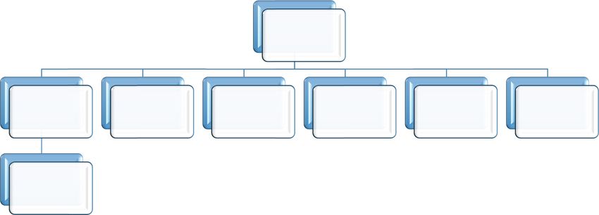
women; and incomplete patient file. Figure 1 presents the
flowchart with the number of patients who were initially
2 Materials and methods included and the number of excluded patients based on
each of the exclusion criteria.
The study was designed as a cross-sectional study. It was The diagnosis of ASAH was made if CT scan showed
approved by the Ethics Committee of the Clinical Center the presence of blood in the subarachnoid space, and DSA
Kragujevac before any of the study procedures was indicated rupture of an aneurysm. CVS was diagnosed by
initiated. The study was conducted according to the DSA [1,2] if the column of contrast in large cerebral arteries
principles of Declaration of Helsinki about experimenta- was decreased by more than 34% in diameter [13].
tion on human subjects and in compliance with national The following variables were extracted from the patient
regulations. Informed consent was obtained from all files: sociodemographic data: age, sex, residence, smoking
individuals included in this study. habit, and caffeine use; symptoms and physical status prior
The patients included in the study satisfied the to admission; scales: modified Rankin scale (MRS) on
following criteria: 18 or more years of age, admitted within discharge [18], Hunt and Hesse scale (HHS) on admission
a period of 24 h of symptom onset, diagnosed and treated [19], Fischer scale (FS) using the initial CT scan [20], and
at Clinical Center Kragujevac, Serbia, during the observa- Glasgow Coma Scale (GCS) on admission; CT findings:
tion period (from 01-01-2014 to 31-12-2018), and suffering hydrocephalus, brain edema, intraventricular hemorrhage,
from SAH caused by the rupture of an intracranial and intracerebral hemorrhage; liquid evacuation: lumbar
89 screened
3 exclluded because 2 excluded because
5 excluded becau use 12 exclud
ded because
of the
t previous they developed 1 excluded becausse
d
66 included of the artefacts on CT of the in
ncomplete
treatment of the serious complicaons
c of the pregnancyy
scan paent file
ruptured aneurysm during h
hospitalizaon
66 completed the
study
Figure 1: The study flowchart.600 Valentina Opancina et al.
puncture and/or ventriculoperitoneal shunt; mechanical regression, for the outcome CVS, are demonstrated in
ventilation after the endovascular procedure; aneurysm Table 2. The univariate regression revealed that acute
features: location, size, existence of a second unruptured hydrocephalus after SAH, maximum recorded interna-
aneurysm; procedure characteristics: duration of fluoroscopy tional normalized ratio (INR) and maximum recorded
and heparin dosage; duration of hospitalization, the time white cell count substantially increased the chances of
from admission to EE; duration of symptoms prior to hospital CVS in post-embolization clinical course (ORs 5.000,
admission; and laboratory analysis (maximum recorded 4.103 and 6.720, respectively). The influence of platelet
values and nadir values): blood count, coagulation tests, count at nadir on CVS was statistically significant, but
and biochemistry analysis. marginal (ORs between 1 and 1.01) (Table 2). Finally, the
maximum recorded level of blood urea nitrogen was also
significantly associated with CVS, with moderate influ-
ence (OR 1.285).
2.1 Statistics The results of multivariable logistic regression are
shown in Table 3. The estimates of the coefficient of
The study data were analyzed using SPSS version 23 software determination according to the Cox and Snell and
(SPSS Inc., Chicago, IL, USA) [21]. Descriptive statistics was Nagelkerke were 0.398 and 0.531, respectively, while
used for primary data processing: continuous variables were the Hosmer–Lemeshow test showed that the observed
described by mean and standard deviation (if normally rate of CVS matched the expected rate of the same
distributed) or by median and interquartile range (if not phenomenon (χ2 = 2.503, p = 0.927). After adjustment,
normally distributed), while categorical variables were the following factors remained significantly associated
presented with rates and percentages. The Kolmogorov– with CVS: maximum recorded white cell count and INR.
Smirnov test was used to test the normality of the data The strength of the association remained similar to that
distribution. Significance of differences in the study groups after univariate analysis, and the direction of the
was tested by the Mann–Whitney test for continuous influence did not change (Table 3).
variables and by contingency tables for categorical variables.
The impact of these variables on the main outcome
(appearance of CVS) was investigated by univariate logistic
regression at first, after which multivariable logistic regres- 4 Discussion
sion was performed using the backward method. The model
of logistic regression was built after several attempts, using Our study showed that the maximum recorded INR
the backward method; each attempt implied different values in patients who were not receiving anticoagulant
combinations of independent and confounding variables. therapy and the maximum recorded WBC were strongly
However, in order to avoid the co-linearity problem, only one associated with cerebrovascular spasm, increasing its
value of the variables that were measured repeatedly (e.g. chances 4.4 and 8.4 times with an increase of each
white cell count before, during and after the EE) was entered integer of the INR value and 1,000 white blood cells
at a time (i.e. repeating measures of the same variable were (WBCs), respectively.
never entered in the same model building attempt). Effects of Elevated INR values in patients not receiving
the variables obtained in the final model were adjusted for anticoagulant therapy were recently linked to an increased
their simultaneous presence. The quality of the multivariable risk of intracerebral aneurysm rupture, although the
logistic regression model was tested by Hosmer–Lemeshow, possible mechanism of action was not proposed [22]. A
Cox–Snell and Nagelkerke tests. similar effect was not observed in patients on anticoagulant
therapy, suggesting the protective role of oral anticoagu-
lants independent of their main pharmacological effect
(vitamin K antagonism) [23]. Although systemic inflamma-
3 Results tion creates a hypercoagulable state through activation of
coagulation, decrease of endogenous anticoagulants in
In total 66 patients were enrolled, and all of them blood, and inhibition of fibrinolysis [24], INR is usually
completed the study, without drop-outs. The main elevated due to the consumption of certain proteins
characteristics of the study group are shown in Table 1, involved in the coagulation cascade [25]. Elevated INR
while for the sake of clarity the rest are listed in the observed in our study may reflect a similar influence of
supplementary file. The results of univariate logistic systemic inflammation (C reactive protein was elevated inRisk factors for cerebral vasospasm 601 Table 1: Characteristics of the study population Risk factors Cerebrovascular spasm (n = 33) No cerebrovascular spasm (n = 33) P value Age (mean ± SD, median [IQR]) 55.45 ± 12.13, 56 [14] 52.55 ± 9.55, 54[17] 0.336 Age category (20–40/40–60/>60 4/16/13 (12.1%/48.5%/39.4%) 5/21/7 (15.2%/63.6%/21.2%) 0.274 years) Gender (male/female, %/%) 8/25 (24.2%/75.8%) 10/23 (30.3%/69.7%) 0.580 Caffeine usage 23 (69.7%) 15 (45.5%) 0.046* Impaired vision 1 (3%) 6 (18.2%) 0.046* Mechanical ventilation 17 (51.5%) 8 (24.2%) 0.022* Intraventricular hemorrhage 23 (69.7%) 13 (39.4%) 0.013* Hydrocephalus 11 (33.3%) 3 (9.1%) 0.016* Aneurysm size (8) 0.148 0.411 0.123–1.373 Hydrocephalus (yes/no) 0.023 5.000* 1.245–20.076 Intraventricular hemorrhage (yes/no) 0.015 3.538* 1.277–9.805 Mechanical ventilation (yes/no) 0.025 3.320* 1.163–9.477 Maximum recorded CRP 0.164 0.444 0.142–1.394 Maximum recorded WBC 0.001 6.720* 2.080-21.708 PLT at nadir (×109/L) 0.029 1.007* 1.001–1.012 Maximum recorded INR 0.008 4.103* 1.455–11.567 Proteins at nadir 0.999 0.000 0.000 Maximum recorded urea (mmol/L) 0.021 1.285* 1.039–1.590 * – statistically significant. CRP – C reactive protein; WBC – white blood cell count; PLT – platelet count; INR – international normalized ratio.
602 Valentina Opancina et al.
Table 3: Multivariate analysis of factors associated with CVS
Risk factors P value Adjusted odds ratio Confidence interval (95%)
PLT at nadir (×10 /L)
9
0.099 1.007 0.999–1.014
Maximum recorded urea (mmol/L) 0.082 1.232 0.974–1.558
Maximum recorded INR 0.027 4.411* 1.181–16.478
Maximum recorded WBC 0.004 8.376* 2.009–34.921
* – statistically significant.
PLT – platelet count; INR – international normalized ratio; WBC – white blood cell count.
tissue after the rupture of an aneurysm that will cause the caffeine-rich beverages or coffee increases platelet aggrega-
release of a multitude of autacoids. At least some of the tion and alters endothelial function reducing the ability of the
released autacoids may activate the receptors on smooth endothelium to mediate vascular relaxation. However, further
muscle cells and produce intense vasospasm, such as research is necessary to confirm this hypothesis.
cysteinyl leukotrienes [28]. The size of cerebral artery aneurysms was previously
The elevated WBC count after the percutaneous inter- connected with the rate of cerebral artery vasospasm after
vention is also reflection of the inflammatory process in the SAH [14], but this is an indirect effect, since CVS is more
brain tissue, and its association with CVS after SAH was not frequent when the extent of subarachnoid bleeding is large.
surprising. It was recently understood that a lot of vascular However, there are no studies finding an association
and neural changes after SAH were caused by inflammation between the size of aneurysms and rate of CVS after
accompanied by the activation of immune cells in brain endovascular embolization. Our study also did not find
parenchyma and blood [29]. There are many autacoids, such an association, but this could be a consequence of
hormones and neurotransmitters that may cause vasospasm, insufficient statistical power, implying the necessity for
and some of them are released from platelets, such as continuous research on this issue.
serotonin, while the others could originate from other blood The limitation of our study was a relatively small
cells destroyed after SAH [30,31]; injured neurons also react sample size, which allowed simultaneous testing of only
with increased firing, so local concentrations of vasoactive up to ten study variables in multivariate logistic
catecholamines, endothelins or other substances may regression, increasing the chances of statistical type 2
mount, causing intense and prolonged vasoconstriction. error. Our study was unicentric, which is an independent
Although mechanical ventilation, hydrocephalus and risk factor for introducing local practice bias in the
intraventricular hemorrhage were more frequent among interpretation of the results.
patients in our study who developed cerebrovascular spasm, In conclusion, SAH after rupture of cerebral aneurysms
after adjustment for confounding factors their influence on creates an endocranial inflammatory state whose intensity
spasm did not reach statistical significance. Intraventricular is probably directly related to the occurrence of vasospasm
hemorrhage was previously linked with inflammation of the and its adverse consequences. Whether pharmacological
brain tissue in several animal studies, causing an upregula- interference for neuroinflammation could be a useful
tion of pro-inflammatory cytokines, attracting WBCs and strategy for the prevention of vasospasm remains to be
activating microglia [32]. On the other hand, hydrocephalus decided by future studies of sufficient size.
could be a consequence of intraventricular hemorrhage
accompanying inflammation and edema. However, intraven- Conflict of interest: The authors state no conflict of
tricular hemorrhage probably was not the sole cause of interest.
neuroinflammation and release of mediators that produced
vasoconstriction in our patients, which, together with a
relatively small number of patients in the study, could
explain why it was not among the significant risk factors
References
after adjustment and multivariate analysis.
Although caffeine usage in our study was not signifi- [1] Muñoz-Guillén NM, León-López R, Túnez-Fiñana I, Cano-
Sánchez A. From vasospasm to early brain injury: new
cantly associated with CVS, some authors did find an
frontiers in subarachnoid haemorrhage research. Neurologia.
association between them [33]. A probable explanation for 2013;28(5):309–16. doi: 10.1016/j.nrl.2011.10.015.
caffeine-induced CVS is a pro-thrombotic state with de- [2] Athar MK, Levine JM. Treatment options for cerebral
creased ability of blood vessels to dilate, since caffeine from vasospasm in aneurysmal subarachnoid hemorrhage.Risk factors for cerebral vasospasm 603
Neurotherapeutics. 2012;9(1):37–43. doi: 10.1007/s13311-011- [16] Juvela S, Siironen J, Kuhmonen J. Hyperglycemia, excess
0098-1. weight, and history of hypertension as risk factors for poor
[3] Dabus G, Nogueira RG. Current options for the management of outcome and cerebral infarction after aneurysmal subarach-
aneurysmal subarachnoid hemorrhage-induced cerebral noid hemorrhage. J Neurosurg. 2005;102(6):998–1003.
vasospasm: a comprehensive review of the literature. Interv doi: 10.3171/jns.2005.102.6.0998.
Neurol. 2013;2(1):30–51. doi: 10.1159/000354755. [17] Shimoda K, Kamiya K, Kano T, Furuichi M, Yoshino A. Cerebral
[4] Etminan N, Vergouwen MD, Ilodigwe D, Macdonald RL. Effect vasospasm and patient outcome after coiling or clipping for
of pharmaceutical treatment on vasospasm, delayed cerebral intracranial aneurysmal subarachnoid hemorrhage.
ischemia, and clinical outcome in patients with aneurysmal J Neuroendovasc Ther. 2019;13:443–8. doi: 10.5797/
subarachnoid hemorrhage: a systematic review and meta- jnet.oa.2019-0023.
analysis. J Cereb Blood Flow Metab. 2011;31(6):1443–51. [18] Nunn A, Bath PM, Gray LJ. Analysis of the modified Rankin
doi: 10.1038/jcbfm.2011.7. scale in randomised controlled trials of acute ischaemic
[5] Lin BF, Kuo CY, Wu ZF. Review of aneurysmal subarachnoid stroke: a systematic review. Stroke Res Treat.
hemorrhage – focus on treatment, anesthesia, cerebral 2016;2016:9482876. doi: 10.1155/2016/9482876.
vasospasm prophylaxis, and therapy. Acta Anaesthesiol [19] Ghosh S, Dey S, Maltenfort M, Vibbert M, Urtecho J, Rincon F,
Taiwan. 2014;52(2):77–84. doi: 10.1016/j.aat.2014.04.005. et al. Impact of Hunt-Hess grade on the glycemic status of
[6] Naraoka M, Matsuda N, Shimamura N, Asano K, Ohkuma H. aneurysmal subarachnoid hemorrhage patients. Neurol India.
The role of arterioles and the microcirculation in the 2012;60(3):283–7. doi: 10.4103/0028-3886.98510.
development of vasospasm after aneurysmal SAH. Biomed Res [20] Oliveira AM, Paiva WS, Figueiredo EG, Oliveira HA, Teixeira MJ.
Int. 2014;2014:253746. doi: 10.1155/2014/253746. Fisher revised scale for assessment of prognosis in patients
[7] Miller BA, Turan N, Chau M, Pradilla G. Inflammation, with subarachnoid hemorrhage. Arq Neuropsiquiatr.
vasospasm, and brain injury after subarachnoid hemorrhage. 2011;69(6):910–3. doi: 10.1590/s0004-282x2011000700012.
Biomed Res Int. 2014;2014:384342. doi: 10.1155/2014/ [21] IBM Corp. Released 2015. IBM SPSS Statistics for Windows,
384342. Version 23.0. Armonk, NY: IBM Corp.
[8] Mijailovic M, Lukic S, Laudanovic D, Folic M, Folic N, [22] Can A, Castro VM, Dligach D, Finan S, Yu S, Gainer V, et al.
Jankovic S. Effects of nimodipine on cerebral vasospasm in Elevated international normalized ratio is associated with
patients with aneurysmal subarachnoid hemorrhage treated ruptured aneurysms. Stroke. 2018;49(9):2046–52.
by endovascular coiling. Adv Clin Exp Med. 2013;22(1):101–9. doi: 10.1161/STROKEAHA.118.022412.
[9] Liu Y, Qiu H, Su J, Jiang W. Drug treatment of cerebral [23] Tarlov N, Norbash AM, Nguyen TN. The safety of antic-
vasospasm after subarachnoid hemorrhage following aneur- oagulation in patients with intracranial aneurysms.
ysms. Chinese Neurosurg J. 2016;2:4. doi: 10.1186/s41016- J Neurointerv Surg. 2013;5(5):405–9. doi: 10.1136/neurint-
016-0023-x. surg-2012-010359.
[10] Leclerc JL, Blackburn S, Neal D, Mendez NV, Wharton JA, [24] Cheng T, Mathews K, Abrams-Ogg A, Wood D. The link
Waters MF, et al. Haptoglobin phenotype predicts the between inflammation and coagulation: influence on the
development of focal and global cerebral vasospasm and may interpretation of diagnostic laboratory tests. Compend Contin
influence outcomes after aneurysmal subarachnoid hemor- Educ Vet. 2011;33(2):E4.
rhage. Proc Natl Acad Sci U S A. 2015;112(4):1155–60. [25] Lyons PG, Micek ST, Hampton N, Kollef MH. Sepsis-associated
doi: 10.1073/pnas.1412833112. coagulopathy severity predicts hospital mortality. Crit Care
[11] Wilson TJ, Stetler Jr WR, Davis MC, Giles DA, Khan A, Med. 2018;46(5):736–42. doi: 10.1097/
Chaudhary N, et al. Intraventricular hemorrhage is associated CCM.0000000000002997.
with early hydrocephalus, symptomatic vasospasm, and poor [26] Al-Tamimi YZ, Bhargava D, Orsi NM, Teraifi A, Cummings M,
outcome in aneurysmal subarachnoid hemorrhage. J Neurol Ekbote UV, et al. Compartmentalisation of the infla-
Surg A Cent Eur Neurosurg. 2015;76(2):126–32. doi: 10.1055/ mmatory response following aneurysmal subarachnoid hae-
s-0034-1394189. morrhage. Cytokine. 2019;123:154778. doi: 10.1016/
[12] Inagawa T, Yahara K, Ohbayashi N. Risk factors associated j.cyto.2019.154778.
with cerebral vasospasm following aneurysmal subarachnoid [27] Chaudhry SR, Stoffel-Wagner B, Kinfe TM, Güresir E, Vatter H,
hemorrhage. Neurol Med Chir. 2014;54(6):465–73. Dietrich D, et al. Elevated systemic IL-6 levels in patients with
doi: 10.2176/nmc.oa.2013-0169. aneurysmal subarachnoid hemorrhage is an unspecific marker
[13] Scherer M, Jung JO, Cordes J, Wessels L, Younsi A, for post-SAH complications. Int J Mol Sci. 2017;18(12):2580.
Schönenberger S, et al. Association of cerebrospinal fluid doi: 10.3390/ijms18122580.
volume with cerebral vasospasm after aneurysmal subarach- [28] Haeggström JZ, Wetterholm A. Enzymes and receptors in the
noid hemorrhage: a retrospective volumetric analysis. leukotriene cascade. Cell Mol Life Sci. 2002;59(5):742–53.
Neurocrit Care. 2019. doi: 10.1007/s12028-019-00878-2. doi: 10.1007/s00018-002-8463-1.
[14] Inagawa T. Risk factors for cerebral vasospasm following [29] Coulibaly AP, Provencio JJ. Aneurysmal subarachnoid hemor-
aneurysmal subarachnoid hemorrhage: a review of the rhage: an overview of inflammation-induced cellular changes.
literature. World Neurosurg. 2016;85:56–76. doi: 10.1016/ Neurotherapeutics. 2020;17(2):436–45. doi: 10.1007/s13311-
j.wneu.2015.08.052. 019-00829-x.
[15] Opancina V. Prevention and treatment of cerebral vasospasm [30] de Oliveira Manoel AL, Macdonald RL. Neuroinflammation as a
after aneurysmal subarachnoid hemorrhage. Ration Ther. Target for Intervention in Subarachnoid Hemorrhage. Front
2016;8(2):35–9. doi: 10.5937/racter8-11415. Neurol. 2018;9:292. doi: 10.3389/fneur.2018.00292.604 Valentina Opancina et al.
[31] Lucke-Wold BP, Logsdon AF, Manoranjan B, Turner RC, Targets. 2017;21(12):1111–22. doi: 10.1080/
McConnell E, Vates GE, et al. Aneurysmal subarachnoid 14728222.2017.1397628.
hemorrhage and neuroinflammation: a comprehensive [33] Grant RA, Cord BJ, Kuzomunhu L, Sheth K, Gilmore E,
review. Int J Mol Sci. 2016;17(4):497. doi: 10.3390/ Matouk CC. Aneurysmal subarachnoid hemorrhage and
ijms17040497. severe, catheter-induced vasospasm associated with exces-
[32] Garton T, Hua Y, Xiang J, Xi G, Keep RF. Challenges sive consumption of a caffeinated energy drink. Interv
for intraventricular hemorrhage research and Neuroradiol. 2016;22(6):674–8. doi: 10.1177/
emerging therapeutic targets. Expert Opin Ther 1591019916660868.You can also read


