EFFECT OF ALKALI BASES ON THE SYNTHESIS OF ZNO QUANTUM DOTS
←
→
Page content transcription
If your browser does not render page correctly, please read the page content below
Open Chemistry 2021; 19: 377–384
Research Article
Xilian Zhang, Shanshan Luo, Xiaodan Wu, Minghui Feng, Yingying Li, Haoyun Han, Wenkui Li*
Effect of alkali bases on the synthesis of ZnO
quantum dots
https://doi.org/10.1515/chem-2021-0027 decades, QDs have been widely studied because of their
received November 23, 2020; accepted February 2, 2021 adjustable size and luminescent properties that make
Abstract: The surface-modified zinc oxide quantum dots them promising agents for biomedicine [1,2], informa-
(ZnO QDs) have broad application prospects in the field tion encryption [3], and optoelectronic devices. Appli-
of biomedicine because of their good water solubility, cations in the biomedical field require ZnO QDs to have
dispersibility, and high fluorescence stability. The alkali good water solubility, dispersion, and fluorescence
bases play important roles in controlling the morphology, stability.
size distribution, dispersity, and fluorescence intensity of The fluorescence color of the QDs is related to their
the synthesized ZnO QDs. In this article, ZnO QDs were size. In the range of 2.5–7 nm, as the particle size increases,
synthesized to induce hydrolysis–condensation reaction. the fluorescence color is blue-violet, blue, green, yellow,
The influences of alkali bases (LiOH, NaOH, and KOH) and orange, and red [4,5]. In 1991, Lubomir and Marc [6]
the ratio of n(Zn2+):n(OH−) on the properties of synthe- prepared ZnO QDs with a size of 3–6 nm for the first
sized ZnO QDs were investigated. The results show that time using zinc acetate and LiOH as the starting materials
the particle size of the ZnO QDs prepared using LiOH and through a sol–gel method. Also, various methods have
NaOH as raw materials are smaller than that using KOH. been used to prepare ZnO QDs, including the sol–gel
ZnO QDs prepared at the ratio of n(Zn2+):n(LiOH) = 1:1 method [7–9], the microemulsion method [10–12], the
have the best fluorescence performance and dispersibility. hydrothermal method [13,14], and so on. The sol–gel
method is widely used in the laboratory research for its
Keywords: zinc oxide, quantum dots, alkaline, fluores- convenience of doping and controllability of reaction
cence properties, water solubility conditions. However, colloidal ZnO QDs are easy to
aggregate or undergo Ostwald ripening because of their
high surface energy. As a result, ZnO QDs are unstable
in the aqueous dispersion used for storage. [15]. To sta-
1 Introduction bilize ZnO QDs, various capping agents have been used,
i.e., polyvinylpyrrolidone, 3-aminopropyltrimethoxysi-
Quantum dots (QDs) are semiconductor materials with a lane (APTES), amines, mercaptocarboxylic acid, etc.
particle size close to or smaller than the de Broglie wave [16,17]. The alkoxyl groups in APTES hydrolyze and react
or the mean free path of electrons. During the past few with the –OH group on the surface of the ZnO to form
a silica capping layer. In addition to being a biocompa-
tible molecule, silica serves two more important func-
* Corresponding author: Wenkui Li, Jiangxi Key Laboratory of tions, namely, controlling the particle size by limiting
Surface Engineering, Jiangxi Science and Technology Normal the growth of ZnO and acting as a side group on the sur-
University, Nanchang 330013, People’s Republic of China, face that can be further conjugated with biomolecules
e-mail: liwenkui1976@163.com
[18]. In the sol–gel method, various factors, including
Xilian Zhang: Jiangxi Key Laboratory of Surface Engineering, Jiangxi
Science and Technology Normal University, Nanchang 330013, doping, the concentration of precursor, the reaction tem-
People’s Republic of China perature, and the molar ratio of the reactants, affect the
Shanshan Luo, Minghui Feng, Yingying Li, Haoyun Han: Jiangxi final performance of the synthesized ZnO QDs. A small
Provincial Key Laboratory of Drug Design and Evaluation, School of amount of Mg2+ doping (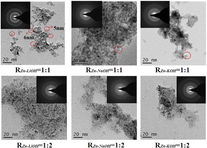
378 Xilian Zhang et al.
Various research studies have been reported on the
synthesis of ZnO nanomaterials with the controlled
crystal size and the surface structure to improve their
properties for a potential application. Few studies have
focused on the influence of the alkali base types on the
size and morphology tunability of ZnO nanomaterials
[22,23]. However, there is no report on the effect of alkali
base types on the dispersibility of water-soluble ZnO QDs
as far as we know. Moreover, the results over the ratio of
hydroxide to zinc ions on the fluorescence properties of
QDs are controversial [21,24]. In this study, ZnO QDs were
prepared by the sol–gel method, and their surface was
modified by APTES to make them water soluble. The
Figure 1: A typical routing of preparing ZnO QDs by the sol–gel
effects of the different alkali bases (LiOH, NaOH, and
method.
KOH) and RZn−OH, which is the ratio of n(Zn2+):n(OH−),
on the size, dispersibility, and fluorescence properties of
ZnO QDs were studied. 85°C until the solution became colorless and transparent,
and then the solution is cooled to room temperature and
added to the ethanol solution of zinc acetate in an ice
bath and reacted for 4 h. The solution quickly turned
2 Materials and methods white and then gradually became clear, indicating that
ZnO QDs were formed. In the second step, 400 µL of
APTES solution was mixed with 2 mL of ultrapure water,
2.1 Materials
and then, the mixed solution was added dropwise to the
aforementioned ZnO QD solution, and the mixed solution
Zinc acetate dihydrate, magnesium acetate tetrahydrate,
was stirred at 60°C for 3 h. APTES undergoes hydrolysis
potassium hydroxide (Shanghai Titan Technology Co.,
to form silica-coated ZnO QDs. As the reaction completed,
Ltd.), sodium hydroxide (Guangdong West Long Science
white ZnO QD precipitates were centrifuged at 4,500 rpm
Co., Ltd.), lithium hydroxide (Shanghai Aladdin Biochemical
for 5 min, followed by washing twice with ethanol to
Technology Co., Ltd.), 3-aminopropyltriethoxysilane
remove unreacted impurities and finally vacuum dried
(Shanghai Yien Chemical Technology Co., Ltd.), ethanol,
at 60°C to obtain ZnO QD powders.
and n-hexane (China National Pharmaceutical Group
Shanghai Chemical Reagent Company) were used in this
study. All the reagents used in this study are of analytical
grade and were used without further purification.
2.3 Characteristics
The crystal structure and composition were measured
2.2 Preparation of ZnO QDs through an X-ray powder diffractometer (Shimadzu,
Japan, XRD-6100). The morphology and particle size
The water-soluble ZnO QDs were prepared according to were observed using a field emission transmission
the sol–gel method [19] with a small modification. A electron microscope (FEI Company, USA). Surface func-
typical two-step synthesis route is shown in Figure 1 tional groups were determined using a Fourier transform
and described as follows: in the first step, 2.20 g zinc infrared spectrometer (IR-960, Tianjin Rui’an Technology
acetate dihydrate and 0.214 g magnesium acetate tetra- Co., Ltd.). The fluorescence performance was tested by
hydrate were dissolved in 100 mL of anhydrous ethanol, a UV spectrophotometer (model UV-2550, Shimadzu,
and then, the solution was refluxed and stirred for Japan), a PL fluorescence spectroscopy (Beijing Zhuoli
150 min at 80°C in a water bath until the solution became Hanguang Instrument Co., Ltd.), and a fluorescence
colorless and transparent. Then, the solution was placed spectrometer (Hitachi High-Tech Co., Ltd., Japan).
in an ice bath. LiOH, NaOH, or KOH was weighed and
dissolved in 150 mL of absolute ethanol, according to Ethical approval: The conducted research is not related to
RZn−OH = 1:1 and 1:2, and then heated and stirred at either human or animal use.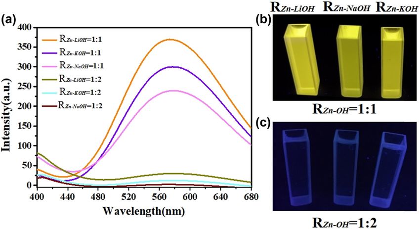
Effect of alkali bases on ZnO quantum dots 379
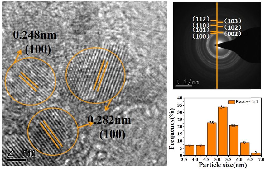
3 Results and discussion plane in the XRD diagram, and the average particle size is
about 5.5 nm, with a uniform distribution.
In the XRD spectrum of ZnO QDs (as shown in Figure 2), The presence of silica capping has been explained
the characteristic peaks of ZnO (100), (002), (101), (102), from the FTIR studies on the samples. To explore the
(110), (103), and (112) are all consistent with the standard functional groups on the surface of the ZnO QDs, infrared
card JCPDS (99-0111), indicating that the crystal structure characterization was used as shown in Figure 5. The
of the synthesized ZnO QDs is wurtzite. Because of the observation of an absorption peak at 900 cm−1, corre-
small particle size of ZnO QDs, the half-width of the dif- sponding to Si–O vibration, suggests the presence of
fraction peak is broadened. The half-width of the diffrac- silica in the capped ZnO QDs. The sketch of the silaniza-
tion peak of the sample with RZn−OH = 1:2 is larger than tion method is shown in Figure 6. In the sample of
that of the sample with RZn−OH = 1:1, which indicates that uncapped ZnO QDs, an absorption peak is observed at
the particle size of the QDs prepared at RZn−OH = 1:2 is 470 cm−1, corresponding to the Zn–O stretching vibra-
smaller than that prepared at RZn−OH = 1:1. When RZn−OH tions. Furthermore, this peak is found to be shifted to
is fixed at 1:1, the ZnO QDs synthesized with LiOH are the the lower wave number 456, 450, and 443 cm−1 for sam-
smallest. ples. The shift of Zn–O peak in all the capped samples has
Figure 3 shows the TEM results of each sample. The been found unidirectional toward the lower wave number
particle size of the ZnO QDs prepared at RZn−OH = 1:2 is side, which is indicative of an increase in the effective
smaller than that prepared at RZn−OH = 1:1. The mor- mass of the Zn–O system [18]. The absorption peak at
phology of the QDs with RZn−OH = 1:1 shows better uni- 3,420 cm−1 is because of the O–H stretching vibrations.
formity. Regardless of RZn−OH, the dispersion of ZnO QDs However, compared with the samples of RZn−OH = 1:1,
prepared with LiOH as the alkali base is better than that there is no N–H stretching vibration peak in the range
prepared with NaOH and KOH. This may be due to the of 3,000–3,500 cm−1 with the samples of RZn−OH = 1:2.
different dissociation constants of the alkali base (KDLiOH < They are derived from the N–H and O–H stretching vibra-
KDNaOH < KDKOH) [25]. As the synthesis process of the QDs is tion absorption peaks. The peak at 1,420 cm−1 is the flex-
a relatively violent reaction process, a low dissociation ural vibration absorption peak of C–H, and the peak at
constant is beneficial to the formation of uniform particle 1,580 cm−1 is the flexural vibration absorption peak of the
morphology. The selected area electron diffraction pat- end group –NH2. The IR spectrum proves that there are
tern (SAED) shows that the ZnO QDs with RZn−OH = 1:1 hydroxyl and amino functional groups on the surface of
has better crystallinity than that with RZn−OH = 1:2. the ZnO QDs. It shows that the high content of OH− is
Figure 4 shows the HRTEM and diffraction ring of harmful to the hydrolysis of APTES to form silica, so the
ZnO QDs prepared with RZn−OH = 1:1 using LiOH as the content of the modified functional groups on the surface
alkali base. Each crystal plane corresponds to the crystal of the ZnO QDs prepared with RZn−OH = 1:2 is less. The
hydroxyl and amino functional groups are hydrophilic
groups, which enhance the stability and solubility of
the ZnO QDs in water.
Figure 7 shows the ultraviolet-visible absorption
spectrum of ZnO QDs. All the ZnO QDs have exciton
absorption peaks because of the relatively larger binding
energy of the exciton (60 mV) [26]. The UV absorption
edge of the samples of RZn−OH = 1:2 has a more significant
blueshift than the samples of RZn−OH = 1:1. The blueshift is
caused by the quantum size effect. The blueshift is more
obvious with a decrease in the particle size. Therefore, it
can be inferred that the particle size of the ZnO QDs pre-
pared with RZn−OH = 1:2 is relatively smaller, which is
consistent with the results of XRD and TEM. RZn−OH has
a significant effect on the UV absorption intensity of ZnO
QDs. Samples with RZn−OH = 1:1 has a stronger absorption
than those with RZn−OH = 1:2, which is consistent with the
Figure 2: XRD pattern of ZnO QDs synthesized under different alkali results of the infrared spectrum. The ultraviolet-visible
bases and RZn−OH values. absorption of ZnO QDs is related to the band gap energy.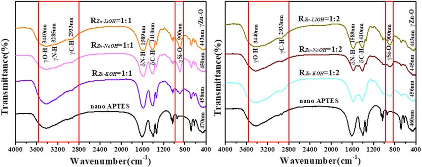
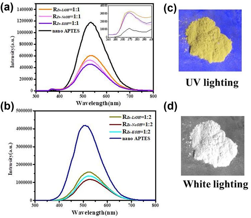
380 Xilian Zhang et al. Figure 3: TEM image and SAED image of ZnO QDs synthesized with different alkali bases and RZn−OH values. Figure 4: HRTEM image, SAED image, and particle size distribution image of ZnO QDs synthesized at RZn−LiOH = 1:1. Because of the quantum size effect of ZnO QDs, the band Figure 8 shows ZnO QDs (LiOH, RZn−OH = 1:1) solid gap energy increases with the decrease in the size, and powder under 365 nm UV lighting and visible light, which the absorption edge is blue shifted in the ultraviolet- shows that ZnO QDs emit a strong yellow fluorescence visible absorption spectrum. The difference in absor- under UV lighting. The solid-state fluorescence spectrum bance is because the water solubility of RZn:OH = 1:1 is of ZnO QDs (EX = 324 nm) is shown in Figure 8. There better than that of RZn:OH = 1:2, and the content in the are two fluorescence emission peaks in the spectrum, solution is higher, so the absorbance is relatively larger. namely, a weak and sharp ultraviolet emission peak
Effect of alkali bases on ZnO quantum dots 381
Figure 5: IR spectra of ZnO QDs synthesized with different alkali bases and different RZn−OH values.
Figure 6: Sketch of the silanization method.
Silica coated with ZnO QDs usually does not affect the
absorption and luminescence properties of the semicon-
ductor nanoparticles, exhibiting good optical transpar-
ency [27]. The luminescence peak at 370 nm is generated
by exciton recombination [28,29], which is caused by the
transition of electrons from the bottom of the conduction
band of ZnO to the valence band, which constitutes the
inherent fluorescence of the material. Several studies
have been reported to explain the origin of broad emis-
sion from ZnO in the visible region [30–32]. It is reported
that this band is an envelope spectrum of multiple emis-
sion bands originating from different defect centers
such as zinc vacancy (VZn), zinc interstitial (Zni), oxygen
vacancy (V+o ), oxygen interstitial (Oi), and antisite oxygen
(OZn). Dijken et al. [32] suggested that the visible emis-
sion from nanocrystalline ZnO particles is because of the
Figure 7: Ultraviolet-visible spectra of ZnO QDs synthesized under
different alkali bases and different RZn−OH values. transition of a photogenerated electron from the conduc-
tion band to a deeply trapped hole (V++ o ). The visible
emission maxima at 530 nm of different sized ZnO QDs
(370 nm) and a strong and broad yellow emission peak observed in the present studies can also be assigned to
(530 nm). It is well known that the intensity of emission the transition of photogenerated electrons from the con-
depends on both size and surface properties of the dots. duction band edge to a deeply trapped level (V++ o centers).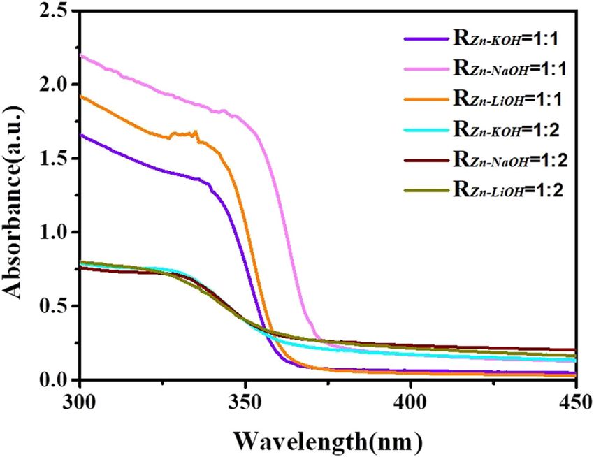
382 Xilian Zhang et al.
aqueous solution, the ZnO QDs prepared with RZn−OH = 1:2
have almost no fluorescence emission because of its poor
water solubility. The ZnO QDs prepared with RZn−OH = 1:1
show a yellow emission peak at about 570 nm. It is different
from the yellow fluorescence emission position measured in
solids because aging caused the peak shift [7]. The digital
photos are ZnO QDs dispersed in the ultrapure water under
365 nm UV light. It can be seen that the ZnO QDs prepared
at RZn−OH = 1:1 emit very strong yellow light, whereas the
samples prepared at RZn−OH = 1:2 hardly emit light.
The growth of the ZnO QDs is first through the direc-
tional attachment and bonding mechanism, and then the
maturation and coarsening of Ostwald ripening [33]. But
when the alkali base is KOH, it will experience the third
type: secondary precipitation of ZnO QDs, which makes
the particles larger. Currently, the formation mechanism
of ZnO QDs is still unclear, and the different processes
Figure 8: Solid-state fluorescence spectra of ZnO QDs synthesized
under different alkali bases and different RZn−OH = 1:1 (a), RZn−OH =
have been proposed because of the difference in the inter-
1:2 (b), and fluorescence of different alkaline and RZn−OH ratios mediate substances [34]. The most common nucleation
under UV irradiation (c and d). and growth mechanism of ZnO QDs were proposed by Jun
et al. [9]. The specific reaction is as follows:
The specific luminescence transition mechanism diagram Zn2 + + OH− → [Zn(OH)]+ , (1)
is shown in Figure 9. The luminescence performance is 2n[Zn(OH)]+ → (ZnO)n + nZn2 + + nH2 O, (2)
also related to the preparation method and react para-
meters, so further deep research studies are needed. (ZnO)n + 2k[Zn(OH)]+ → (ZnO)n + k + k Zn2 + + k H2 O, (3)
Regardless of the alkali base, the luminescence intensity where (ZnO)n represents the crystal nucleus, and (ZnO)n+k
of ZnO QDs at RZn−OH = 1:2 is stronger than that at RZn−OH = represents the QD grown from the crystal nucleus.
1:1 in the range of 500–530 nm, indicating that the defect Equation (1) indicates that the reaction occurs when
concentration of ZnO QDs prepared at RZn−OH = 1:2 is high. Zn2+ and OH− mix, then generating [Zn(OH)]+. Equations
This indicates that the hydrolysis reaction of APTES is (2) and (3) represent nucleation and growth, respectively.
inhibited under the condition of RZn−OH = 1:2, resulting Only when the [Zn(OH)]+ concentration reaches a certain
in an incomplete silica covering layer and numerous sur- value, the reaction in equation (2) can be triggered. Once
face defects on the surface of the QDs. equation (2) is triggered, the reaction in equation (3) will
Figure 10 shows the difference between the liquid fluor- proceed simultaneously.
escence spectrum of ZnO QDs (EX = 350 nm) and the solid The alkali bases have different dissociation constants
fluorescence spectrum of ZnO QDs (EX = 324 nm). In the (KDLiOH < KDNaOH < KDKOH), which affect the content of OH−.
When the RZn–OH is kept as a constant, low dissociation
constants result in small reaction driving force, which
reduces the growth efficiency of ZnO QD, and the parti-
cles are relatively dispersible. The difference in yield also
follows the order of dissociation constants of LiOH, NaOH,
and KOH. The smaller the number of QDs generated at the
same time, the less agglomeration formed to a certain extent.
4 Conclusions
ZnO QDs have a potential application in the medical
Figure 9: Schematic diagram of luminescence mechanism of QDs. fields, but the preparation of high-quality ZnO QDs is still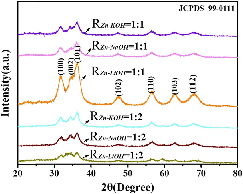
Effect of alkali bases on ZnO quantum dots 383
Figure 10: Liquid-state fluorescence spectra of ZnO QDs synthesized under different alkali bases and different RZn−OH values (a). Images of
the ZnO QDs under UV lighting and white lighting conditions (b and c).
a challenge. The alkali bases and RZn−OH are vital factors Competing interest: The authors have declared that no
influencing the morphology and performance of ZnO QDs competing interests exist.
in the sol–gel method. This article studies the influence of
the ratio of different alkali bases (LiOH, NaOH, and KOH) Data availability statement: The datasets generated
and RZn−OH on the performance of ZnO QDs. The results during and/or analyzed during the current study are
show that ZnO QDs can be synthesized successfully using available from the corresponding author on reasonable
any one of the three alkali bases, and higher OH− con- request.
centration is beneficial for forming a smaller particle size,
but harmful for the water solubility and the fluorescence
intensity. The difference in dissociation constants may be References
the major reason that influences the reaction process. The
particle size of the ZnO QDs prepared using LiOH and
[1] Dai-Xin Y, Ying-Ying M, Wei Z, Hong-Mei C, Ji-Lie K, Huan-
NaOH as raw materials is smaller than that prepared Ming X, et al. ZnO-based nanoplatforms for labeling and
using KOH. ZnO QDs prepared at the ratio of n(Zn2+): treatment of mouse tumors without detectable toxic side
n(LiOH) = 1:1 have the best fluorescence performance effects. ACS Nano. 2016;10(4):4294–300. doi: 10.1021/
and dispersibility. acsnano.5b07846.
[2] Chuan X, Yan Z, Peng W, Bo Z, Yukun Z. Novel surface mod-
ification of ZnO QDs for paclitaxel-targeted drug delivery for
Funding information: This work was financially supported lung cancer treatment. Dose-Response. 2020;18(2):1–7. doi:
by the Foundation of Natural Science Foundation of Jiangxi 10.1177/1559325820926739.
Province (Grant No. 20202BABL203041), the Research [3] Tsukazaki A, Ohtomo A, Onuma T, Ohtani M, Makino T,
Project of Jiangxi Provincial Department of Education Sumiya M, et al. Repeated temperature modulation epitaxy for
p-type doping and light-emitting diode based on ZnO. Nat
(GJJ190600), and the Open Fund of Pharmacy department,
Mater. 2005;4(1):42–6. doi: 10.1038/nmat1284.
Jiangxi Science & Technology Normal University (Grant No. [4] Bruchez M, Moronne M, Gin P, Weiss S, Alivisatos AP.
JFCEC-KF-1902). Semiconductor nanocrystals as fluorescent biological labels.
Science. 1998;281(5385):2013–6. doi: 10.1126/
Author contributions: Xilian Zhang and Shanshan Luo: science.281.5385.2013.
data curation and original draft. Xiaodan Wu: review and [5] Xiaosheng T, Eugene GC, Ling L, Jun D, Junmin X. Synthesis of
ZnO nanoparticles with tunable emission colors and their cell
editing. Minghui Feng: investigation. Yingying Li: meth-
labeling applications synthesis of ZnO nanoparticles with
odology. Haoyun Han: visualization. Wenkui Li: concep- tunable emission colors and their cell labeling applications.
tualization, review, and editing. Chem Mater. 2010;22(11):3383–8. doi: 10.1021/cm903869r.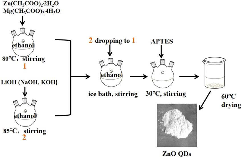
384 Xilian Zhang et al.
[6] Lubomir S, Marc AA. Semiconductor clusters in the sol–gel [20] Park WI, Yi GC, Kim MY, Pennycook SJ. Quantum confinement
process: quantized aggregation, gelation, and crystal growth observed in ZnO/ZnMgO nanorod heterostructures. Adv Mater.
in concentrated ZnO colloids. J Am Chem Soc. 2003;15(6):526–9. doi: 10.1002/adma.200390122.
1991;113(8):2826–33. doi: 10.1021/ja00008a004. [21] Ye YF. Photoluminescence property adjustment of ZnO
[7] Zhong C, XiaoXia L, Guoping D, Nan C, Andy YS. A sol–gel quantum dots synthesized via sol–gel method. J Mater Sci-
method for preparing ZnO quantum dots with strong blue Mater Electron. 2018;29(6):4967–74.
emission. J Lumines. 2011;131(10):2072–7. doi: 10.1016/ [22] Singh J, Bhartimittu D, Chauhan A, Singla ML. Role of alkali
j.jlumin.2011.05.009. metal hydroxide in controlling the size of ZnO nanoparticles in
[8] Li-Li H, Lan C, WeiHua W, JiangLong W, XiWen D. On the origin non-aqueous medium. Int J Fundam Appl Sci. 2012;1(4):91–3.
of blue emission from ZnO quantum dots synthesized by a [23] Anzlovar A, Kogej K, Orel Z-C. Impact of inorganic hydroxides
sol–gel route. Semicond Sci Technol. 2012;27(6):065020–7. on ZnO nanoparticle formation and morphology. Cryst Growth
[9] Jun Z, Suqing Z, Kun Z, Jianqing Z, Yanfei C. A study of Des. 2014;14(9):4262–9. doi: 10.1021/cg401870e.
photoluminescence properties and performance improvement [24] Huang WL, Lv XW, Tan JL, Huang QM, Cheng H, Feng J, et al.
of Cd-doped ZnO quantum dots prepared by the sol–gel Regulable preparation of the oxygen vacancy of ZnO QDs and
method. Nanoscale Res Lett. 2012;7:1–7. doi: 10.1186/1556- their fluorescence performance. J Mol Structure.
276X-7-405. 2019;1195:653–8. doi: 10.1016/j.molstruc.2019.05.105.
[10] Panigrahi S, Bera A, Basak D. Ordered dispersion of ZnO [25] Caetano BL, Silva MN, Santili CV, Briois V, Pulcinelli SH.
quantum dots in SiO2 matrix and its strong emission proper- Unified ZnO Q-dot growth mechanism from simultaneous
ties. J Colloid Interface Sci. 2011;353(1):30–8. doi: 10.1016/ UV–Vis and EXAFS monitoring of sol–gel reactions induced by
j.jcis.2010.09.055. different alkali base. Opt Mater. 2016;61:92–7. doi: 10.1016/
[11] Ensafi AA, Zakery M, Rezaei B. An optical sensor with specific j.optmat.2016.06.038.
binding sites for the detection of thioridazine hydrochloride [26] Tang ZK, Wong GKL, Yu P, Kawasaki M, Ohtomo A. Room-
based on ZnO-QDs coated with molecularly imprinted polymer. temperature ultraviolet laser emission from self-assembled
Spectrochim Acta A Mol Biomol Spectrosc. 2019;206:460–5. ZnO microcrystallite thin films. Appl Phys Lett.
doi: 10.1016/j.saa.2018.08.040. 1998;72(25):3270–2. doi: 10.1063/1.121620.
[12] Haghani SH, Ensafi AA, Kazemifard N, Rezaei B. Development [27] Lu Y, Yin Y, Li Z-Y, Xia Y. Synthesis and self-assembly of
of a selective and sensitive chlorogenic acid fluorimetric Au@SiO2 core−shell colloids. Nano Lett. 2002;2(7):785–8.
sensor using molecularly imprinted polymer ZnO quantum doi: 10.1021/nl025598i.
dots. IEEE Sens J. 2020;20(11):5691–7. [28] Yu SH, Oshimura MY, Calderon-Moreno JM, Fujiwara T,
[13] Ali F, Mona A, Shiva T. Degradation of toxin via ultraviolet and Fujino T, Teranishi R. In situ fabrication and optical properties
sunlight photocatalysis using ZnO quantum dots/CuO of a novel polystyrene/semiconductor nanocomposite
nanosheets composites: preparation and characterization embedded with CdS nanowires by a soft solution processing
studies. J Mater Sci-Mater Electron. 2017;28(21):16397402. route. Langmuir. 2001;17(5):1700–7. doi: 10.1021/la000941p.
doi: 10.1007/s10854-017-7550-x. [29] Wang X, Yang S, Wang J, Li M, Jiang X, Du G, et al. Structural
[14] Lei S, Zhixian L, Xiongtu Z, Yongai Z, Tailiang G. Synthesis of and optical properties of ZnO film by plasma-assisted MOCVD.
Cu-doped ZnO quantum dots and their applications in field Opt Quantum Electron. 2002;34(9):883. doi: 10.1023/
emission. J Alloy Compd. 2016;671:473–8. doi: 10.1016/ A:1019956323462.
j.jallcom.2016.02.136. [30] Mohanta A, Thareja RK. Photoluminescence study of ZnO
[15] Talapin DV, Rogach AL, Shevchenko EV. Dynamic distribution nanowires grown by thermal evaporation on pulsed laser
of growth rates within the ensembles of colloidal II–VI and deposited ZnO buffer layer. J Appl Phys. 2008;104(4):044906.
III–V semiconductor nanocrystals as a factor governing their doi: 10.1063/1.2969908.
photoluminescence efficiency. J Am Chem. [31] Lin B, Fu Z, Jia Y. Green luminescent center in undoped zinc
2002;124(20):5782–90. doi: 10.1021/ja0123599. oxide films deposited on silicon substrates. J Appl Phys.
[16] Zhang J, Zhang R, Zhao L-H, Sun S-Q. Synthesis of water- 2001;79(7):943–5. doi: 10.1063/1.1394173.
soluble γ-aminopropyl triethoxysilane-capped ZnO:MgO [32] van Dijken A, Meulenkamp EA, Vanmaekelbergh D,
nanocrystals with biocompatibility. Cryst Eng Comm. Meijerink A. The luminescence of nanocrystalline ZnO parti-
2012;14:613–9. doi: 10.1039/C1CE05941F. cles: the mechanism of the ultraviolet and visible emission.
[17] Karakoti A-S, Shukla R, Shanker R. Surface functionalization of J Lumines. 2004;87–89:454–6. doi: 10.1016/S0022-2313(99)
quantum dots for biological applications. Adv Colloid 00482-2.
Interface. 2015;215:28–45. doi: 10.1016/j.cis.2014.11.004. [33] Pesika NS, Setbe KJ, Searson PC. Relationship between
[18] Patra MK, Manoth M, Singh VK, Siddaramana Gowd G, absorbance spectra and particle size distributions for
Choudhry VS, Vadera SR, et al. Synthesis of stable dispersion quantum-sized nanocrystals. J Phys Chem B.
of ZnO quantum dots in aqueous medium showing visible 2003;107:10412–5. doi: 10.1021/jp0303218.
emission from bluish green to yellow. J Lumines. [34] Patra MK, Manzoor K, Manoth M, Choudhry VS, Vadera SR,
2009;129(3):320–4. doi: 10.1016/j.jlumin.2008.10.014. Kumar N. Optically transparent colloidal suspensions of single
[19] Zhao L, Zhang R, Zhang J, Sun S-Q. Synthesis and character- crystalline ZnO quantum dots prepared by simple wet-chem-
ization of biocompatible ZnO nanoparticles. CrystEngComm. istry. J Optoelectron Adv Mater. 2008;10(10):2588–91.
2012;14(3):945–50. doi: 10.1039/C1CE05621B. doi: 10.1021/ja028416v.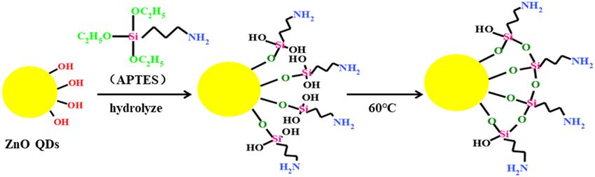
You can also read


