Expression and Significance of Matrix Metalloproteinase-2 and Matrix Metalloproteinas-9 in Endometriosis
←
→
Page content transcription
If your browser does not render page correctly, please read the page content below
DOI: 10.25122/jml-2020-0117                           Journal of Medicine and Life Vol. 13, Issue 3, July-September 2020, pp. 314–320

              Expression and Significance of Matrix Metalloproteinase-2 and Matrix
                             Metalloproteinas-9 in Endometriosis
      Adrian Mykhailovych Barbe1*, Andrii Mykolaiovych Berbets1, Igor Sviatoslavovych Davydenko2, Halyna Danylivna Koval3,
                                 Viktoriia Oleksandrivna Yuzko1, Oleksandr Mykhailovych Yuzko1
              1. Department of Obstetrics and Gynecology, Bukovinian State Medical University, Chernivtsi, Ukraine
                  2. Department of Pathologic Anatomy, Bukovinian State Medical University, Chernivtsi, Ukraine
   3. Department of Clinical Immunology, Allergology and Endocrinology, Bukovinian State Medical University, Chernivtsi, Ukraine

                                                         * Corresponding Author:
                                                        Adrian Mykhailovych Barbe,
                                                         2 Teatralna Sq, Chernivtsi,
                                                               Ukraine, 58000.
                                                          Phone: +380953912201
                                                      E-mail: adryanbarbe@gmail.com

                                             Received: April 14th, 2020 – Accepted: July 9th, 2020

Abstract
Endometriosis is a chronic benign hormone-dependent condition when the endometrial tissue, identical with the endometrium by its
morphological and functional properties, grows outside the borders of the uterine mucous membrane. Recent studies have pointed
to the possible role of matrix metalloproteinases (MMPs) in the pathogenesis of endometriosis. We suggested a hypothesis that
increased expression of MMPs activity in eutopic and ectopic endometrium of patients with endometriosis might correlate with the
presence of endometriotic lesions.
The aim of the study was to evaluate the level of MMP-2 and MMP-9 expression in the ectopic endometrium of women with visible
endometriotic lesions and eutopic endometrium in patients with no signs of endometriosis.
The study was conducted on 43 patients. They were divided into two groups. Group 1 included 31 patients with peritoneal/ovarian en-
dometriosis who had undergone laparoscopy and hysteroscopy. Group 2 consisted of 12 patients with leiomyoma, endometrial polyps
or relatively healthy patients who had undergone hysterectomy or polypectomy and endometrial curettage.
This study showed statistically higher expression of MMP-2 (1.7783 ± 0.22 immunohistochemistry (IHC) optical density score com-
pared to the control group – 1.41± 0.34, p = 0.0017) and MMP-9 (1.352 ± 0.067 versus 1.85 ± 0.26 in the control group, p = 0.001) in
ectopic and eutopic endometrium samples from patients with endometriosis compared to samples taken from patients without endo-
metriosis. A strong correlation between expression of the above-mentioned MMPs (r=0.74 for MMP-2 and r=0.88 for MMP-9) in ectopic
and eutopic endometrium might be of promising diagnostic value.

Keywords: Endometriosis, MMP-2, MMP-9, metalloproteinase, neo-angiogenesis, cellular matrix.

Abbreviations: CA-125 – carbohydrate antigen-125; DAB – Diaminobenzidine; HE4 – human epididymis protein 4; HRP – horserad-
ish peroxidase; IHC – immunohistochemistry; MMP – matrix metalloproteinase; TIMP – tissue inhibitors of metalloproteinase; VEGFR
– vascular endothelial growth factor receptor.

Introduction                                                              Nowadays, the pathogenetic theory of retrograde outflow
                                                                          of endometrial cells into the peritoneal cavity is gaining
                                                                          increasing support [5, 6]. Since retrograde menstruation
Endometriosis is a chronic benign hormone-dependent                       occurs in many women, it is postulated that endometriosis
condition when the endometrial tissue, identical with the                 develops as a consequence of disturbances in the balance
endometrium by its morphological and functional prop-                     between the amount of menstrual blood and capacity of
erties, grows outside the borders of the uterine mucous                   the “clearance” system in the peritoneal milieu. Recent
membrane. It leads to clinical symptoms able to affect the                studies have pointed to the possible role of matrix metallo-
physical condition, psychological status, and social status               proteinases (MMPs) in the pathogenesis of endometriosis.
of the patient [1-4]. According to research data, endometri-              In particular, Wenzl and Heinzl [7, 8] suggested that ectop-
osis is diagnosed in 5-10% of the female population. There                ic endometrium maintains its protease secretory capacity,
are approximately 176 million women with endometriosis                    which, in turn, allows the invasion of surrounding tissues
in the world, mainly of reproductive age.                                 and the subsequent formation of endometrial foci [8].

                                                                    314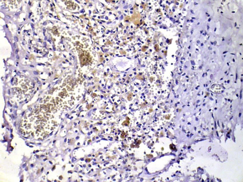
Journal of Medicine and Life Vol. 13, Issue 3, July-September 2020, pp. 314–320

Matrix metalloproteinases (MMPs) are a family of endo-                 Clinical Maternity Hospital No. 1 and the Medical Center of
peptidases playing a specific role in the degradation and              Infertility Treatment (Chernivtsi, Ukraine). All the patients
turnover of the extracellular matrix (ECM). These zinc-de-             were premenopausal.
pendent enzymes, including collagenases, gelatinases,                       They were divided into two groups. Group 1 included
and stromelysins, are capable of degrading all the ECM                 31 patients with peritoneal/ovarian endometriosis who had
components. Tissue inhibitors of metalloproteinases                    undergone laparoscopy and hysteroscopy. Patients were
(TIMPs), which affect normal and pathologic matrix remod-              selected by the existence of visible peritoneal endometri-
eling, regulate the activity of MMPs [9, 10].                          otic lesions (stage II, III) (ASRM, 1997), characterized as
      The implantation of endometrium cells results from im-           red- or gland-like lesions as well as red vesicles.
balance and excretion of growth factors. Adequate blood                     The age of the patients ranged from 22 to 48 years, with
supply and neo-angiogenesis play an essential role in the              an average of 33.8 years. The diagnosis of endometriosis
successful implantation and occurrence of ectopic foci [11-            was confirmed histologically by detecting endometrial-like
14]. However, for triggering this process, primarily, there is         glands in the heterotopic locations (peritoneum or ovarium).
a need for endometrium cell adhesion, proteolytic activity,            The patients with comorbid gynecologic diseases, including
and extracellular matrix alteration. MMPs and their natural            leiomyoma and endometrial polyps, were excluded.
inhibitors – tissue inhibitors of metalloproteinases (TIMPs)                Group 2 consisted of 12 patients with leiomyoma, en-
play an important role in the remodeling of the endometrial            dometrial polyps, or relatively healthy patients who had
tissue during the normal menstrual cycle [15, 16], as well             undergone hysterectomy or polypectomy and endometrial
as in endometriosis [1, 17, 18]. MMPs and TIMPs play a                 curettage. None of the patients had identifiable adenomyo-
specific role in testicular development and maturation [19]            sis by ultrasonography imaging.
and in the ovary during ovulation [20].                                     All the endometrial samples were collected in prolif-
      The peritoneal fluid of women with endometriosis is a            erative menstrual cycle phases. The day of the menstrual
specific microenvironment containing numerous substanc-                cycle was established from the women’s menstrual history
es that can act on the endometriotic tissue [21]. In particu-          and was confirmed by endometrial dating using the crite-
lar, it contains an increased number of macrophages and                ria of Noyes et al. For each case of endometriosis, repre-
their secretory products, such as growth factors, cytokines,           sentative slides of eutopic and ectopic endometrium were
and angiogenic factors [22]. On the other hand, increased              selected. For cases of the control group, representative
metalloproteinase activity in the endometrium, in its turn,            slides of eutopic endometrium in the proliferative phase
might facilitate the breakdown of the peritoneal extracel-             were selected.
lular matrix and establishment of endometriotic foci in the
peritoneal cavity [23].
      These observations have prompted us to suggest a
                                                                       Immunohistochemistry
hypothesis that increased expression of MMPs activity in               MMP-2 and MMP-9 in the eutopic and ectopic endometrial
eutopic endometrium combined with increased expression                 tissue were identified by means of immunoassay diagnos-
of vascular endothelial growth factor receptor 2 (VEGFR-2)             tic kits based on the antibodies specific for the mentioned
in the peritoneum, eutopic and ectopic endometrium of pa-              antigens and receptors (manufactured by Abcam, Cam-
tients with endometriosis might correlate with the presence            bridge, United Kingdom).
of endometriotic lesions [24]. If this is the case, a simple                Visualization of antibodies was performed by
endometrium biopsy might prove useful in assessing the                 mouse-specific HRP/DAB Detection IHC Kit with diamin-
early stages of endometriosis.                                         obenzene ink (manufactured by Abcam, Cambridge, United
      The aim of the study was to evaluate the level of MMP-           Kingdom). Cell nuclei were additionally stained with May-
2 and MMP-9 expression in ectopic and eutopic endometri-               er’s hematoxylin solution. The immunohistochemistry (IHC)
um of women with visible endometriotic lesions and eutop-              optical density score assessment of immunohistochemical
ic endometrium in patients with no signs of endometriosis.             staining of MMP-2 and MMP-9 was performed (see below).
We also examined the possible correlation between MMP-
2 and MMP-9 levels in eutopic and ectopic endometrium
in patients with endometriosis. As expected, our findings
                                                                       Assessment of MMP-2, MMP-9 Staining
will provide a theoretical basis for a better understanding            Cytoplasmic staining was defined as positive. Semiquanti-
of the role of matrix metalloproteinases as possible early             tative and quantitative analysis of the immunostainings for
diagnostic markers of endometriosis.                                   MMP-2 and MMP-9 was performed for each case. MMP-2
                                                                       and MMP-9 expression was evaluated in the glandular and
                                                                       stromal cells. The staining of the superficial epithelium and
                                                                       stroma was scored according to the intensity for both MMP-
Material and Methods                                                   2 and MMP-9. The results of staining were assessed by one
                                                                       highly qualified pathologist as an observer in a blind fashion.

Subjects                                                               Image acquisition
The study was conducted on 43 patients who were treated                Images were captured using the bright field technique with
at the Gynecological Department of Chernivtsi Municipal                a binocular microscope (Olympus СХ-21).

                                                                 315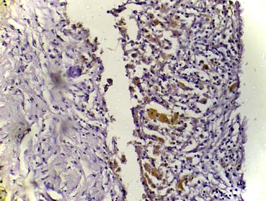
Journal of Medicine and Life Vol. 13, Issue 3, July-September 2020, pp. 314–320

Images were captured at ×10 magnification using color                     Results
photo camera Olympus С450 attached to a computer sys-
tem. The field was selected with good contrast between
the DAB chromogen and hematoxylin, which is considered
                                                                          Patients’ characteristics
the region of interest. After capturing the images, the color             Patients’ characteristics of each of the groups studied are
density and white balance were standardized for all the                   presented in Table 1. Our study has shown that the number
images. All the acquired images were saved as a JPEG                      of pregnancies significantly differed between the groups
format with minimal compression.                                          (0.87 in the study group vs. 1.66 in the control group), as
                                                                          many studies have shown before [26-28]. However, no
                                                                          significant differences between the studied groups were
ImageJ analysis                                                           found concerning age, number of childbirths, number of
For IHC image analysis, the 1.48 version of ImageJ (free                  abortions and number of miscarriages.
software, NIH, Bethesda, Maryland) (Java 1.8.9) was ap-
plied. IHC profiler plugin with cytoplasmic-stained mode
was used for qualifying images. Images were qualified as
                                                                          MMP-2 expression
high positive, positive, low positive, and negative with the              The measurement results of the IHC optical density score
corresponding percentage of cells. As a result, the IHC Op-               of immunohistochemical staining of MMP-2 in human eu-
tical Density Score was calculated by using the following                 topic endometrium obtained from patients with endometri-
formula [25]:                                                             osis and patients without endometriosis are represented in
     IHC optical density score = (percentage contribution of              Figure 1. The IHC optical density score of MMP-2 in eutop-
high positive × 4 + percentage contribution of positive × 3               ic endometrium obtained from women with endometriosis
+ percentage contribution of low positive × 2 + percentage                was significantly higher (1.7783 ± 0.22 IHC optical densi-
contribution of negative × 1) / 100.                                      ty score) than in the eutopic endometrium obtained from
                                                                          women without endometriosis (1.41 ± 0.34, p = 0.0017).
                                                                               As for the comparison of MMP-2 expression between
Statistical Analysis                                                      ectopic endometrium in group 1 (with endometriosis) and
Statistical data were calculated and compared using the                   eutopic endometrium in group 2 (control group), there was
MedCalc software, developed by “MedCalc Software” (Os-                    a significant difference between them (1.82 ± 0.27 IHC
tend, Belgium).                                                           optical density score vs. 1.41 ± 0.34 in the control group,
     The immunohistochemical data are reported as the                     p = 0.001).
mean ± SEM. The data obtained were statistically pro-                          The correlation in the group 1 between ectopic and
cessed using the Mann-Whitney U test. Bivariate corre-                    eutopic IHC optical density score was obtained. It showed
lation between variables was determined by Pearson’s                      a high score with r = 0.74, which indicates a strong cor-
correlation coefficients. A P-value < 0.05 was considered                 relation between the expression of MMP-2 in eutopic and
significant. Results are represented as mean ± SD.                        ectopic endometrium (Figure 2).
                                                                               On IHC images, MMP-2 stained areas were located
                                                                          mainly in the stroma and around the vessels (in eutopic en-
Ethical approval                                                          dometrium) or directly under the epithelial layer (in ectopic
The study was approved by the Biological and Medical                      endometrium) (Figure 3 and 4).
Ethics Committee of the Higher State Educational Estab-
lishment of Ukraine “Bukovinian State Medical University”
(the minutes No. 6 dated September 15th, 2016). It was
                                                                          MMP-9 expression
carried out strictly following The Code of Ethics of the                  The measurement results of the IHC optical density score
World Medical Association (Declaration of Helsinki, 1983)                 of immunohistochemical staining of MMP-9 in human eu-
for medical research involving human subjects, including                  topic endometrium obtained from patients with and without
research on identifiable human material and data.                         endometriosis are represented in Figure 5.

Table 1: Patients’ characteristics of the studied groups.
                                                     Group 1, n=31          Control group, n=12           p
                     Age, years                             32.61                  37.00                 0.07
                     Number of pregnancies                  0.87                    1.66                0.022
                     Number of childbirths                  0.77                    1.25                0.139
                     Number of abortions                     0                       0                    -
                     Number of miscarriages                 0.06                    0.16                 0.35
Note: The values are expressed as mean (SD).

                                                                    316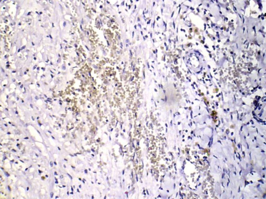
Journal of Medicine and Life Vol. 13, Issue 3, July-September 2020, pp. 314–320
Figure 1: MMP-2 IHC optical density score comparison between group 1 (endometriosis, eutopic endometrium) and group 2 (the
control group).
Figure 2: Correlation of MMP-2 IHC optical density between eutopic endometrium and ectopic endometriosis foci (correlation coeffi-
cient r = 0.74).
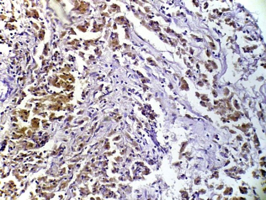
Figure 3: The visualization of immunohistochemical staining of Figure 4: The visualization of immunohistochemical staining of
MMP-2 in ectopic endometriosis foci: a light microscopic view. MMP-2 in eutopic endometrium in endometriosis: a light micro-
Stained areas are located mainly in the stroma and under the scopic view. Stained areas are located mainly in the stroma and
epithelial layer (x400). around the vessels (x400).
317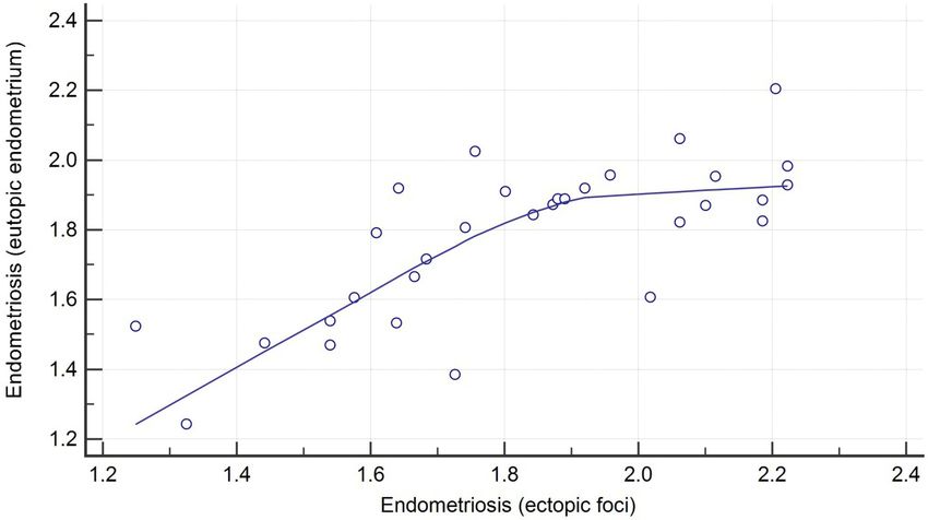
Journal of Medicine and Life Vol. 13, Issue 3, July-September 2020, pp. 314–320

Figure 5: MMP-2 IHC optical density score comparison between group 1 (endometriosis, eutopic endometrium) and group 2 (the
control group).

Figure 6: Correlation of MMP-9 IHC optical density between eutopic endometrium and ectopic endometriosis foci (correlation coeffi-
cient r = 0.88).

The IHC optical density score of MMP-9 in eutopic endo-                On IHC images, MMP-9 stained areas were located in the
metrium obtained from women with endometriosis was                     stroma and around the vessels (in eutopic endometrium)
significantly higher (1.85 ± 0.26 IHC optical density score)           or in the sub-epithelial and epithelial layer (in ectopic en-
than that in the eutopic endometrium obtained from women               dometrium) (Figure 7 and 8).
without endometriosis (1.3528 ± 0.067, p = 0.001). This
indicates a significantly higher activity of MMP-9 in patients
with endometriosis.
     When comparing MMP-9 expression between ectopic
                                                                       Discussion
endometrium in group 1 (with endometriosis) and eutopic
endometrium in group 2 (control group), a significant dif-             Matrix metalloproteinases (MMPs) are a family of endo-
ference between them was established (1.87 ± 0.30 IHC                  peptidases playing a specific role in the degradation and
optical density score vs. 1.35 ± 0.06 in the control group,            turnover of the extracellular matrix (ECM). These zinc-de-
p < 0.001).                                                            pendent enzymes, including collagenases, gelatinases
     The correlation determined in group 1 between ectopic             and stromelysins, are capable of degrading all the com-
and eutopic IHC optical density score showed a high score              ponents of the ECM. Tissue inhibitors of metalloprotein-
with r = 0.88, which indicates a strong correlation between            ases (TIMPs), which affect normal and pathologic matrix
expression of MMP-9 in eutopic and ectopic endometrium                 remodeling, regulate the activity of MMPs [29, 30]. MMP
as in case of MMP-2 (Figure 6).                                        is supposed to enable the endometriotic tissue to be di-

                                                                 318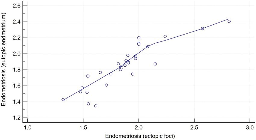
Journal of Medicine and Life Vol. 13, Issue 3, July-September 2020, pp. 314–320

Figure 7: The visualization of immunohistochemical staining of            Figure 8: The visualization of immunohistochemical staining of
MMP-9 in ectopic endometriosis foci: a light microscopic view.            MMP-9 in eutopic endometrium in endometriosis: a light micro-
Stained areas are located in stroma, subepithelial and epithelial         scopic view. Stained areas are located in the stroma and around
layer (x400).                                                             the vessels (x400).

gested into the peritoneal ECM and underlying connective                  garding the limitations of the studies conducted, it should
tissue. Endometrial remodeling and MMP expression are                     be mentioned that we used a limited of research objects,
well known to occur during the proliferative and menstru-                 which may slightly impair the accuracy of the results.
al phases of the cycle, and that progesterone is a strong
suppressor of MMPs [31]. The production of MMPs and
their inhibitors takes place in the endometrial stroma and
epithelium, as well as in polymorphic mononuclear leuko-
                                                                          Conclusions
cytes [32].
     Taking into account the fact that the production of                  Our study demonstrates a statistically higher expression of
MMPs (as well as their inhibitors) takes place in the endo-               MMP-2 and MMP-9 both in ectopic and eutopic endome-
metrial stroma, according to our findings, we suggest a hy-               trium samples compared to samples taken from patients
pothesis that MMPs activity in eutopic endometrium could                  without endometriosis. A strong correlation between the
correlate with the activity in ectopic foci of endometriosis.             expression of the above-mentioned MMPs in ectopic and
This peculiarity could improve the chances of non-invasive                eutopic endometrium might be of promising diagnostic val-
early diagnostics of endometriosis. This statement is also                ue.
based on the opinion of Chung et al., who considers that
increased proteolytic activity of the endometrial and endo-
metriotic tissue may be one of the reasons for the invasive
properties of the endometrium [33]. On this basis, we can
                                                                          Conflict of Interest
assume that the endometrium could be a predominant
source of MMP-9, as well as of MMP-2.                                     The authors declare that there is no conflict of interest.
     In the current study, we demonstrate that the expres-
sion of endometrium MMP-2 and MMP-9 are increased in
patients with endometriosis. Similar conclusions were ex-
pressed by Meng-Hsing Wu et al. [34]. They found that
                                                                          References
MMP-9 plays a pivotal role in the peritoneal macrophage’s                 1.   Guidice LC. Endometriosis. Lancet. 2010;362:2389-98 https://doi.
ability to degrade the basement membrane and, thus, its                        org/10.1016/S0140-6736(04)17403-5.
capability of phagocytosis. Liu, H et al. claim that plasma               2.   Brown J, Farquhar C. An overview of treatments for endometriosis.
MMP-9 directly correlates with the severity of endome-                         The Journal of the American Medical Association. 2015;313(3):296-
triosis and could be used as a biochemical marker [35].                        8 http://doi.org/10.1001/jama.2014.17119.
However, we found only a few studies in which authors                     3.   Dunselman G, Vermeulen N, Becker C, Calhaz-Jorge C, D’Hooghe
investigate MMP-2 expression in endometriosis [36].                            T, De Bie B, et al. ESHRE guideline: management of women with
     Our study has particular strengths. First of all, we                      endometriosis. Human Reproduction. 2014;29:400-12 https://doi.
                                                                               org/10.1093/humrep/det457.
have established a correlation between ectopic and eu-
                                                                          4.   Cheong YC, Smotra G, Williams AC. Non-surgical interventions
topic endometrium MMPs expression, which could help in                         for the management of chronic pelvic pain. Cochrane Database
the future in the early non-invasive diagnostic of endome-                     Systematic Review. 2014;3 http://doi.org/10.1002/14651858.
triosis. Second, immunohistochemistry, a simple, reliable                      CD008797.pub2.
and informative method, has been used, which resulted in                  5.   Macer ML, Taylor HS. Endometriosis and Infertility: A review of
good visualization and the possibility of extensive use. Re-                   the pathogenesis and treatment of endometriosis-associated

                                                                    319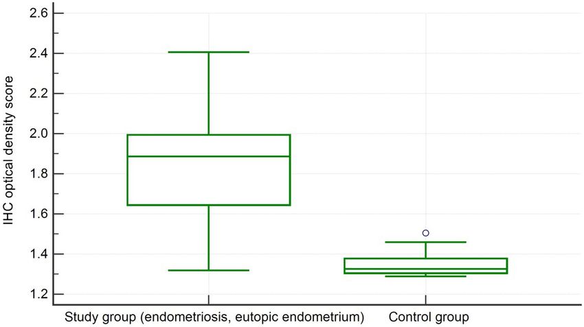
Journal of Medicine and Life Vol. 13, Issue 3, July-September 2020, pp. 314–320

      infertility. Obstetrics and Gynecology Clinics of North America.             22. International Journal of Endocrinology. 2016(8) https://doi.org/10.2
      2012;39(4):535-49 https://doi.org/10.1016/j.ogc.2012.10.002.                     2141/22240721.8.80.2016.89539.
6.    Laux-Biehlmann A, d’Hooghe T, Zollner T. Menstruation pulls the              23. Koval H, Chopyak VV, Kamyshnyi O, Kurpisz M. Transcription reg-
      trigger for inflammation and pain in endometriosis. Trends in Phar-              ulatory factor expression in T-helper cell differentiation pathway in
      macological Sciences. 2015;36(5):270-6 https://doi.org/10.1016/j.                eutopic endometrial tissue samples of women with endometriosis
      tips.2015.03.004.                                                                associated with infertility. Clinical immunology. 2018;1(43) https://
7.    Wenzl R, Heinzl H. Localization of matrix metalloproteinase-2 in                 doi.org/10.5114/ceji.2018.74878.
      uterine endometrium and ectopic implants. Gynecol Obstet Invest.             24. Sharpe-Timms K, Cox K. Paracrine regulation of matrix metallopro-
      1998(45):253–7 https://doi.org/10.1159/000009978.                                teinase expression in endometriosis. Ann NY Acad Sci 2002(955):147-
8.    Osteen K, Bruner-Tran K, Ong D, Eisenberg E. Paracrine mediators                 56 https://doi.org/10.1111/j.1749-6632.2002.tb02775.x.
      of endometrial matrix metalloproteinase expression. Potential tar-           25. Barbe A, Berbets A, Davydenko I, Yuzko V, Yuzko O. The effects of
      gets for progestin-based treatment of endometriosis. Ann NY Acad                 certain angioneogenesis inhibitors in experimental endometriosis
      Sci. 2002(955):139–46 https://doi.org/10.1111/j.17496632.2002.                   in rats. Cell and Organ Transplantology. 2019;2(7):140-7 https://
      tb02774.x.                                                                       doi.org/10.22494/cot.v7i2.101.
9.    Murphy G, Knauper V, Cowell S, Hembry R, Stanton H, Butler G,                26. Jafari S, Hunger R. IHC Optical Density Score: A New Practical
      et al. Evaluation of some newer matrix metalloproteinases. Ann NY                Method for Quantitative Immunohistochemistry Image Analysis.
      Acad Sci 1999(878):25-39 https://doi.org/10.1111/j.1749-6632.1999.               Appl Immunohistochem Mol Morphol. 2017;1(25):e12-e3 https://
      tb07672.x.                                                                       doi.org/10.1097/pai.0000000000000370.
10.   Nagase H, Woessner JJ. Matrix metalloproteinases. J Biol Chem.               27. Tomassetti C, D’Hooghe T. Endometriosis and Infertility: Insights
      1999(274):21491– 4 https://doi.org/10.1074/jbc.274.31.21491.                     Into the Causal Link and Management Strategies. Best Pract Res
11.   Ranney B. Endometriosis: pathogenesis, sympyoms, and findings.                   Clin Obstet Gynaecol. 2018(51):2533 https://doi.org/10.1016/j.
      Clinical Obstetrics & Gynecology. 1988;23(3):865-74.                             bpobgyn.2018.06.002.
12.   Donnwez J. Endometriosis: pathogenesis and pathophysiology.                  28. Singh S, Suen M. Surgery for Endometriosis: Beyond Medical
      New Jersey: RW Shaw; 1990.                                                       Therapies. Fertil Steril. 2017;3(107) https://doi.org/10.1016/j.fertn-
13.   Lu Z, Zhang W, Jiang S, Zou J, Li Y. Effect of oxygen tensions on                stert.2017.01.001.
      the proliferation and angiogenesis of endometriosis heterograft in           29. Tanbo T, Peter Fedorcsak P. Endometriosis-associated Infertility:
      severe combined immunodeficiency mice. Fertility and Sterility.                  Aspects of Pathophysiological Mechanisms and Treatment Op-
      2014;101(2):568-76 https://doi.org/10.1016/j.fertnstert.2013.10.039.             tions. Acta Obstet Gynecol Scand. 2017;6(96):659-67 https://doi.
14.   Signorile P, Baldi A. Endometriosis: New Concepts in the Pathogen-               org/10.1111/aogs.13082.
      esis. Int J Biochem Cell Biol. 2010;6(42) https://doi.org/10.1016/j.         30. Matrisian LM. The matrix degrading metalloproteinases. Bioes-
      biocel.2010.03.008.                                                              says. 1992(14):455– 63.
15.   Rodgers W, Matrisian L, Giudice L, Dsupin B, Cannon P, Svitek C,             31. Jeziorska M, Salamonsen LA, Woolley DE. Mast cell and eosino-
      et al. Patterns of matrix metalloproteinase expression in cycling                phil distribution and activation in human endometrium throughout
      endometrium imply differential functions and regulation by steroid               the menstrual cycle. Biol Reprod. 1995(53):312–20 https://doi.
      hormones. J Clin Invest. 1994(94):946–53 https://doi.org/10.1172/                org/10.1095/biolreprod53.2.312.
      jci117461.                                                                   32. Osteen KG, Bruner KL, Sharpe-Timms KL. Steroid and growth
16.   Salamonsen L, Wooley D. Matrix metalloproteinases in nor-                        factor regulation of matrix metalloproteinase expression and endo-
      mal menstruation. Hum Reprod. 1996(11):124–33 https://doi.                       metriosis. Semin Reprod Endocrinol. 1996(14):247–55 https://doi.
      org/10.1093/humrep/11.suppl_2.124.                                               org/10.1055/s-2007-1016334.
17.   Koks CA, Groothuis PG, Slaats P. Matrix metalloproteinases and               33. Vercellini P, Viganò P, Somigliana E, Fedele L. Endometri-
      their inhibitors in antegradely shed menstruum and peritoneal                    osis: pathogenesis and treatment. Nature Rev Endocrinol.
      fluid. Fertil Steril. 2000(73):604–12 https://doi.org/10.1016/S0015-             2014(10):261-75 https://doi.org/10.1038/nrendo.2013.255.
      0282(99)00566-X.                                                             34. Chung H-W, Wen Y, Chun S-H. Matrix metalloproteinase-9 and
18.   Gottschalk C, Malberg K, Arndt M. Matrix metalloproteinases and                  tissue inhibitor of metalloproteinase-3 mRNA expression in ectopic
      TACE play a role in the pathogenesis of endometriosis. Adv Exp Med               endometrium in women with endometriosis: a rationale for endo-
      Biol. 2000(477):483–6 https://doi.org/10.1007/0-306-46826-3_49.                  metriotic invasiveness. Fertil Steril. 2001(75):152–9 34. Wu M-H,
19.   Slongo M, Zampieri M, Onisto M. Expression of matrix met-                        Shoji Y, M-C W, Chuang P-C, Lin C-C, M-F H, et al. Suppression of
      alloproteases (MMP-2, MT1-MMP) and their tissue inhibitor                        Matrix Metalloproteinase-9 by Prostaglandin E2 in Peritoneal Mac-
      (TIMP-2) by rat Sertoli cells in culture: implications for spermat-              rophage Is Associated with Severity of Endometriosis. American
      ogenesis. Biol Chem. 2002(383):235-9 https://doi.org/10.1515/                    Journal of Pathology. 2005;167(4).
      bc.2002.025.                                                                 35. Liu H, Wang J, Wang H, Tang N, Li Y. The plasma and peritoneal flu-
20.   Curry TJ, Osteen K. Cyclic changes in the matrix metalloprotein-                 id levels of MMP-9 are elevated in patients with endometriosis. Ann
      ase system in the ovary and uterus. Biol Reprod. 2001(64):1285–                  Clin Biochem. 2016 https://doi.org/10.1177/0004563215626458.
      96 https://doi.org/10.1095/biolreprod64.5.1285.                              36. Sui X, Li Y, Sun Y, Chunyan L, Li X, Zhang G. Expression and
21.   Koval HD, Chopyak VV, Yuzko OM. Dependence of the Effective-                     Significance of Autophagy Genes LC3, Beclin1 and MMP-2 in
      ness of Infertility Treatment in Women with Endometriosis on the                 Endometriosis. Exp Ther Med. 2018;3(16) https://doi.org/10.3892/
      State of Immunogenetic Regulation.                                               etm.2018.6362.

                                                                             320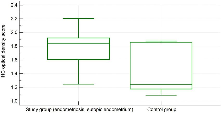
You can also read


