Congenital angiolipoma of the chest wall in a child: A case report and review of literature - Net Journals
←
→
Page content transcription
If your browser does not render page correctly, please read the page content below
International Research Journal of Medicine and Medical Sciences
                                                                       Vol. 9(1), pp. 43-51, March 2021
                                                                               ISSN: 2354-211X
                                                                                  Case Report

Congenital angiolipoma of the chest wall in a child: A
case report and review of literature
Parichehr Tootoonchi
Pediatrics Ward, Amir Al Momenin Teaching Hospital, Faculty of Medicine, Islamic Azad University of Medical
Sciences, Tehran, Iran.
Accepted 1 March, 2021

      ABSTRACT

      Angiolipoma is the most common soft tissue tumor in human trunk and extremities, mostly affecting young
      male patients; however, in children, it occurs rarely and the most involved area has not been determined. A
      9 year old girl was admitted to the pediatrics ward because of a newly arousal of pain associated with
      suddenly rapid increasing of the size of an old mass on the right side of her chest wall for almost one month
      without any history of trauma to the area; she had the lesion since her birthday. Otherwise, her physical
      examination and laboratory tests were unremarkable. Findings of different types of imaging of the mass
      were in favor of a benign chest wall lesion. A surgical excision was performed and histopathologic report of
      the lesion was consistent with typical features of a non-infiltrating angiolipoma. The patient recovered
      uneventfully with complete resolution of lesion. No recurrence of the tumor was observed in the patient in
      the following 30 months. This report seems to be the first documented case of congenital non-infiltrating
      angiolipoma of the chest wall in a child. Review of literature has shown that in children, angiolipoma is
      mostly a single, benign lesion commonly appears in the face or the spinal region, with complete recovery
      after a simple surgical excision and no recurrence.

      Keywords: Angiolipoma, non-infiltrating, child, chest wall, review.
      Email: parichehr.tootoonchi@gmail.com.

INTRODUCTION

Angiolipoma is a rare type of soft tissue tumors presents      choice method for diagnosing small and superficial
as a vascular variant of lipoma and represents                 lesions (Choong, 2004; Bang et al., 2012; Sheybani et
approximately 5 to 17% of all lipomas in humans (Jo and        al., 2016; Shin et al., 2016), whereas, Magnetic
Fletcher, 2013; Choong, 2004; Bang et al., 2012). It is        Resonance Imaging (MRI) is the best modality for
composed of mature adipocytes with prominent vascular          diagnosing of the larger and deeper masses (Jo and
supply including small vessels and capillaries which           Fletcher, 2013; Murphey et al., 2004; Sheybani et al.,
characteristically contain fibrin thrombi (Kransdorf et al.,   2016; Navarro, 2009; Navarro et al., 2009). Besides, it is
1991; Gupta et al., 2016). Angiolipomas are slowly             the most sensitive imaging procedure for excluding the
growing benign subcutaneous lesions, most commonly             invasion of the surrounding structures by the mass
affecting young male patients in the second and third          (Matsuoka et al., 1988). Ultimately, in most of cases, a
decades of life (Bang et al., 2012). It is the most frequent   definite diagnosis is made by histopathologic section of
tumor in the trunk and the extremities of young people         the lesion (Sheybani et al., 2016; Navarro, 2009).
(Lee et al., 2011); however, its pathogenesis remains          Despite, there is a broad spectrum of benign soft tissue
unclear. Generally, these masses can appear in the skin        tumors presented in children, including vascular and/or
or deeply seated within underneath soft tissues (Murphey       adipocytic lesions, just a few reports of angiolipoma in
et al., 2004). In spite of primary clinical diagnosis of the   children mentioned in the literature; nevertheless,
mostly superficial mass lesions, the diagnosis of deep         congenital variants have rarely reported in the articles.
seated soft tissue tumors often requires performing            Angiolipoma may present as painless soft tissue masses
imaging methods in the patient. Ultra-sonography is the        or according to its size and location may compress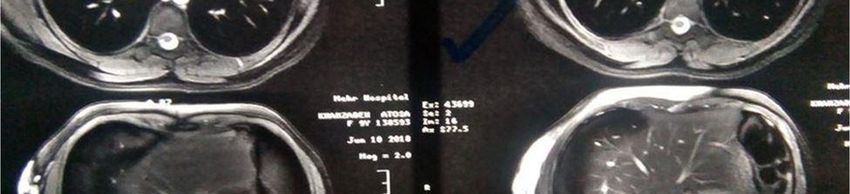
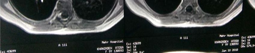
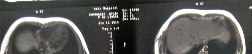
Int Res J Med Med Sci      44

nearby structures. This tumor has been barely reported in
the chest wall, and as we searched the literature, it
sounds that it has not been reported as a congenital
lesion in a child’s chest wall. The author presents the
case and reviews childhood angiolipomas reported in the
literature.

CASE PRESENTATION

A 9 year old girl presented to our clinic with a mass on
the right side of her chest wall. She complained of newly
arousal of pain in the lesion with suddenly rapid
increasing of its size since almost one month earlier. The
pain was not permanent and had no referral to anywhere.
Her parents mentioned that the mass has existed since
her birthday as a greenish almost 2*2 centimeter size
lump with gradually slow growth over time, but with no
previous history of any pain or tenderness. Every history
of recent trauma to the mass or surrounding area was
denied. Other personal or family history of the case was
unremarkable. On physical examination, there was a
relatively firm well-circumscribed 5*7 centimeter smooth
protuberance with tenderness associated with greenish
color changes on some parts of overlying skin, located at
the right anterior part of the chest wall at the mid-axillary
line direction (Figure 1A and B).
   No adhesion to the surrounding or underneath tissues
as well as overlying skin was found. There was no
evidence of bruising or bleeding near the mass or in other
parts of the body. No evidence of hepatosplenomegaly or
lymphadenopathy was detected on the physical
examination. Other aspects of physical examination were
normal. All the laboratory tests including Protrombin Time
(PT) and Partial Thromboplastin Time (PTT) as well as
International Normalizing Ratio (INR) were within normal        Figure 1A. The mass lesion from anterior view.
ranges. The report of superficial ultra-sonography of soft
tissue has revealed a hyper echoic heterogenous mass
with 50*40*15 millimeter in dimension included cystic and       signal tubular structures were observed in subcutaneous
tubular areas in favor of dilated vessels. Moreover, no         fatty tissues with significant enhancement in contrast
invasion of the mass to pectorals muscles or underneath         images (Figure 2A and B).
ribs was seen. Furthermore, no pathologic lymph node in           T2-weighted sections without contrast revealed fluid
axillary or supra clavicular region was detected. Color         signals inside the high signal tubular structures with no
Doppler surveys showed no vascular markings. These              significant enhancement in contrast images (Figure 2C
findings recommended a benign chest wall lesion                 and D).
including vascular mass with low flow veno lymphatic            These findings supported the diagnosis of dilated vessels
malformation. A spiral computed tomography scan (CT-            too. Therefore, a decision was made in order to remove
scan) of lungs and mediastinum with and without contrast        the mass by surgical excision. During the operation, it
in bone window has ordered and has shown an image of            was revealed that the mass was placed under the sub-
soft tissue mass in the right lateral side of the chest wall    dermal fatty tissue, over pectoralis major muscle with
which was over pectorals and serratus anterior muscles          some involvement of the muscle fascia, but no invasion
with almost 47*30*18 millimeter in dimension. There was         to underneath muscles, nerves or vessels. It grossly
no invasion to deep internal organs as well as no               appeared without a capsule or any vascularity. The mass
obviously enhancement in post contrast images. These            was resected on bloc and has sent for biopsy. In
features were in support of the ultra-Sonographic findings      histopathology report of the lesion, the specimen was
and brought up a benign chest wall lesion. A Magnetic           well-circumscribed and unencapsulated, contained
Resonance Imaging (MRI) with and without contrast was           subcutaneous fatty tissue with 7.5*4.5*2.5 centimeter in
performed. In T1-weighted sections without contrast, low        dimension. Its external surface was unremarkable.
Tootoonchi   45

Figure 1B. The mass lesion in left lateral decubitus view.

Figure 2A. MRI of the chest. T1- weighted, without contrast and without fat suppression.
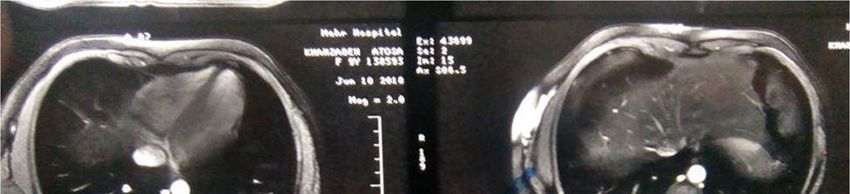
Int Res J Med Med Sci   46

Figure 2B. MRI of the chest. T1- weighted, with contrast and without fat suppression.

Figure 2C. MRI of the chest. T2-weighted, without contrast and without fat suppression.
Tootoonchi 47
Figure 2D. MRI of the chest.T2-weighted, with contrast and with fat suppression.
The consistency of the mass lesion was rubbery and
areas of cystic formation, brownish discoloration and
myxoid changes were identified, which distance from
nearest margin was 0.3 centimeter. The lesion was
entirely composed of mature adipocytes arranged in
irregular lobes interspersed throughout vascular spaces
circumscribed with thin fibrose septa and areas of
abundant small sized blood vessels that were more
prominent at the peripheral localization. In addition, fibrin
thrombi were noted within occasional vascular lumens
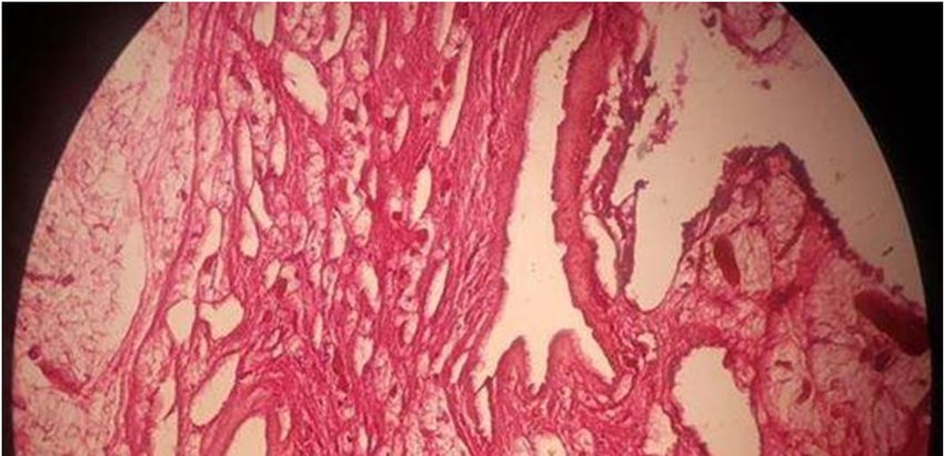
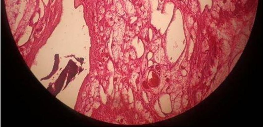
(Figure 3A and B).
These findings are consistent with typical features of a
non-infiltrating angiolipoma. After operation, the surgeon
prescribed an elastic bandage over the area of removed
lesion in order to prevent fluid accumulation. In a follow
up visit, after one month, there was some mass formation
on the lesion removal site with cyst consistency which
was the result of no applying of the bandage by the
patient. A MRI was ordered again and the images have
revealed a cystic collection of fluid in favor of seroma.
The cyst lesion was aspirated and the bandage was
applied. At the next follow up visit performed two weeks
later, the site of operation was completely closed, flatted Figure 3A. Photo of biopsy section of the mass lesion. Based on
and cleared. The patient recovered uneventfully with the histomorphologic findings, fibrin thrombi were noted in some
complete resolution of pain. No recurrence of tumor was vessels and the sections showing adipose tumor containing
observed in the following 30 months. proliferation of capillary vessels.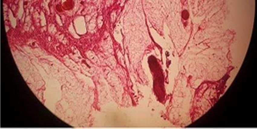
Int Res J Med Med Sci 48
mesenchimal tumors most commonly affected young
males. This article introduces a case of congenital non-
infiltrating angiolipoma of the chest wall in a 9 year old
girl presented to our clinic with newly arousal of pain and
tenderness in an old mass as well as rapidly enlargement
of its size for almost one month with seemingly no history
of trauma. The tumor has appeared in the chest wall
since her birthday and had a slow growth over time with
no history of pain or tenderness. As we searched the
literature, there were reported a few cases (50 cases) of
angiolipoma in childhood (Table 1).
Generally, twenty five cases (52%) appeared in males
versus twenty three cases (48%) in females. The sex of
two cases not determined because of no access to the
two non-English articles’ full text. Twenty six cases (53%)
occurred before 11 years of age and twenty four cases
(47%) occurred from 11 till 20 years of age in children.
According to the location, the frequency of lesions has
occurred more than one time was as follows: 18 cases in
the facial region (37.5%), 16 cases in spinal regions
(33%), 4 cases in knee, 2 cases in neck, 2 cases in chest
wall, 2 cases in back or lower back, 2 cases in ankle, 2
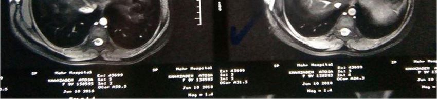
Figure 3B. Photo of biopsy section of the mass lesion. Mature cases in orbit and 2 cases in forearm. In each of the
adipocytes arranged in irregular lobes interspersed throughout following location just 1 case has been reported: lower
vascular spaces as well as fibrin thrombi were noted in some extremity, thigh, foot, back of the tongue, frontal bone,
vessels. mandible, upper lip, nose, eyelid, hard palate, and parotid
as well as pectoralis major muscle. In the face, cheek
was the most common area for tumor presentation (5
DISCUSSION cases, 30%). Therefore, it sounds that the pattern of
tumor involvement in children is different from adults in
Angiolipomas are benign acquired subcutaneous whom most of the cases appeared on the trunk or
Table 1. Summary of Reported Angiolipomas in childhood in the literature.
Author Age Sex Location Histopathology type Additional finding
Caucci (1959) newborn ? Forearm ? Congenital
Cvetinović et al. (1988) 5 month ? Neck ?
Reilly et al. (1988) 6 month Female Parotid Noninfiltrating
Shahi et al. (2014) 9 month Female Buccal mucosa Infiltrating
Sandvik et al. (2015) 1 year Male Spinal Noninfiltrating
Takahashi et al. (1998) 1 year Female Tongue Noninfiltrating
Jaiswal et al. (2020) 1.5 year Female Spinal epidural Noninfiltrating
Maier (1962) 1.5 year Female Spinal epidural Infiltrating
Stimpson (1971) 2 year Female Pectoralis major muscle Infiltrating
Feinfield et al. (1988) 3 year Male Eyelid Noninfiltrating
Carruth and Meyer (2015) 3 year Male Orbit Noninfiltrating
Gonzalcz-Crussi et al. (1996) 3 year Male Knee Infiltrating
Weitzner and Moynihan (1978) 4 year Male Cheek Noninfiltrating
Gelabert-González et al. (2002) 4 year Male Thoracic spinal Noninfiltrating
Turgut (2004) 4 year Male Thoracic spinal Noninfiltrating
Arenaz-Búa et al., (2010) 5 year Male Cheek Noninfiltrating
Kasper and Cowan (1931) 6 year Male Spinal epidural Noninfiltrating
Chew et al. (1980) 6 year Male Knee Infiltrating
Gonzalcz-Crussi et al. (1996) 6.5 year Female Knee Infiltrating
Koopmann (1988) 7 year Male Nose Noninfiltrating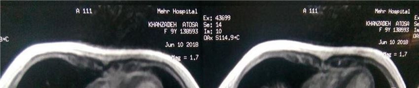
Tootoonchi           49

Table 1. Continues.

Aljerian et al. (2019)           7 year     Female      Orbit                      Noninfiltrating
Yeo et al. (2018)                7 year     Male        Foot                       Infiltrating
Regan et al. (1946)              8 year     Male        Lower extremity            Infiltrating            Multiple
Flaggert et al. (1986)           8 year     Female      Hard palate                Noninfiltrating
Sah et al. (2012)                9 year     Female      Upper lip                  Noninfiltrating         Congenital
Alvi et al. (1998)               10 year    Female      Cheek                      Noninfiltrating
Aniceto et al. (1990)            11 year    Male        Cheek                      Noninfiltrating
Chew et al. (1980)               11 year    Female      Ankle                      Infiltrating
Michilli et al. (1993)           12 year    Male        Spinal extradural          Noninfiltrating
Akhaddar et al. (2000)           12 year    Female      Spinal epidural            Noninfiltrating
Ali and El-Zuebi (1996)          13 year    Female      Cheek                      Noninfiltrating
Shetty and Prabhu (2009)         13 year    Female      Face                       Noninfiltrating
İlyas et al. (2016)              13 year    Male        Knee                       Noninfiltrating
Matsuoka et al. (1988)           14 year    Female      Neck                       Infiltrating
Raghavendra et al. (2007)        14 year    Male        Spinal                     Noninfiltrating
Fernández et al. (1994)          14 year    Female      Spinal epidural            Noninfiltrating
Kumar et al. (2013)              15 year    Male        Forearm & back             Noninfiltrating         Multiple
Shetty and Prabhu (2009)         16 year    Male        Mandible                   Noninfiltrating
Gelabert-González and García-    16 year    Male        Spinal extradural          Noninfiltrating
Allu (2009)
Lo Re and Michelacci, 1969       16 year    Male        Spinal epidural            Noninfiltrating         Congenital
Petrella et al. (2005)           16 year    Male        Spinal epidural            Noninfiltrating
Atilgan et al. (2014)            16 year    Female      Frontal bone               Infiltrating
Pearson et al. (1970)            17 year    Female      Spinal extradural          Infiltrating
Lacour et al. (2018)             17 year    Male        Thoracic spinal epidural   Noninfiltrating
Labram et al. (1999)             17 year    Male        Spinal epidural            Noninfiltrating
Vijay et al. (2015)              17 year    Female      Ankle                      Infiltrating
Komatsu et al. (2013)            18 year    Male        Chest wall                 Infiltrating
Deviri et al. (1987)             18 year    Female      Chest wall & lower back    Noninfiltrating         Multiple
Shetty and Prabhu (2009)         19 year    Female      Face                       Noninfiltrating
Chew et al. (1980)               19 year    Female      Thigh                      Infiltrating

extremities (Lee et al., 2011; Lin and Lin, 1974; Howard          male (Hamano et al., 2013), a postero-lateral thoraco-
and Helwig, 1960). It sounds that this is the first report of     abdominal wall mass in a 41 year old male (Biondetti et
a chest-wall non-infiltrating angiolipoma in a child              al., 1982), a tumor in a 42 year old female (Mayooran et
presented since birthday. According to other studies, just        al., 2016) and a tumor in right chest wall in a 68 year old
three cases have appeared congenitally in childhood               female (Sakamoto et al, 2019). All of the cases except
(Caucci, 1959; Sah et al., 2012; Lo Re and Michelacci,            one case (Sakamoto et al, 2019) had definite diagnosis of
1969). Therefore, almost all the reported cases in the            infiltrating angiolipoma and half of them were described
literature were acquired (47 cases, 94%). Furthermore,            as asymptomatic (Mayooran et al., 2016; Sakamoto et al,
despite multiple lesions are frequently appeared in adults        2019). Contrary to the type of reported cases in adults’
(Lin and Lin, 1974), in children occurrence of multiple           chest wall, present case was of non-infiltrating type and
masses are rare; only 3 cases include an eight year old           has appeared as a congenital lesion.
boy with two huge lesions in lower extremity (Regan et               According to the literature, in adulthood these lesions
al., 1946), a fifteen year old boy with multiple painful skin     are seen more commonly in a younger age group;
lesions over forearm and back (Kumar et al., 2013) and            generally in pubescent patients and are rare before
an eighteen year old girl with two lesions of chest wall          puberty. They present as painless or tender
and lower back (Komatsu et al., 2013) had been                    subcutaneous nodules and have no tendency to
reported. It is interesting that, as we could find, there         recurrence (Arenaz-Búa et al., 2010; Howard and Helwig,
were just four case reports of chest wall angiolipoma in          1960); however, infiltrating angiolipomas usually appear
adults mentioned in the literature; all of them were              in patients over 30 years of age, and there is a
acquired; 75% of the cases appeared in young adult                recurrence rate of 50%. Besides, infiltrating angiolipomas
patients. The cases included a tumor in a 25 year old             have not been found to undergo malignant transformation
Int Res J Med Med Sci                    50

(Arenaz-Búa et al., 2010; Lin and Lin, 1974; Howard and             Ali MH, El-Zuebi F, 1996. Angiolipoma of the cheek: report of a case. J
                                                                       Oral Maxillofac Surg, 54: 213–215.
Helwig, 1960). In the review of literature of children with
                                                                    Aljerian K, Fathaddin A, Almajed N, Kalantan H, 2019. Angiolipoma of
angiolipoma, just 15 cases (30%) were of infiltrative type             the orbit. Ophthalmic Plast Reconstr Surg, 35(3), e81-e82.
and most of the reported cases (33 cases, 59%) like our             Alvi A, Garner C, Thomas W, 1998. Angiolipoma of the head and neck.
case had non-infiltrating histopathology. It is noteworthy             J Otolaryngol, 27: 100–103.
that similar to adults, the treatment of an angiolipoma in          Aniceto GS, Saez RS, Penin AG, 1990. Angiolipoma of the cheek:
                                                                       report of a case. J Oral Maxillofac Surg, 48: 512–515.
children is surgical excision (Arenaz-Búa et al., 2010; Lin         Arenaz-Búa J, Luáces R, Lorenzo Franco F, García-Rozado A, Crespo
and Lin, 1974; Howard and Helwig, 1960) which ranges                   Escudero JL, Fonseca Capdevila E, López-Cedrún JL, 2010.
from simple excision in non-infiltrating cases to wide                 Angiolipoma in head and neck: report of two cases and review of the
                                                                       literature. Int J Oral Maxillofac Surg, 39(6): 610-615.
surgical excision in infiltrating angiolipoma. Furthermore,
                                                                    Atilgan AO, Terzi A, Agildere M, Caner H, Ozdemir BH, 2014.
the studies have shown that non-infiltrating cases have                Intraosseous angiolipoma of the frontal bone with a unique location:
no tendency to recur (Arenaz-Búa et al., 2010; Lin and                 A clinical and pathological case illustration and review of the
Lin, 1974; Howard and Helwig, 1960). Like to reported                  literature. Indian J Pathol Microbial, 57(2): 301-304.
                                                                    Bang M, Kang BS, Hwang JC, Weon YC, Choi SH, Shin SH, Kwon WJ,
cases in the literature, a simple surgical excision of the
                                                                       Hwang CM, Lee SY, 2012. Ultrasonographic analysis of
lesion has performed on our case; identical to other                   subcutaneous angiolipoma. Skeletal Radiol, 41(9): 1055-1059.
reported children, she hadn’t experienced any recurrence            Biondetti PR, Fiore D, Perin B, Ravasini R, 1982. Infiltrative
on follow up visits.                                                   angiolipoma of the thoracoabdominal wall. J Comput Assist Tomogr,
                                                                       6: 847.
                                                                    Carruth BP, Meyer DR, 2015. Angiolipoma of the orbit. A rare tumor in
                                                                       an unusual location. Ophtal Plast Reconstr Surg, 31(6): e142-e145.
CONCLUSION                                                          Caucci M, 1959. Voluminous angiolipoma of the forearm in a newborn
                                                                       infant [Article in Italian]. Minerva Chir, 30(14): 1144-6.
                                                                    Chew FS, Hudson TM, Hawkins Jr IF, 1980. Radiology of infiltrating
In conclusion, angiolipomas are rarely seen in the chest
                                                                       angiolipoma. Am J Roentgenol, 135(4): 781-787.
wall region of the children, especially as a congenital             Choong KKL, 2004. Sonographic Appearance of Subcutaneous
mass. The presented case showed the typical clinical,                  Angiolipomas. J Ultrasound Med, 23(5): 715-717.
imaging and histological findings of a non-infiltrating             Cvetinović M, Jović N, Petrović Z, Knezević M, Vukanić D, 1988.
angiolipoma. Furthermore, its behavior regards to natural              Giant angiolipoma of the neck in a 5-month-old child [Article in
                                                                       Croatian]. Chir Maxillofac Plast, 18(1-3): 75-80.
history, response to surgical excision and recurrence is            Deviri E, Levinsky L, Shaklai M, Lavie G, Levy MJ, 1987. Total excision
identical to other non-infiltrating tumors. The more striking          of a giant angiolipoma of chest wall with A-V malformation and with
features in this case are its existence since the birthday             the use of an autotransfusion system. J Cardiovasc Surg (Torino),
as a single lesion, the uncommon location as well as the               28(5): 546-8.
                                                                    Feinfield RE, Hesse RJ, Scharfenberg JC, 1988. Orbital angiolipoma.
unusual way of its presentation after a long period of its             Arch Ophthalmol, 106(8): 1093-1095.
asymptomatic existence with slow growth. Furthermore,               Fernández JJ, Abad RM, Ribas T, García CPJ, Mostaza SAL, Vihuela
review of literature has shown that children with                      LJ, Mazabel M, 1994. Spinal angiolipoma causing acute paraplegia.
                                                                       Report of two cases. Neurocirugia, 5: 242–245.
angiolipoma mostly presented with single acquired
                                                                    Flaggert III JJ, Heldt LV, Keaton WM, 1986. Angiolipoma of the palate:
benign lesion which appears most commonly in the face                  Report of a case. Oral Surg Oral Med Oral Pathol, 61(4): 333-336.
or spinal region. The lesions usually have non-infiltrating         Gelabert-González M, García-Allu A, 2009. Spinal extradural
histopathology and recovered uneventfully with simple                  angiolipoma: report of two cases and review of the literature. Eur
surgical excision without occurring of any recurrence.                 Spine J, 18(3):324–335.
                                                                    Gelabert-González M, Agulleiro-Díaz J, Reyes-Santías RM, 2002.
                                                                       Spinal extradural angiolipoma with a literature review. Childs Nerv
                                                                       Syst, 18(12): 725-8.
Patient consent for publication                                     Gonzalez-Crussi F, Enneking WF, Arean VM, 1966. Infiltrating
                                                                       angiolipoma. J Bone Joint Surg Am, 48(6): 1111-1124.
                                                                    Gupta P, Potti TA, Wuertzer SD, Lenchik L, Pacholke DA, 2016.
Written informed consent for publication of the case                   Spectrum of fat-containing soft-tissue masses at MR Imaging: The
report and any accompanying images, without any                        common, the uncommon, the characteristic, and the sometimes
potential identifying information, was provided by the                 confusing. Radiographics, 36(3): 753-66
                                                                    Hamano A, Suzuki K, Saito T, Kuwatsuru R, Oh S, Suzuki K, 2013.
parents of the patient.                                                Infiltrating angiolipoma of the thoracic wall: a case report. Open J Clin
                                                                       Diagn, 3(2): 19–22.
                                                                    Howard WR, Helwig EB, 1960. Angiolipoma. Arch Dermatol, 82:924-
Conflicts of interest                                                  931.
                                                                    İlyas G, Turgut A, Ayaz D, Kalenderer Ö, 2016. Intraarticular Giant Size
                                                                       Angiolipoma of the Knee Causing Lateral Patellar Dislocation. Balkan
The author declares no conflict of interest.                           Med J, 33(6): 691-694.
                                                                    Jaiswal PA, Divakar G, Krishnakumar K, Karthikayan A, Sawakare Y,
                                                                       Mhatre R, Abraham M, 2020. Spinal angiolipoma-a rare but
REFERENCES                                                             reversible cause of paraplegia in a child. Childs Nerv Syst, 36(6):
                                                                       1121-1125.
Akhaddar A, Gazzaz M, Derraz S, Rifi L, Amarti A, Aghzadi A, El     Jo VY, Fletcher CDM, 2013. WHO classification of tumors of soft tissue
 Ouahabi A, El Khamlichi A, 2000. Spinal epidural angiolipomas: a      and bone. 4th ed. Lyon, France: IARC Press.
 rare cause of spinal cord compression. A report of 8 cases and     Kasper JA, Cowan A, 1931. Extradural lipoma of the spinal canal. J
 review of the literature. Neurochirurgie, 46(6): 523-533.             Nerv Ment Dis, 74(4) - p 564.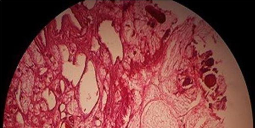
Tootoonchi               51

Komatsu T, Takahashi K, Fujinaga T, 2013. Chest wall angiolipoma              Shahi AK, Ash H, Chatterji K, Singh R, 2014. Cellular infiltrative
  complicating von Recklinghausen disease. Ann Thorac Surg, 96:                  angiolipoma of cheek in an infant. Natl J Maxillofac Surg, 5(2): 202-
  e73–4.                                                                         205.
Koopmann CH Jr, 1988. The “Pinocchio’’ nasal deformity—                       Shetty SR, Prabhu S, 2009. Angiomatosis in the head and neck-3
  hemangioma vs. agiolipoma: esthetic correction and etiology. J                 Case Reports. Head and Neck Pathol, 3: 54–8.
  Otolaryngol, 17: 169–172.                                                   Sheybani EF, Eutsler EP, Navarro OM, 2016. Fat-containing soft-tissue
Kransdorf MJ, Moser RP Jr, Meis JM, Meyer CA, 1991. Fat-containing               masses in children. Pediatr Radiol, 46(13): 1760-1773.
  soft-tissue masses of the extremities. Radiographics, 11(1): 81-106.        Shin YS, Kim YJ, Park IS, Chu YC, Kim JH, Lee HY, Lee KH, Kang YH,
Kumar SP, Kamath SM, Prasad AL, Mysorekar VV, Sumathy TK, 2013.                  2016. Sonographic Differentiation Between Angiolipomas and
  Acute-Onset of Multiple Painful Nodules over Forearms and Back. J              Superficial Lipomas. J Ultrasound Med, 35(11): 2421-2429.
  Clin Diagn Res, 7(10): 2314-2316.                                           Stimpson N, 1971. Infiltrating angiolipomata of skeletal muscle. Br J
Labram EK, el-Shunnar K, Hilton DA, Robertson NJ, 1999. Revisited:               Surj, 58: 464-466.
  spinal angiolipoma, three additional cases. Br J Neurosurg, 13: 25–         Takahashi M, Kurokawa H, Ando T, Sato Y, Noguchi I, 1998. A case of
  29.                                                                            angiolipoma of the tongue in a child. Jap J Oral Maxillofacial Surg,
Lacour M, Gilard V, Marguet F, Curey S, Perez A, Derrey S, 2018.                 44(10): 817-819.
  Sudden paraplegia due to spontaneous bleeding in a thoracic                 Turgut MR, 2004. Spinal extradural angiolipoma, with a literature
  epidural angiolipoma and literature review. Neurochirurgie, 64(1): 73-         review. Child s Nervous System, 20(2): 73-74.
  75.                                                                         Vijay V, Srivastava N, Kumar Yadav Y, Shukla S, 2015. Infiltrating
Lee JY, Kim SM, Fessell DP, Jacobson JA, 2011. Sonography of                     Angiolipoma around Ankle: A Case Report. Sch J Med Case Rep,
  benign palpable masses of the elbow. J Ultrasound Med, 30(8):                  3(7): 598-601.
  1113-1119.                                                                  Weitzner S, Moynihan PC, 1978. Angiolipoma of the cheek in a child.
Lin JJ, Lin F, 1974. Two entities in angiolipoma (a study of 459 cases           Oral Surg Oral Med Oral Pathol, 45: 95–97.
  of lipoma with review of literature on infiltrating angiolipoma). Cancer,   Yeo ED, Chung BM, Kim EJ, Kim WT, 2018. Infiltrating angiolipoma of
  34: 720–727.                                                                   the foot: magnetic resonance imaging features and review of the
Lo Re F, Michelacci M, 1969. Clinical and surgical findings on various           literature. Skeletal Radiol, 47(6): 859-864.
  lumbo-sacral abnormalities associated with angiolipoma. Arch Putti
  Chir Organi Mov, 24: 70–85.
Maier HC, 1962. Extradural and intrathoracic lipoma causing spinal
  cord compression: successful treatment by surgical excision. JAMA,
  181: 610–612.
Matsuoka Y, Kurose K, Nakagawa O, Katsuyama J, 1988. Magnetic
  resonance imaging of infiltrating angiolipoma of the neck. Surg
  Neurol, 29(1): 62–66.
Mayooran N, Tarazi M, O'Brien O, Hinchion J, 2016. Infiltrating
  angiolipoma of the chest wall: a rare clinical entity. J Surg Case Rep,
  1: 1-3.
Michilli      R, Tzonos      P, Iglesias-Rozas      JR,     1993.    Spinal
  extradural angiolipoma: case report and literature review.
  Neurochirurgia (Stuttg), 36(2): 63-65.
Murphey MD, Carroll JF, Flemming DJ, Pope TL, Gannon FH,
  Kransdorf MJ, 2004. Benign musculoskeletal lipomatous lesions.
  Radiographics, 24(5): 1433–1466.
Navarro OM, 2009. Imaging of benign pediatric soft tissue tumors.
  Semin Musculoskelet Radiol, 13(3): 196-209.
Navarro OM, Laffan EE, Ngan BY, 2009. Pediatric Soft-Tissue Tumors
  and Pseudo-Tumors: MR imaging features with pathologic
  correlation: part 1. imaging approach, pseudotumors, vascular
  lesions, and adipocytic tumors. Radiographics, 29(3): 887-906.
Pearson J, Stellar S, Feigin I, 1970. Angiolipoma-long-term cure
  following radical approach to malignant-appearing benign intraspinal
  tumor. J Neurosurg, 33: 466-470.
Petrella G, Tamburrini G, Lauriola L, Di Rocco C, 2005. Spinal epidural
  angiolipoma complicated by an intratumoral abscess. Case report. J
  Neurosurg, 103(2 Suppl): 166-9.
Raghavendra S, Krishnamoorthy T, Ashalatha R, Kesavadas C, 2007.
  Spinal angiolipoma with acute subarachnoid hemorrhage. J Clin
  Neurosci, 14(10): 992-4.
Regan JM, Bickle W H, Brothers AC, 1946. Infiltrating benign lipomas of
  the extremities. West J Surg Obstet Gynecol, 54: 87-93.
Reilly JS, Kelly DR, Royal SA, 1988. Angiolipoma of the parotid: Case
  report and review. Laryngoscope, 98(8 Pt 1): 818-821.
Sah K, Kadam A, Sunita JD, Chandra S, 2012. Non-infiltrating
  angiolipoma of the upper lip: A rare entity. J Oral Maxillofac Pathol,
  16(1): 103–106.
Sakamoto R Tanaka T, Murakami J, Nakamura T, 2019. A case of
  non-infiltrating angiolipoma of the chest wall mimicking radiological
  infiltration of the ribCT. J Jpn Assoc Chest Surg, 33(5): 555-559.          Citation: Tootoonchi P, 2021. Congenital angiolipoma of the chest wall
Sandvik U, Svensdotter E, Gustavsson B, 2015. Spinal cavernous                in a child: A case report and review of literature. Int Res J Med Med
  extradural angiolipoma manifesting as a spontaneous spinal epidural         Sci, 9(1): 43-51.
  hematoma in a child. Childs Nerv Syst, 31(8): 1223-6.
You can also read