THROMBOSIS AND THROMBOCYTOPAENIA AFTER CHADOX1 NCOV-19 VACCINATION: A SINGLE UK CENTRE EXPERIENCE
←
→
Page content transcription
If your browser does not render page correctly, please read the page content below
Case report
BMJ Case Rep: first published as 10.1136/bcr-2021-243894 on 13 July 2021. Downloaded from http://casereports.bmj.com/ on August 14, 2021 by guest. Protected by copyright.
Thrombosis and thrombocytopaenia after ChAdOx1
nCoV-19 vaccination: a single UK centre experience
Fehmida Bano , Buddikha Badugama, Deepak Chandra
Haematology, University SUMMARY were diagnosed with cerebral venous sinus throm-
Hospitals of North Staffordshire We report clinical findings of three patients presenting bosis (CVST) and developed secondary cerebral
NHS Trust, Stoke-on-Trent, UK with thrombosis and thrombocytopaenia 10–16 days haemorrhage, which proved fatal. One patient
following the first dose of the ChAdOx1 nCoV-19 vaccine presented with a large pulmonary embolism (PE)
Correspondence to and went on to make a full recovery. We present all
Dr Fehmida Bano;
against SARS-CoV-2. All patients presented to a major
fehmida.bano@uhnm.nhs.u k university teaching hospital in the UK over a 5-day these cases in order of presentation to our hospital
period and were found to have high-titre antibodies which occurred over a 5-day period (19–23 March
Accepted 22 June 2021 against platelet factor 4 (PF4) without previous exposure 2021) which was prior to widespread recognition of
to heparin. All three patients presented with extensive the syndrome. Several countries have since reported
venous thrombosis, significant thrombocytopaenia, cases of VITT, leading to restriction of this vaccine
elevated D-dimer and borderline low fibrinogen. Two in the younger age group in many countries.
had fatal intracerebral haemorrhage secondary to
cavernous venous sinus thrombosis and one had PE. CASE PRESENTATION
Reference laboratory testing of serum demonstrated Patient 1: presentation
anti-PF4 antibodies in all three patients. The clinical A 61- y ear-
old woman presented with a 3- day
and laboratory findings confirmed vaccine-induced history of progressive dyspnoea, pain and
thrombotic thrombocytopaenia (VITT) which was swelling in the right leg. She had received the
poorly described at the time of presentation. We were first dose of ChAdOx1 nCoV-19 16 days before
able to manage successfully one patient with PE with presentation and had a history of asthma and
intravenous immunoglobulin and corticosteroids. hypertension. Her thrombotic risk factors
included a high body mass index of 38 kg/m2
and current use of hormone replacement therapy
BACKGROUND which had been ongoing for 20 years. The two-
Following regulatory approval by the UK Medi- level Wells Score predicted a high probability for
cines and Healthcare products Regulatory Agency PE. D-dimer was elevated at 9376 ng/mL, and
(MHRA) in December 2020, the UK was one of the platelet count at presentation was 25×10⁹/L. A
first countries worldwide to introduce large-scale CT pulmonary angiogram confirmed bilateral PE
non-trial vaccination with ChAdOx1 nCoV-19 for (figure 1) with right heart strain. Prothrombin
adults over 18 years of age to prevent COVID-19.1 time (PT) international normalised ratio (INR)
ChAdOx1 nCoV-19 is a recombinant adenoviral and activated partial thromboplastin time
vector encoding the spike protein of SARS-CoV-2. (aPTT) were normal, and fibrinogen was low
The safety and efficacy of the ChAdOx1 nCoV-19 at 1.26 g/L. Biochemical tests were unremark-
vaccine were confirmed following four randomised able. Thrombotic thrombocytopenic purpura
controlled trials in the UK, South Africa and Brazil, (TTP) and antiphospholipid syndrome (APLS)
none of which reported an increased incidence of were excluded after appropriate screening
thrombosis or thrombocytopaenia.2 The MHRA investigations.
granted temporary authorisation for prophylactic
use of this vaccine for adults over 18 years of age. Patient 1: further investigations and management
Initial vaccination was prioritised for the elderly At presentation, the patient was transfused one
(over 70 years of age) and frontline health and social unit of platelets and commenced on two times
care workers. Subsequently, adults of all ages have per day low-molecular-weight heparin (LMWH).
been invited for vaccination, prioritised according Due to the unusual presentation, the possibility
coexisting medical problems and descending age of autoimmune heparin- induced thrombocyto-
order. As of 8 April 2021, over 31 million individ- paenia (HIT) with no prior heparin exposure
uals have received the first dose of a SARS-CoV-2 was considered. As such, a blood sample was
vaccine in the UK,3 the majority of which have taken for an HIT screen.
© BMJ Publishing Group
Limited 2021. No commercial
been ChAdOx1 nCoV-19. A rare complication of The HIT screening test with a rapid particle
re-use. See rights and thrombosis associated with thrombocytopaenia has gel immunoassay (ID-PaGIA Heparin/PF4 Anti-
permissions. Published by BMJ. been reported 5–28 days following the ChAdOx1 body Test, Bio- R ad) was weak positive. The
nCoV-19 vaccine and is now termed vaccine- sample was sent for HIT ELISA and functional
To cite: Bano F, Badugama B,
Chandra D. BMJ Case induced thrombotic thrombocytopaenia (VITT).4–7 assay. LMWH was stopped and anticoagulation
Rep 2021;14:e243894. We describe this syndrome in three patients who was switched to treatment dose fondaparinux
doi:10.1136/bcr-2021- presented to our centre 10–16 days after receiving after 12 hours of presentation. Further platelet
243894 the first dose of ChAdOx1 nCoV-19. Two patients transfusion was withheld. The patient was
Bano F, et al. BMJ Case Rep 2021;14:e243894. doi:10.1136/bcr-2021-243894 1Case report
BMJ Case Rep: first published as 10.1136/bcr-2021-243894 on 13 July 2021. Downloaded from http://casereports.bmj.com/ on August 14, 2021 by guest. Protected by copyright.
Patient 2: presentation
A 53-year-old woman was referred to our centre’s neurosurgical
department from another centre where she had presented a few
hours prior with sudden onset headache and facial weakness.
She gave a history of worsening headaches for the past 3 days
and weakness of the right arm and leg. She had received the
first dose of ChAdOx1 nCoV-19 14 days prior to presentation.
She had a medical history of fibromyalgia. Her initial blood tests
showed an elevated D-dimer of 5620 ng/ml and a platelet count
of 24×10⁹/L. A CT scan of the head showed extensive intra-
cerebral haemorrhage. She further had a CT venogram, which
confirmed CVST (figure 3). PT INR and aPTT were normal
with a borderline low fibrinogen of 1.9 g/L. Biochemical tests
were normal. TTP and APLS were again excluded after relevant
investigations.
Patient 2: further investigation and management
Twenty-four hours into admission, her level of consciousness
deteriorated and she required intubation. Three units of platelets
Figure 1 CT pulmonary angiogram demonstrating acute thrombus were transfused before urgent neurosurgical intervention. Anti-
as filling defect (arrow) involving the right pulmonary artery branch coagulation was not started due to the risk of life-threatening
extending into the right inferior pulmonary artery and segmental bleeding. Following referral to clinical haematology, a blood
branches. sample was taken for an in-house HIT screen by rapid particle
gel immunoassay, which was reported as negative. However, our
experience with patient 1 and emerging case reports prompted
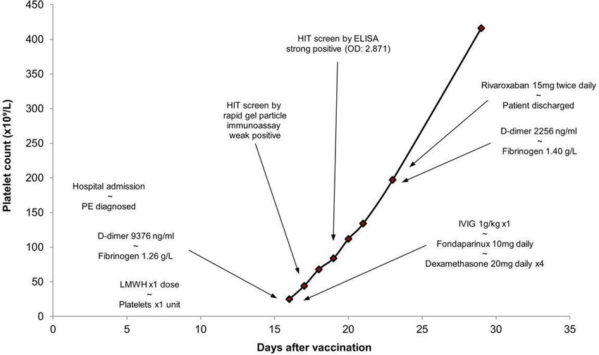
treated with intravenous immunoglobulin (IVIG) 1 g/kg a high clinical suspicion for VITT. As such, a sample was sent to
single dose and pulsed dexamethasone 20 mg once daily for the reference laboratory. Her HIT ELISA was reported as strong
4 days. Her platelet count improved steadily after 2 days of positive (OD 2.631). Platelet functional assay was negative in
treatment. The HIT ELISA was reported as strong positive this case.
for anti-platelet factor 4 (PF4)/heparin antibodies by IgG-
specific ELISA immunoassays (optical density (OD) 2.871;
Patient 2: outcome and follow-up
cut-o ff for positive reaction >0.4). In the functional assay in
Her repeat scans showed further bleeding, with signs of
which platelet activation was tested with serum and heparin
increased intracranial pressure (ICP). She further deteriorated
in high and low concentration, her platelet activation was
with seizures and coning and further neurosurgical intervention
inhibited by excess heparin effectively confirming strong
was not attempted due to futility. Her platelet count remained
positive antibodies againstPF4.
low at 23×10⁹/L, and she died on the 16th day after vaccination.
Figure 4, table 1 provide an outline of her clinical presentation
Patient 1: outcome and follow-up and management.
She was discharged on day 7 with a platelet count of 197×10⁹/L
after switching anticoagulation to rivaroxaban for 3 months. At
follow-up 1 week post discharge, she was asymptomatic with a Patient 3: presentation
normal full blood count. She has been advised not to take the A 55-year-old man with no comorbidities presented to the emer-
second dose of the COVID-19 vaccine. Figure 2, table 1 provide gency department with a 2-day history of headache followed by
an outline of her clinical presentation and management. the onset of dysphasia, right arm weakness and discoordination
in the preceding few hours. He had received his first dose of
ChAdOx1 nCoV-19 10 days prior to admission. At presentation,
there was isolated thrombocytopaenia with a platelet count of
21×10⁹/L and a very high D-dimer of 47 881 ng/ml. CT scan of
the head showed extensive CVST with the extension of thrombus
into the left internal jugular vein and secondary subarachnoid
haemorrhage (figure 5). The PT INR and aPTT were normal,
and fibrinogen was borderline low at 1.9 g/L. Biochemical tests
were normal. TTP and APLS were again excluded after relevant
investigations. As with patient 2, the in-house rapid particle gel
immunoassay HIT screen was negative, but due to high clinical
suspicion, samples were sent to the reference laboratory.
Patient 3: further investigation and management
Prior to the results of the reference laboratory testing, the
patient was managed empirically for VITT with dexamethasone
Figure 2 Time course and events in the clinical presentation of 20 mg and anticoagulation with argatroban, given its revers-
patient 1. HIT, heparin-induced thrombocytopaenia; IVIG, intravenous ibility. Platelets and cryoprecipitate were transfused to maintain
immunoglobulin; LMWH, low-molecular-weight heparin; PE, pulmonary the count above 50×10⁹/L and fibrinogen above 1.5 g/L. IVIG
embolism. 0.5 g/kg was planned to be given. HIT ELISA was reported as
2 Bano F, et al. BMJ Case Rep 2021;14:e243894. doi:10.1136/bcr-2021-243894Case report
BMJ Case Rep: first published as 10.1136/bcr-2021-243894 on 13 July 2021. Downloaded from http://casereports.bmj.com/ on August 14, 2021 by guest. Protected by copyright.
Table 1 Clinical and laboratory features of three patients
Characteristics Patient 1 Patient 2 Patient 3
Age (years) 61 53 55
Sex Female Female Male
Comorbid conditions Asthma, hypertension, high body Fibromyalgia Nil
mass index
Time from vaccination to admission (days) 16 14 10
Medication on admission Hormone replacement therapy, None None
indapamide
Presenting symptoms Shortness of breath, pain and Headaches, facial weakness, Headaches, dysphasia, right arm
swelling in the right leg hemiparesis weakness
Site of thrombosis Right main pulmonary artery Superior sagittal sinus, right sigmoid, Cortical veins, superior sagittal sinus,
extending into upper, middle and right transverse sinus left transverse, left sigmoid sinus and
lower pulmonary arteries and left left internal jugular vein
segmental pulmonary arteries
Platelet count nadir (×10⁹/L (reference range 150–450)) 25 24 21
D-dimer peak (ng/mL (reference range 0–350)) 9376 5620 47 881
PT INR peak (normal range 0.80–1.20) 1.0 1.2 1.1
aPTTR peak 1.1 0.9 0.97
(normal range 0.80–1.17)
Fibrinogen (Clauss) nadir (g/L (reference range 1.9–4.8)) 1.26 1.90 1.33
SARS-CoV-2 PCR test result Not detected Not detected Not detected
Rapid particle gel immunoassay Weak positive Negative Negative
PF4-ELISA (optical density (cut-off for positive reaction 2.871 2.631 2.423
>0.400))
Platelet functional assay Positive Negative Negative
Anticoagulation therapy Initial split dose of LMWH then Nil Alteplase
fondaparinux
Platelet transfusion 1 unit Multiple Multiple
Other treatment IVIG (1 g/kg) None Dexamethasone 20 mg
Dexamethasone (20 mg once daily Cryoprecipitate
for 4 days)
Outcome Full recovery Death Death
aPTT, activated partial thromboplastin time; IVIG, intravenous immunoglobulin; LMWH, low molecular-weight heparin; PF4, platelet factor 4; PT INR, prothrombin time international
normalised ratio.
strong positive (OD=2.423) confirming antibodies against PF4. or any schistocytes in the blood film of all three patients. APLS
The platelet functional assay was negative. and TTP were excluded. All three patients tested negative for
SARS-CoV-2 by PCR on a nasopharyngeal swab.
Patient 3: outcome and follow-up Reference laboratory testing of serum demonstrated anti-PF4
Despite intensive management he developed seizures and dete- antibodies in all three patients despite no prior heparin exposure.
riorated further with falling level of consciousness. Repeat brain Of concern, the in-house rapid gel particle immunoassay was
imaging showed a new subarachnoid haemorrhage causing only weak positive in one patient and negative in the other two.
significant midline shift and compression of the midbrain. He All three patients tested strongly positive for anti-PF4/heparin
underwent emergency decompressive craniectomy and ICP antibodies by IgG-specific ELISA immunoassays but only patient
bolt insertion under the cover of platelet transfusion. High ICP 1 tested positive on platelet functional assay.
persisted and the patient deteriorated. Following discussion VITT has emerged as a novel disease in the first part of 2021
within the medical team and with the family, treatment was secondary to vaccination with ChAdOx1 nCoV-19 against
withdrawn and he died within 24 hours of presentation. Table 1 SARS-CoV-2.
and figure 6 provide an outline of his clinical presentation and The clinical picture of severe thrombocytopaenia and
management. extensive thrombosis is akin to that of HIT where the antigen
recognition domain of IgG antibodies binds to complexes of
DISCUSSION PF4 and heparin.8 The Fc domain of these antibodies binds
We have summarised the clinical and laboratory characteris- to platelet FcγRII-A resulting in platelet activation, throm-
tics of three patients who presented to our centre with VITT bosis, platelet consumption and life- threatening throm-
10–16 days after receipt of the SARS-CoV-2 vaccine, ChAdOx1 bosis.9 10 The term ‘autoimmune HIT’ has been used to
nCoV-19. describe this pathological process which can occur in the
All three patients presented with extensive venous thrombosis: absence of heparin but it is a rare clinical entity.11 In our
two had fatal intracerebral haemorrhage secondary to CVST report, all three patients demonstrated strikingly high levels
and one had PE. All three patients at admission had elevated of IgG antibodies to PF4–polyanion complexes by ELISA
D-dimer and borderline low fibrinogen. PT and aPTT were with an OD of >2, similar to other case reports and were
within the normal range. There were no features of haemolysis the pathological cause of extensive thrombosis.12
Bano F, et al. BMJ Case Rep 2021;14:e243894. doi:10.1136/bcr-2021-243894 3Case report
BMJ Case Rep: first published as 10.1136/bcr-2021-243894 on 13 July 2021. Downloaded from http://casereports.bmj.com/ on August 14, 2021 by guest. Protected by copyright.
Figure 5 CT venogram demonstrating ‘empty delta’ sign in superior
sagittal sinus (solid arrow) with thrombus extension into bilateral
cortical veins (dashed arrows). This is complicated by extensive
subarachnoid haemorrhage bilaterally seen as high attenuation lining
the cerebral sulci (*).
such as danaparoid, argatroban, direct oral anticoagulants
Figure 3 CT venogram demonstrating filling defect and classic ‘empty and fondaparinux is recommended. 14 In unstable patients,
delta’ sign (solid arrow) in the superior sagittal sinus. This is complicated we have favoured argatroban due to its short half-life. The
by large volume left-sided intraparenchymal haemorrhage (*) with initiation of anticoagulation in a thrombocytopaenic patient
rightward midline shift (dashed arrows demonstrate shift of the falx with thrombosis necessitates careful evaluation of the risks
cerebri). and benefits. In the presence of catastrophic haemorrhage,
this judgement is especially challenging. However, due to
Managing such patients is challenging and requires timely the pathophysiology of VITT, the balance of risk is in great
intervention. Involvement of a multidisciplinary team with favour of commencing therapeutic anticoagulation and
urgent referral to haematology at the earliest suspicion is the avoiding platelet transfusion. Despite this, in some circum-
key to achieving a good outcome. Radiological, neurosur- stances, anticoagulation will be contraindicated and these
gical, pathological and pharmacological support is manda- patients will likely be those with the most severe clinical
tory to save lives by active intervention, especially during presentations. Moreover, although prompt recognition
the first few hours. HIT ELISA testing should be done at and management is important, for some patients with cata-
the earliest suspicion and clinicians should not be falsely strophic presentations, the efficacy of medical interventions
reassured by a negative rapid particle gel immunoassay or will be limited and it is likely that VITT will remain a fatal
functional assay. complication of vaccination in isolated cases. Table 2 high-
The optimal treatment of VITT is not fully established but lights potential diagnostic and therapeutic strategies and
national and international guidelines recommend IVIG, ther- pitfalls.
apeutic anticoagulation and the avoidance of platelet trans- Further investigation of the underlying pathophysiology
fusion.13 Anticoagulation with non-h eparin anticoagulants and epidemiology of VITT is urgently underway to inform the
Figure 4 Time course and events in the clinical presentation of Figure 6 Time course and events in the clinical presentation of
patient 2. CVST, cerebral venous sinus thrombosis; HIT, heparin-induced patient 3. CVST, cerebral venous sinus thrombosis; HIT, heparin-induced
thrombocytopaenia. thrombocytopaenia.
4 Bano F, et al. BMJ Case Rep 2021;14:e243894. doi:10.1136/bcr-2021-243894Case report
BMJ Case Rep: first published as 10.1136/bcr-2021-243894 on 13 July 2021. Downloaded from http://casereports.bmj.com/ on August 14, 2021 by guest. Protected by copyright.
Table 2 Managing patients with suspected or confirmed vaccine induced thrombotic thrombocytopaenia (VITT)
Dos Don’ts
Laboratory testing by PF4 antibody assay (ELISA HIT assay) Do not exclude VITT based on a negative HIT screen from a rapid particle gel
immunoassay
Commence therapeutic anticoagulation with non-heparin-based therapies Avoid heparin, including heparin-based flushes. Fondaparinux can be used
Urgently treat with IVIG Avoid platelet transfusion
Consider giving corticosteroids Avoid thrombopoietin receptor agonists
Report to regulatory health agencies Do not give a second dose of any vaccine for SARS-CoV-2 until further data are available
HIT, heparin-induced thrombocytopaenia; IVIG, intravenous immunoglobulin; PF4, platelet factor 4.
dynamic risk assessment which must balance individual and Funding The authors have not declared a specific grant for this research from any
public health implications of vaccination and this rare side effect. funding agency in the public, commercial or not-for-profit sectors.
Following these three cases over 5 days, our institution devel- Competing interests None declared.
oped a local guideline to raise awareness across the hospital and Patient consent for publication Obtained.
to trigger involvement of haematology at the earliest opportunity. Provenance and peer review Not commissioned; externally peer reviewed.
This article is made freely available for use in accordance with BMJ’s website
Patient’s perspective terms and conditions for the duration of the covid-19 pandemic or until otherwise
determined by BMJ. You may use, download and print the article for any lawful,
I had my vaccine about 2 weeks before I attended the hospital. I non-commercial purpose (including text and data mining) provided that all copyright
suffered from aches and pains shortly after the vaccine but they notices and trade marks are retained.
got better. A few days later I had pain in my right leg and bad ORCID iD
headache which persisted. The next day I had pain in the chest Fehmida Bano http://orcid.org/0000-0001-8260-7604
and shortness of breath when I attended the hospital. I was
diagnosed of a blood clot in my lungs. I am now much better
after the blood thinning treatment and my walking is improving. REFERENCES
I am walking my dog and getting back to normal. I have read 1 Gov.UK. Regulatory approval of COVID-19 Vaccine AstraZeneca [Internet]. GOV.UK,
2021. Available: https://www.gov.uk/government/publications/r egulatory-approval-of-
about these vaccine complications but never thought I will be covid-19-vaccine-a strazeneca [Accessed 7 Apr 2021].
affected. I am just glad that it has got better. 2 Voysey M, Clemens SAC, Madhi SA, et al. Safety and efficacy of the ChAdOx1
nCoV-19 vaccine (AZD1222) against SARS-CoV-2: an interim analysis of
four randomised controlled trials in Brazil, South Africa, and the UK. Lancet
Learning points 2021;397:99–111.
3 .The official UK government website for data and insights on coronavirus (COVID-19).
►► Clinicians should have a high suspicion of investigating any Available: https://coronavirus.data.gov.u k/ [Accessed 13 Apr 2021].
4 Greinacher A, Thiele T, Warkentin TE, et al. Thrombotic thrombocytopenia after
acute onset thrombosis associated with thrombocytopaenia ChAdOx1 nCov-19 vaccination. N Engl J Med 2021;384:2092–101.
presenting 5–28 days following the ChAdOx1 nCov-19 5 Scully M, Singh D, Lown R, et al. Pathologic antibodies to platelet factor 4 after
vaccine. ChAdOx1 nCoV-19 vaccination. N Engl J Med 2021;384:2202–11.
►► Check baseline full blood count, D-dimer and fibrinogen 6 Blauenfeldt RA, Kristensen SR, Ernstsen SL, et al. Thrombocytopenia with acute
levels in all suspected cases. ischemic stroke and bleeding in a patient newly vaccinated with an adenoviral vector-
based COVID-19 vaccine. J Thromb Haemost 2021;19:1771–5.
►► Seek advice from an experienced haematologist at earliest 7 Pottegård A, Lund LC, Karlstad Øystein, et al. Arterial events, venous
suspicion. thromboembolism, thrombocytopenia, and bleeding after vaccination with Oxford-
►► Screen for platelet factor 4 antibodies by ELISA heparin- AstraZeneca ChAdOx1-S in Denmark and Norway: population based cohort study.
induced thrombocytopaenia assay and treat positive cases BMJ 2021;373:n1114.
8 Arepally GM. Heparin-Induced thrombocytopenia. Blood 2017;129:2864–72.
with intravenous immunoglobulin. 9 Amiral J, Marfaing-Koka A, Poncz M, et al. The biological basis of immune heparin-
►► Further investigations are required to prove the link induced thrombocytopenia. Platelets 1998;9:77–91.
between the ChAdOx1 nCov-19 vaccine and immune 10 Greinacher A, Juhl D, Strobel U, et al. Heparin‐induced thrombocytopenia: a
thrombocytopaenia with associated thrombosis. prospective study on the incidence, platelet-activating capacity and clinical
significance of antiplatelet factor 4/heparin antibodies of the IgG, IgM, and IgA
classes. J Thromb Haemost 2007;5:1666–73.
Acknowledgements The authors would like to thank Dr Richard Buka, resident 11 Greinacher A, Selleng K, Warkentin TE. Autoimmune heparin-induced
haematologist, in addition to the medical staff of the Neurosurgical and Intensive thrombocytopenia. J Thromb Haemost 2017;15:2099–114.
12 Schultz NH, Sørvoll IH, Michelsen AE, et al. Thrombosis and thrombocytopenia after
care Department at the University Hospital of North Midlands for their valuable
ChAdOx1 nCoV-19 vaccination. N Engl J Med 2021;384:2124–30.
contributions in management of these patients. The authors would also like to thank
13 B-s-h.org.uk, 2021. Available: https://b-s-h.org.uk/media/19530/guidance-version-
Dr James Davies, resident radiology, for providing help for radiology images. 13-on-mngmt-of-thrombosis-with-thrombocytopenia-occurring-after-c-19-vaccine_
Contributors FB is the lead author for this case report, leading the care and 20210407.pdf [Accessed 7 Apr 2021].
management of patients 1 and 2 as well leading the writing of the manuscript. BB 14 The ObG Project. ASH Guidelines: Diagnosis and Management of COVID-19
is the consultant incharge of patient 3 and was actively involved in decision-making Vaccine-Induced Thrombosis with Thrombocytopenia - The ObG Project [Internet],
and patient treatment, also she contributed to the manuscript. DC is the lead 2021. Available: https://www.obgproject.com/2021/04/2 5/ash-guidelines-diagnosis-
consultant supervising the management of all three patients as well he reviewed and and-management-of-covid-19-v accine-induced-thrombosis-with-thrombocytopenia
edited the manuscript. [Accessed 25 Apr 2021].
Bano F, et al. BMJ Case Rep 2021;14:e243894. doi:10.1136/bcr-2021-243894 5Case report
BMJ Case Rep: first published as 10.1136/bcr-2021-243894 on 13 July 2021. Downloaded from http://casereports.bmj.com/ on August 14, 2021 by guest. Protected by copyright.
Copyright 2021 BMJ Publishing Group. All rights reserved. For permission to reuse any of this content visit
https://www.bmj.com/company/products-services/rights-and-licensing/permissions/
BMJ Case Report Fellows may re-use this article for personal use and teaching without any further permission.
Become a Fellow of BMJ Case Reports today and you can:
►► Submit as many cases as you like
►► Enjoy fast sympathetic peer review and rapid publication of accepted articles
►► Access all the published articles
►► Re-use any of the published material for personal use and teaching without further permission
Customer Service
If you have any further queries about your subscription, please contact our customer services team on +44 (0) 207111 1105 or via email at support@bmj.com.
Visit casereports.bmj.com for more articles like this and to become a Fellow
6 Bano F, et al. BMJ Case Rep 2021;14:e243894. doi:10.1136/bcr-2021-243894You can also read