Rising Concern over Cosmetic Tattoos - ORIGINAL ARTICLE
←
→
Page content transcription
If your browser does not render page correctly, please read the page content below
ORIGINAL ARTICLE

Rising Concern over Cosmetic Tattoos
ARISA E. ORTIZ, MD,*          AND   TINA S. ALSTER, MD†

BACKGROUND A rise in popularity of cosmetic tattoos has led to an increase in adverse reactions. Due to
more pressing concerns, the Food and Drug Administration (FDA) has not traditionally enforced its author-
ity over tattoo inks.
OBJECTIVE       To raise awareness of the dangers of cosmetic tattoos.
MATERIALS AND METHODS We reviewed FDA policies regarding tattoo ink, different ink components,
adverse reactions, and various treatment options for cosmetic tattoo removal.
RESULTS AND CONCLUSION An increase in consumer complaints has prompted FDA investigation into
tattoo inks and their safety. It is important that further complications be reported to the FDA to promote
regulation of cosmetic tattoo inks.
The authors have indicated no significant interest with commercial supporters.

C      osmetic tattoos, often referred to as perma-
       nent makeup, have become increasingly pop-
ular since the late 1970s. Permanent makeup is
                                                                  tation. There is also variability in the setting in
                                                                  which these procedures are performed, the methods
                                                                  of anesthesia, sterility, and artistic ability. With the
generally used to replace traditional temporary eye               gain in popularity of micropigmentation, societies
liner, lip liner, blush, or eyebrow pencil. Individu-             such as the American Academy of Micropigmenta-
als may choose to undergo cosmetic tattooing to                   tion have been established to improve the quality
save time or as an adjunct to reconstructive sur-                 of practice through a certification process in this
gery, commonly after breast surgery. Cosmetic tat-                technique.
toos may also be applied to camouflage conditions
such as vitiligo or alopecia. Although cosmetic tat-
                                                                  Tattoo Regulation
toos are intended to enhance facial features, they
ironically do not age well. For example, a lip liner              The pigments in tattoo inks contain color additives,
tattoo that once traced a youthful full lip will                  which are defined as any material that can impart
become displaced outside the lip’s border as the lip              color to a food, a drug, a cosmetic, a medical
thins with age.                                                   device, or the human body. The color additives used
                                                                  in inks require premarket approval under the fed-
The process by which tattoo inks are injected into                eral Food, Drug, and Cosmetic Act to ensure that
the dermis to give the appearance of temporary                    they are used safely and appropriately. Approved
makeup is called micropigmentation. Currently, a                  color additives are listed in the U.S. Code of Federal
variety of professionals and nonprofessionals,                    Regulations (21 CFR Parts 73, 74, 82), but this
including physicians, nurses, cosmetologists, esthe-              approval does not extend to injected use. No color
ticians, and makeup artists, perform micropigmen-                 additives are Food and Drug Administration (FDA)

*Department of Dermatology, University of California at Irvine, Irvine, California; †Washington Institute of
Dermatologic Laser Surgery, Washington, District of Columbia

© 2011 by the American Society for Dermatologic Surgery, Inc.  Published by Wiley Periodicals, Inc.
ISSN: 1076-0512  Dermatol Surg 2011;1–6  DOI: 10.1111/j.1524-4725.2011.02202.x

                                                                                                                             1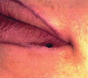
REVIEW OF COSMETIC TATTOOS

    approved for injection into the skin (21 CFR 70.5b)
                                                              TABLE 1. Tattoo Pigment Components
    (www.fda.gov). Therefore, no tattoo pigments are
                                                              Color          Pigment
    approved for use. The majority of tattoo ink is
                                                              Red            Mercury sulfide (cinnabar), cadmium
    industrial-grade color intended for use as printer
                                                                               selenide (cadmium red), sienna (red
    ink or automobile paint. Although tattoo ink is                            ochre, ferric hydrate and ferric
    subject to regulation by the FDA, state and local                          sulfate), azo dyes, hematite
    health authorities regulate the practice of tattooing,    Yellow         Cadmium sulfide (cadmium yellow),
                                                                               ochre, curcumin yellow, azo dyes,
    including those performed in salons and tattoo par-
                                                                               limonite, anthraquinone
    lors. These departments mainly regulate sanitation        Green          Chromium oxide (casalis green),
    requirements and prohibit tattooing minors.                                hydrated chromium sesquioxide
                                                                               (guignet green), malachite green,
                                                                               lead chromate, ferro-ferric cyanide,
    In the past, tattoo ink regulation has not been a                          curcumin green, phthalocyanine
    priority because of other, more-pressing public                            dyes (copper salts with yellow coal
    health concerns. The FDA has not traditionally                             tar dyes)
                                                              Blue           Cobalt aluminate (azure blue),
    enforced its authority over tattoo inks or the pig-
                                                                               phthalocyanine, ferric ferrocyanide,
    ments found within them, but in recent years, there                        indigoid
    has been increasing concern regarding adverse reac-       Violet         Manganese violet, indigoid
    tions to tattoo ink pigment.                              White          Titanium dioxide, zinc oxide,
                                                                               corundum
                                                              Tan            Iron oxides
                                                              Brown          Ochre
    Tattoo Pigment Components
                                                              Black          India ink, carbon, iron oxide, logwood
    Tattoo pigments are composed of inorganic and                              extract, magnetite

    synthetic organic pigments (Table 1).1 Inorganic
    tattoo pigments come from mineral sources such           or ethanol, which facilitate the dyeing process in
    as metal oxides, salts, and minerals. Magnetite          the skin.
    (FeO·Fe2O3) and charcoal (C) are often found in
    black tattoo ink; hematite (Fe2O3) and cinnabar
                                                             Adverse Reactions
    (HgS) are used in red ink; limonite
    (FeO·OH·nH2O) is used for yellow pigment;                Between 1988 and 2003, only five cases of adverse
    corundum (Al2O3), rutile (TiO2), and zincite             reactions were reported to the FDA. More recently,
    (ZnO) are used for white pigment, and blue               there has been a tremendous increase in consumer
    pigment can be achieved with ferric ferrocyanide         complaints, with more than 150 adverse reactions
    (Fe4[Fe(CN)6]3) and cobaltous aluminate                  to permanent makeup procedures reported to the
    (CoAl2O4). These compounds are naturally occur-          FDA in 2003 and 2004. The FDA and Centers for
    ring, but they may fade or change color over             Disease Control and Prevention (CDC) identified
    time. Mercury and cadmium salts are no longer            101 of these patients as having adverse reactions at
    found in tattoo inks because of their toxicity.          their tattoo sites. The most commonly reported
    Synthetic organic pigments, such as anthraquinone        reactions were tenderness and itching associated
    (yellow), phthalocyanine (blue, green), azo (mostly      with allergic reactions and bumps secondary to
    yellow, orange, red, magenta, purple), and indig-        granulomatous reactions.2 After investigation by
    oid (violet–blue), are synthesized chemical com-         the FDA and CDC, it was found that most of these
    pounds that create brighter, more-diverse colors.        reactions were due to tattoo ink manufactured by
    Newer fluorescent inks may even glow under               a single company (Premier Products, Arlington,
    black light. In addition to pigment, tattoo inks         TX). The company voluntarily recalled the associ-
    contain diluents and preservatives, such as glycerin     ated ink pigments in September 2004. This has

2   DERMATOLOGIC SURGERY
ORTIZ AND ALSTER
prompted FDA investigation of tattoo ink safety at tory responses to mercury-free pigments still
the National Center for Toxicological Research. occur.11,31–33
This laboratory is currently investigating the chem-
ical composition of tattoo inks, how the body Some manufacturers promote alcohol- and preser-
metabolizes them, short- and long-term safety, and vative-free tattoo inks, but these inks run the risk of
interactions with light and lasers. It has been found microbial contaminants. In 2004, Starbrite Colors
that azo pigments, such as pigment red (PR) 9, PR tattoo inks were taken off the market in Belgium
22, and pigment yellow 74, decompose into known because of microbial contamination with Pseudo-
carcinogens with exposure to light and laser irradi- monas aeruginosa and Acremonium mold (www.
ation.3,4 fda.gov/downloads/AboutFDA/Transparency/
Basics/UCM246800.pdf).
Safety risks of unsterilized needles in tattoos have
been well established, but studies are lacking on the
Treatment of Tattoos
safety of the tattoo ink itself. Several histologic
reactions to tattoo ink have been described, includ- The removal of cosmetic tattoos, similar to treat-
ing pseudolymphomatous,5 lichenoid,6 granuloma- ment of other decorative tattoos, is often more
tous,5,7,8 mild acanthosis,9 scleroderma-like,10 costly and complicated than their original acquisi-
pseudoepitheliomatous hyperplasia,11,12 and aller- tion. Laser treatment requires multiple painful
gic contact dermatitis.13 The lichenoid pattern is sessions that are expensive and sometimes incom-
the most common and is thought to represent a pletely successful. With an increase in the number
delayed hypersensitivity reaction.14 Coincidental of ink colors, tattoo removal is becoming increas-
lesions such as sarcoidosis,15,16 B-cell lymphoma,17 ingly difficult.
pseudolymphoma,18,19 melanoma,20–22 basal cell
carcinoma,23 non-Hodgkin’s lymphoma,24 and Historically, removal of undesired tattoos included
squamous cell carcinoma25,26 have also been tissue-destructive techniques such as dermabrasion
reported to occur. Magnetic resonance imaging and salabrasion,34–37 cryosurgery,38 electrosur-
(MRI) may interact with tattoo ink, causing irrita- gery,39,40 and surgical excision.41,42 Although
tion.27 Nevertheless, MRI is still recommended effective at removing the tattoo ink, these treat-
when indicated regardless of the presence of a cos- ments often led to scarring and unwanted skin pig-
metic tattoo. Tattoo pigment may complicate evalu- mentation changes. Thus, more-specific (laser)
ation of metastatic disease in people with malignant technologies that minimize untoward side effects
melanoma by migrating into regional lymph nodes, have replaced them. Early laser systems (e.g.,
mimicking metastases.28,29 ruby,43 carbon dioxide [CO2],44 argon45) were
initially used to vaporize tattooed skin, but they
Red pigment is most commonly implicated in also resulted in significant scarring and hypopig-
adverse reactions, but the safety of tattoo pigment mentation. Intense pulsed light devices also lead to
components for injection into the skin is not well scarring and are not appropriate for tattoo
established because no tattoo ink or additive is removal.46 The concept of selective photothermoly-
FDA approved. Mercury contained in red mercuric sis revolutionized the treatment of tattoos by pref-
sulphide (cinnabar) is well known to be the causa- erentially targeting the tattoo pigment with specific
tive agent of allergy in red tattoos.9,30,31 Mercury- wavelengths and pulse durations of laser light that
free dyes such as cadmium red (cadmium selenide), the tattoo ink particles selectively absorb while
sienna and red ochre (ferric hydrate), and organic adjacent structures are left essentially unharmed.47
vegetable dyes (e.g., Brazilwood) have largely Tattoo ink particles are small and therefore
replaced mercury-containing dyes, but inflamma- require Q-switched (QS) laser systems with brief
2011 3REVIEW OF COSMETIC TATTOOS
(A)
Figure 2. Tattoo ink darkening in permanent lip liner after
(B) Q-switched laser irradiation.
Cosmetic tattoos can be more difficult to treat
because they generally contain red, brown, flesh-
colored, and white inks containing iron oxides
and titanium dioxide, which may turn irreversibly
black after QS laser irradiation (Figure 2).50
Chemical reduction of ferric oxide to ferrous oxide
is thought to be responsible for the potentially
permanent darkening of tattoos. It is impossible to
predict which pigments will darken upon QS laser
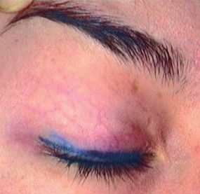
Figure 1. (A) Eye liner cosmetic tattoo before treatment. irradiation or if the darkened pigment will respond
(B) Resolution of tattoo after Q-switched alexandrite laser
to further laser treatment. Therefore, one must
treatment.
proceed with caution when using QS lasers to
(nanosecond) pulse durations. The high energy treat pale-colored tattoo pigments containing
delivered over an ultrashort time period results in metallic oxides and properly educate patients of
shattering of the ink particles, which are then their risks. Nevertheless, it is possible to treat the
engulfed by tissue macrophages and cleared by the paradoxical darkening with continued QS laser
lymphatic system or through transepidermal elimi- treatments.51 To preclude paradoxical darkening,
nation. alternative treatments may include pulsed CO252
and erbium-doped YAG lasers,53 which have been
The QS 694-nm ruby laser was the first laser to shown to be effective in vaporizing red, pink, and
selectively destroy tattoo ink without peripheral flesh-colored cosmetic tattoos. Newer techniques
tissue damage. Other QS lasers such as the 532- for tattoo removal involve combinations of QS
and 1,064-nm neodymium-doped yttrium alumi- pigment-specific (red and infrared) lasers with
num garnet (Nd:YAG) and 755-nm alexandrite ablative fractional laser resurfacing, which have
lasers allow for removal of a variety of tattoo ink been reported to enhance the rate of pigment
colors (Figure 1). Red ink absorbs the 532-nm Nd: clearance and decrease risk of vesiculation.54
YAG laser, and the red and infrared wavelengths Other novel technologies include the picosecond
of the 755-nm alexandrite and 1,064-nm Nd:YAG laser, which has been shown to be better in tattoo
lasers are each effective for the treatment of black, pigment clearance than the nanosecond lasers in
blue, and green inks.48,49 Yorkshire pigs.55
4 DERMATOLOGIC SURGERYORTIZ AND ALSTER
Conclusions 15. Kargi E, Aslan G, Erdogan B. Squamous cell carcinoma arising
from a hydrochloric acid burn. Plast Reconstr Surg
A rise in the number of cosmetic tattoo procedures 1999;103:2086.
being performed has prompted further FDA inves- 16. Weidman AI, Andrade R, Franks AG. Sarcoidosis. Report of a
case of sarcoid lesions in a tattoo and subsequent discovery of
tigation of tattoo ink safety. Adverse reactions to pulmonary sarcoidosis. Arch Dermatol 1966;94:320–5.
tattoo inks are becoming more common, and the 17. Sangueza OP, Yadav S, White CR Jr, Braziel RM. Evolution
number of complaints is likely greatly underreport- of B-cell lymphoma from pseudolymphoma. A
multidisciplinary approach using histology,
ed. Consumers and medical professionals should be immunohistochemistry, and Southern blot analysis. Am J
encouraged to report adverse reactions from per- Dermatopathol 1992;14:408–13.
manent makeup to the FDA to promote FDA regu- 18. Rijlaarsdam JU, Bruynzeel DP, Vos W, Meijer CJ, et al.
lation of cosmetic tattoo inks (http://www.fda.gov/ Immunohistochemical studies of lymphadenosis benigna cutis
occurring in a tattoo. Am J Dermatopathol 1988;10:518–23.
ora/fed_state/Small_business/sb_guide/regions.htm).
19. Zinberg M, Heilman E, Glickman F. Cutaneous
pseudolymphoma resulting from a tattoo. J Dermatol Surg
Oncol 1982;8:955–8.
References
20. Soroush V, Gurevitch AW, Peng SK. Malignant melanoma in a
1. Bolognia JL, Jorizzo JL, Rapini RP. Dermatology. Elsevier: tattoo: case report and review of the literature. Cutis
Spain, 2008; p. 2107. 1997;59:111–2.
2. Straetemans M, Katz LM, Belson M. Adverse reactions after 21. Kirsch N. Malignant melanoma developing in a tattoo. Arch
permanent-makeup procedures. N Engl J Med 2007;356:2753. Dermatol 1969;99:596–8.
3. Jemec GB. Comment on: tattooing of skin results in 22. Kircik L, Armus S, van den Broek H. Malignant melanoma in a
transportation and light-induced decomposition of tattoo tattoo. Int J Dermatol 1993;32:297–8.
pigments. Exp Dermatol 2010;19:61–2.
23. Wiener DA, Scher RK. Basal cell carcinoma arising in a tattoo.
4. Cui Y, Spann AP, Couch LH, Gopee NV, et al. Cutis 1987;39:125–6.
Photodecomposition of Pigment Yellow 74, a pigment used in
tattoo inks. Photochem Photobiol 2004;80:175–84. 24. Armiger WG, Caldwell EH. Primary lesion of a non-Hodgkin’s
lymphoma occurring in a skin tatoo: case report. Plast Reconstr
5. Blumental G, Okun MR, Ponitch JA. Pseudolymphomatous Surg 1978;62:125–7.
reaction to tattoos. Report of three cases. J Am Acad Dermatol
1982;6:485–8. 25. Ortiz A, Yamauchi PS. Rapidly growing squamous cell
carcinoma from permanent makeup tattoo. J Am Acad
6. Clarke J, Black MM. Lichenoid tattoo reactions. Br J Dermatol Dermatol 2009;60:1073–4.
1979;100:451–4.
26. McQuarrie DG. Squamous-cell carcinoma arising in a tattoo.
7. Vagefi MR, Dragan L, Hughes SM, Klippenstein KA, et al. Minn Med 1966;49:799–801.
Adverse reactions to permanent eyeliner tattoo. Ophthal Plast
Reconstr Surg 2006;22:48–51. 27. van der Velden EM, Defranq J, Baruchin AM. Cosmetic and
reconstructive medical tattooing. Curr Opin Otolaryngol Head
8. Verdich J. Granulomatous reaction in a red tattoo. Acta Derm Neck Surg 2005;13:349–53.
Venereol 1981;61:176–7.
28. Anderson LL, Cardone JS, McCollough ML, Grabski WJ, et al.
9. Bagley MP, Schwartz RA, Lambert WC. Hyperplastic reaction Tattoo pigment mimicking metastatic malignant melanoma.
developing within a tattoo. Granulomatous tattoo reaction, Dermatol Surg 1996;22:92–4.
probably to mercuric sulfide (cinnabar). Arch Dermatol
1987;123:1557, 60–1. 29. Kurle S, Schulte KW, Homey B. [Accumulation of tattoo
pigment in sentinel lymph nodes]. Hautarzt 2009;60:781–3.
10. Kluger N, Mathelier-Fusade P, Moguelet P. Scleroderma-like
reaction restricted to the red parts of a tattoo. Acta Derm 30. McGrouther DA, Downie PA, Thompson WD. Reactions to red
Venereol 2009;89:95–6. tattoos. Br J Plast Surg 1977;30:84–5.
11. Goldstein N. Mercury-cadmium sensitivity in tattoos. A 31. Mortimer NJ, Chave TA, Johnston GA. Red tattoo reactions.
photoallergic reaction in red pigment. Ann Intern Med Clin Exp Dermatol 2003;28:508–10.
1967;67:984–9.
32. Yazdian-Tehrani H, Shibu MM, Carver NC. Reaction in a red
12. Balfour E, Olhoffer I, Leffell D, Handerson T. Massive tattoo in the absence of mercury. Br J Plast Surg 2001;54:
pseudoepitheliomatous hyperplasia: an unusual reaction to a 555–6.
tattoo. Am J Dermatopathol 2003;25:338–40.
33. Sowden JM, Byrne JP, Smith AG, Hiley C, et al. Red tattoo
13. Kaur RR, Kirby W, Maibach H. Cutaneous allergic reactions to reactions: X-ray microanalysis and patch-test studies. Br J
tattoo ink. J Cosmet Dermatol 2009;8:295–300. Dermatol 1991;124:576–80.
14. Winkelmann RK, Harris RB. Lichenoid delayed hypersensitivity 34. Scutt RW. The chemical removal of tattoos. Br J Plast Surg
reactions in tattoos. J Cutan Pathol 1979;6:59–65. 1972;25:189–94.
2011 5REVIEW OF COSMETIC TATTOOS
35. Manchester GH. The removal of commercial tattoos by 48. Alster TS. Q-switched alexandrite laser treatment (755 nm) of
abrasion with table salt. Plast Reconstr Surg 1974;53:517–21. professional and amateur tattoos. J Am Acad Dermatol
1995;33:69–73.
36. Clabaugh W. Removal of tattoos by superficial dermabrasion.
Arch Dermatol 1968;98:515–21. 49. Fitzpatrick RE, Goldman MP. Tattoo removal using the
alexandrite laser. Arch Dermatol 1994;130:1508–14.
37. Koerber WA Jr, Price NM. Salabrasion of tattoos A correlation
of the clinical and histological results. Arch Dermatol 50. Anderson RR, Geronemus R, Kilmer SL, Farinelli W, et al.
1978;114:884–8. Cosmetic tattoo ink darkening. A complication of Q-switched
and pulsed-laser treatment. Arch Dermatol 1993;129:1010–4.
38. Dvir E, Hirshowitz B. Tattoo removal by cryosurgery. Plast
Reconstr Surg 1980;66:373–9. 51. Kirby W, Kaur RR, Desai A. Paradoxical darkening and
removal of pink tattoo ink. J Cosmet Dermatol 2010;9:149–51.
39. Colver GB, Cherry GW, Dawber RP, Ryan TJ. Tattoo removal
using infra-red coagulation. Br J Dermatol 1985;112:481–5. 52. Mafong EA, Kauvar AN, Geronemus RG. Surgical pearl:
removal of cosmetic lip-liner tattoo with the pulsed carbon
40. Groot DW, Arlette JP, Johnston PA. Comparison of the dioxide laser. J Am Acad Dermatol 2003;48:271–2.
infrared coagulator and the carbon dioxide laser in the removal
of decorative tattoos. J Am Acad Dermatol 1986;15:518–22. 53. Wang CC, Huang CL, Yang AH, Chen CK, et al. Comparison
of two Q-switched lasers and a short-pulse erbium-doped
41. Buncke HJ Jr, Conway H. Surgery of decorative and traumatic yttrium aluminum garnet laser for treatment of cosmetic tattoos
tattoos. Plast Reconstr Surg (1946) 1957;20:67–77. containing titanium and iron in an animal model. Dermatol
42. Bailey BN. Treatment of tattoos. Plast Reconstr Surg Surg 2010;36:1656–63.
1967;40:361–71. 54. Weiss ET, Geronemus RG. Combining fractional resurfacing
43. Goldman L, Rockwell RJ, Meyer R, Otten R, et al. Laser and Q-switched ruby laser for tattoo removal. Dermatol Surg
treatment of tattoos. A preliminary survey of three year’s 2011;37:97–9.
clinical experience. JAMA 1967;201:841–4. 55. Izikson L, Farinelli W, Sakamoto F, Tannous Z, et al. Safety
44. Bailin PL, Ratz JL, Levine HL. Removal of tattoos by CO2 and effectiveness of black tattoo clearance in a pig model after
laser. J Dermatol Surg Oncol 1980;6:997–1001. a single treatment with a novel 758 nm 500 picosecond laser: a
pilot study. Lasers Surg Med 2010;42:640–6.
45. Apfelberg DB, Maser MR, Lash H, White DN, et al. Comparison
of argon and carbon dioxide laser treatment of decorative
tattoos: a preliminary report. Ann Plast Surg 1985;14:6–15.
46. Bernstein EF. Laser tattoo removal. Semin Plast Surg Address correspondence and reprint requests to:
2007;21:175–92. Tina S. Alster, MD, Washington Institute of
47. Anderson RR, Parrish JA. Selective photothermolysis: precise
Dermatologic Laser Surgery, 1430 K Street NW
microsurgery by selective absorption of pulsed radiation. Suite 200, Washington DC 20005, or e-mail:
Science 1983;220:524–7. talster@skinlaser.com
6 DERMATOLOGIC SURGERYYou can also read