Single versus dual elastic nails for closed reduction and antegrade intramedullary nailing of displaced fifth metacarpal neck fractures
←
→
Page content transcription
If your browser does not render page correctly, please read the page content below
www.nature.com/scientificreports
OPEN Single versus dual elastic nails
for closed reduction and antegrade
intramedullary nailing of displaced
fifth metacarpal neck fractures
Langqing Zeng1,5, Lulu Zeng2,5, Xiaogang Miao1, Yunfeng Chen3, Weiguo Liang4* &
Yuwen Jiang1*
Closed reduction and internal fixation with antegrade intramedullary nails is a feasible and effective
treatment for displaced fifth metacarpal neck fractures (FMNFs). The present study aimed to compare
clinical and radiological outcomes in patients with displaced FMNFs after treatment with single or
dual antegrade elastic intramedullary nails (AEIMNs). Thirty-three patients were treated with a single
2.0 mm AEIMN and 34 patients were treated with two 1.5 mm AEIMNs. Clinical and radiological
outcomes included grip strength, active range of motion (ROM), active flexion and extension of the
fifth metacarpophalangeal (MCP) joint, dorsal angulation loss, and metacarpal shortening of the
fifth metacarpal at 12 months after treatment. No significant difference was observed between the
two groups with respect to grip strength, ROM or flexion of the fifth MCP joint. The average values
of dorsal angulation loss, metacarpal shortening, and extension of the fifth MCP joint of the dual
nails group were better than those of the single nail group (dorsal angulation loss, 2.79 ± 1.93° vs.
4.05 ± 1.59°, P = 0.009; metacarpal shortening, 1.66 ± 0.80 mm vs. 2.12 ± 0.88 mm, P = 0.028; extension
of the fifth MCP joint, 7.71 ± 4.43° vs. 4.82 ± 4.09°, P = 0.012). In conclusion, dual AEIMNs fixation
provided better MCP extension and radiological outcomes than single AEIMN fixation.
Fifth metacarpal neck fractures (FMNFs) are considered the most common type of hand fractures; they account
for approximately 20% of all fractures in the hand and are more common in males1,2. The majority of FMNFs are
simple and closed and are generally treated with conservative m ethods3. However, severe palmar displacement
and shortening or rotational deformity of the fifth metacarpal fracture may result in a considerable decrease in
grip strength and range of motion (ROM), and surgical treatment is recommended for such c ases4,5. Cadaveric
studies suggest that metacarpal head angulations greater than 30° result in dysfunction of small finger motion at
the metacarpophalangeal (MCP) joint6. Various techniques are available for treating FMNFs, including closed
reduction with percutaneous pinning, antegrade or retrograde intramedullary nailing, open reduction and inter-
nal fixation with plates and/or screws, and transverse pinning with k-wires4,5,7–10. However, there is as yet no
consensus on the most ideal technique of fixation.
The goal of surgical treatment is to restore hand function, and not to simply heal the hand as observed in
normal radiographs. In general, the surgical technique should be able to minimize soft tissue disruption and allow
early motion of the hand8. Recently, the use of single or dual antegrade elastic intramedullary nailing (AEIMN)
has gained increasing interest because it is relatively simple, causes minimal trauma, is cosmetically acceptable,
reduces the risk of soft tissue adhesions, and shows good to excellent clinical o utcomes4,11–14. Yammine et al.12
reported that antegrade intramedullary nailing provided better clinical and radiological outcomes than percu-
taneous transverse pinning or miniplate fixation in their meta-analysis of treatment procedures for FMNFs. The
features of internal fixation, including the number and diameter of the nail, are associated with the stability of
1
Department of Orthopaedics, Zhuhai People’s Hospital, Zhuhai Hospital Affiliated With Jinan University,
Guangdong, China. 2Department of Anesthesiology, Zhuhai People’s Hospital, Zhuhai Hospital Affiliated
With Jinan University, Guangdong, China. 3Department of Orthopaedic Surgery, Shanghai Sixth People’s
Hospital, Shanghai Jiao Tong University Affiliated Sixth People’s Hospital, Shanghai Jiao Tong University,
Shanghai, China. 4Department of Orthopaedics, Guangzhou Red Cross Hospital, Medical College, Jinan
University, Guangzhou, China. 5These authors contributed equally: Langqing Zeng and Lulu Zeng. *email:
liangweiguo2020@163.com; jywdoctor1971@sina.com
Scientific Reports | (2021) 11:1778 | https://doi.org/10.1038/s41598-021-81242-3 1
Vol.:(0123456789)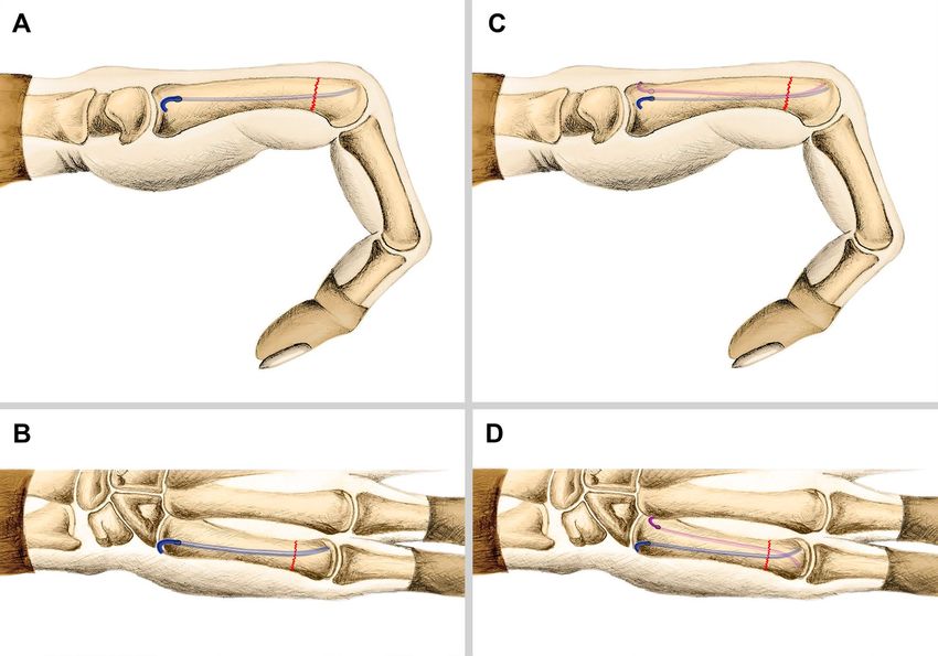
www.nature.com/scientificreports/

                                            Figure 1.  Schematic illustration of an FMNF fixed with single AEIMN on lateral view (A) and anteroposterior
                                            view (B), and fixed with dual AEIMNs on lateral view (C) and anteroposterior view (D).

                                            fixation and therefore may further affect the secondary displacement and prognosis. However, to the best of our
                                            knowledge, no previous studies have compared both single and dual elastic intramedullary nailing techniques
                                            in terms of the stability of fixation and clinical and radiological outcomes. In this study, FMNFs were surgically
                                            treated with a closed reduction and percutaneous antegrade intramedullary nailing fixation with single or dual
                                            elastic nails. The authors evaluated whether the number of nails affected the stability of elastic intramedullary
                                            nailing fixation and the clinical and radiological outcomes.

                                            Materials and methods
                                            This retrospective study was conducted in accordance with the Declaration of Helsinki and was approved by
                                            the Ethics Committee of Zhuhai People’s Hospital. Informed consent was obtained from all donors. Between
                                            February 2012 to July 2018, 78 patients with isolated displaced FMNFs were treated with closed reduction and
                                            percutaneous antegrade elastic intramedullary nailing (Double Medical Technology INC, Xiamen, China).
                                                Inclusion criteria were as follows: patients over 18 years of age who had an isolated FMNF (≤ 14 days), dorsal
                                            angulation of the metacarpal neck of ≥ 30°, or presence of rotational deformity of the fifth finger upon flexion.
                                            Patients meeting any of the following criteria were excluded: any injuries on tendons, ligaments, vessels, and
                                            nerves on the ipsilateral upper limbs; multi-fragmentary fractures, open fractures, or noncooperative patients.
                                            Eleven patients were excluded due to incompletion of a 12-month follow-up.
                                                Sixty-seven patients (54 males and 13 females) completed the study. Thirty-three patients were treated with
                                            single AEIMN (single nail group). Thirty-four patients were treated with dual AEIMNs (dual nails group). All
                                            surgeries were performed by the same two surgical orthopaedic specialists (LQ Zeng and YW Jiang). Surgeries
                                            were performed under general anaesthesia in 15 patients and under brachial plexus block in 52 patients. All
                                            surgeries were performed under fluoroscopic guidance, and all fractures were closed reduced.
                                                For single AEIMN, a small incision was made at the dorsal-ulnar aspect of the metacarpal base. The fifth
                                            metacarpal base was approached by a subcutaneous blunt dissection. An awl was used to open the dorsal-ulnar
                                            cortex in an oblique manner. An elastic nail of 2.0 mm in diameter was selected, and it was inserted into the
                                            medullary cavity and advanced antegrade to the fracture. After the fracture was reduced, the nail was advanced
                                            until the nail tip reached the subchondral bone of the metacarpal head, with the tip of the nail directed at the
                                            dorsal surface of the metacarpal head. This allows for a 3-point fixation that increases the stability of fixation
                                            (Fig. 1A,B). The proximal end of the nail was bent and cut, leaving approximately 8 mm extending out of the
                                            fifth metacarpal, and then buried subcutaneously (Fig. 2A–C).
                                                For dual AEIMNs, two small incisions were made at the dorsal-ulnar and dorsal-radial aspect of the meta-
                                            carpal base. The fifth metacarpal base was approached by a subcutaneous blunt dissection, and an awl was used
                                            to open the dorsal-ulnar and dorsal-radial cortex in an oblique manner. Two elastic nails of 1.5 mm in diameter

          Scientific Reports |   (2021) 11:1778 |                https://doi.org/10.1038/s41598-021-81242-3                                                 2

Vol:.(1234567890)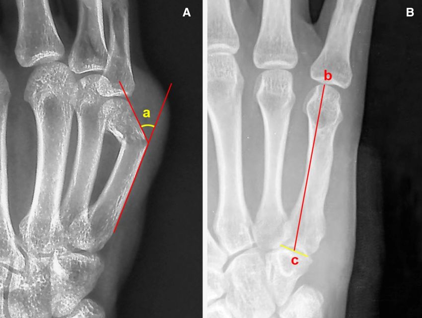
www.nature.com/scientificreports/
Figure 2. Clinical case of an FMNF treated by single AEIMN. Preoperative oblique view radiograph (A).
Postoperative radiographs showing a good reduction of the fracture (B). Radiograph at 8 weeks after the
operation shows union of the fracture (C).
were inserted into the medullary cavity and advanced antegrade to the fracture. After the fracture was reduced,
the nails were advanced until their tips reached the subchondral bone of the metacarpal head. The surgeon
adjusted the direction of the nail s’ tips so that they were directed at the dorsal surface of the metacarpal head
(Fig. 1C) to form a cross configuration at the level of the nail tips (Fig. 1D). The proximal ends of the nails were
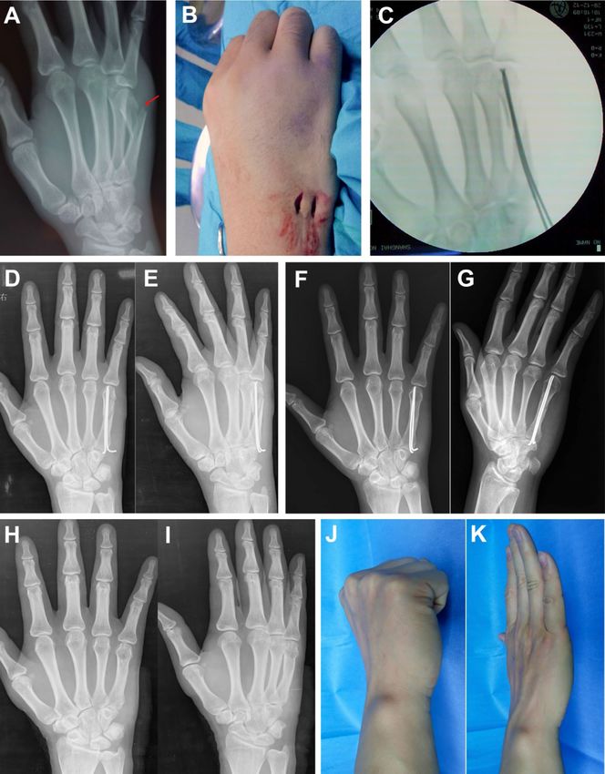
bent and cut, leaving 5 mm extension out of the fifth metacarpal, and then buried subcutaneously (Fig. 3A–K).
Postoperatively, the MCP joints were left free (without using the splint), and gentle passive motion was initi-
ated. After 1–2 weeks, active motion was started. At 4 weeks, strengthening exercises and light daily activities
such as writing and computer work were allowed. Activities requiring power grips such as sports and heavy works
were allowed after union of the fractures. The patients were followed up at 2, 4, and 6 weeks, and at 3, 6, and
12 months. Radiographic follow-up included an anteroposterior view and a 30° oblique view hand X-ray. Clinical
union was defined as the absence of tenderness at the fracture s ite15. In both groups, the elastic nails were removed
3–6 months later under locoregional anaesthesia in the operating room of the outpatient surgery department.
Demographic parameters, including age, gender, injury mechanism, dominant hand, time to treatment,
and operative time, were recorded for both groups. An independent assessor blinded to patient details assessed
the radiological and clinical outcomes. For radiological assessments, dorsal angulation and metacarpal length
were radiologically measured using a picture archiving and communication system. The dorsal angulation was
measured on the 30° pronated oblique view in the true lateral hand position (Fig. 4A), and the length of the
fifth metacarpal was measured on the anteroposterior view (Fig. 4B)16. The secondary dorsal angulation loss
and metacarpal shortening of the fifth metacarpal from postoperative radiographs acquired immediately after
surgery to 12 months after operation were calculated. Clinical functions of the hand were assessed at 12 months
after operation. For clinical assessment, the short version of the Disabilities of the Arm, Shoulder, and Hand
questionnaire (Quick-DASH score) was u sed15,17, and the visual analogue scale (VAS) was used to evaluate pain
in the hand. Grip strength in percentage (%) compared to the contralateral side was measured using a Jamar’s
dynamometer (Asimov Engineering, Los Angeles, CA, USA)18. The time to return to work was recorded. The
active ROM of the fifth MCP joint was measured by a handheld goniometer, in terms of active flexion and exten-
sion of the MCP j oint19.
Complications were noted at all follow-ups, including loss of reduction, non-union, penetration of the nail
tip through the metacarpal head, infection, tendon irritation, tendon adhesion, skin irritation, and injury to the
dorsal cutaneous branch of the ulnar nerve. Loss of reduction was defined as the secondary angulation of the
metacarpal neck of ≥ 30° in the follow-up.
Descriptive statistics were expressed using mean ± standard deviation for normally distributed variables and
median (interquartile range [IQR]) for non-normally distributed variables. Comparisons between groups were
analysed by the chi-square test and Fisher’s exact test for categorical outcomes, and Student’s t test and nonpara-
metric Wilcoxon rank-sum test for continuous outcomes. P < 0.05 was considered statistically significant. SAS
11.0 (SAS Institute Inc., Cary, NC, USA) was used for statistical analysis.
Scientific Reports | (2021) 11:1778 | https://doi.org/10.1038/s41598-021-81242-3 3
Vol.:(0123456789)www.nature.com/scientificreports/
Figure 3. Clinical case of an FMNF treated by dual AEIMNs. Preoperative oblique view radiograph (A).
Intraoperative photograph showing two incisions in the metacarpal base (B). Intraoperative fluoroscopy (C).
Postoperative radiographs showing good reduction of the fracture on the anteroposterior view (D) and oblique
view (E). Radiograph at 8 weeks after the operation shows union of the fracture (F, G). Radiograph at 6 months
after removal of the nails (H, I). Photographs of the hand in flexion (J) and extension (K) at 12 months after
operation.
Scientific Reports | (2021) 11:1778 | https://doi.org/10.1038/s41598-021-81242-3 4
Vol:.(1234567890)www.nature.com/scientificreports/
Figure 4. Methods of radiological measurements. Dorsal angulation was defined as an acute angle (a) between
the line drawn on the dorsal cortex of the fifth metacarpal shaft and the second line on the dorsal cortex of
the fifth metacarpal head/neck (A). The metacarpal length was defined as the length between the most distal
articular surface of the fifth metacarpal head (b) and the mid-point (c) of both corners of the fifth metacarpal
base (B)16.
Characteristics Single nail group (n = 33) Dual nails group (n = 34) Statistics P
Age (years) 30.30 ± 8.69 31.85 ± 11.11 0.63 0.528
Sex (male/female) 27/6 27/7 0.062 0.803
Side of injury (right/left) 22/11 21/13 0.175 0.676
Injured dominant hand (n) 23 24 0.006 0937
Time to operation (days) 3.09 ± 1.01 2.88 ± 0.88 − 0.90 0.371
Angulation pre-op (°) 45.87 ± 9.20 45.17 ± 9.18 − 0.31 0.756
Operation time (min) 25.91 ± 4.96 37.27 ± 8.53 6.63 < .000
Injury mechanism (n)
Fall 4 3
Crush 6 7 0.232 0.972
Punch 18 19
Sports 5 5
Table 1. Patient demographic data of both groups.
Results
There were 33 patients with single AEIMN fixation and 34 patients with dual AEIMNs fixation. In our series,
54 patients were males and 13 patients were females. The average age of this series of patients was 31.09 ± 9.94
(18–67 years). The right hand was affected in 43 patients, and the dominant hand was affected in 47 patients.
There were no significant differences between the two groups with respect to age, sex, side of injury, domi-
nant hand, time from initial trauma to operation, preoperative dorsal angulation, and mechanism of injury
(Table 1). However, the operative time was significantly longer in the dual nails group than in the single nail
group (P < 0.000) (Table 1).
Scientific Reports | (2021) 11:1778 | https://doi.org/10.1038/s41598-021-81242-3 5
Vol.:(0123456789)www.nature.com/scientificreports/
Characteristics Single nail group (n = 33) Dual nails group (n = 34) Statistics P
Angulation post-op (°) 10.60 ± 3.63 10.34 ± 3.69 − 0.30 0.766
Angulation post-op. 12 months (°) 14.65 ± 3.53 13.13 ± 3.70 − 1.71 0.091
Angulation loss (°) 4.05 ± 1.59 2.79 ± 1.93 − 2.70 0.009
Metacarpal shortening (mm) 2.12 ± 0.88 1.66 ± 0.80 − 2.25 0.028
Bone union time (weeks) 10.06 ± 2.03 10.35 ± 2.00 0.59 0.555
Quick-DASH score 7.27 ± 5.08 6.62 ± 4.52 − 0.56 0.579
VAS score 0.55 ± 0.79 0.56 ± 0.75 − 0.23 0.820
MCP extension (°) 4.82 ± 4.09 7.71 ± 4.43 2.57 0.012
MCP flexion (°) 90.27 ± 5.56 90.50 ± 7.61 0.14 0.887
ROM of MCP (°) 95.09 ± 6.89 98.21 ± 7.52 1.78 0.079
Grip strength (%/healthy side) 91.46 ± 9.09 91.62 ± 10.43 0.07 0.946
Time return to work (weeks) 8.82 ± 2.38 8.74 ± 2.60 − 0.14 0.892
Complication rate (n%) 7 (21.21%) 6 (17.65%) 0.14 0.712
Table 2. Clinical and radiological outcomes of both groups. DASH Disabilities of the arm, shoulder, and hand
questionnaire; VAS visual analogue scale; MCP metacarpophalangeal joint; ROM range of motion.
No significant difference was observed between the two groups with respect to the dorsal angulation imme-
diately post-operation, the dorsal angulation at 12 months after operation, bone union time, Quick-DASH
score, VAS score, MCP joint flexion, ROM of the MCP joint, grip strength, and time to return to work (Table 2).
The dorsal angulation loss was significantly greater in the single nail group (4.05 ± 1.59°) than in the dual nails
group (2.79 ± 1.93°) (P = 0.009). The single nail group showed significantly greater metacarpal shortening
(2.12 ± 0.88 mm) than the dual nails group (1.66 ± 0.80 mm) (P = 0.028). However, the MCP joint extension was
significantly better in the dual nails group (7.71 ± 4.43°) than in the single nail group (4.82 ± 4.09°) at 12 months
post-operation (P = 0.012) (Table 2).
There was no loss of reduction, non-union or malunion, infection, or tendon adhesion complications in
any patient in this study. The most common complication was skin irritation at the entry point of the nails (six
patients), including three patients each in the single and dual nail groups. These six patients were successfully
treated by a second operation to remove the nails under locoregional anaesthesia. Migration of the nail tip into
the MCP joint was observed in three patients (including two patients in the single nail group and one patient in
the dual nails group), but it did not influence the final outcome. Three patients had injury to the dorsal cutane-
ous branch of the ulnar nerve, including two patients in the single nail group and one patient in the dual nails
group. In addition, one patient experienced tendon irritation of the extensor digitorum minimi tendon at the
end of the dorsal-radial nail in the dual nails group. This patient was successfully treated by a second operation
to remove the nails. No significant difference was noted in the rates of complications between the two groups
(P = 0.712) (Table 2).
Discussion
Various surgical modalities have been reported for treating FMNFs, and each modality has its own potential
advantages and disadvantages. Percutaneous K-wire fixation offers limited soft tissue disruption, but it could also
lead to an increase in superficial infection and irritation of the skin20. In addition, K-wires may lead to unstable
fracture reduction and require auxiliary immobilization by splint after operation, which delays the ability for
early motion. Plate osteosynthesis has the advantage of immediate stabilization of the fracture21. However, this
technique has the disadvantage of excessive soft tissue damage, which may lead to complications such as ten-
don adhesion, severe tendon irritation, and joint s tiffness22,23. Retrograde intramedullary nailing fixation with
K-wires, or elastic nails have been reported for the treatment of FMNFs, but these techniques cause tendon irrita-
tion and MCP s tiffness24. The use of a retrograde headless intramedullary screw has shown good o utcomes25,26.
The main advantage of this technique is faster recovery to perform daily living and work-related activities, with
no serious c omplications27. However, this technique damages articular cartilage. Ten Berg et al.28 reported that
the articular surface injury caused by the screws in the metacarpal head has a relatively low relevance (4% for
the 2.4 mm screw and 5% of the total joint surface for the 3.0 mm screw). To date, there have been no reports of
mid-term osteoarthritic degeneration at the metacarpal head following the use of this technique.
Antegrade intramedullary osteosynthesis with K-wires or elastic nails offers limited soft tissue stripping, does
not affect the joint capsule, reduces the risk of soft tissue adhesion, has excellent functional and cosmetic results,
and lowers severe complication rate4,12,29. Thus, these techniques have become a commonly used method for the
fixation of FMNFs. The intramedullary nailing fixation acted on a three-point intramedullary fixation, thereby
providing adequate stability. Although the biomechanical data showed that intramedullary nailing fixation was
less stable than plate osteosynthesis, the former is significantly stronger in monocyclic loading than crossed
K-wire osteosynthesis30. Heo et al.16 and Foucher31 suggested that intramedullary nailing fixation can provide
adequate stability to allow early mobilization. It is important that early postoperative mobilization is allowed
to reduce the risk of stiffness. Previously, Kim et al. reported that the antegrade intramedullary pinning group
showed better recovery in the ROM of the fifth MCP joint, grip strength, and DASH score than the retrograde
group at 3 months after surgery32. Winter et al.6 and Sletten et al.22 also reported that this minimally invasive
Scientific Reports | (2021) 11:1778 | https://doi.org/10.1038/s41598-021-81242-3 6
Vol:.(1234567890)www.nature.com/scientificreports/
intramedullary nailing fixation had a better functional recovery than transverse fixation33,34. A meta-analysis
conducted by Yammine and Harvey showed that antegrade intramedullary pinning provided better grip strength,
fifth digit ROM, lower pain scores, and fewer complications than percutaneous transverse pinning or miniplate
fixation for the treatment of FMNFs12.
In the present study, we showed that the functional outcomes of both single and dual nail groups were satisfac-
tory at 12 months after surgery and the complication rate in both groups was similar and acceptable. These results
were found to be similar to those reported in the literature when using single AEIMN or dual AEIMNs4,11,13,35. On
the other hand, we found that the fixation with dual elastic intramedullary nails could reduce the secondary dis-
placement (both dorsal angulation loss and metacarpal shortening) and improve the MCP extension as compared
to that with single elastic intramedullary nailing fixation. However, the dual nails group needed more operative
time than the single nail group, because the intraoperative manipulation of dual AEIMNs is relatively compli-
cated. Calder et al. treated FMNFs with a blunt 1.6 mm K-wire, and they reported an average volar angulation of
3.7° and an average metacarpal shortening of 3.8 mm36. Boonyasirikool C and Niempoog S in their anatomical
study showed that the average metaphyseal widths of metacarpal bone were between 11.42 and 16.42 mm and
the a medullary canal widths were between 3.05 and 6.74 m m37. Therefore, intramedullary fixation with two
1.5 mm elastic nails is a practicable technique for the majority of adult FMNFs. On the basis of our experience,
elastic nails are easier to manipulate during the operation than K-wires, because the nails are more flexible and
the distal tips of the nails have a natural curve; thus, they do not require bending. In addition, fixation with two
1.5 mm elastic intramedullary nails may provide more biomaterial stability than fixation with a single 2.0 mm
elastic intramedullary nail. Hence, for patients requiring an early return to activity, fixation with two 1.5 mm
elastic intramedullary nails may be preferred. Malik et al.38 stated that the normal angulation of the fifth meta-
carpal head to the neck is 15 degrees. In our opinion, an adequate reduction of the fractures should be achieved
with dorsal angulation less than 15 degrees and without rotational deformity. The entry holes of the elastic nails
were made in the dorsal cortex of the fifth metacarpal base (Fig. 1). When the fixation is being completed, the
surgeon needs to adjust the direction of the nail’s tip toward the dorsal surface of the metacarpal head (Fig. 1).
Thus, a three-point fixation is created with two dorsal contact points at the base and head of the fifth metacarpal
and one palmar contact point at the fifth metacarpal’s shaft (Fig. 1). This configuration may provide more bio-
material stability because it is in the opposite direction to the natural dorsal convexity of the fifth metacarpal15.
Furthermore, the surgeon needs to adjust the direction of the nails’ tips to form a cross configuration at the level
of the tips for dual nail fixation (Fig. 1D). This configuration may provide more biomaterial stability, because the
nails’ tips point in opposite directions. No severe complications occurred in any patient. Both single and dual
AEIMN fixations had complications of skin irritation, protrusion of the nail tip into the MCP joint, and injury
to the dorsal cutaneous branch of the ulnar nerve. We believe that the tail of the nails should be bent, cut to an
appropriate length, and buried subcutaneously. The bent tail might prevent forward migration of the nail tip
and avoid being covered with bone. The direction of the nail’s tail may need to be adjusted to avoid its contact
with tendons and to reduce its prominence after skin closure. These manipulations might reduce the risk of skin
and tendon irritation. On the other hand, instead of direct puncture, we could make dissection incisions and
spread subcutaneous tissue bluntly to avoid iatrogenic injury to the dorsal cutaneous branch of the ulnar n erve4.
This study has several limitations. First, although the radiological outcomes were blinded assessment, there
may have been a measurement error in radiological parameters. Second, the quality of radiographs might have
influenced the measurement results, because it was difficult to acquire the oblique radiograph under certain con-
ditions. Finally, because the number of patients was relatively small and the follow-up time was only 12 months,
the results may not be reproducible in other centres with different surgical indications. A larger, long-term,
multi-centre, prospective study is therefore required to appropriately address these issues.
Conclusion
The present study showed that both single and dual AEIMN fixations are safe and effective treatment options for
FMNFs. Better MCP extension and less dorsal angulation loss and metacarpal shortening are advantages of dual
AEIMN fixation over single AEIMN fixation. Hence, for patients requiring an early return to activity, fixation
with two 1.5 mm elastic intramedullary nails may be preferred. Early motion after AEIMN fixation should be
performed carefully because complications related to articular perforation or reduction loss may occur.
Received: 26 February 2020; Accepted: 29 December 2020
References
1. Sun, T. et al. Epidemiological investigation of adult metacarpal fractures from 2003 through 2012 in the Third Hospital to Hebei
Medical University. Chin. J. Orthop. Trauma 16, 603–606 (2017).
2. Hunter, J. M. & Cowen, N. J. Fifth metacarpal fractures in a compensation clinic population. A report on one hundred and thirty-
three cases. J. Bone Jt. Surg. Am. 52, 1159–1165 (1970).
3. Harris, A. R., Beckenbaugh, R. D., Nettrour, J. F. & Rizzo, M. Metacarpal neck fractures: Results of treatment with traction reduc-
tion and cast immobilization. Hand 4, 161–164 (2009).
4. She, Y. & Xu, Y. Treatment of fifth metacarpal neck fractures with antegrade single elastic intramedullary nailing. BMC Musculoskel.
Dis. 18, 238 (2017).
5. Zhu, H., Bao, X. & Zheng, X. Three-screw versus two-screw fixation of distal fragment in fifth metacarpal neck fractures stabilized
with locking plate. Sci. Rep. 7, 12516 (2017).
6. Ali, A., Hamman, J. & Mass, D. P. The biomechanical effects of angulated boxer’s fractures. J. Hand Surg. Am. 24, 835–844 (1999).
7. Zhang, X., Huang, X. & Shao, X. Reduction of fifth metacarpal neck fractures with a Kirschner wire. J. Hand Surg. Am. 40,
1225–1230 (2015).
8. Ben-Amotz, O. & Sammer, D. M. Practical management of metacarpal fractures. Plast. Reconstr. Surg. 136, 370e (2015).
Scientific Reports | (2021) 11:1778 | https://doi.org/10.1038/s41598-021-81242-3 7
Vol.:(0123456789)www.nature.com/scientificreports/
9. Assi, C., Mansour, J., Samaha, C., Ajjoub, S. & Yammine, K. A single antegrade intramedullary k-wire for fifth metacarpal neck
fractures. Eur. J. Trauma Emerg. Surg. 46, 389–395 (2019).
10. Zhu, H., Xu, Z., Wei, H. & Zheng, X. Locking plate alone versus in combination with two crossed Kirschner wires for fifth meta-
carpal neck fracture. Sci. Rep. 7, 46109 (2017).
11. Boussakri, H. et al. Fractures of the neck of the fifth metacarpal bone, treated by percutaneous intramedullary nailing: surgical
technique, radiological and clinical results study (28 cases). Pan. Afr. Med. J. 18, 187 (2014).
12. Yammine, K. & Harvey, A. Antegrade intramedullary nailing for fifth metacarpal neck fractures: A systematic review and meta-
analysis. Eur. J. Orthop. Surg. Traumatol. 24, 273–278 (2014).
13. Pogliacomi, F. et al. Fifth metacarpal neck fractures: fixation with antegrade locked flexible intramedullary nailing. Acta Biomed.
88, 57–64 (2017).
14. Zeng, L. et al. Retrospective comparison of intramedullary antegrade double elastic nail and mini-plate repair of unstable fifth
metacarpal fractures. Chin. J. Hand Surg. 35, 59–61 (2019).
15. Galal, S. & Safwat, W. Transverse pinning versus intramedullary pinning in fifth metacarpal’s neck fractures: A randomized con-
trolled study with patient-reported outcome. J. Clin. Orthop. Trauma 8, 339–343 (2017).
16. Heo, Y. M., Kim, S. B., Yi, J. W., Kim, T. G. & Lim, B. G. Radiologic changes by early motion in neck fractures of the fifth metacarpal
treated with antegrade intramedullary fixation. J. Hand Surg. Asian Pac. 21, 30–36 (2016).
17. Beaton, D. E., Wright, J. G. & Katz, J. N. Development of the QuickDASH: comparison of three item-reduction approaches. J. Bone
Jt. Surg. Am. 87, 1038–1046 (2005).
18. Lazarus, P. et al. Transverse and oblique fractures of the diaphysis of the fifth metacarpal: Surgical outcomes for antegrade intramed-
ullary pinning versus combined antegrade and retrograde intramedullary pinning. Eur. J. Orthop. Surg. Traumatol. 30, 425–433
(2019).
19. Sletten, I. N. et al. Conservative treatment has comparable outcome with bouquet pinning of little finger metacarpal neck fractures:
A multicentre randomized controlled study of 85 patients. J. Hand Surg. Eur. 40, 76–83 (2015).
20. Greeven, A. P., Bezstarosti, S., Krijnen, P. & Schipper, I. B. Open reduction and internal fixation versus percutaneous transverse
Kirschner wire fixation for single, closed second to fifth metacarpal shaft fractures: A systematic review. Eur. J. Trauma Emerg.
Surg. 42, 169–175 (2016).
21. Curtis, B. D., Fajolu, O., Ruff, M. E. & Litsky, A. S. Fixation of metacarpal shaft fractures: Biomechanical comparison of intramedul-
lary nail crossed K-wires and plate-screw constructs. Orthop. Surg. 7, 256–260 (2015).
22. Padegimas, E. M., Warrender, W. J., Jones, C. M. & Ilyas, A. M. Metacarpal neck fractures: A review of surgical indications and
techniques. Arch. Trauma Res. 5, e32933 (2016).
23. Kollitz, K. M., Hammert, W. C., Vedder, N. B. & Huang, J. I. Metacarpal fractures: Treatment and complications. Hand (N Y) 9,
16–23 (2014).
24. Poumellec, M. A. & Dreant, N. Elastic retrograde intramedullary percutaneous pinning for fifth metacarpal neck fractures: A
series of 32 patients. Hand Surg. Rehabil. 36, 250–254 (2017).
25. Ruchelsman, D. E., Puri, S., Feinberg-Zadek, N., Leibman, M. I. & Belsky, M. R. Clinical outcomes of limited-open retrograde
intramedullary headless screw fixation of metacarpal fractures. J. Hand Surg. Am. 39, 2390–2395 (2014).
26. Doarn, M. C., Nydick, J. A., Williams, B. D. & Garcia, M. J. Retrograde headless intramedullary screw fixation for displaced fifth
metacarpal neck and shaft fractures: Short term results. Hand 10, 314–318 (2015).
27. Beck, C. M., Horesh, E. & Taub, P. J. Intramedullary screw fixation of metacarpal fractures results in excellent functional outcomes:
A literature review. Plast. Reconstr. Surg. 143, 1111–1118 (2019).
28. Tenberg, P. W., Mudgal, C. S., Leibman, M. I., Belsky, M. R. & Ruchelsman, D. E. Quantitative 3-dimensional CT analyses of
intramedullary headless screw fixation for metacarpal neck fractures. J. Hand Surg. Am. 38, 322–330 (2013).
29. Shen, K., Cai, H., Wang, Z. & Xu, Y. Elastic stable intramedullary nailing for severely displaced distal tibial fractures in children.
Medicine 95, e4980 (2016).
30. Gick, S., Oppermann, J., Owerst, I., Pennig, D. & Dargel, J. Biomechanical comparison of six different fixation techniques for
treatment of metacarpal neck fractures. Unfallchirurg 122, 587–595 (2019).
31. Foucher, G. “Bouquet” osteosynthesis in metacarpal neck fractures: A series of 66 patients. J. Hand Surg. Am. 20, S86-90 (1995).
32. Kim, J. K. & Kim, D. J. Antegrade intramedullary pinning versus retrograde intramedullary pinning for displaced fifth metacarpal
neck fractures. Clin. Orthop. Relat. Res. 473, 1747–1754 (2015).
33. Winter, M., Balaguer, T., Bessiere, C., Carles, M. & Lebreton, E. Surgical treatment of the boxer’s fracture: Transverse pinning
versus intramedullary pinning. J. Hand Surg. Eur. 32, 709–713 (2007).
34. Sletten, I. N. et al. Isolated, extra-articular neck and shaft fractures of the 4th and 5th metacarpals: A comparison of transverse
and bouquet (intra-medullary) pinning in 67 patients. J. Hand Surg. Eur. 37, 387–395 (2012).
35. Huang, J., Chen, Y., Luo, Y. & Zhang, C. Analysis of the clinical efficacy of closed reduction and antegrade double elastic nail fixa-
tion of metacarpal fractures. Chin. J. Hand Surg. 29, 263–266 (2013).
36. Calder, J. D., O’Leary, S. & Evans, S. C. Antegrade intramedullary fixation of displaced fifth metacarpal fractures. Injury 31, 47–50
(2000).
37. Boonyasirikool, C. & Niempoog, S. Locked intramedullary nail: Metacarpal geometry study in adults. J. Med. Assoc. Thail. 97(Suppl
8), S194–S198 (2014).
38. Malik, S., Herron, T. & Rosenberg, N. Fifth metacarpal fractures. StatPearls [Internet]. https://www.ncbi.nlm.nih.gov/books/
NBK470428/ (2020).
Acknowledgements
We thank International Science Editing (http://www.intern
ation
alsci encee ditin
g.com) for editing this manuscript.
Author contributions
L.Z. and Y.J. were responsible for clinical studies and data collection. L.Z. was responsible for data analysis and
manuscript writing. Y.C. was responsible for the literature search. X.M. was responsible for follow up. W.L. was
responsible for study design.
Competing interests
The authors declare no competing interests.
Additional information
Correspondence and requests for materials should be addressed to W.L. or Y.J.
Scientific Reports | (2021) 11:1778 | https://doi.org/10.1038/s41598-021-81242-3 8
Vol:.(1234567890)www.nature.com/scientificreports/
Reprints and permissions information is available at www.nature.com/reprints.
Publisher’s note Springer Nature remains neutral with regard to jurisdictional claims in published maps and
institutional affiliations.
Open Access This article is licensed under a Creative Commons Attribution 4.0 International
License, which permits use, sharing, adaptation, distribution and reproduction in any medium or
format, as long as you give appropriate credit to the original author(s) and the source, provide a link to the
Creative Commons licence, and indicate if changes were made. The images or other third party material in this
article are included in the article’s Creative Commons licence, unless indicated otherwise in a credit line to the
material. If material is not included in the article’s Creative Commons licence and your intended use is not
permitted by statutory regulation or exceeds the permitted use, you will need to obtain permission directly from
the copyright holder. To view a copy of this licence, visit http://creativecommons.org/licenses/by/4.0/.
© The Author(s) 2021
Scientific Reports | (2021) 11:1778 | https://doi.org/10.1038/s41598-021-81242-3 9
Vol.:(0123456789)You can also read