Posterior fixation can further improve the segmental alignment of lumbar degenerative spondylolisthesis with oblique lumbar interbody fusion - BMC ...
←
→
Page content transcription
If your browser does not render page correctly, please read the page content below
Wu et al. BMC Musculoskeletal Disorders    (2021) 22:218
https://doi.org/10.1186/s12891-021-04086-y

 RESEARCH ARTICLE                                                                                                                                    Open Access

Posterior fixation can further improve the
segmental alignment of lumbar
degenerative spondylolisthesis with oblique
lumbar interbody fusion
Jingye Wu, Tenghui Ge, Ning Zhang, Jianing Li, Wei Tian and Yuqing Sun*

  Abstract
  Background: For patients with degenerative spondylolisthesis, whether additional posterior fixation can further
  improve segmental alignment is unknown, compared with stand-alone cage insertion in oblique lumbar interbody
  fusion (OLIF) procedure. The aim of this study was to compare changes of the radiographical segmental alignment
  following stand-alone cage insertion and additional posterior fixation in the same procedure setting of OLIF for
  patients with degenerative spondylolisthesis.
  Methods: A retrospective observational study. Selected consecutive patients with degenerative spondylolisthesis
  underwent OLIF procedure from July 2017 to August 2019. Five radiographic parameters of disc height (DH), DH-
  Anterior, DH-Posterior, slip ratio and segmental lordosis (SL) were measured on preoperative CT scans and
  intraoperative fluoroscopic images. Comparisons of those radiographic parameters prior to cage insertion, following
  cage insertion and following posterior fixation were performed.
  Results: A total of thirty-three patients including six males and twenty-seven females, with an average age of
  66.9 ± 8.7 years, were reviewed. Totally thirty-six slipped levels were assessed with thirty levels at L4/5, four at L3/4
  and two at L2/3. Intraoperatively, with only anterior cage support, DH was increased from 8.2 ± 1.6 mm to 11.8 ±
  1.7 mm (p < 0.001), DH-Anterior was increased from 9.6 ± 2.3 mm to 13.4 ± 2.1 mm (p < 0.001), DH-Posterior was
  increased from 6.1 ± 1.9 mm to 9.1 ± 2.1 mm (p < 0.001), the slip ratio was reduced from 11.1 ± 4.6% to 8.3 ± 4.4%
  (p = 0.020) with the slip reduction ratio 25.6 ± 32.3%, and SL was slightly changed from 8.7 ± 3.7° to 8.3 ± 3.0°(p =
  1.000). Following posterior fixation, the DH was unchanged (from 11.8 ± 1.7 mm to 11.8 ± 2.3 mm, p = 1.000), DH-
  Anterior and DH-Posterior were slightly changed from 13.4 ± 2.1 mm and 9.1 ± 2.1 mm to 13.7 ± 2.3 mm and 8.4 ±
  1.8 mm respectively (P = 0.861, P = 0.254), the slip ratio was reduced from 8.3 ± 4.4% to 2.1 ± 3.6% (p < 0.001) with
  the slip reduction ratio 57.9 ± 43.9%, and the SL was increased from 8.3 ± 3.0° to 10.7 ± 3.6° (p = 0.008).
  Conclusions: Compared with stand-alone cage insertion, additional posterior fixation provides better segmental
  alignment improvement in terms of slip reduction and segmental lordosis in OLIF procedures in the treatment of
  lumbar degenerative spondylolisthesis.
  Keywords: Spondylolisthesis, Interbody fusion, OLIF, Sagittal alignment, Slip reduction, Segmental lordosis

* Correspondence: syuqing2004@126.com
Department of Spine Surgery, Beijing Jishuitan Hospital, No. 31, Xinjiekou
East Street, Xicheng District, Beijing 100035, People’s Republic of China

                                         © The Author(s). 2021 Open Access This article is licensed under a Creative Commons Attribution 4.0 International License,
                                         which permits use, sharing, adaptation, distribution and reproduction in any medium or format, as long as you give
                                         appropriate credit to the original author(s) and the source, provide a link to the Creative Commons licence, and indicate if
                                         changes were made. The images or other third party material in this article are included in the article's Creative Commons
                                         licence, unless indicated otherwise in a credit line to the material. If material is not included in the article's Creative Commons
                                         licence and your intended use is not permitted by statutory regulation or exceeds the permitted use, you will need to obtain
                                         permission directly from the copyright holder. To view a copy of this licence, visit http://creativecommons.org/licenses/by/4.0/.
                                         The Creative Commons Public Domain Dedication waiver (http://creativecommons.org/publicdomain/zero/1.0/) applies to the
                                         data made available in this article, unless otherwise stated in a credit line to the data.
Wu et al. BMC Musculoskeletal Disorders (2021) 22:218 Page 2 of 9 Background before surgeries. Patients with isthmic spondylolisthesis, Lumbar degenerative spondylolisthesis is the anterior slip of high grade spondylolisthesis (greater than Meyerding Grade one vertebral body on another in the presence of an intact 2 spondylolisthesis), degenerative scoliosis (Cobb angle > neural arch [1] with underlying pathologies of segmental in- 30°) and L5 degenerative spondylolisthesis were excluded. stability, anterior slip and spinal stenosis [2, 3]. Neural de- Selected patients underwent OLIF procedure with percu- compression and segmental stabilization are aims of surgical taneous pedicle screw fixation for indirect decompression. treatment, thus traditional surgical techniques involve direct Indications for indirect decompression include: intermittent neural decompression and instrumented fusion. As a re- neurological symptoms can be resolved by lying down and cently developed minimally invasive technique for degenera- rest;
Wu et al. BMC Musculoskeletal Disorders     (2021) 22:218                                                        Page 3 of 9

Table 1 Patient characteristics
No. of patients                                                                                                 33
Age (years)                                                                                                     66.9 ± 8.7
Female: Male                                                                                                    27:6
Fusion levels
  One-level fusion                                                                                              20
  Two-level fusion                                                                                              10
  Multi-level fusion(≥3 levels fusion)                                                                          3
Slipped level
  L2/3                                                                                                          2
  L3/4                                                                                                          4
  L4/5                                                                                                          30
Preoperative slip ratio (%)                                                                                     11.1 ± 4.6
Average surgical time (minutes)                                                                                 199 ± 49
Estimated blood loss (ml)                                                                                       209 ± 99
Indirect decompression with percutaneous pedicle screw fixation                                                 11 (12 levels)

    fluoroscopic view. DH was defined as R × a/b mm.              OLIF procedure
    DH-Anterior was defined as the vertical distance              After general anesthesia, the patient was placed in right
    from posteroinferior corner of L4 vertebral body to           decubitus position. The center of intervertebral disc of
    superior endplate of L5. DH-Posterior was defined             the index level was identified under fluoroscopy and
    as the vertical distance from anterosuperior of L5 to         marked on skin. A skin incision was made 4–10 cm an-
    the inferior endplate of L4.                                  terior to the center of index intervertebral disc. The
   Slip ratio: A perpendicular line was drawn from the           musculature of abdominal wall was bluntly divided along
    posteroinferior point of L4 vertebral body to the L5          the muscle fibers until the retroperitoneal space was
    superior endplate and the distance of from the                reached. The psoas major and abdominal aorta were pal-
    intersection point to posterosuperior point of L5             pated and bluntly dissected through the interval between
    vertebral body was measured and recorded as c mm              them by the surgeon’s fingers. The tip of guiding wire
    on the lateral fluoroscopic view. The slip ratio was          was placed at the intervertebral space and confirmed
    defined as the ratio of c to the length of L5 superior        under fluoroscopy. Afterwards, a serial of dilators was
    endplate.                                                     placed and the final pathway was established. The disc
   Slip reduction ratio: defined as the change of slip           was incised and removed, followed by endplate prepara-
    ratio following cage insertion or posterior fixation          tions, implant trialing and grafting. Appropriate size of
    divided by slip ratio prior to cage insertion.                cage with 6 degrees of lordosis (Clydesdale Spinal Sys-
   Segmental lordosis (SL): defined as angulation                tem, Medtronic) was chosen and inserted into proper
    between the parallel lines of L4 superior endplate            position which was confirmed under fluoroscopy.
    and L5 superior endplate on the lateral fluoroscopic            Percutaneous pedicle screw fixation was performed in
    view.                                                         selected patients who met the criteria for indirect de-
                                                                  compression, which was guided by computed navigation
                                                                  system or robotic system through stab incisions. Percu-
  Preoperative CT scans and intraoperative fluoro-                taneous reduction was performed following the manuals
scopic images were transferred into Carestream PACS               of the provider (Viper MIS Spine System, DePuy Spine).
(Version 11.0) and OsiriX Lite (Version 10.0.5) re-               A pistol-grip reducer was used to apply internal reduc-
spectively. To ensure reliability of measurement, two             tion for the cranial screw after the caudal screws were
observers received training of measurement on soft-               tightened. If direct decompression was planned, poster-
ware workstation and the inter-observer reliability               ior midline dissection and exposure was performed,
were assessed by interclass correlation coefficient               followed by pedicle screw fixation and partial laminec-
(ICC). The ICC value were greater than 0.75 which                 tomy. Reduction maneuvers by screw-rod construct were
indicated good reliability of measurement, and the                attempted for all cases. Following rod bending and tight-
average value of two observations was calculated for              ening the caudal screw heads, the cranial screw heads
statistical analysis.                                             were gradually tightened during reduction maneuver. No
Wu et al. BMC Musculoskeletal Disorders (2021) 22:218 Page 4 of 9
Fig. 1 The methods of measurement for Disc Height (DH), slip ratio and Segmental Lordosis (SL). Ia. The length of posterior wall of L4 vertebral
body were chosen as a reference, R mm. Ib. Draw a perpendicular line from the midpoint of superior endplate of L5. The distance between
midpoint of L5 superior endplate and intersection point of this perpendicular line and L4 inferior endplate was measured as a mm. Measure the
length of posterior wall of L4 vertebral body as b mm. DH was R × a/b mm. DH-A and DH-P were anterior and posterior distances of the
overlapped disc space. II. draw a perpendicular line from the posteroinferior point of L4 vertebral body to the L5 superior endplate and measure
the distance of intersection point and posterosuperior point of L5 vertebral body as c mm. The slip ratio defined as the ratio of c to the length of
L5 superior endplate. III. angulation between the lines of L4 superior endplate and L5 superior endplate
compressive force across pedicle screw heads to increase statistically significant. Sub-group analysis was per-
segmental lordosis was applied for all patients. formed to compare the effect of open and percutaneous
techniques of pedicle screw fixation on radiographic pa-
Statistical analysis rameters by using ANOVA with repeated measures and
Statistical analysis was performed using SPSS Version Student t test. Student paired t-test was used to compare
23.0 (IBM Corp, Chicago, Illinois). Categorical data were the preoperative and postoperative pain and disability
presented as numbers and/or ratio, while numerical data scores to assess the clinical improvement.
as mean and standard deviation. Statistical significance
level was defined as P < 0.05 on the basis of two-sided Results
hypothesis test. Intraoperatively, with only anterior cage support, DH
One-way ANOVA was used to compare the radio- was increased from 8.2 ± 1.6 mm to 11.8 ± 1.7 mm (p <
graphic parameters prior to cage insertion, following 0.001), DH-Anterior was increased from 9.6 ± 2.3 mm to
cage insertion and following reduction maneuver. Mul- 13.4 ± 2.1 mm (p < 0.001), DH-Posterior was increased
tiple comparisons were performed for radiographic pa- from 6.1 ± 1.9 mm to 9.1 ± 2.1 mm (p < 0.001), the slip
rameters prior to cage insertion and following cage ratio was reduced from 11.1 ± 4.6% to 8.3 ± 4.4% (p =
insertion, as well as following cage insertion and follow- 0.020) with the slip reduction ratio 25.6 ± 32.3%, and SL
ing posterior fixation if one-way ANOVA result was was unchanged (from 8.7 ± 3.7° to 8.3 ± 3.0°, p = 1.000).Wu et al. BMC Musculoskeletal Disorders (2021) 22:218 Page 5 of 9
Following posterior fixation, the DH was unchanged The postoperative complications were also evaluated,
(from 11.8 ± 1.7 mm to 11.8 ± 2.3 mm, p = 1.000), DH- 4 patients complained transient weakness of hip flexion
Anterior and DH-Posterior were slightly changed from and numbness over the anterior thigh which disappeared
13.4 ± 2.1 mm and 9.1 ± 2.1 mm to 13.7 ± 2.3 mm and within 3 months. Six patients complained of residual
8.4 ± 1.8 mm respectively (P = 0.861, P = 0.254), the slip neurological deficit which was not relieved at postopera-
ratio was reduced from 8.3 ± 4.4% to 2.1 ± 3.6% (p < tive 1 year. No obvious endplate subsidence (> 2 mm
0.001) with the slip reduction ratio 57.9 ± 43.9%, and the subsidence) or cage migration or pedicle screw loosening
SL was increased from 8.3 ± 3.0° to 10.7 ± 3.6° (p = were observed on the lumbar radiographs at postopera-
0.008). Radiographic parameters at different stages of tive 1 year.
OLIF procedures were shown in Table 2 and Fig. 2.
Two case examples of OLIF with open and percutan-
Discussion
eous pedicle screw fixation were shown in Figs. 3 and 4.
In patients with degenerative spondylolisthesis, anterior
The differences between the two groups of open and
displacement of inferior articular processes and osteo-
percutaneous techniques of pedicle screw fixation were
phyte formation at superior articular process lead to lat-
analyzed. Among these five radiographic parameters,
eral recess stenosis, while the displacement of the disc
only DH-Anterior showed significant difference between
and thickening of ligamentum flavum cause central
two groups (P = 0.005) following ANOVA with multiple
canal stenosis [3]. Choosing lumbar fusion for patients
measures. The results of DH-Anterior prior to cage in-
with symptomatic degenerative spondylolisthesis was
sertion and following posterior fixation were different
challenged [12, 13], especially for degenerative spondylo-
between two groups (P = 0.019 and P = 0.012) following
listhesis without segmental instability. If lumbar fusion
Student t test. The detailed results were shown on
was necessary with the evidence of segmental instability,
Table 3.
using mini-open oblique lateral approach, OLIF allows
Preoperative Visual Analogue Scale (VAS) for back
for large cage insertion which can result in reduction of
pain was 4.8 ± 3.45, VAS for leg pain 6.1 ± 1.6, Japanese
disc bulging, the elongation of ligamentum flavum and
Orthopaedic Association (JOA) score 15.9 ± 6.1 and
thus enlarging lumbar spinal canal [6, 14, 15]. That’s
Oswestry Disability Index (ODI) 47.9 ± 20.1%. Postoper-
why indirect neural decompression could be achieved
ative VAS for back pain was 1.6 ± 1.5, VAS for leg pain
through this method.
1.3 ± 1.5, JOA score 22.6 ± 5.3 and ODI 23.8 ± 17.6% at
3-month follow-up. Postoperative VAS for back pain
was 0.8 ± 1.0, VAS for leg pain 1.1 ± 1.7, JOA score The slip reduced by OLIF
24.1 ± 5.6, and ODI 14.2 ± 13.5 at 1-year follow-up. All The degree of slip negatively correlates with canal size
the differences between preoperative and postoperative and patients’ quality of life preoperatively [16, 17]. Al-
VAS, JOA score and ODI at 1-year follow-up were though attempt for complete reduction of slip was not
shown statistically significant by paired Student t tests. necessary in direct decompression procedure in terms of
The treatment effects at 1-year follow-up were 3.7 (95% improving patient-reported outcomes [18], complete re-
confidential interval [CI], 2.6–4.8) for VAS of back pain, duction of slip and restoring the normal anatomy of this
4.7 (95% CI, 3.8–5.5) for VAS of leg pain, 8.5 (95% CI, segment can increase the canal size, meanwhile it can
5.8–11.2) for JOA score and 30.1 (95% CI, 24.7–37.3) for achieve indirect decompression in OLIF procedure with
ODI. percutaneous pedicle screw fixation.
Table 2 Radiographic parameters of segmental alignment at different stages of OLIF procedures
Prior to cage Following cage Following P Value Comparison prior to Cage Comparison following cage
insertion insertion posterior fixation insertion and following cage insertion and following
insertion (P Value) posterior fixation (P value)
Disc height (mm) 8.2 ± 1.6 11.8 ± 1.7 11.8 ± 2.3 < < 0.001* 1.000
0.001*
Disc height-Anterior 9.6 ± 2.3 13.4 ± 2.1 13.7 ± 2.3 < < 0.001* 0.861
(mm) 0.001*
Disc height-Posterior 6.1 ± 1.9 9.1 ± 2.1 8.4 ± 1.8 < < 0.001* 0.254
(mm) 0.001*
Slip ratio (%) 11.1 ± 4.6 8.3 ± 4.4 2.1 ± 3.6 < 0.020* < 0.001*
0.001*
Segmental lordosis 8.7 ± 3.7 8.3 ± 3.0 10.7 ± 3.6 0.006* 1.000 0.008*
(°)
*Means statistically significantWu et al. BMC Musculoskeletal Disorders (2021) 22:218 Page 6 of 9 Fig. 2 Changes of radiographic parameter (DH, DH-Anterior, DH-Posterior, SL and slip ratio) at different stages of OLIF procedures. The values were expressed as means and standard deviations Sato et al [15] compared axial canal diameter, sagittal Inserting large-size cage raises the disc height, canal diameter and spinal canal cross-sectional area be- hence stretches the ligamentous structures around the fore and after OLIF procedures with posterior fixation slipped level and reduces the slip. In this study, fol- for degenerative spondylolisthesis. Slip ratio was reduced lowing cage insertion, disc height was improved from from 14% preoperatively to 5% postoperatively and all 8.2 mm to 11.8 mm, while the slip ratio was improved those parameters of canal size were increased with slip from 11.1 to 8.3% and slip reduction ratio was 25.6% reduction. As a result, reducing slip as much as possible on average, which meant one quarter of slip were re- can decompress the nerve impingement to the greatest duced by stand-alone cage insertion. These findings extent, particularly necessary for indirect decompression supported the mechanism of slip reduction by cage in OLIF procedures. insertion alone. However, residual slip of 8.3% limits Fig. 3 The case example of OLIF with open pedicle screw fixation for degenerative spondylolisthesis. a. Prior to cage insertion. Disc height (DH) 6.7 mm; Slip ratio 20.1%; Segmental lordosis (SL) 14.7°. b. Following cage insertion. DH 8.4 mm; Slip ratio 24.7%; SL 18.5°. c. Following posterior fixation. DH 8.4 mm; Slip ratio 6.5%; SL 23.8°
Wu et al. BMC Musculoskeletal Disorders (2021) 22:218 Page 7 of 9
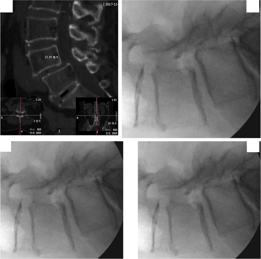
Fig. 4 The case example of OLIF with percutaneous pedicle screw fixation for degenerative spondylolisthesis. a. Prior to cage insertion. Disc
height (DH) 4.4 mm; Slip ratio 23.0%; Segmental lordosis (SL) 0.7°. b. Following cage insertion. DH 11.2 mm; Slip ratio 16.4%; SL 9.0°. c. Following
posterior fixation. DH 12.7 mm; Slip ratio 0%; SL 17.4°
the stand-alone technique in terms of capacity of in- Segmental lordosis improved by OLIF
direct nerve decompression. Segmental lordosis can be improved by insertion of cage
In this study, following posterior fixation, the slip was with lordotic angle design [19]. The magnitude of improve-
further reduced to 2.1%, slip reduction ratio was 57.9% ment correlates with preoperative segmental lordosis and
on average, which meant greater than half of slip reduc- anteroposterior position of cage [20]. This current study,
tion was achieved by posterior fixation. Therefore, add- however didn’t show increase the segmental lordosis by
itional posterior fixation and reduction maneuver could cage insertion alone (8.7°to 8.3°). The likely cause was that
reduce the slip to larger extent than stand-alone tech- the large preoperative segmental lordosis (8.7°) limited the
nique could. capacity of anterior realignment due to tightness of anterior
Table 3 Radiographic parameters of segmental alignment between open and percutaneous techniques of pedicle screw fixation
Radiographic parameters Prior to cage insertion Following cage insertion Following posterior fixation
Disc height (mm)
Open 8.7 ± 1.7a 11.9 ± 1.9 11.4 ± 2.4
Percutaneous 7.3 ± 1.2a 11.5 ± 1.3 12.5 ± 2.1
Total 8.2 ± 1.6 11.8 ± 1.7 11.8 ± 2.3
Disc height-Anteriorb (mm)
Open 10.3 ± 2.3a 13.9 ± 2.2 14.3 ± 2.2a
Percutaneous 8.4 ± 2.0a 12.6 ± 1.5 12.3 ± 1.8a
Total 9.6 ± 2.3 13.4 ± 2.1 13.7 ± 2.3
Disc height-Posterior (mm)
Open 6.4 ± 2.1 9.2 ± 2.1 8.5 ± 2.0
Percutaneous 5.5 ± 1.1 8.9 ± 2.1 8.2 ± 1.4
Total 6.1 ± 1.9 9.1 ± 2.1 8.4 ± 1.8
Slip ratio (%)
Open 10.8 ± 4.4 8.3 ± 4.4 2.1 ± 3.7
Percutaneous 11.6 ± 5.1 8.3 ± 4.7 2.2 ± 3.4
Total 11.1 ± 4.6 8.3 ± 4.4 2.1 ± 3.6
Segmental lordosis (°)
Open 9.5 ± 3.5 8.7 ± 3.2 10.9 ± 3.8
Percutaneous 7.1 ± 3.7 7.4 ± 2.5 10.4 ± 3.3
Total 8.7 ± 3.7 8.3 ± 3.0 10.7 ± 3.6
a
Means comparison of open and percutaneous techniques showed statistically significant
b
Means statistically significant following multivariate analysis between open and percutaneous techniquesWu et al. BMC Musculoskeletal Disorders (2021) 22:218 Page 8 of 9
longitudinal ligament. Additional posterior fixation short- fixation probably result in difference alignment
ened the posterior column and further increased the seg- changes. However, separate analysis showed consist-
mental lordosis (10.7°) in this study, which indicated that ent outcome between two groups, making the com-
posterior fixation could further improve segmental lordosis bined analysis reasonable.
even if anterior realignment reached its limit.
Conclusions
Stand-alone versus additional posterior fixation Stand-alone cage insertion did have some degree of slip
Stand-alone cage insertion of OLIF procedure without reduction and restoration of disc height. However, com-
posterior fixation is advocated by some surgeons [7, pared with stand-alone cage insertion, additional poster-
8]. Several clinical results favoring standalone tech- ior fixation provides better segmental alignment
nique were reported in the literature [7, 21], and improvement in terms of slip reduction and segmental
those favorable outcomes depend on inserting a large lordosis in OLIF procedures in the treatment of lumbar
cage which can both achieve indirect decompression degenerative spondylolisthesis.
effect and provide instant stability by axial loading
[6]. However, some drawbacks of stand-alone tech- Abbreviations
OLIF: Oblique lumbar interbody fusion; DH: Disc height; SL: Segmental
nique were shown during follow-ups. Cage subsidence lordosis
and subsequent loss of correction may occur without
posterior fixation [8]. The effect of indirect decom- Acknowledgements
Not applicable.
pression was also decreased during follow-up in some
patients undergoing stand-alone techniques [10]. A Authors’ contributions
recent meta-analysis showed the reoperation rate and JYW collected, analyzed, and interpreted the data and wrote the draft. YQS
occurrence of cage migration was higher for standa- performed the surgery, designed the protocol, revised the draft. THG, NZ
measured the radiographic parameters. All the authors have read and
lone technique [9]. approved the final manuscript.
OLIF with posterior fixation can enhance the seg-
mental stability, decrease the rate of cage subsidence Funding
This work was supported by Beijing Municipal Science & Technology
and migration, and maintain the instant indirect de- Commission (Z191100004419007) and Beijing JST Research Funding
compression effect by cage insertion [9]. Additionally, (XKGG201811).
as this study revealed, posterior fixation for patients
with degenerative spondylolisthesis can further reduce Availability of data and materials
The data used to support the findings of this study are available from the
the slip that maximizes the effect of indirect decom- corresponding author upon request.
pression, together with improvement of segmental lor-
dosis. Therefore, OLIF with additional posterior Ethics approval and consent to participate
This study protocol was established according to the ethical guidelines of
fixation was recommended for patients with degenera- the Helsinki Declaration and was approved by the Human Ethics Committee
tive spondylolisthesis. of Beijing Jishuitan Hospital. Written informed consent was obtained from
each participant.
Limitations
Consent for publication
Although this study allows to demonstrate the Written informed consent for publication was obtained from each
changes of segmental alignment within the same participant.
procedure setting, this retrospective observational
Competing interests
study has some limitations. Firstly, all the slip were The authors declare that they have no known competing financial interests
Grade I spondylolisthesis (slip ratio: 3.5 to 23%) with or personal relationships that could have appeared to influence the work
most slipped levels located at the L4/5 level, even if reported in this paper.
the inclusion criteria included Grade I and II slips, Received: 18 August 2020 Accepted: 15 February 2021
which may constrain drawing conclusion for Grade
II slip or other segments. Secondly, additional fix-
ation did improve the segmental alignment together References
1. Steven R, Garin FJE, Bell GR. Rothman-SIMEONE and HERKOWITZ’S the spine.
with favorable symptoms and disability improve- 7th ed. Philadelphia: Elsevier; 2018.
ments in short-term, however, whether the improved 2. Koreckij TD, Fischgrund JS. Degenerative Spondylolisthesis. J Spinal Disord
segmental alignment or the superiority of posterior Tech. 2015;28(7):236–41.
3. Sengupta DK, Herkowitz HN. Degenerative spondylolisthesis: review of current
fixation can be maintained is still uncertain in long- trends and controversies. Spine (Phila Pa 1976). 2005;30(6 Suppl):S71–81.
term. Thirdly, the result of sagittal alignment 4. Mayer HM. A new microsurgical technique for minimally invasive anterior lumbar
changes was derived from combined analysis of open interbody fusion. Spine (Phila Pa 1976). 1997;22(6):691–9 discussion 700.
5. Silvestre C, Mac-Thiong JM, Hilmi R, Roussouly P. Complications and
and percutaneous pedicle screw fixation due to rela- morbidities of mini-open anterior retroperitoneal lumbar Interbody fusion:
tively small sample size. Open and percutaneous oblique lumbar Interbody fusion in 179 patients. Asian Spine J. 2012;6(2):89–97.Wu et al. BMC Musculoskeletal Disorders (2021) 22:218 Page 9 of 9
6. Fujibayashi S, Hynes RA, Otsuki B, Kimura H, Takemoto M, Matsuda S. Effect
of indirect neural decompression through oblique lateral interbody fusion
for degenerative lumbar disease. Spine (Phila Pa 1976). 2015;40(3):E175–82.
7. Malham GM, Ellis NJ, Parker RM, Blecher CM, White R, Goss B, Seex KA.
Maintenance of segmental Lordosis and disk height in stand-alone and
instrumented extreme lateral Interbody fusion (XLIF). Clin Spine Surg. 2017;
30(2):E90–8.
8. Marchi L, Abdala N, Oliveira L, Amaral R, Coutinho E, Pimenta L.
Radiographic and clinical evaluation of cage subsidence after stand-alone
lateral interbody fusion. J Neurosurg Spine. 2013;19(1):110–8.
9. Alvi MA, Alkhataybeh R, Wahood W, Kerezoudis P, Goncalves S, Murad MH,
Bydon M. The impact of adding posterior instrumentation to transpsoas
lateral fusion: a systematic review and meta-analysis. J Neurosurg Spine.
2018;30(2):211–21.
10. Ozgur BM, Agarwal V, Nail E, Pimenta L. Two-year clinical and radiographic
success of minimally invasive lateral transpsoas approach for the treatment
of degenerative lumbar conditions. SAS J. 2010;4(2):41–6.
11. Fujiwara A, Tamai K, Yamato M, An HS, Yoshida H, Saotome K, Kurihashi
A. The relationship between facet joint osteoarthritis and disc
degeneration of the lumbar spine: an MRI study. Eur Spine J. 1999;8(5):
396–401.
12. Forsth P, Olafsson G, Carlsson T, Frost A, Borgstrom F, Fritzell P, Ohagen P,
Michaelsson K, Sanden B, Randomized A. Controlled trial of fusion surgery
for lumbar spinal stenosis. N Engl J Med. 2016;374(15):1413–23.
13. Sigmundsson FG, Jonsson B, Stromqvist B. Outcome of decompression with
and without fusion in spinal stenosis with degenerative spondylolisthesis in
relation to preoperative pain pattern: a register study of 1,624 patients.
Spine J. 2015;15(4):638–46.
14. Oliveira L, Marchi L, Coutinho E, Pimenta L. A radiographic assessment of
the ability of the extreme lateral interbody fusion procedure to indirectly
decompress the neural elements. Spine (Phila Pa 1976). 2010;35(26 Suppl):
S331–7.
15. Sato J, Ohtori S, Orita S, Yamauchi K, Eguchi Y, Ochiai N, Kuniyoshi K, Aoki Y,
Nakamura J, Miyagi M, et al. Radiographic evaluation of indirect
decompression of mini-open anterior retroperitoneal lumbar interbody
fusion: oblique lateral interbody fusion for degenerated lumbar
spondylolisthesis. Eur Spine J. 2017;26(3):671–8.
16. Kanno H, Aizawa T, Ozawa H, Koizumi Y, Morozumi N, Itoi E. An increase in
the degree of olisthesis during axial loading reduces the dural sac size and
worsens clinical symptoms in patients with degenerative spondylolisthesis.
Spine J. 2018;18(5):726–33.
17. Wegmann K, Gundermann S, Siewe J, Eysel P, Delank KS, Sobottke R.
Correlation of reduction and clinical outcome in patients with
degenerative spondylolisthesis. Arch Orthop Trauma Surg. 2013;133(12):
1639–44.
18. Fan G, Zhang H, Guan X, Gu G, Wu X, Hu A, Gu X, He S. Patient-reported
and radiographic outcomes of minimally invasive transforaminal lumbar
interbody fusion for degenerative spondylolisthesis with or without
reduction: a comparative study. J Clin Neurosci. 2016;33:111–8.
19. Melikian R, Yoon ST, Kim JY, Park KY, Yoon C, Hutton W. Sagittal plane
correction using the lateral Transpsoas approach: a biomechanical study on
the effect of cage angle and surgical technique on segmental Lordosis.
Spine (Phila Pa 1976). 2016;41(17):E1016–21.
20. Otsuki B, Fujibayashi S, Takemoto M, Kimura H, Shimizu T, Murata K,
Matsuda S. Analysis of the factors affecting lumbar segmental Lordosis
after lateral lumbar Interbody fusion. Spine (Phila Pa 1976). 2020;45(14):
E839–46.
21. Ahmadian A, Bach K, Bolinger B, Malham GM, Okonkwo DO, Kanter AS,
Uribe JS. Stand-alone minimally invasive lateral lumbar interbody fusion:
multicenter clinical outcomes. J Clin Neurosci. 2015;22(4):740–6.
Publisher’s Note
Springer Nature remains neutral with regard to jurisdictional claims in
published maps and institutional affiliations.You can also read


