RSNA COVID-19 Task Force: Best Practices for Radiology Departments during COVID-19
←
→
Page content transcription
If your browser does not render page correctly, please read the page content below
RSNA COVID-19 Task Force: Best Practices for Radiology
Departments during COVID-19
M. Mossa-Basha, J. Azadi, J. Ko, J. Klein, C. Meltzer, COVID-19 Task Force*

      Given the emerging and evolving nature of the situation, many institutions, hospitals, and clinics have also
      established their own local guidelines. We urge you to follow the evolving Centers for Disease Control and
      Prevention (CDC) recommendations and your local requirements. The information in this document is sub-
      ject to change as information regarding COVID-19 changes.
      RSNA COVID-19 Task Force
      The goal of this guidance document is to protect health care workers (HCW) and the general public from
      exposure and dissemination of COVID-19, while maintaining critical radiology functions and preserving
      personal protective equipment (PPE) and other critical care resources during the COVID-19 pandemic. These
      guidelines apply in the setting of sufficient resource availability, and in the setting of a surge when resources
      are in short supply; mitigating steps and alternative approaches should be considered to provide the best care
      for patients while protecting all HCW.

RSNA  COVID
RSNA COVID TaskTask
                Force:Force:
                      April 27, April 27, 2020
                                2020 rsna.org/COVID-19          1                                         rsna.org/COVID-19
Radiology Elective Imaging
o Postponement of nonurgent outpatient imaging
All screening radiology examinations including mammography, lung cancer screening, and CT colonos-
copy as well as DEXA scans and coronary artery calcium scoring
Other elective diagnostic examinations as determined by the institution and in discussion between radiolo-
gist, ordering health care provider (HCP), and patient
• Critical and time-sensitive imaging, necessary for treatment decision making, should be per-
formed within the needed time frame.
o At radiology front desk or hospital/outpatient center entrance, outpatients scheduled for imaging or procedures
should be screened for symptoms including fever, new dry cough, dyspnea, and sore throat.
Prohibit accompanying visitors except in cases where a single family member and/or caregiver is needed for
the patient’s ambulation, understanding of, and/or cooperation with the examination.
Symptomatic patients, if not already masked (some states/regions require everyone to wear masks public-
ly), are given surgical masks with use of droplet and contact precautions, and isolated from other patients
while awaiting scanner availability.
• Patient is imaged with portable equipment when possible (eg, chest x-ray) in isolation room.
• If portable is not possible, after droplet/contact precaution is instituted, patient is imaged using
main rooms.
o Dedicated CT scanners and interventional suites can be used, ideally with negative pres-
sure, as institutional space and resources allow.
o Hand hygiene
Hand washing with soap and water for 20 seconds is preferable over use of hand sanitizer, especially when
considering sanitizer shortages (1); however, hand sanitizers with at least 60% ethanol and 70% isopropyl
alcohol effectively inactivate coronaviruses (2).
Avoid touching face
o PPE training video: https://www.youtube.com/watch?v=bG6zISnenPg
COVID-19 Suspected or Confirmed Case Precautions (Figure 1)
o Develop a method to identify patients as positive for PUI-COVID-19 or COVID-19, such as in electronic medical
record or by report order by HCP.
If possible, institutions should consider COVID-19 testing prior to procedures and sometimes imaging
studies, especially those that require sedation, are aerosol generating, or are for intubated patients.
o Staff with direct patient contact with COVID-19 and persons under investigation (PUIs) should be fit tested for
N95 masks if fit testing has never been performed or has expired.
It is recommended that those with beards consider shaving their facial hair for appropriate N95 mask fitting.
N95 masks may be preserved for reuse by wearing overlying surgical/isolation masks and/or face shield or
by central cleaning per institutional practice.
Note that N95 masks may have a metal strip, risking loss of seal in the MRI room; nonferrous N95 masks
may be available.
Minimize staff in the room with COVID-19 and PUIs to those needed to perform the procedure.
o Droplet/contact precautions with eye protection
Majority of cases
Patients wear surgical masks
HCW wear fitted N95, PAPR, or surgical mask and face shield and contact precaution gear (gown and
gloves) and appropriate hand hygiene
o Airborne/contact precautions
Aerosol-generating procedures per Society of Interventional Radiology guidelines (3):
• Lung biopsy, lung ablation, thoracentesis, pleural drain, chest tube, bronchial artery emboliza-
tion, bronchial stenting, nasogastric or orogastric tube placement, gastrostomy, gastrojejunos-
tomy, jejunostomy, gastrointestinal stent placement
• Intubation/extubation, open suction, tracheostomy, moderate or general anesthesia
• Esophagograms, upper gastrointestinal fluoroscopic studies, modified swallow fluoroscopic stud-
ies, contrast enemas, and virtual CT colonography should also be considered aerosol generating
Patients wear surgical masks.
HCW wear N95 masks (or PAPR) and face shield (if N95 mask) and contact precaution gear (gown and
gloves) and appropriate hand hygiene
RSNA COVID
RSNA COVID Task Force:
Task Force: AprilApril 27,rsna.org/COVID-19
27, 2020 2020 2 rsna.org/COVID-19• Limited supply of PAPR hoods at this time
• PAPR hoods cannot be used in MRI suites.
PPE for Staff
o Determined along with institutional guidelines, with resource availability and conservation taken into consideration
o Consider education of staff through communication of guidelines for use of PPE, easy-to-read charts
o Surgical/isolation masks should be worn by all HCW while in clinical facilities, whether performing direct clinical
care or not, with consideration for institutional availability and governmental and institutional guidelines.
If soiled or exposed during droplet precautions without a covering face shield, the mask should be disposed
of, with use of a new mask for the next patient interaction.
o Surgical masks with covering face shield (if available) should be worn for exposure to patients with respiratory symp-
toms.
o Encourage judicious use of HCW PPE for conservation in accordance with institutional policy and supplies.
Postimaging Room Cleaning
o Room cleaning is multifactorial, with need for consideration for room air exchange rates
Please communicate with local infection control to evaluate workflows for room clean-up for imaging of
patients positive for or suspected of having COVID-19.
The higher the air exchange rate per hour, the shorter duration needed for room closure (4).
o Droplet/contact precautions
Standard antiseptic wipe down of equipment using quaternary ammonium/alcohol-impregnated wipes or
other Environmental Protection Agency (EPA)–approved disinfectants (5)
No need for room closure if adequate air circulation in the room
o Airborne/contact precaution
Standard wipe down
Room closed for 1 hour after imaging/procedure for rooms with greater than 6 air exchanges per hour,
which includes cleaning time
• Use high-efficiency particulate air (HEPA) filter if possible and available to accelerate air circula-
tion and shorten room closure.
o HEPA filtration is not compatible with MRI suites.
Patient Screening for COVID-19
o Patient screening may be performed with reverse-transcription–polymerase chain-reaction (RT-PCR) and is espe-
cially recommended prior to image-guided procedures with risk of aerosolization.
o With a negative RT-PCR result in a PUI for COVID-19, high-risk cases are retested.
o Chest imaging currently is not typically used for screening or for follow-up if RT-PCR is available.
Consideration for CT use if patient surge exceeds capacity for PCR testing and quick throughput is
needed.
Imaging should also be kept in mind for patients with atypical presentations.
Assess the role of imaging and determine reporting language from chest imaging studies in PUI or con-
firmed COVID-19 cases in consultation with HCP.
Chest Imaging in COVID-19 (Figure 1) (6)
o Fleischner Society Guidelines for Imaging in Patient Management during the COVID-19 Pandemic (7)
Imaging can be considered in patients:
• With mild clinical features of COVID-19 who have risk factors for progression and are either
positive for COVID-19 or have moderate/high pretest probability of COVID-19 (RT-PCR
unavailable)
• With moderate to severe clinical features (any pretest probability), regardless if RT-PCR positive
(to assess severity) or if RT-PCR negative or testing not performed (to identify any alternative
diagnosis)
• Setting of a surge of patients suspected of having COVID-19
Risk factors: Patients with high likelihood of poor outcomes with COVID infection
• Age greater than 65 years, hypertension, diabetes mellitus, immunocompromised, cardiovascular
disease, and chronic respiratory disease (7)
RSNARSNA
COVID Task
COVID TaskForce: April
Force: April 27, 27,
20202020
rsna.org/COVID-19 3 rsna.org/COVID-19Imaging in Patients with Confirmed or Suspected COVID-19
o Nonscreening imaging is reserved for those in which there will be impact on patient management.
This will typically apply to cases where alternative diagnoses are considered or acute patient worsening is
considered due to an alternative diagnosis, such as pulmonary embolism.
Patients with high pretest probability of COVID-19 despite negative PCR tests
If patients require emergent imaging for other reasons (suspected of having appendicitis, cholecystitis,
stroke, etc), the targeted imaging for diagnosis (eg, CT abdomen/pelvis for appendicitis) will also be
performed.
Nonemergent imaging for inpatients will be delayed until diagnosis is confirmed and the patient has
recovered from the illness and is considered noncontagious based on clearance from infection control.
Identify imaging units/scanners that are to be used for PUI-COVID 19 and patients positive for CO-
VID-19 who undergo imaging
o When possible, chest radiography is performed in patient rooms using portable equipment (6).
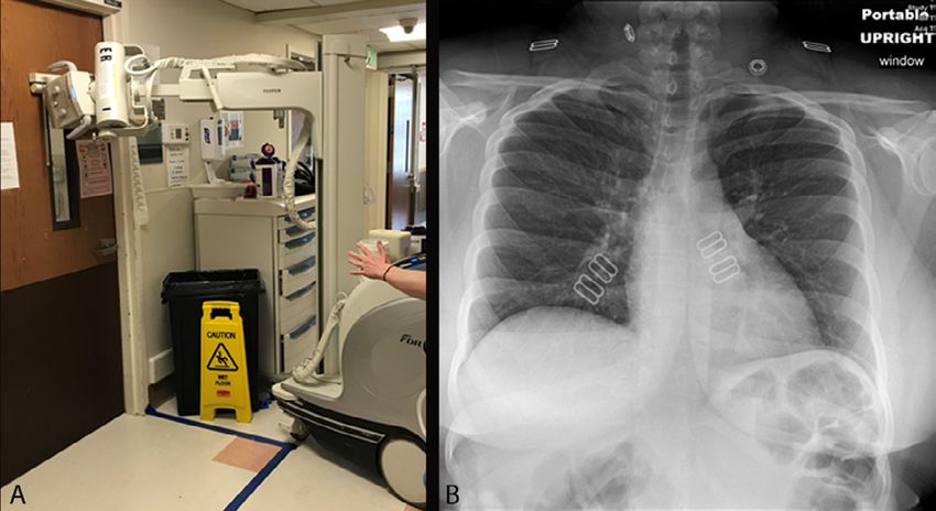
Radiographic imaging can be performed through the glass of isolation rooms (Figure 2), where only the
cassette enters the room and the source remains outside. This reduces equipment cleaning time, PPE use,
and potentially exposures.
o Transports will observe droplet/contact or airborne/contact precaution.
Elective Procedure Cancellations
o Elective nonemergent procedures should be postponed or cancelled during the pandemic surge.
o The goal is to conserve resources (PPE, staff resources), conserve inpatient capacity that may become needed, con-
solidate staff for mobilization, and protect our patients from exposure.
o This should not affect emergent/critical procedures.
Decisions to proceed, postpone, or cancel image-guided procedures should be made jointly with the refer-
ring HCP.
o Scheduling of future procedures can be performed in coordination with the ordering physician, with placement of
procedures into nonurgent (can be postponed for months), time-sensitive (short delay is acceptable), and critical
(performed as soon as possible) categories (Table 1).
Pregnancy and COVID-19
o As of now, there is no evidence to indicate teratogenic effects or detrimental effects on pregnancy from COVID-19
infection; however, there have been reports of transmission during late stages of pregnancy (8,9).
o Recommend redeploying pregnant HCW to sites with minimized exposure to patients with COVID-19 and PUIs
Meetings
o Meetings should be held via teleconferencing software such as Zoom, Cisco’s WebEx, Microsoft Teams, or Skype.
Safe social distancing at small meetings (few attendees) if not virtual
o Encourage frequent and potentially daily updates for the entire department and within sections/teams, depending
on the evolving situation.
Radiologist Workflows
o Section faculty, trainees, and essential staff can be scheduled to work in exclusive teams that alternatively cover
weeks to minimize exposures across the section and maintain the workforce.
A backup workforce can be established for shifts, particularly in high-exposure areas; HCWs may work in
teams in which Team A and Team B alternate working for 2 weeks followed by 2 weeks out.
o Social distancing of 6 feet is recommend to reduce risk of potential person-to-person transmission.
o Reconfigure reading rooms and radiologists in the reading room to maintain distancing.
o Radiologists should sterilize shared workstation equipment before and after use; ideally, workstations should be
dedicated to a single radiologist or resident/fellow for full shift with cleaning between shifts.
o Phone communications between radiologists and other staff are preferable to in-person communications.
o Radiologists who can perform their clinical duties remotely should be permitted to do so.
o Create single-radiologist and single-workstation reading rooms, if feasible.
o Consider workflows for virtual consults with ordering clinical teams, including phone and telecommunication capa-
bilities.
o Consider development of radiology PACS/RIS worklists and approach to reporting language for chest imaging
studies for PUIs or confirmed COVID-19 cases in consultation with HCP and communication of findings.
RSNA COVID
RSNA COVID Task Force:
Task Force: AprilApril 27,rsna.org/COVID-19
27, 2020 2020 4 rsna.org/COVID-19Screening, Evaluation, and HCW Return-to-Work Criteria
o Recommend screening employees and patients at all entrances (symptoms within last 72 hours and potential SARS-
CoV-2 exposure) and consider temperature checks
o Employees to complete daily attestation of health and lack of symptoms (fever, new dry cough, sore throat, new
shortness of breath, new muscle aches, loss of taste or smell)
Employees with these symptoms should stay home and contact occupational/employee health officer for
need for potential further testing.
Per CDC guidelines, two options for return-to-work criteria for HCW that are positive for COVID-19
(10)
• Test-based strategy
o Can return to work after resolution of fever, resolution of respiratory symptoms, and at
least two negative PCR tests from nasopharyngeal swab separated by at least 24 hours
• Non–test-based strategy
o Requirement to stay home for 7 days from symptom onset AND 72 hours after symp-
tom resolution (fever and respiratory symptoms)
• After return to work, HCW should wear a face mask at all times while in the health care facility
until all symptoms are completely resolved or until 14 days after symptom onset (whichever is
longer)
• Restricted from contact with severely immunocompromised patients for 14 days
o Another approach is having COVID-affected HCW to continue to wear a mask for 7
days after symptom resolution.
Patients with these symptoms should be considered PUIs.
Formation of a Radiology Crisis Management Team/Task Force
o Consider formation of a departmental crisis management team comprised of leadership from radiologist, technolo-
gist, and staff with standing meetings to assess situation and adapt staffing and other approaches according to time
within surge. This task force will provide quick decision making, policy discussion, information gathering, and
policy dissemination.
o Surveillance of number of examinations performed, emergency department visits for PUI-COVID-19 for technolo-
gist and radiologist staffing
o Daily communication within sections, technologist groups, department, and institution to disseminate policies
o Departmental point person in charge of guidelines/policies and communication updates
Social Distancing within Radiology
o In addition to workspace areas, emphasis on social distancing throughout the department, including in break
rooms, congregation areas, and scanner unit console areas
o Reconfiguration of seating in these areas, as well as reduced staffing and staggered breaks, can be considered.
o Staff education and engagement to explain the importance of social distancing should be conducted.
Staff Engagement and Well-Being
o Radiology leadership should monitor for staff anxiety or burnout.
o Constant communication and engagement with staff and faculty
o Daily virtual meetings with sectional, trainee, and operational leadership to communicate policy and
informational updates, as well as to check on team morale and listen to questions and concerns
o Weekly departmental meetings to listen to questions and concerns and to communicate policy updates
References
1. Centers for Disease Control and Prevention. Show Me the Science – When & How to Use Hand Sanitizer in Community Set-
tings. https://www.cdc.gov/handwashing/show-me-the-science-hand-sanitizer.html. Published 2020.
2. Centers for Disease Control and Prevention. CDC Statement for Healthcare Personnel on Hand Hygiene during the Response
to the International Emergence of COVID-19. https://www.cdc.gov/coronavirus/2019-ncov/hcp/hand-hygiene.html?CDC_AA_
refVal=https%3A%2F%2Fwww.cdc.gov%2Fcoronavirus%2F2019-ncov%2Finfection-control%2Fhcp-hand-sanitizer.html. Published
2020.
RSNARSNA
COVID Task
COVID TaskForce: April
Force: April 27, 27,
20202020
rsna.org/COVID-19 5 rsna.org/COVID-193. Society of Interventional Radiology. Aerosol Generating Procedures Performed by Interventional Radiology Clinical Notification
from the Society of Interventional Radiology. https://www.sirweb.org/practice-resources/covid-19-resources/covid-19-clinical-notifica-
tion-3-26-20/. Published 2020.
4. Centers for Disease Control and Prevention. Airborne Contaminant Removal. https://www.cdc.gov/infectioncontrol/guidelines/
environmental/appendix/air.html#tableb1 Published 2019.
5. Environmental Protection Agency. List N: Disinfectants for Use Against SARS-CoV-2. https://www.epa.gov/pesticide-registration/
list-n-disinfectants-use-against-sars-cov-2. Published 2020.
6. Mossa-Basha M, Medverd J, Linnau K, Lynch JB, Wener MH, Kicska G, Staiger T, Sahani D. Policies and Guidelines for CO-
VID-19 Preparedness: Experiences from the University of Washington. Radiology 2020:201326. doi: 10.1148/radiol.2020201326
7. Rubin GD, Ryerson CJ, Haramati LB, Sverzellati N, Kanne JP, Raoof S, Schluger NW, Volpi A, Yim JJ, Martin IBK, Anderson
DJ, Kong C, Altes T, Bush A, Desai SR, Goldin J, Goo JM, Humbert M, Inoue Y, Kauczor HU, Luo F, Mazzone PJ, Prokop M,
Remy-Jardin M, Richeldi L, Schaefer-Prokop CM, Tomiyama N, Wells AU, Leung AN. The Role of Chest Imaging in Patient
Management during the COVID-19 Pandemic: A Multinational Consensus Statement from the Fleischner Society. Radiology
2020:201365. doi: 10.1148/radiol.2020201365
8. Alzamora MC, Paredes T, Caceres D, Webb CM, Valdez LM, La Rosa M. Severe COVID-19 during Pregnancy and Possible
Vertical Transmission. Am J Perinatol 2020. doi: 10.1055/s-0040-1710050
9. Murphy S. Newborn baby tests positive for coronavirus in London. The Guardian. https://www.theguardian.com/world/2020/
mar/14/newborn-baby-tests-positive-for-coronavirus-in-london. Published 2020.
10. Centers for Disease Control and Prevention. Return to Work for Healthcare Personnel with Confirmed or Suspected COVID-19.
https://www.cdc.gov/coronavirus/2019-ncov/healthcare-facilities/hcp-return-work.html. Published 2020.
Support Figures
Figure 1: Imaging workflow and indications in patients suspected of having COVID-19.
RSNA COVID
RSNA COVID Task Force:
Task Force: AprilApril 27,rsna.org/COVID-19
27, 2020 2020 6 rsna.org/COVID-19Figure 2: Chest radiography through glass. A, Technologists position the portable x-ray unit outside the patient room, with the tube
peering through the mesh wire–reinforced isolation room window. B, Anteroposterior chest radiograph obtained is of diagnostic quality.
Category Designation Description Evaluator
Category 1 Elective/Nonurgent If delayed, will not harm patients
in the next 2-6 months. These
procedures can be delayed until
after postponement period. Radiologist in con-
Category 2 Time-Sensitive Short delay (eg, 2–4 weeks) is sensus with ordering
acceptable within a certain time clinician
frame.
Category 3 Critical Cannot be delayed. Schedule
these procedures right away.
*COVID-19 Task Force
Mahmud Mossa-Basha, MD, University of Washington Medical Center (Chair)
Javad R. Azadi, MD, Johns Hopkins Medicine
Christopher Filippi, MD, North Shore LIJ Health System
Maryellen L. Giger, PhD, University of Chicago
Jeffrey S. Klein, MD, University of Vermont
Jane Ko, MD, New York University Langone Health
Brian S. Kuszyk, MD, Eastern Radiologists
Christine O. Menias, MD, Mayo Clinic, Arizona
Richard E. Sharpe, Jr, MD, Kaiser Permanente, Denver
Bien Soo Tan, MD, Singapore General Hospital
Erik M. Velez, MD, University of Southern California
Carolyn C. Meltzer, MD, Emory University (RSNA Board liaison)
rsna.org/COVID-19
RSNARSNA
COVID Task
COVID TaskForce: April
Force: April 27, 27,
20202020
rsna.org/COVID-19 7 rsna.org/COVID-19You can also read