Neuroimaging manifestations of epidermal nevus syndrome
←
→
Page content transcription
If your browser does not render page correctly, please read the page content below
Case Report

Neuroimaging manifestations of epidermal nevus syndrome
Andrea De Vito1, Ajay Taranath2, Hisham Dahmoush3, Shankar Srinivar Ganapathy4, Sniya Sudhakar5,
Kshitij Mankad5
1
Department of Neuroradiology, H. S. Gerardo Monza, Monza, Italy; 2Department of Radiology, Women’s and Children’s Hospital, North Adelaide,
South Australia, Australia; 3Department of Radiology, Lucile Packard Children’s Hospital, Stanford University School of Medicine, Stanford, CA, USA;
4
Department of Radiology, Akron Children’s Hospital, Akron, OH, USA; 5Department of Radiology, Great Ormond Street Hospital, London, UK

Correspondence to: Kshitij Mankad, MD. Department of Radiology, Great Ormond Street Hospital, Great Ormond Street, London WC1N 3JH, UK.
Email: drmankad@googlemail.com.

                  Abstract: Epidermal nevus syndrome (ENS) represents a diverse group of rare neurocutaneous
                  diseases associated with the presence of characteristic epidermal nevi (EN) in the skin and extracutaneous
                  manifestations in the eyes, skeletal, urogenital and central nervous systems. We present a case series of 7
                  children with ENS, with specific attention to the neuroradiological characteristics of this entity.

                  Keywords: Central nervous system diseases; hemimegalencephaly (HME); linear sebaceous nevus syndrome

                  Submitted May 05, 2020. Accepted for publication Aug 24, 2020.
                  doi: 10.21037/qims-20-634
                  View this article at: http://dx.doi.org/10.21037/qims-20-634

Introduction                                                                 can be grey matter heterotopia, atrophy with dystrophic
                                                                             calcification and hamartomas (7,8). Cerebellar involvement
Epidermal nevus syndrome (ENS) is a term used to
                                                                             has also been described. Vascular abnormalities are rare,
represent a diverse group of neurocutaneous diseases in
                                                                             including vessel dysplasia and infarction (9). The molecular
which one of the subtypes of epidermal nevi (EN) are found
                                                                             basis of ENS spectrum is unclear. The syndrome has been
in association with extracutaneous abnormalities involving
                                                                             observed sporadically in the relatives of affected patients.
the eyes, nervous, skeletal, and urogenital systems (1).
                                                                             It has been hypothesized that it is a form of mosaicism of a
Most EN are present at birth and typically follow the
                                                                             lethal autosomal dominant gene (5,10). Recent reports of
lines of Blaschko (2). Blaschko’s lines refer to the S-shaped
                                                                             somatic mosaicism in the pathogenesis of ENS have been
or V-shaped whorled, streaked, and linear patterns that
                                                                             reported, along with the identification of HRAS and KRAS
are believed to represent the dorsoventral migratory
                                                                             mutations (11,12).
pathways of the neuroectoderm during embryogenesis (2).
The incidence of EN has been reported to range from 1 to
3 per 1,000 live births, without gender predominance (2,3).                  Case presentation
Numerous phenotypes with different clinical appearances
                                                                             Case 1
and histopathological features have been described (4), such
as the nevus sebaceous syndrome (NS), nevus comedonicus                      A 7-year-old girl presented with new-onset of generalized
syndrome (NC), phakomatosis pigmentokeratotica (PPK),                        seizures and developmental delay in multiple domains.
proteus syndrome, and congenital hemidysplasia with                          Physical examination revealed an epidermal nevus on the
ichthyosiform erythroderma and limb defects (CHILD)                          left trunk. Ophthalmological assessment showed bilateral
syndrome (5). In terms of CNS abnormalities however,                         optic nerve colobomata and corneal opacification. Brain
hemimegalencephaly (HME) is the most striking finding,                       MRI demonstrated two small lipomas on the dorsal surface
typically found on the side of the skin lesion, associated often             of the cervicomedullary junction (Figure 1A). There was
with polymicrogyria (4,6). Other neuroimaging findings                       also moderate generalised cerebral atrophy (Figure 1B) with

© Quantitative Imaging in Medicine and Surgery. All rights reserved.    Quant Imaging Med Surg 2021;11(1):415-422 | http://dx.doi.org/10.21037/qims-20-634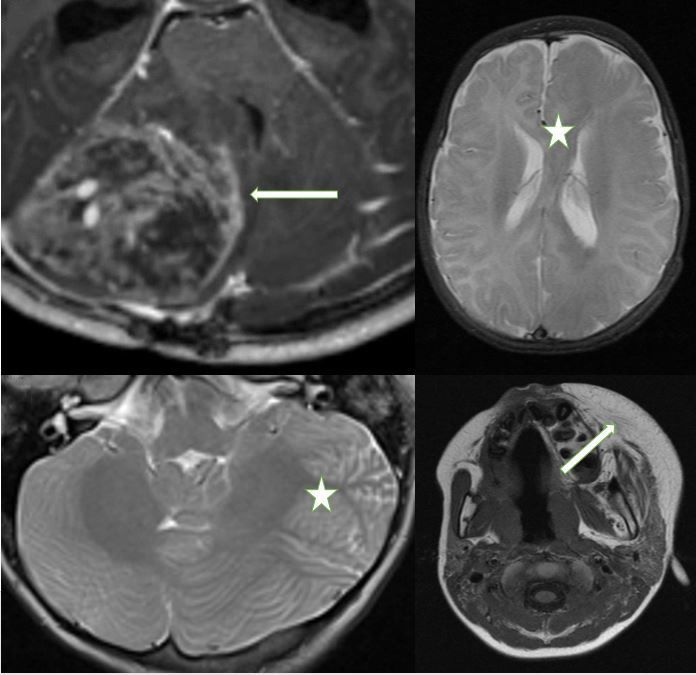
416 De Vito et al. Neuroradiological findings of ENS
A B
C D
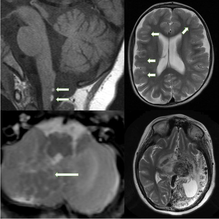
Figure 1 MRI scans showing cerebral and cerebellar abnormalities. (A) Sagittal T1-WI showing two dorsal cervico-medullary lipomas. (B)
Axial T2-WI showing generalized brain atrophy with some areas of cortical dysplasia (arrows). (C) Axial T2-WI showing right cerebellar
grey matter heterotopia (arrow). (D) Axial T2-WI showing left hemispheric proliferative arteriopathy.
mild prominence of the subarachnoid spaces overlying both 9 of life showed an extensive right cerebral and cerebellar
hemispheres and inferior cerebellar vermian dysplasia. HME. He underwent functional hemispherectomy at the
age of 12 months.
Case 2
Case 4
A female infant presenting with right-sided seizures at week
1 of life, with evidence of an epidermal nevus of the neck A 7-year-old girl was diagnosed with a large left neck
and right cheek noticed at 6 months of age. Right hemifacial and cheek epidermal nevus. Left cerebral arterio-venous
hypertrophy, prominent right eye with associated exotropia malformation was diagnosed when she presented with right
and congenital nystagmus were noted on examination. Brain sided weakness and refractory seizures. She underwent
MRI at 1 month of age showed right cerebral HME with multiple embolizations. Historical images were not available
extensive areas of cortical dysplasia and associated neuronal but CT scan and MRI scan at age 18 showed a left-sided
migrational abnormality within the cerebellum, consistent extensive proliferative arteriopathy with associated brain
with heterotopic grey matter (Figure 1C). atrophy (Figure 1D).
Case 3 Case 5
A 3-day-old boy with right facial hypertrophy, right An 8-year-old boy presenting with vomiting and headache.
epidermal nevus, right ocular enlargement, nystagmus and He was noted to have a right facial nevus. Brain MRI revealed
developmental delay presented with medical refractory a large medulloblastoma in the posterior right cerebellar
right-sided seizures. CT and MRI scan performed at day hemisphere which was resected in 2015 (Figure 2A). He had
© Quantitative Imaging in Medicine and Surgery. All rights reserved. Quant Imaging Med Surg 2021;11(1):415-422 | http://dx.doi.org/10.21037/qims-20-634Quantitative Imaging in Medicine and Surgery, Vol 11, No 1 January 2021 417
A B
C D
Figure 2 MRI scans showing supra- and infratentorial abnormalities. (A) Axial T1-WI after contrast administration showing a right cerebellar
hemispheric medulloblastoma. (B) Axial T2-WI showing left HME involving the corpus callosum (star); (C) Axial T2-WI showing left
cerebellar hemimegalencephaly (HME) with associated cortical dysplasia (star); (D) Axial T1-WI showing left facial lipomatous hypertrophy.
a large local recurrence 3 years later. categorized together (10). Several specific syndromes with
overlapping features have been described, namely, the NS
syndrome, Proteus syndrome, PPK (coexistence of nevus
Case 6
sebaceous with melanocytic nevi) and Keratinocytic ENS
An 8-year-old boy with left facial hypertrophy and (10-14). These have been summarized in Table 1.
ipsilateral neck and trunk nevus presented with new The cutaneous features of ENS depend on the
onset left-sided seizures and developmental delay. Brain predominant cell type involved histologically, the degree of
MRI showed a left cerebral and cerebellar hemispheric cellular differentiation, the body site involved, and the age
(Figure 2B) HME with extensive cortical dysplasia within of the patient. EN typically follow several well recognized
both structures (Figure 2C). patterns of cutaneous mosaicism, typically “the lines of
Blaschko” (2). Various methods of classifying EN have
been proposed over the years. The concept that nevi may
Case 7
show differentiation towards various skin structures and
A 15-month-old boy presenting with new-onset focal glands, comprising of pilosebaceous, apocrine, eccrine
left frontal epilepsy and developmental delay. Physical glands and keratinocytes, has been the basis of most clinical
examination showed a left facial nevus. Brain MRI revealed classifications to date (5,15). The two main subtypes
an extensive left cerebral HME (Figure 2D). are Nevus sebaceus (with excessive sebaceous glands
components) and Keratinocytic EN (lesions without any
adnexal components).
Discussion
In terms of the genetic basis of ENS, the published
ENS is not one disease, but rather a heterogeneous and evidence suggests that the clinical expression of ENS is
diverse cluster of different disorders which have been based on genomic mosaicisms (16). The primary genes
© Quantitative Imaging in Medicine and Surgery. All rights reserved. Quant Imaging Med Surg 2021;11(1):415-422 | http://dx.doi.org/10.21037/qims-20-634418 De Vito et al. Neuroradiological findings of ENS
Table 1 Classification of epidermal nevus syndrome
Syndrome Type of epidermal nevus Salient clinical features Genetics
Nevus Sebaceous syndrome Nevus Sebaceous Nevus Sebaceous and epibulbar lipodermoid KRAS
Phakomatosis Pigmentokeratotica Nevus Sebaceus Additional presence of nevus spilus papulosus, HRAS
arranged in a checkerboard pattern
Nevus Comedonicus syndrome Nevus Comedonicus Ipsilateral cataract and skeletal anomalies FGFR2
Becker Nevus syndrome Becker Nevus Breast hypoplasia in females, hypertrichosis in Undefined
males, no CNS involvement
Proteus syndrome Keratinocytic Nevus Cerebriform connective tissue nevi of palms or AKT
soles; asymmetric macrodactyly
CHILD syndrome CHILD Nevus Lateralized, inflammatory skin lesions; NSDHL mutations
ptychotropism; ipsilateral limb defects
Keratinocytic Nevus syndrome Keratinocytic Nevus Presence of keratinocytic nevus of the FGFR3
common type; absence of lesions of
disproportionate overgrowth
FGFR, fibroblast growth factor receptor; AKT, protein kinase B; NSDHL, NAD (P) dependent steroid dehydrogenase-like.
thought to be responsible for ENS are the ones involved in developed in consultation with a librarian with expertise
the Ras/MAPK signaling pathways; hence the term mosaic in health research and systematic reviews, included terms
RASopathies is commonly used (11). The genes involved related to ENS HME (in studies published from 1995).
in the pathway include KRAS, HRAS, NRAS and FGFR1 A broad approach was taken through the inclusion of
(11,17,18). FGFR2 and FGFR3 mutations have also been all possible synonyms and abbreviations for the terms
reported to be involved in some cases (11,19). of interest, and controlled vocabulary/ subject headings
Sebaceous Nevi are mostly located on the scalp or face, (including MeSH, EmTree).
although they can be more widespread. They have a salmon
to yellow color and a characteristic smooth waxy surface
HME
and can be complicated by eczematous reactions (14).
Keratinocytic EN (KN) are another type of EN typically HME is the most common structural brain abnormality
found on the trunk and extremities. They appear as linear encountered in the different subsets of ENS. As a
or whorled skin-colored to pink or slightly hyperpigmented corollary, ENS is the most common neurocutaneous
plaques that may be unilateral or bilateral. disorder associated with HME (28). The distinctive
Central nervous system involvement has been noted to triad of hemifacial EN, ipsilateral HME, and lipoma
be the most commonly described systemic association with was first described by Gross and Uiberrak in 1955, who
ENS, presenting with a constellation of signs and symptoms introduced the term “hemimegalencephaly.” Pavone
ranging from headaches, epilepsy, focal motor deficits and colleagues also reported HME in 17 out of 60
and developmental delay (20). Ophthalmological findings patients with ENS, their study being a review of the
described include epibulbar lipodermoid, coloboma, corneal literature and based on pathology or CT studies (28).
opacities and defects of the optic nerves, while associated Menascu et al. (25) described MRI abnormalities in
skeletal abnormalities are craniofacial defects, frontal bossing, 3 patients with ENS showing HME, polymicrogyria
kyphoscoliosis, hip dislocation and limb deformities (21,22). and cortical heterotopia while Pavlidis et al. (26)
Spanning the last 20 years, some cases of ENS have been reported a boy with right ‘total’ HME, with increased
published describing their neuroimaging manifestations, volume of both cerebellar and cerebral right hemisphere.
as summarized in Table 2. Our search strategy on PubMed, In our series, HME was found in 4 patients out of 7,
© Quantitative Imaging in Medicine and Surgery. All rights reserved. Quant Imaging Med Surg 2021;11(1):415-422 | http://dx.doi.org/10.21037/qims-20-634Quantitative Imaging in Medicine and Surgery, Vol 11, No 1 January 2021 419
Table 2 Literature review
Authors No. of patients Neuroimaging findings
Dodge et al. 1995 (23) 1 Left HME, pachygyria, absence of the corpus
callosum, and type A Dandy-Walker malformation
Booth et al. 2002 (9) 2 Cervicomedullary subpial lipomas, cerebral cortex dysplasias
Abdelhalim et al. 2003 (6) 1 Asymmetric bilateral hemispheric enlargement and diffuse pachygyria. The right
cerebellar
hemisphere was large, with disorganized cerebellar folia
Neumann et al. 2003 (7) 1 Asymmetry with cortical hypoplasia on
the left side and giant cystic enlargement of Virchow-Robin spaces.
Zhang et al. 2003 (4) 3 Hemimegalencephaly, heterotopia, calcifications and gyral fusions
Canygit et al. 2006 (24) 1 Intracranial left orbital and left cerebellopontine angle cistern lipomas, thinning of
the left internal carotid artery, left cerebral HME
Menascu et al. 2008 (25) 2 Hemimegalencephaly, heterotopia, polymicrogyria
Pavlidis et al. 2011 (26) 1 Right ‘total’ HME, with increased volume of both
cerebellar and cerebral right hemisphere, PMG
Okumura et al. 2012 (8) 1 Medulloblastoma associated with supratentorial cortical dysplasias, a
dysmorphic enlarged midbrain and a small cerebellum with dysplastic folias
Ullah et al. 2016 (27) 1 Cortical dysplasia and multiple hamartomas in the medial temporal lobe,
thalamus, and
periventricular region on right side and cerebellar atrophy with Dandy-Walker
variant
HME, hemimegalencephaly; PMG, polymicrogyria.
mostly right-sided. 2 of these patients showed both cerebral manifestations of ENS; 2 patients from Booth et al. (9)
and cerebellar involvement. Furthermore, HME was always showed the presence of subpial lipomas dorsal to the
ipsilateral to the epidermal nevus. cervicomedullary junction, in association with very subtle
dysplasias involving the cerebral cortex ipsilateral to the
nevi. Left orbital and left cerebellopontine angle cistern
Cerebellar involvement
lipomas were present in the series from Canyigit et al. (24).
Dodge et al. showed severe hypoplasia of the cerebellum with We found 2 dorsal cervicomedullary lipomas in 1 patient,
upward rotation of the vermis and a large retrocerebellar in association with cerebral and cerebellar atrophy.
cyst in a 6-month old girl with infantile spasms (23). Also,
Ullah et al. (27) reported cerebellar atrophy and cerebral
Neoplasms
cortical dysplasia in a 25-year-old girl. Abdelhalim et al. (6)
reported an infant with ENS characterized by enlargement Bourdeaut et al. reported an oncogenic KRAS mutation in a
of both cerebral hemispheres, malformed basal ganglia, patient with ENS associated with rhabdomyosarcoma (12)
and unilateral enlargement of cerebellar hemisphere with while Okumura et al. (8) showed a 51-day-old boy with neonatal
disorganized folia. medulloblastoma associated with cerebral and cerebellar
Infratentorial anomalies were shown in 5 patients of cortical dysplasia. A right lateral medulloblastoma was present
our series, represented by HME, atrophy (unilateral and in 1 patient from our series, along with an ipsilateral nevus.
bilateral) and cerebellar heterotopia.
Vascular abnormalities
Lipomas
Juan et al. (29) revealed multiple sites of intra-thoracic
Intracranial and intraspinal lipomas represent rare and abdominal arterial stenoses and aneurysm formations
© Quantitative Imaging in Medicine and Surgery. All rights reserved. Quant Imaging Med Surg 2021;11(1):415-422 | http://dx.doi.org/10.21037/qims-20-634420 De Vito et al. Neuroradiological findings of ENS
in association with ENS in one patient, while Canyigit org/10.21037/qims-20-634). Dr. KM serves as an unpaid
et al. (24) reported a 27-month-old boy with thinning of Associate Editor of Quantitative Imaging in Medicine and
the left internal carotid artery (ICA) with occlusion at the Surgery. The other authors have no conflicts of interest to
T-junction. declare.
We had one case with the presence of a large hemispheric
arteriovenous malformation that was embolized on multiple Ethical Statement: Written informed consent was obtained
occasions. from the patients for publication of this study and any
accompanying images. A copy of the written consent is
available for review by the Editor-in-Chief of this journal.
Differential diagnosis
ENS overlaps with HME spectrum of disorders, some Open Access Statement: This is an Open Access article
entities within both these two groups being caused by distributed in accordance with the Creative Commons
similar mutations in the mTOR pathway. As such, HME Attribution-NonCommercial-NoDerivs 4.0 International
with the co-existence of an epidermal nevus is very specific License (CC BY-NC-ND 4.0), which permits the non-
for ENS. commercial replication and distribution of the article with
Further, within the neurocutaneous metameric disorders, the strict proviso that no changes or edits are made and the
Sturge-Weber syndrome (SWS), PHACES, MCAP original work is properly cited (including links to both the
(Megalencephaly-Capillary malformation), Parry-Romberg formal publication through the relevant DOI and the license).
Syndrome (PRS) and encephalocraniocutaneous lipomatosis See: https://creativecommons.org/licenses/by-nc-nd/4.0/.
(ECCL) may all present as potential differentials, but
associated CNS manifestations like a pial angioma (SWS)
or characteristic segmental hemangiomas with posterior References
fossa malformations, ocular and cerebral arteries anomalies 1. Solomon LM, Fretzin DF, Dewald RL. The epidermal
(PHACES), or hemifacial atrophy with both brain nevus syndrome. Arch Dermatol 1968;97:273-85.
parenchyma anomalies (PRS), or presence of craniofacial 2. Solomon LM, Esterly NB. Epidermal and other
lipomas with regional brain malformation (ECCL) (30) help congenital organoid nevi. Curr Probl Pediatr 1975;6:1-56.
differentiate these entities from ENS. 3. Alper J, Holmes LB, Mihm MC Jr. Birthmarks
ENS is a rare spectrum of neurocutaneous disorders, all with serious medical significance: nevocullular nevi,
characterized by the presence of different types of EN with sebaceous nevi, and multiple café au lait spots. J Pediatr
associated extracutaneous manifestations. CNS symptoms 1979;95:696-700.
are common and ocular, skeletal and urogenital findings 4. Zhang W, Simos PG, Ishibashi H, Wheless JW,
can be associated. Intracranial involvement is best evaluated
Castillo EM, Breier JI, Baumgartner JE, Fitzgerald
with MR imaging and neuroimaging findings range widely,
ME, Papanicolau AC. Neuroimaging features of
with the most common finding being HME. Rarer findings
epidermal nevus syndrome. AJNR Am J Neuroradiol
include intracranial/intraspinal lipomas, vascular anomalies
2003;24:1468-70.
and neoplasms.
5. Happle R, Rogers M. Epidermal nevi. Adv Dermatol
2002;18:175-201.
Acknowledgments 6. Abdelhalim AN, Moritani T, Richfield E, Ekholm
SE, Westesson PL. Epidermal nevus syndrome:
Funding: None.
megalencephaly with bihemispheric and cerebellar
involvement: imaging and neuropathologic correlation.
Footnote
J Comput Assist Tomogr 2003;27:534-7.
Conflicts of Interest: All authors have completed the ICMJE 7. Neumann LM, Scheer I, Kunze J, Stöver B. Cerebral
uniform disclosure form (available at http://dx.doi. manifestations, hemihypertrophy and lymphoedema
© Quantitative Imaging in Medicine and Surgery. All rights reserved. Quant Imaging Med Surg 2021;11(1):415-422 | http://dx.doi.org/10.21037/qims-20-634Quantitative Imaging in Medicine and Surgery, Vol 11, No 1 January 2021 421
of one leg in a child with epidermal nevus syndrome mutations affecting codon 146 cause oculoectodermal
(Schimmelpenning-Feuerstein-Mims). Pediatr Radiol syndrome and encephalocraniocutaneous lipomatosis.
2003;33:637-40. Clin Genet 2016;90:334-42.
8. Okumura A, Lee T, Ikeno M, Shimojima K, Kajino 18. Peacock JD, Dykema KJ, Toriello HV, Mooney MR,
K, Inoue Y, Yoshikawa N, Suganuma H, Suzuki Scholten DJ 2nd, Winn ME, Borgman A, Duesbery
M, Hisata K, Shoji H, Takanashi J, Barkovich AJ, N, Hiemenga JA, Liu C, Campbell S, Nickoloff
Shimizu T, Yamamoto T, Hayashi M. A severe BP, Williams BO, Steensma M. Oculoectodermal
form of epidermal nevus syndrome associated with syndrome is a mosaic RASopathy associated with KRAS
brainstem and cerebellar malformations and neonatal alterations. Am J Med Genet A 2015;167:1429-35.
medulloblastoma. Brain Dev 2012;34:881-5. 19. Munro CS, Wilkie AO. Epidermal mosaicism
9. Booth TN, Rollins NK. MR imaging of the spine in producing localised acne: somatic mutation in FGFR2.
epidermal nevus syndrome. AJNR Am J Neuroradiol Lancet 1998;352:704-5.
2002;23:1607-10. 20. Baker RS, Ross PA, Baumann RJ. Neurologic
10. Happle R. Epidermal nevus syndromes. Semin complications of the epidermal nevus syndrome. Arch
Dermatol 1995;14:111-21. Neurol 1987;44:227-32.
11. Groesser L, Herschberger E, Ruetten A, Ruivenkamp 21. Happle R. The group of epidermal nevus syndromes
C, Lopriore E, Zutt M, Langmann T, Singer S, Part I. Well defined phenotypes. J Am Acad Dermatol
Klingseisen L, Schneider-Brachet W, Toll A, Real F, 2010;63:1-22; quiz 23-4.
Landthaler M, Hafner C. Postzygotic HRAS and KRAS 22. Happle R. The group of epidermal nevus syndromes
mutations cause nevus sebaceous and Schimmelpenning Part II. Less well defined phenotypes. J Am Acad
syndrome. Nat Genet 2012;44:783-7. Dermatol 2010;63:25-30; quiz 31-2.
12. Bourdeaut F, Hérault A, Gentien D, Pierron G, Ballet 23. Dodge NN, Dobyns WB. Agenesis of the corpus
S, Reynaud S, Paris R, Schleiermacher G, Baumann C, callosum and Dandy-Walker malformation associated
Philippe-Chomette P, Gauthier-Villars M, Peuchmaur, with hemimegalencephaly in the sebaceous nevus
M, Radvaniy F, Delattre O. Mosaicism for oncogenic syndrome. Am J Med Genet 1995;56:147-50.
G12D KRAS mutation associated with epidermal 24. Canyigit M, Oguz KK. Epidermal nevus syndrome
nevus, polycystic kidneys and rhabdomyosarcoma. J with internal carotid artery occlusion and intracranial
Med Genet 2010;47:859-62. and orbital lipomas. AJNR Am J Neuroradiol
13. Vidaurri-de la Cruz H, Tamayo-Sánchez L, Durán- 2006;27:1559-61.
McKinster C, de la Luz Orozco-Covarrubias M, Ruiz- 25. Menascu S, Donner EJ. Linear nevus sebaceous
Maldonado R. Epidermal nevus syndromes: clinical syndrome: case reports and review of the literature.
findings in 35 patients. Pediatr Dermatol 2004;21:432-9. Pediatr Neurol 2008;38:207-10.
14. Asch S, Sugarman JL. Epidermal nevus syndromes: 26. Pavlidis E, Cantalupo G, Boria S, Cossu G, Pisani
New insights into whorls and swirls. Pediatr Dermatol F. Hemimegalencephalic variant of epidermal nevus
2018;35:21-9. syndrome: case report and literature review. Eur J
15. Molho-Pessach V, Schaffer JV. Blaschko lines and Paediatr Neurol 2012;16:332-42.
other patterns of cutaneous mosaicism. Clin Dermatol 27. Ullah W, Abdullah HMA, Shahzad MA, Sadiq MA,
2011;29:205-25. Ahmad E, Khan S. First Reported Case of “Epidermal
16. Happle R. Lethal genes surviving by mosaicism: a Nevus Syndrome” with a Triad of Central Nervous
possible explanation for sporadic birth defects involving System Deformities. Cureus 2016;8:e916.
the skin. J Am Acad Dermatol 1987;16:899-906. 28. Pavone L, Curatolo P, Rizzo R, Micali G, Incorpora
17. Boppudi S, Bögershausen N, Hove HB, Percin EF, G, Garg BP, Dunn DW, Dobyns WB. Epidermal
Aslan D, Dvorsky R, Kayhan G, Li Y, Cursiefen C, nevus syndrome: a neurologic variant with
Tantcheva-Poor I, Toft PB, Bartsch O, Lissewski hemimegalencephaly, gyral malformation, mental
C, Wieland I, Jakubiczka S, Wollnik B, Ahmadian retardation, seizures, and facial hemihypertrophy.
MR, Heindl LM, Zenker M. Specific mosaic KRAS Neurology 1991;41:266-71.
© Quantitative Imaging in Medicine and Surgery. All rights reserved. Quant Imaging Med Surg 2021;11(1):415-422 | http://dx.doi.org/10.21037/qims-20-634422 De Vito et al. Neuroradiological findings of ENS
29. Juan YH, Nagpal P, Saboo SS, Lin -C, Khandelwal A. Scotti G. Encephalocraniocutaneous lipomatosis:
Vascular manifestations and post-treatment changes of complete neuroradiologic evaluation and follow-up of
epidermal nevus syndrome. QJM 2015;108:135-7. two cases. AJNR Am J Neuroradiol 1999;20:173-6.
30. Parazzini C, Triulzi F, Russo G, Mastrangelo M,
Cite this article as: De Vito A, Taranath A, Dahmoush H,
Ganapathy SS, Sudhakar S, Mankad K. Neuroimaging
manifestations of epidermal nevus syndrome. Quant Imaging
Med Surg 2021;11(1):415-422. doi: 10.21037/qims-20-634
© Quantitative Imaging in Medicine and Surgery. All rights reserved. Quant Imaging Med Surg 2021;11(1):415-422 | http://dx.doi.org/10.21037/qims-20-634You can also read