Cranial Osteopathy: Obscurantism and Enlightenment - Cureus
←
→
Page content transcription
If your browser does not render page correctly, please read the page content below

Open Access Review
Article DOI: 10.7759/cureus.4730
Cranial Osteopathy: Obscurantism and
Enlightenment
Bruno Bordoni 1 , Bruno Morabito 2 , Marta Simonelli 3
1. Cardiology, Foundation Don Carlo Gnocchi, Milan, ITA 2. Osteopathy, School of Osteopathic Centre
for Research and Studies, Milan, ITA 3. Osteopathy, French-Italian School of Osteopathy, Pisa, ITA
Corresponding author: Bruno Bordoni, bordonibruno@hotmail.com
Disclosures can be found in Additional Information at the end of the article
Abstract
The application of cranial osteopathic manipulative medicine (OMM) is always controversial in
the literature. Primary respiration related to the movement of spheno-basilar synchondrosis in
the adult goes against the knowledge of complete ossification that occurs at this articulation
after the pubertal phase. The idea that the operator's hands can communicate with the
meninges is difficult to accept. The anatomy shows us that the fascial system involves the
meninges and that from the microcellular point of view there are no layers that divide one
tissue from another. The backing of new sciences, such as quantum physics, suggest that cranial
palpation allows the osteopath to come into contact with the meninges. Recent scientific
evidence shows that meningeal afferents can affect extracranial areas and that the pericranial
musculature itself is able to influence these afferents. The article highlights some reflections in
support of cranial osteopathy, based on scientific information that could help the osteopath to
improve clinical work.
Categories: Medical Physics, Physical Medicine & Rehabilitation, Anatomy
Keywords: osteopathic, fascia, quantum physics, neurophysiology, cranio
Introduction And Background
In the scientific landscape, the approach with the cranial osteopathic manipulative medicine
(OMM) is much debated [1]. At present, there is still no absolute recognition of the effect of
manual cranial manipulations in the field of international literature. The latest revisions on the
OMM are negative, describing the fallacious scientific depth and the poor methodology applied
in carrying out the research, thus relegating this medical discipline to the borders of the
credible [2-3]. We know that the synchondrosis between the occipital bone and the sphenoid
bone, articulation underlying the concept of primary respiration and cranial bone movement,
begins to ossify from 11-13 years to complete ossification at the end of puberty [4]. The process
Received 05/12/2019
begins at the level of the endocranial surface, to finally continue ectocranially [4]. From this
Review began 05/15/2019
Review ended 05/15/2019 point of view, the cranial model devised by Dr Sutherland should be reviewed. Brain meninges
Published 05/23/2019 are subject to physiological calcification, with age or due to previous head trauma or
craniocervical surgery [5-7]. The ossification puts into question the manual techniques for
© Copyright 2019
Bordoni et al. This is an open access draining the dura mater's sinus or for influencing the cerebrospinal fluid (CSF) or the lymph of
article distributed under the terms of the glymphatic system [8-10]. The choroid plexus region can ossify bilaterally [5]. This
the Creative Commons Attribution information reminds us that the production, circulation and absorption of liquor is still a
License CC-BY 3.0., which permits
matter of debate by researchers [11]. Probably, liquor does not circulate, but disperses
unrestricted use, distribution, and
reproduction in any medium, provided
depending on the molecular weight of its components, and is absorbed differently by the
the original author and source are ventricles and sub-arachnoid space [11]. Scholars who are not used to putting their hands on
credited. the patient's skull are inclined to rely only on this information, in order to demonize the OMM,
but every coin has always two sides. To understand a phenomenon one must not be prejudicial,
How to cite this article
Bordoni B, Morabito B, Simonelli M (May 23, 2019) Cranial Osteopathy: Obscurantism and Enlightenment.
Cureus 11(5): e4730. DOI 10.7759/cureus.4730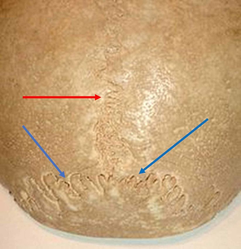
but follow every information and scientific field to correctly define the contours of the event to
                                   be studied. If such a research strategy is lacking, full awareness cannot be reached (knowing or
                                   not being able to know), but a self-imposed scientific limit is reached, which does not
                                   correspond to the final goal of the scientist and scholar. In this way, obscurantism is created.

                                   Review
                                   Metanalytic reviews forget which are the foundations of evidence-based medicine, that is, the
                                   fusion of the operator's clinical experience, the patient's experience and experimental research:
                                   "External clinical evidence can inform, but can never replace, individual clinical expertise, and
                                   it is this expertise that decides whether external evidence applies to individual patients at all,
                                   if, how it should be integrated into a clinical decision" [12]. The synchondrosis between the
                                   occipital bone and the sphenoid bone when ossified does not create movement (flexion-
                                   extension), but the most recent scientific notions show that most cranial sutures or
                                   synarthrosis are patent, even in very old subjects (Figure 1) [4].

2019 Bordoni et al. Cureus 11(5): e4730. DOI 10.7759/cureus.4730                                                                 2 of 7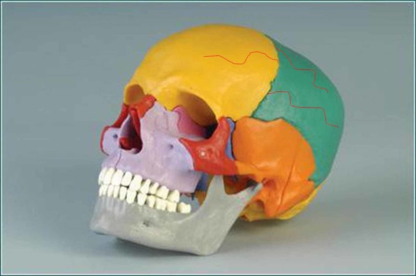
FIGURE 1: The figure illustrates the synchondrosis articulation
                                      between the occipital bone and the sphenoid bone, with a view
                                      of the base of the skull. The red arrow indicates the
                                      synchondrosis articulation; the yellow arrow indicates the
                                      basiocciput; the blue arrow indicates the body of the sphenoid.

                                   The sutures consist of extracellular matrix, proteoglycans, collagen fibres and water; the
                                   synarthrosis with interdigitations, for example, the occipitoparietal, have a modulus of
                                   elasticity and absorption of mechanical stresses are greater (Figure 2) [4].

                                      FIGURE 2: The figure illustrates the occipitoparietal sutures.
                                      The red arrow shows the suture between the parietal bones;
                                      the blue arrows show the occipitoparietal sutures.

                                   The sutures play different mechanical roles, including cushioning the extracranial tensions

2019 Bordoni et al. Cureus 11(5): e4730. DOI 10.7759/cureus.4730                                                                 3 of 7
towards the skull and those intracranial towards the outside, thanks to the presence of cranial
meninges [4]. We know that the cerebral mass moves (caudomedially and craniolaterally),
through the solicitation of the heartbeat and the respiratory diaphragm [13]. The same cerebral
mass, in particular, the neocortex and the limbic area oscillate during inspiration. The dura
mater can change the mechanical tension of the extracranial musculature and the cervical tract,
just as the deep pericranial musculature influences the mechanical dural tension. As regards the
bi-univocal relationship of the dura mater and the pericranial musculature/fascia, the presence
of trigeminal dural nerve endings external to the skull has been demonstrated; the latter cross
the sutures and innervate the myofascial system of the skull (muscles and tendons) (Figure 3)
[14-15].
FIGURE 3: The figure shows a model of the skull with red lines
drawn above the frontal bone and the parietal bone. The figure
demonstrates the presence of trigeminal dural nerve endings
external to the skull, whose terminations cross the sutures
and innervate the myofascial system of the skull.
To give an example of the myofascial presence above the bone tissue of the skull, the
occipitofrontalis muscle has a very large tendinous arch below the galea capitis, which connects
the muscular occipital body with the frontal one [16]. This muscle and other contractile and
connective tissue districts that superficially cover the bones of the skull are involved by the
dural afferents [17]. We cannot exclude the presence of sympathetic and parasympathetic
terminations running parallel to the dural endings, with influence also on the intra/extracranial
vascular pathways [17]. The dural afferents, beyond the extracranial area, reach the arachnoid
space and the pia mater. These afferents/efferences function in an orthodromic and antidromic
manner: there is a close relationship between the cranial myofascial system and the dura mater
[17-19]. The relationship of the subtentorial dural area with the first cervical roots is always bi-
univocal; the mechanical tension of the cervical musculature can be altered by dural
information and the tentorial dural area can undergo a tension modification by the cervical
musculature [20]. This happens due to afferents coming from the cervicovascular area and from
2019 Bordoni et al. Cureus 11(5): e4730. DOI 10.7759/cureus.4730 4 of 7non-trigeminal tentorial neural afferents reaching the cervical muscular portion [20]. The
meningeal system, in which we recognize the dura mater, the arachnoid mater and the pia
mater, is connective tissue; its fibroblasts can alter their tensional state, increasing or
decreasing it [21]. By altering the dural tensional state, the secretion of some substances by
fibroblasts (prostaglandins) is stimulated; from the sympathetic, parasympathetic and
myelinated fibres terminations, multiple neuropeptides (noradrenaline, acetylcholine) are
produced, as well as from the trigeminal meningeal terminations (calcitonin gene-related
peptide - CGRP) [22-23]. These substances, depending on the perceived physiological or non-
physiological stress, will influence the metabolic state of the brain and the brain's immune
status, the repair capacity of the neuronal cells, as well as the nociceptive or analgesic
meningeal response [22-23]. Recall that the meninges are rich in immune cells; the pia mater,
with its outermost layer (glial limitans) and near the brain parenchyma, comes into contact
with neuronal cells and astrocytes [23]. Palpation can be trained up to perceive measurable
objects in microns and considering the close relationship of the cranial myofascial system and
the intracranial meningeal system, we can throw new scientific reflections on the validity of
osteopathic manipulative medicine (OMM). The tension of the hands resting on the
skull/myofascial system is perceived by the extracranial trigeminal afferents, for example by
improving brain oxygenation [24]. It must be remembered that there is a close relationship
between the dural tissue and the blood vessels; an intra/extracranial mechanical stimulus easily
alters dural vascular behaviour (permeability, vasodilation, vasoconstriction) [23]. The dura
mater is vascularized and with the presence of lymphatic vessels; blood vessels pass molecules
of low molecular weight or kilodalton (up to a maximum of 45kDa), which reach the arachnoid
mater. The passage of larger molecules will depend on trigeminal intervention (vasodilation).
The arachnoid mater, through the tight junctions, transports the blood molecules into the
vessels of the subarachnoid space (in space we find different immune substances) [23]. The
vessels of the subarachnoid space will enter the brain with the protection of the pia mater. The
blood vessels and their perivascular connective tissue are considered a continuation of the pia
mater [23]. We can hypothesize that the stimulation of extracranial trigeminal afferents could
improve blood transport, from dura to pia, improving arterial vasodilation. By improving
extracranial tension, it improves the intracranial trigeminal response [25]. It must be
considered that at the cellular level of the different cranial tissues there are no layers, but an
absolute anatomical and functional continuity [26-27]. Quantum physics helps us further.
Palpation is interactive communication between the operator and the patient, and all the
palpated and not palpated tissues are aware of the mechanical information that comes from the
hands placed on the skull (quantum entanglement) [27]. Magnetobiology relates
electromagnetic fields and living cells [27]. The operator's hand emits electromagnetic fields
(such as on the patient's skull), and these magnetic forces or vibrations deform the morphology
of the cell, becoming a mechanical stimulus felt by extracranial terminations trigeminal. The
electromagnetic fields travel at higher speeds than the electric flow, crossing the whole body;
the touch of the osteopath goes beyond the skull [27]. The fact that the cranial sutures are still
patent in old age, could mean that the millimetre movement of the brain during
systole/diastole and the contractions of the diaphragm muscle, is amortized by cranial
synarthrosis. Probably, the oscillations of some brain areas, such as the hippocampus and the
limbic area during breathing, could influence the same movement of the brain mass. Does OMM
affect the liquids in the skull? All cells oscillate and aggregate to form tissues; liquids are an
important fascial component and form the liquid fascia [28]. Thus we have the extracellular
matrix, the interstitial fluids, blood, lymph, liquor and the same cells that are full of water [27].
The oscillations of the cells create further alterations of mechanical tension that travel faster in
the liquid tissues, creating a wet network [27]. It is very probable, relying on the notions of
physics that the OMM is able to enter into communication with the liquor or the cranial lymph.
Can we feel the movement of the cranial bones? The overall movement of the cranial bones,
allowed by the patency of the various sutures, is measured in microns, with an amplitude that is
around 10-50 μm [29]. The palpatory sensitivity of the operator trained to listen to the smallest
movements and variations of tension coincides with the measure of cranial movement [29]. We
2019 Bordoni et al. Cureus 11(5): e4730. DOI 10.7759/cureus.4730 5 of 7still need a lot of information on what happens between the osteopath, his hands and the
patient's skull, but it is this need to know which creates the research, the right orientation
towards knowledge. The question must always be neutral in order to obtain an efficient
scientific response and move forward: this is the enlightenment of research.
Conclusions
The cranial osteopathic manipulative medicine is not always positively shared by the scientific
world, but it cannot even be rejected by scholars and scientists since non-knowledge does not
preclude the end of knowledge. The article discussed an orientation of the current literature
that goes against the scientific nature of the osteopathic manipulative medicine (OMM),
showing, however, that there are further reflections that are able to be made in support of
osteopathic cranial therapy.References
Additional Information
Disclosures
Conflicts of interest: In compliance with the ICMJE uniform disclosure form, all authors
declare the following: Payment/services info: All authors have declared that no financial
support was received from any organization for the submitted work. Financial relationships:
All authors have declared that they have no financial relationships at present or within the
previous three years with any organizations that might have an interest in the submitted work.
Other relationships: All authors have declared that there are no other relationships or
activities that could appear to have influenced the submitted work.
References
1. King HH: Cranial osteopathic manipulative medicine's growing evidence base . J Am Osteopath
Assoc. 2012, 112:9.
2. Guillaud A, Darbois N, Monvoisin R, Pinsault N: Reliability of diagnosis and clinical efficacy of
cranial osteopathy: a systematic review. PLoS One. 2016, 11:0167823. Accessed: May 23,
2019: 10.1371/journal.pone.0167823
3. Jäkel A, von Hauenschild P: Therapeutic effects of cranial osteopathic manipulative medicine:
a systematic review. J Am Osteopath Assoc. 2011, 111:685-93.
4. Bordoni B, Zanier E: Sutherland's legacy in the new millennium: the osteopathic cranial
model and modern osteopathy. Adv Mind Body Med. 2015, 29:15-21.
5. Xu Z, Su C, Xiao Y: A massive calcification and ossification of the transverse sinus and the
neighbouring dura mimicking meningioma. BMC Neurol. 2013, 13:143. Accessed: May 23,
2019: 10.1186/1471-2377-13-143
6. Li B, Guo S, Qiu G, Li W, Liu Y, Zhao Y: A potential mechanism of dural ossification in
ossification of ligamentum flavum. Med Hypotheses. 2016, 92:1-2.
10.1016/j.mehy.2016.03.011
7. Guida L, Mazzoleni F, Bozzetti A, Sganzerla E, Giussani C: Extensive dural ossification after
decompressive posttraumatic craniectomy: a case report and review of the literature. World
Neurosurg. 2018, 120:59-62. 10.1016/j.wneu.2018.08.115
8. Hitscherich K, Smith K, Cuoco JA, Ruvolo KE, Mancini JD, Leheste JR, Torres G: The
glymphatic-lymphatic continuum: opportunities for osteopathic manipulative medicine. J Am
Osteopath Assoc. 2016, 116:170-7. 10.7556/jaoa.2016.033
9. Warren C, Keys J, Pierce-Talsma S: Osteopathic cranial manipulative medicine in the setting
of concussion. J Am Osteopath Assoc. 2018, 118:41-42. 10.7556/jaoa.2018.088
10. Żurowska A, Malak R, Kołcz-Trzęsicka A, Samborski W, Paprocka-Borowicz M: Compression
of the fourth ventricle using a craniosacral osteopathic technique: a systematic review of the
clinical evidence. Evid Based Complement Alternat Med. 2017, 2017:2974962.
10.1155/2017/2974962
11. Akai T, Hatta T, Shimada H, Mizuki K, Kudo N, Hatta T, Otani H: Extracranial outflow of
particles solved in cerebrospinal fluid: fluorescein injection study. Congenit Anom (Kyoto).
2019 Bordoni et al. Cureus 11(5): e4730. DOI 10.7759/cureus.4730 6 of 72018, 58:93-98. 10.1111/cga.12257
12. Masic I, Miokovic M, Muhamedagic B: Evidence based medicine - new approaches and
challenges. Acta Inform Med. 2008, 16:219-225. 10.5455/aim.2008.16.219-225
13. Bordoni B, Purgol S, Bizzarri A, Modica M, Morabito B: The influence of breathing on the
central nervous system. Cureus. 2018, 10:2724. Accessed: May 23, 2019: 10.7759/cureus.2724
14. Kosaras B, Jakubowski M, Kainz V, Burstein R: Sensory innervation of the calvarial bones of
the mouse. J Comp Neurol. 2009, 515:331-48. 10.1002/cne.22049
15. Lee SH, Hwang SJ, Koh KS, Song WC, Han SD: Macroscopic innervation of the dura mater
covering the middle cranial fossa in humans correlated to neurovascular headache. Front
Neuroanat. 2017, 11:127. Accessed: May 23, 2018: 10.3389/fnana.2017.00127
16. Bordoni B, Zanier E: Clinical and symptomatological reflections: the fascial system . J
Multidiscip Healthc. 2014, 7:401-11. Accessed: May 23, 2019: 10.2147/JMDH.S68308
17. Schueler M, Neuhuber WL, De Col R, Messlinger K: Innervation of rat and human dura mater
and pericranial tissues in the parieto-temporal region by meningeal afferents. Headache.
2014, 54:996-1009. 10.1111/head.12371
18. Schueler M, Messlinger K, Dux M, Neuhuber WL, De Col R: Extracranial projections of
meningeal afferents and their impact on meningeal nociception and headache. Pain. 2013,
154:1622-31. 10.1016/j.pain.2013.04.040
19. Burstein R, Blake P, Schain A, Perry C: Extracranial origin of headache . Curr Opin Neurol.
2017, 30:263-271. 10.1097/WCO.0000000000000437
20. Noseda R, Melo-Carrillo A, Nir RR, Strassman AM, Burstein R: Non-trigeminal nociceptive
innervation of the posterior dura: implications to occipital headache. J Neurosci. 2019,
39:1867-1880. 10.1523/JNEUROSCI.2153-18.2018
21. Maikos JT, Elias RA, Shreiber DI: Mechanical properties of dura mater from the rat brain and
spinal cord. J Neurotrauma. 2008, 25:38-51. 10.1089/neu.2007.0348
22. Zhao J, Levy D: The CGRP receptor antagonist BIBN4096 inhibits prolonged meningeal
afferent activation evoked by brief local K+ stimulation but not cortical spreading depression-
induced afferent sensitization. Pain Rep. 2017, 3:632. Accessed: May 23, 2019:
10.1097/PR9.0000000000000632
23. Rua R, McGavern DB: Advances in meningeal immunity . Trends Mol Med. 2018, 24:542-559.
10.1016/j.molmed.2018.04.003
24. Shi X, Rehrer S, Prajapati P, Stoll ST, Gamber RG, Downey HF: Effect of cranial osteopathic
manipulative medicine on cerebral tissue oxygenation. J Am Osteopath Assoc. 2011, 111:660-
6.
25. Nöbel M, Feistel S, Ellrich J, Messlinger K: ATP-sensitive muscle afferents activate spinal
trigeminal neurons with meningeal afferent input in rat - pathophysiological implications for
tension-type headache. J Headache Pain. 2016, 17:75. 10.1186/s10194-016-0668-z
26. Bordoni B, Marelli F, Morabito B, Sacconi B: The indeterminable resilience of the fascial
system. J Integr Med. 2017, 15:337-343. 10.1016/S2095-4964(17)60351-0
27. Bordoni B, Simonelli M: The awareness of the fascial system . Cureus. 2018, 10:3397.
Accessed: May 23, 2019: 10.7759/cureus.3397
28. Bordoni B, Marelli F, Morabito B, Castagna R: A new concept of biotensegrity incorporating
liquid tissues: blood and lymph. J Evid Based Integr Med. 2018, 23:2515690-18792838.
29. Kasparian H, Signoret G, Kasparian J: Quantification of motion palpation . J Am Osteopath
Assoc. 2015, 115:604-10. 10.7556/jaoa.2015.121
2019 Bordoni et al. Cureus 11(5): e4730. DOI 10.7759/cureus.4730 7 of 7You can also read