Abstract Isolated Sixth Nerve Palsy: A Case of Pseudotumor Cerebri and an Overview of the Evolutionary Dynamic Geometry of Dorello's Canal
←
→
Page content transcription
If your browser does not render page correctly, please read the page content below
Open Access Case
                             Report                                                DOI: 10.7759/cureus.15340

                                             Isolated Sixth Nerve Palsy: A Case of
                                             Pseudotumor Cerebri and an Overview of the
                                             Evolutionary Dynamic Geometry of Dorello's
                                             Canal
                                             Hassan Kesserwani 1

                                             1. Neurology, Flowers Medical Group, Dothan, USA

                                             Corresponding author: Hassan Kesserwani, neuro1815@yahoo.com

                                             Abstract
                                             The dynamics of increased intracranial pressure (ICP) and sixth cranial nerve palsy has undergone a
                                             paradigm shift, with emphasis shifting from a length hypothesis to a theory based on novel anatomic
                                             findings pertaining to the geometry of Dorello's canal. In particular, the sixth cranial nerve resides in a
                                             transfixed coaxial cylinder within the canal. The cisternal portion of the nerve is intradural and the rest of
                                             the nerve is extradural; therefore, with increased ICP, the former is stretched, thereby pulling on the rest of
                                             the nerve, which is anchored in Dorello's canal. We present a case of pseudotumor cerebri secondary to
                                             minocycline presenting with an isolated sixth nerve palsy. This case is used as a platform to segue into the
                                             recent findings outlined above, in particular, the evolutionary transformation of Dorello's canal from a
                                             circular outline with a bony roof to an elliptic profile with a fibro-osseus roof during hominid basocranial
                                             expansion. The fibro-osseus roof, being elastic, is particularly susceptible to the influence of raised ICP,
                                             thereby narrowing the canal and injuring the sixth cranial nerve.

                                             Categories: Neurology, Ophthalmology, Anatomy
                                             Keywords: severe headache, sixth cranial nerve palsy, benign intracranial hypertension, headache in children,
                                             ophthalmology

                                             Introduction
                                             The sixth cranial nerve is quite unique: a pure motor nerve, the second longest cranial nerve after the
                                             trochlear nerve, entirely dedicated to supplying the lateral rectus muscle, thereby allowing abduction of the
                                             eye. Its course at the base of the brain is remarkable - after leaving the ponto-medullary junction, it enters
                                             the subarachnoid space (the cisternal segment), and after a variable distance, it penetrates the dura
                                             (intradural segment), leaves the intramural space, and travels through the osteofibrous Dorello's canal under
                                             the petrosphenoidal ligament (PSL), also known as Gruber's ligament, stretching between the petrous
                                             apex (spine) and the accessory clinoid process (ACP) [1]. While here it is anchored by a meningeal tube - a
                                             rigid immobile cylinder within Dorello's canal - as a co-axial cylinder [2]. It then climbs up the wall of the
Review began 05/24/2021
                                             clivus and penetrates the cavernous sinus, riding along with the inferior pertrosal sinus. In the cavernous
Review ended 05/30/2021
Published 05/30/2021                         sinus, it lies immediately lateral to the pericarotid sympathetics. After leaving the cavernous sinus, it makes
                                             a sharp, almost ninety-degree turn upwards and forwards and then enters the superior orbital fissure at the
© Copyright 2021
                                             orbital apex surrounded by the annulus of Zinn. The latter is a fibrous ring formed by the tendons of the four
Kesserwani. This is an open access article
distributed under the terms of the           recti muscle. Through it also enter cranial nerves III, IV, the ophthalmic division of V, ophthalmic vein, and
Creative Commons Attribution License         sympathetic fibers. The optic nerve, ophthalmic artery, and central retinal vein pass through the optic canal
CC-BY 4.0., which permits unrestricted       [3].
use, distribution, and reproduction in any
medium, provided the original author and
source are credited.                         During this treacherous journey, the sixth cranial nerve is highly vulnerable to injury, especially with high
                                             intracranial pressure. This was thought to be due to its long intracranial course, however, a study of 26
                                             cadavers showed that the sixth cranial nerve is one-third the length of the trochlear nerve, and the former is
                                             more likely to be injured by raised ICP [4]. The sixth cranial nerve is tethered at two points: at its exit from
                                             the pontomedullary junction and at Dorello's canal. As the pontomedullary junction descends during
                                             transtentorial herniation due to raised ICP, the sixth nerve is stretched like a bow-string and pulled at
                                             Dorello's canal, leading to ischemic necrosis. In the pontine cistern, the sixth cranial nerve is traversed and
                                             indented by branches of the basilar artery, especially the anterior inferior cerebellar artery (AICA), and
                                             posterior fossa tumors may distort the anatomy and lead to a sixth nerve palsy by a pressure-mass effect. The
                                             parallel course of the sixth cranial nerve along the clivus may lead to a palsy from the mass effect of a
                                             chordoma [5]. With the cavernous sinus syndrome from a malignancy such as a meningioma, carotid siphon
                                             aneurysm or fistula, fungal infection, vasculitis or the Tolosa-Hunt syndrome, a sixth nerve palsy may arise
                                             from direct pressure or inflammation. The close proximity of the sixth cranial nerve to the peri-carotid
                                             sympathetics may lead to a Horner's syndrome, the combination of a sixth nerve palsy and a Horner
                                             syndrome being virtually pathognomic of a cavernous sinus syndrome [6,7].

                                             The differential diagnosis of an isolated sixth nerve palsy includes ocular myasthenia gravis, thyroid

                              How to cite this article
                              Kesserwani H (May 30, 2021) Isolated Sixth Nerve Palsy: A Case of Pseudotumor Cerebri and an Overview of the Evolutionary Dynamic Geometry
                              of Dorello's Canal. Cureus 13(5): e15340. DOI 10.7759/cureus.15340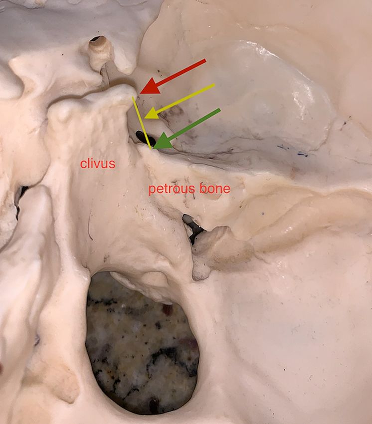
ophthalmopathy, and microvascular disease from diabetes and hypertension. For the sake of completion, we
                                                 will mention the brainstem syndromes of Raymond, Millard-Gubler, and Foville syndrome as they are not
                                                 etiologies of isolated sixth nerve palsy and also involve other lower cranial nerves, gaze centers, and long
                                                 tracts. Gradenigo's syndrome with petrous apex spread from otitis media via the inferior petrosal sinus
                                                 involves cranial nerves V, VI, VII, and VIII, and is of historical interest [8].

                                                 Lastly, as in our case report, a sixth nerve palsy can be an isolated finding in patients with pseudotumor
                                                 cerebri/idiopathic intracranial hypertension and can be seen in up to 30% of cases. The injury is likely due to
                                                 stretching of the sixth cranial nerve but is almost always reversible and therefore a neuropraxia [9].

                                                 Case Presentation
                                                 A 17-year-old young woman presented with an insidious onset of unremitting severe bilateral occipital
                                                 headaches associated with a roaring sound in her left ear. The severity of the headache required her to seek
                                                 care in the emergency room twice. One week into her illness, a fixed horizontal diplopia with maximal
                                                 separation of images with left direction of gaze had developed, necessitating the use of an eye patch. She did
                                                 not report any transient visual obscurations or other vision loss. Despite being overweight, no increase in
                                                 weight was recorded over the previous year. Her past medical history was significant for acne, for which was
                                                 treated with monocyline 100 milligrams (mg) daily for the last three months. Otherwise she was a healthy
                                                 young woman.

                                                 On examination, her blood pressure is 112/68 mmHG, with a pulse of 98 beats per minute. Her height is 5
                                                 feet and 5 inches with a weight of 241 pounds and a body mass index of 40.1 kg/m2. Gait cadence, tandem-
                                                 walking, and heel-and-toe walking were normal. Ocular motion reveals a frank left sixth nerve palsy (Figure
                                                 1).

                                                   FIGURE 1: Failure of abduction of the left eye consistent with a left sixth
                                                   nerve palsy (red arrow).

                                                 No Horner's syndrome or Marcus-Gunn pupil was noted. Consensual and accommodative reflexes were
                                                 normal. Funduscopic examination with pupil dilation was performed by an ophthalmologist and revealed
                                                 bilateral papilledema (Figure 2).

                                                   FIGURE 2: Bilateral optic disc edema with blurring of optic disc margins
                                                   and reduction of blood vessels (yellow arrows). 2A: left eye, 2B: right
                                                   eye.

2021 Kesserwani et al. Cureus 13(5): e15340. DOI 10.7759/cureus.15340                                                                                              2 of 7
Facial sensation and power were symmetric and preserved. The rest of the cranial nerve examination was
normal. Power testing with the medical research council (MRC) scale was 5/5 throughout both upper and
lower limbs. Deep tendon reflexes were lively in upper and lower limbs. Touch-pressure, vibratory, and joint
position sense were normal in the fingers and toes.
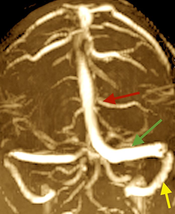
A magnetic resonance imaging (MRI) of the brain was normal with and without contrast. A magnetic
resonance venogram (MRV) showed patent venous sinuses (Figure 3).
FIGURE 3: Magnetic resonance venography (coronal section) showing
patent major venous sinuses bilaterally. Left superior sagittal sinus (red
arrow), transverse sinus (green arrow) and sigmoid sinus (yellow
arrow).
MRV: magnetic resonance venography
An MRI of the brain and orbits with and without contrast showed bilateral optic nerve sheath distention
with flattening of the posterior bulge of the sclera (lamina cribrosa) consistent with optic disc edema (Figure
4).
2021 Kesserwani et al. Cureus 13(5): e15340. DOI 10.7759/cureus.15340 3 of 7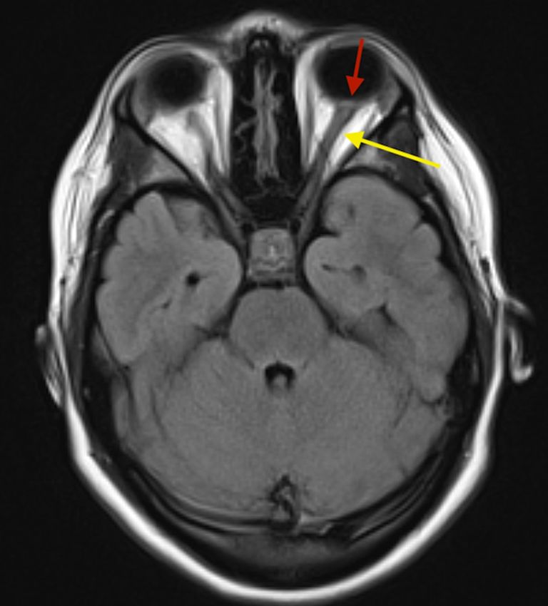
FIGURE 4: FLAIR MRI of the brain (axial section) showing thickened
                                                   optic nerve sheath (yellow arrow) and flattened lamina cribrosa of
                                                   sclera (red arrow).
                                                   FLAIR MRI: Fluid-attenuated inversion recovery magnetic resonance imaging

                                                 A supine lumbar puncture in the lateral decubitus position revealed a high cerebrospinal fluid (CSF)
                                                 pressure, greater than 55 centimeter (cm) of water pressure (normal: less than 25 cm of water). CSF revealed
                                                 normal protein and no pleocytosis. The patient was diagnosed with pseudotumor cerebri secondary to
                                                 minocyline therapy, a well-established and tight association. Minocycline was discontinued and the patient
                                                 was started on a slowly escalating dose of 250 mg of acetazolamide daily with a target dose of 750 mg daily,
                                                 as tolerated. By the end of the first week, the headache had completely resolved, and by the end of the
                                                 second week, the diplopia had resolved. At the two-month ophthalmologic visit, the optic disc edema had
                                                 also resolved and the patient was weaned off acetazolamide.

                                                 Discussion
                                                 Dorello's canal, also known as the petroclival segment, marks the transition between the posterior and
                                                 middle cranial fossa. This canal has undergone evolutionary change being fused superiorly by a bony arch in
                                                 non-hominid primates, the union of the petrous apex (spine) and the ACP. With hominid brain expansion
                                                 and growth of the basicranium, the non-union of the petrous apex and ACP was replaced by an elastic fibro-
                                                 osseus ligament, the PSL, covering the roof of the foramen. The canal is elliptic in shape in humans and
                                                 rounded in non-human primates, the expansion of the cranial base converting it from a circle to an ellipse
                                                 (Figure 5) [10].

2021 Kesserwani et al. Cureus 13(5): e15340. DOI 10.7759/cureus.15340                                                                                           4 of 7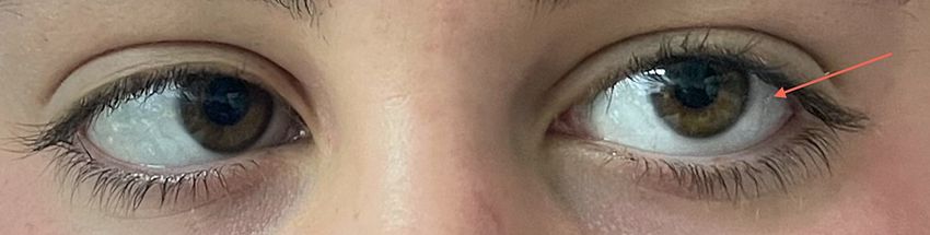
FIGURE 5: Expansion of the basicranium and conversion of a circular
                                                   Dorello's canal to an elliptic canal in humans with replacement of a
                                                   bony roof (violet arrow) to a fiber-ligamentous roof (red line), PSL, as
                                                   we transition from a non-hominid primate to a hominid primate.
                                                   PSL: petrosphenoid ligament

                                                 The elastic nature of the PSL allows high ICP to squeeze and compress the PSL and constrict the sixth cranial
                                                 nerve in Dorello's canal leading to a sixth nerve palsy. Furthermore, the PSL is butterfly-shaped, with a
                                                 narrowing or stricture in the middle in 78% of cases. Meanwhile, the sixth cranial nerve runs in the middle of
                                                 the canal, where it is susceptible to compression by the vertical bending forces on the middle portion of the
                                                 elastic PSL into Dorello's canal [11]. In non-hominid primates, this is not possible as the roof of Dorello's
                                                 canal is bony.

                                                 The cisternal segment of the sixth cranial nerve is intradural and therefore subject to raised ICP. Meanwhile
                                                 the rest of the sixth cranial nerve, including the petroclival and cavernous segments, are extradural and not
                                                 subject to raised ICP. Therefore, with a diffuse and isotropic increase in ICP, the nerve is subject to a
                                                 differential pressure with the proximal cisternal segment of the nerve pulling or tugging on the rest of the
                                                 nerve; this being a tentative explanation of a neuropraxia of the nerve. Contrast this to transtentorial
                                                 herniation where there is global stretch of the nerve with descent of the pontomedullary junction and
                                                 massive increases in ICP (plateau waves) and subsequent necrosis of the nerve due to stretching of the nerve
                                                 beyond its yield point (Hooke's law) [12,13].

                                                 With Dorello's canal being defined as the space or trough between the clivus and petrous bone bounded
                                                 superiorly by the the PSL, anatomic variations do occur and this space is often referred to as the petroclival
                                                 region (Figure 6).

2021 Kesserwani et al. Cureus 13(5): e15340. DOI 10.7759/cureus.15340                                                                                             5 of 7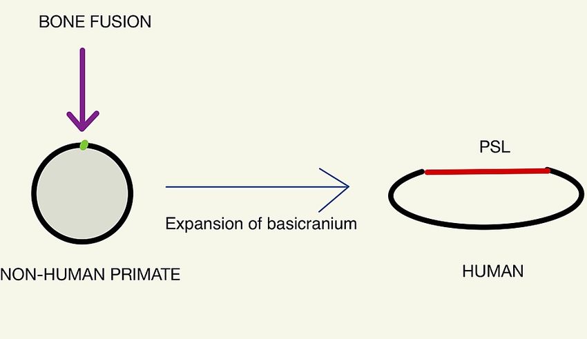
FIGURE 6: Oblique view of right Dorello's canal - posterior clinoid
                                                   process (red arrow), petrous spine (green arrow), petro-clinoid ligament
                                                   (yellow arrow).

                                                 There are instances wherein the sixth cranial nerve courses over Dorello's canal and some authors have
                                                 suggested an alternative appellation; the petroclival venous confluence (PVC). This region is akin to
                                                 chamber within the confines of the meningeal and periosteal layers of the dura with the confluence of
                                                 multiple venous sinuses draining into the inferior petrosal sinus [14]. The PVC is divided into two
                                                 compartments: an upper compartment carries the sixth cranial nerve and the lower compartment, the
                                                 inferior petrosal sinus. This is similar to the situation seen with the jugular foramen, which is divided into
                                                 two compartments: the pars venosa (internal jugular vein and cranial nerves X, XI, and Arnold's nerve) and
                                                 pars nervosa (cranial nerve IX, and Jacobsen's nerve) [15].

                                                 Conclusions
                                                 Evolutionary developmental biology or "evo-devo" is an exciting field that helps explain functional anatomy
                                                 pertaining to evolutionary descent. The expansion of the basicranium parallels the growth of the brain
                                                 during hominid evolution. The morphological changes are many, one of which pertains to a classic and
                                                 frequent presentation in neurological practice: a sixth nerve palsy. Localization of a sixth nerve palsy has a
                                                 well-established differential diagnosis and is an exercise of neurological finesse and beauty. However,
                                                 classical neurologists have also noted the "false localizing sign" of a sixth nerve palsy in cases of diffuse and
                                                 isotropic increase in ICP. Here there is no focal disease but a global process, which as a quirk of
                                                 neuroantomy has predisposed this nerve to compression at Dorello's canal due to the transformation of the
                                                 roof from an osseus structure to a fibro-elastic and compressible tent as a result of brain expansion during
                                                 hominid evolution. This is the classic "false localizing sign" of a sixth cranial nerve palsy, which now has a
                                                 plausible and beautiful explanation.

2021 Kesserwani et al. Cureus 13(5): e15340. DOI 10.7759/cureus.15340                                                                                                6 of 7
Additional Information
Disclosures
Human subjects: Consent was obtained or waived by all participants in this study. Conflicts of interest: In
compliance with the ICMJE uniform disclosure form, all authors declare the following: Payment/services
info: All authors have declared that no financial support was received from any organization for the
submitted work. Financial relationships: All authors have declared that they have no financial
relationships at present or within the previous three years with any organizations that might have an
interest in the submitted work. Other relationships: All authors have declared that there are no other
relationships or activities that could appear to have influenced the submitted work.
References
1. Joo W, Yoshioka F, Funaki T, Rhoton AL Jr: Microsurgical anatomy of the abducens nerve . Clin Anat. 2012,
25:1030-42. 10.1002/ca.22047
2. Tubbs RS, Radcliff V, Shoja MM, et al.: Dorello canal revisited: an observation that potentially explains the
frequency of abducens nerve injury after head injury. World Neurosurg. 2012, 77:119-21.
10.1016/j.wneu.2011.03.046
3. Lieber S, Fernandez-Miranda JC: Anatomy of the orbit . J Neurol Surg B Skull Base. 2020, 81:319-32.
10.1055/s-0040-1715096
4. Hanson RA, Ghosh S, Gonzalez-Gomez I, Levy ML, Gilles FH: Abducens length and vulnerability? .
Neurology. 2004, 62:33-6. 10.1212/WNL.62.1.33
5. Marinkovic SV, Gibo H, Stimec B: The neurovascular relationships and the blood supply of the abducent
nerve: surgical anatomy of its cisternal segment. Neurosurgery. 1994, 4:1017-26. 10.1097/00006123-
199406000-00010
6. Keane JR: Cavernous sinus syndrome. Analysis of 151 cases . Arch Neurol. 1996, 53:967-71.
10.1001/archneur.1996.00550100033012
7. Bhatkar S, Goyal MK, Takkar A, Mukherjee KK, Singh P, Singh R, Lal V: Cavernous sinus syndrome: a
prospective study of 73 cases at a tertiary care centre in Northern India. Clin Neurol Neurosurg. 2017,
155:63-9. 10.1016/j.clineuro.2017.02.017
8. Azarmina M, Azarmina H: The six syndromes of the sixth cranial nerve . J Ophthalmic Vis Res. 2013, 8:160-
71.
9. Kesserwani H: Space flight-associated neuroocular syndrome, idiopathic intracranial hypertension, and
pseudotumor cerebri: phenotypic descriptions, pathogenesis, and hydrodynamics. Cureus. 2021, 13:e14103.
10.7759/cureus.14103
10. Marom A: A new look at an old canal . Skull Base. 2011, 21:53-8. 10.1055/s-0030-1263282
11. Icke C, Ozer E, Arda N: Microanatomical characteristics of the petrosphenoidal ligament of Gruber . Turk
Neurosurg. 2010, 20:323-7. 10.5137/1019-5149.JTN.2921-10.0
12. Tillett RL, Afoke A, Hall SM, Brown RA, Phillips JB: Investigating mechanical behaviour at a core-sheath
interface in peripheral nerve. J Peripher Nerv Syst. 2004, 9:255-62. 10.1111/j.1085-9489.2004.09411.x
13. Kwan MK, Wall EJ, Massie J, Garfin SR: Strain, stress and stretch of peripheral nerve. Rabbit experiments in
vitro and in vivo. Acta Orthop Scand. 1992, 63:267-72. 10.3109/17453679209154780
14. Kshettry VR, Lee JH, Ammirati M: The Dorello canal: historical development, controversies in microsurgical
anatomy, and clinical implications. Neurosurg Focus. 2013, 34:E4. 10.3171/2012.11.FOCUS12344
15. Duque PJE, Barco RJ, Mendoza ZJ: Dorello’s canal or abducens nerve canal: Constancy or inconstancy? . Int.
J. Morphol. 2017, 35:233-5.
2021 Kesserwani et al. Cureus 13(5): e15340. DOI 10.7759/cureus.15340 7 of 7You can also read