Cerebellar ataxia with coenzyme Q10 deficiency: Diagnosis and follow-up after coenzyme Q10 supplementation
←
→
Page content transcription
If your browser does not render page correctly, please read the page content below
Journal of the Neurological Sciences 246 (2006) 153 – 158
                                                                                                                                 www.elsevier.com/locate/jns

                                                                 Short communication

         Cerebellar ataxia with coenzyme Q10 deficiency: Diagnosis and
                 follow-up after coenzyme Q10 supplementation
    Rafael Artuch a,*, Gloria Brea-Calvo b, Paz Briones c, Asunción Aracil d, Marta Galván d,
     Carmen Espinós e, Jordi Corral f, Victor Volpini f, Antonia Ribes c, Antoni L. Andreu g,
          Francesc Palau e, José A. Sánchez-Alcázar b, Plácido Navas b, Mercè Pineda d
                                           a
                                               Biochemistry Department, Hospital Sant Joan de Déu, Barcelona, Spain
                                   b
                                    Centro Andaluz de Biologı́a del Desarrollo. Universidad Pablo de Olavide, Sevilla, Spain
                                    c
                                       Institut de Bioquı́mica Clı́nica-Corporació Sanitaria Clı́nic y CSIC, Barcelona, Spain
                                              d
                                                Neurology Department, Hospital Sant Joan de Déu, Barcelona, Spain
                                                         e
                                                           Instituto de Biomedicina, CSIC, Valencia, Spain
                               f
                                   Centre de Diagnòstic Genètic-Molecular-IDIBELL Htal. Durán i Reynals, Barcelona, Spain
                                     g
                                        Unidad de Enfermedades Mitocondriales, Hospital Vall d’Hebrón, Barcelona, Spain

                               Received 14 October 2005; received in revised form 6 January 2006; accepted 9 January 2006
                                                              Available online 3 May 2006

Abstract

   Our aim was to report a new case with cerebellar ataxia associated with coenzyme Q10 (CoQ) deficiency, the biochemical findings caused
by this deficiency and the response to CoQ supplementation.
Patient: A 12-year-old girl presenting ataxia and cerebellar atrophy.
Biochemical studies: Coenzyme Q10 in muscle was analysed by HPLC with electrochemical detection and mitochondrial respiratory chain
(MRC) enzyme activities by spectrophotometric methods. CoQ biosynthesis in fibroblasts was assayed by studying the incorporation of
radiolabeled 4-hydroxy[U-14C] benzoic acid by HPLC with radiometric detection.
Results: Mitochondrial respiratory chain enzyme analysis showed a decrease in complex I + III and complex II + III activities. CoQ
concentration in muscle was decreased (56 nmol/g of protein: reference values: 157 – 488 nmol/g protein). A reduced incorporation of
radiolabeled 4-hydroxy[U-14C] benzoic acid was observed in the patient (19% of incorporation respect to the median control values). After
16 months of CoQ supplementation, the patient is now able to walk unaided and cerebellar signs have disappeared.
Conclusions: Cerebellar ataxia associated with CoQ deficiency in our case might be allocated in the transprenylation pathway or in the
metabolic steps after condensation of 4-hydroxybenzoate and the prenyl side chain of CoQ. Clinical improvement after CoQ supplementation
was remarkable, supporting the importance of an early diagnosis of this kind of disorders.
D 2006 Elsevier B.V. All rights reserved.

Keywords: Coenzyme Q10; Mitochondrial diseases; Cerebellar ataxia; Treatment; Mitochondrial respiratory chain

                                                                                    1. Introduction

                                                                                       Coenzyme Q10 (CoQ) is a lipid-soluble component of
                                                                                    cell membranes, which transports electrons from complexes
                                                                                    I and II to complex III of the mitochondrial respiratory chain
    DOI of original article: 10.1016/j.jns.2006.03.017.                             (MRC). It also has a key role as a free radical scavenger,
 * Corresponding author. Biochemistry Department Hospital Sant Joan de
Déu, Passeig Sant Joan de Déu, 2, 08950 Esplugues, Barcelona, Spain. Tel.:
                                                                                    regenerating other antioxidants in other cellular membranes
+34 93 2806169; fax: +34 93 2803626.                                                such as plasma membrane [1]. Coenzyme Q10 is composed
    E-mail address: rartuch@hsjdbcn.org (R. Artuch).                                of a benzoquinone ring, synthesized from tyrosine through
0022-510X/$ - see front matter D 2006 Elsevier B.V. All rights reserved.
doi:10.1016/j.jns.2006.01.021
154 R. Artuch et al. / Journal of the Neurological Sciences 246 (2006) 153 – 158
4-hydroxybenzoate, and a polyprenyl side-chain, generated electromyography were normal. Brain MRI showed severe
from acetyl-CoA through the mevalonate pathway, which is cerebellar atrophy (Fig. 1A). The patient’s father also
common for the synthesis of other compounds, such as showed such cerebellar atrophy (Fig. 1B). Clinical evolution
cholesterol and dolichol-phosphate [2]. remained stable until 12 years of age.
Since the first description of a mitochondrial encephalo- Routine laboratory tests including complete blood count,
myopathy associated with CoQ deficiency [3], several blood chemistry and thyroid function were normal. Labo-
patients have been reported. This is an autosomal recessive ratory tests for screening of inborn errors of metabolism in
disorder with a clinical spectrum that encompasses several blood and urine (including analysis of sialotransferrin and
main phenotypes [4]: a myopathic form, with myoglobi- urine organic acids) were all normal. No hyperlactacidemia
nuria, epilepsy and ataxia [3,5]; a severe infantile syndrome or hyperalaninemia were observed during the evolution of
with encephalopathy and renal disease [6]; and an ataxic the patient. Plasma CoQ concentration (0.57 Amol/L) was
form presenting with cerebellar atrophy [7]. However, other also within the reference ranges (0.41 – 1.15 Amol/L).
phenotypes have been reported, such as a neonatal Inherited causes of ataxia were also ruled out (ataxia –
presentation with fatal evolution [8], Leigh syndrome in telangiectasia, Friedreich ataxia, ataxia with vitamin E
adulthood [9], and liver failure plus Leigh syndrome [10]. A deficiency and abetalipoproteinemia). Muscle and skin
common feature in these cases is the presence of a variable biopsies were taken at 12 years of age, and studies for
degree of CoQ deficiency in muscle and/or fibroblasts diagnosis of a mitochondrial disorder were started. Muscle
causing decreased activities of NADH:cit c oxidoreductase and skin biopsies from the father were also studied.
and succinate:cit c oxidoreductase of the MRC. Interesting-
ly, treatment response has been remarkable in most cases,
highlighting the importance of an early diagnosis of these
disorders.
Here we present the diagnosis of a new case of cerebellar
ataxia associated with CoQ deficiency. We also report the
biochemical findings caused by this deficiency and the
clinical response after CoQ supplementation.
2. Materials and methods
2.1. Case report
A 12-year-old girl, born after a term pregnancy and
normal delivery of non-consanguineous parents. Her elder
brother and her mother both have long Q-T syndrome. Her
father (45 years old) described a history of clumsiness,
frequent falls, tremor and nistagmus from the early years of
life. After a normal newborn period and early motor
development, at 15 months of age it was noticed that the
patient was clumsy fell frequently when walking unaided.
At 5 years of age, general physical examination was normal.
Neurological exam showed slight dysarthria with slurring
speech, rapid alternating movements and finger – nose
testing with oscillating movement, without decomposition.
Finger –finger test evidenced mild instability for the action
with no action tremor. On knee –tibia test, movement was
decomposed in several phases and was abnormally slow
with no tremor. Pronation – supination alternating move-
ments were slow and irregularly performed. Ocular pursuit
was saccadic. Balance evidenced slight oscillations with feet
together, but she was unable to walk with feet in tandem
position. Gait speed was slightly reduced with broad base.
Limb tone and tendon reflexes were decreased. No muscle
weakness was observed. Plantar response was flexor.
Normal oculi fundi and cognitive level were present. Fig. 1. Cranial MRI from the index case at 12 years of age (A) and her
Electrocardiogram, motor, sensory nerve conduction and father at 35 years of age (B). Cerebellar atrophy was observed.R. Artuch et al. / Journal of the Neurological Sciences 246 (2006) 153 – 158 155
To study CoQ biosynthesis in fibroblasts, incorporation
of radiolabeled 4-hydroxy[U- 14 C] benzoic acid (4-
[U-14C]HB), a precursor of the quinone ring in CoQ10,
was assayed. After incubation of fibroblasts, the CoQ
generated was analysed by HPLC with radiometric
detection.
Mitochondrial function on fibroblasts was studied by
membrane potential assessment (MitoTracker Red; Molec-
ular Probes, Eugene, OR) and cytochrome c immunostain-
ing. Oxidative stress in fibroblasts was assayed by
incubating cells for 30 min with 1 Amol/L dihydrorodh-
amine. The fluorescence produced was measured by flow
cytometry.
2.2.3. Genetic studies
DNA was isolated from peripheral white blood cells.
Mutation screening of the aprataxin gene was performed by
Fig. 2. Electron microscopy examination of muscle biopsy, which showed direct sequencing of purified PCR products (Qiage, Hilden,
the presence of an important subsarcolemmic mitochondrial accumulation.
Germany), according to a previously reported procedure
[16]. Spinocerebellar ataxia (SCA1, 2, 3, 6, 7, 8, 12 and 17)
Samples from patients were obtained after written genes were investigated. To determine the individual’s SCA
permission. The Ethics Committee of the Hospital Sant genotypes, primer sequences and polymerase chain reaction
Joan de Déu approved the study. (PCR) conditions were used for known CAG repeats of
SCA genes as previously described [17]. PCR products
2.2. Measurements were analysed using an ABI Prism 3100 sequencer (PE
Applied Biosystems, Foster City, CA, USA).
Neurological assessment of ataxia was performed by
means of the International Cooperative Ataxia Rating Scale
(ICARS) [11]. 3. Results
2.2.1. Histological studies 3.1. Histological and biochemical studies in muscle
Both optic (trichromic stain and inmunohistochemistry of
respiratory chain complexes) and electronic microscopy Optic microscopy studies did not reveal pathological
analysis were performed on muscle biopsies. changes (data not shown). Electron microscopy examination
of muscle biopsy showed the presence of an important
2.2.2. Biochemical studies subsarcolemmic mitochondrial accumulation (Fig. 2). Bio-
Coenzyme Q10 analysis in serum, muscle and fibro- chemical analysis of MRC enzyme activities showed a clear
blasts were performed by reverse phase HPLC with decrease in both NADH:cytochrome c oxidoreductase and
electrochemical detection (Coulochem II, ESA, USA), succinate:cytochrome c reductase activities, with normal
according to previously reported procedure [12]. Briefly, results for the other MRC complexes (Table 1), suggesting
CoQ from serum and muscle and fibroblast homogenates CoQ deficiency. These results led us to measure CoQ
was extracted with n-hexane, evaporated under nitrogen concentration in muscle, showing a clear decrement (Table
stream and dissolved in ethanol. 50 AL was injected onto 1). However, the father did not show any abnormalities,
the HPLC, and CoQ (ubiquinone and ubiquinol) was including normal muscle MRC enzyme activities and CoQ
separated in a nucleosil C-18 column (25 cm, Teknok-
roma) with a mobile phase consisting of 55:45 methanol/ Table 1
ethanol (v/v) plus 20 mmol/L of lithium perchlorate). Results of muscle mitochondrial respiratory chain enzyme and CoQ
analysis of a patient with CoQ deficiency
Activities of NADH:cytochrome c oxidoreductase (com-
plex I + CoQ + complex III), succinate:cytochrome c reduc- Case 1 Reference values
tase (complex II + CoQ + complex III), succinate Complex I + CoQ + complex III 103 107 – 560
dehydrogenase (complex II), decylubiquinol:cytochrome c Complex II + CoQ + complex III 51 75 – 149
Complex II 67 33 – 69
oxidoreductase (complex III), cytochrome c oxidase Complex III 1128 610 – 1760
(complex IV), and citrate synthase were determined Complex IV 641 590 – 1300
according to described spectrophotometric methods: CoQ 56 157 – 488
[13,14]. Protein was determined according to Lowry et Activities are reported related to citrate synthase (mU/U CS) and CoQ in
al. [15]. nmol/g protein.156 R. Artuch et al. / Journal of the Neurological Sciences 246 (2006) 153 – 158
content (211 nmol/g protein). Histological analysis did After 3 months of supplementation, ICARS scale evaluation
neither reveal pathological changes. showed a decreased score in fine motor and kinetic
functions, ocular disorders and dysarthria (Fig. 4). After
3.2. Biochemical studies in fibroblasts 16 months of treatment (current doses are 1000 mg/day) the
patient is now able to walk unaided and cerebellar signs
Coenzyme Q10 deficiency was confirmed in fibroblasts have disappeared. Monitoring of CoQ treatment was
(44 nmol/g protein; control values: 124 – 277 nmol/g). performed, and high plasma concentrations were observed
Furthermore, a clearly decreased incorporation of radio- compared with reference values over the duration of
labeled 4-[U-14C]HB measured as cpm/mg of protein was treatment (Fig. 4).
observed (19% of incorporation respect to the median
control values (n = 4)). A decreased incorporation of radio-
labeled 4-[U-14C]HB (24% of incorporation with respect to 4. Discussion
the median control values) was also observed in fibroblasts
of the father. The clinical features of our patient were similar to those
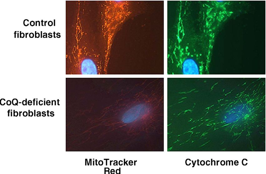
Mitochondrial membrane potential assessment (Mito- previously reported [7,18], and the main signs are probably
Tracker Red) and cytochrome c immunostaining results are related to the cerebellar involvement. Biochemical and
reported in Fig. 3. A clear decrease of both parameters was histological investigation results in muscle biopsy of the
observed when comparing the patient with a control index case also showed impaired results, but still without
analysed simultaneously. Furthermore, fibroblast incubation muscle clinical involvement. These observations suggest that
with dihydrorodhamine showed an increased fluorescence the clinical phenotypes of this disorder may present a
(3.5 a.u.) compared with control values (1.3 – 1.6; medi- continuous spectrum of symptoms and signs depending on
an = 1.5), suggesting increase in ROS production. the different degrees of CoQ deficiency in muscle or brain [7].
In our patient, ataxia was associated with a partial CoQ
3.3. Genetic studies deficiency in both muscle and fibroblasts, affecting MRC
enzyme activities as previously reported [7]. It has been
No mutations were observed neither in the aprataxin suggested that myopathic forms of CoQ deficiency would
gene after screening for each coding exon and flanking only show decreased CoQ values in muscle, while patients
intronic sequences nor in any of the SCA studied genes. with predominant brain involvement would show decreased
CoQ values both in muscle and fibroblasts [7]. Partial
3.4. CoQ supplementation and follow-up deficiencies causing mild phenotypes would only affect the
cerebellum, since antioxidant defenses and CoQ content are
Oral CoQ supplementation was started (2500 mg/day), very limited in this brain area [19].
divided in three doses. The doses were decreased every 3 Coenzyme Q10 biosynthesis is controlled by nuclear
months according to plasma CoQ monitoring data (Fig. 4). genes [20]. However, to our knowledge, the molecular
Fig. 3. Cultured fibroblasts from a healthy control (upper panels) and a patient with cerebellar ataxia and CoQ10-deficiency (lower panels). Membrane potential
was assessed using MitoTracker Red (Molecular Probes, Eugene, OR) in the left panels. Cytochrome c was visualized by immunostaining in the right panels. A
clear decrement of both parameters was observed in the patient fibroblasts.R. Artuch et al. / Journal of the Neurological Sciences 246 (2006) 153 – 158 157 Fig. 4. Effect of CoQ supplementation and evaluation of ICARS scale and plasma CoQ concentration over 16 months of follow-up in case 1. Above columns, plasma CoQ concentrations (Amol/L) and doses applied (mg/day in brackets) are stated. A write test was also performed, showing a clear improvement after CoQ supplementation. defects causing this disorder are still unknown. Only Rotig pothesis that the molecular defect might be allocated in the et al. [6], by incubating fibroblasts with 3H-labelled genes controlling the transprenylation pathway or in the mevalonate, hypothesized that the defect might be allocated metabolic steps after condensation of 4-hydroxybenzoate in the transprenylation part of the CoQ biosynthethic and the prenyl side chain of CoQ. pathway. Similar conclusions were obtained by Rahman Concerning the physiopathological mechanisms in- et al. [8], who showed normal organic acid profile volved in CoQ deficiency, it seems clear that this and glycosylation of proteins, ruling out defects in the deficiency reduces the flux of electrons from both complex mevalonate pathway and in the transformation of tyrosine to I and II to complex III, and therefore, causes a mitochon- 4-hydroxybenzoate. Furthermore, the possibility of an drial dysfunction [7]. These findings were further supported increased destruction of CoQ as responsible of CoQ both by the histological changes observed in muscle biopsy deficiency has been ruled out by Boitier et al., since normal and mitotrack and by the cytochrome c staining analysis in expression of CoQ-binding protein was observed by these fibroblasts, suggesting a mitochondrial oxidative phosphor- authors [21]. Recently, it has been reported an association ylation dysfunction, and probably, a reduction in ATP between cerebellar ataxia and CoQ deficiency with apra- synthesis. Furthermore, in vitro measurement of free radical taxin mutations [22]. Our case did neither reveal abnormal- generation also showed higher values than those observed ities in the organic acid profile or in the glycosylation status in controls, supporting the potential involvement of of transferrin, suggesting that an impaired mevalonate increased oxidative stress in this disorder. However, there pathway (which would cause organic acid accumulation are few controversial, reports concerning oxidative damage and altered N-glycosylation of transferrin) or biosynthesis of in CoQ deficiency [4,23] and this aspect deserves further 4-hydroxybenzoate is unlikely. Moreover, no mutations investigations. were found in the aprataxin gene, and autosomal dominat Patients with CoQ deficiency may benefit from CoQ cerebellar ataxias were ruled out. Furthermore, the experi- supplementation [7,18], although the more severe pheno- ments with incorporation of radiolabeled 4-[U-14C]HB types did not show such clear improvement in the clinical confirmed these preliminary findings, supporting the hy- manifestations [8]. Our case showed a very good CoQ
158                                    R. Artuch et al. / Journal of the Neurological Sciences 246 (2006) 153 – 158

supplementation response, with the main symptoms related                               and reduces apoptosis in familial Q10 deficiency. Neurology 2001;
with cerebellar dysfunction disappearing, and the ICARS                                57:515 – 8.
                                                                                 [5]   Sobreira C, Hirano M, Shanske S, Haller RG, Davidson E, Santorelli
scores decreasing after 16 months of supplementation. This                             FM, et al. Mitochondrial encephalomyopathy with coenzyme Q10
improvement has not been observed in all patients with                                 deficiency. Neurology 1997;48:1238 – 43.
ataxia and CoQ deficiency previously reported [7,18], and                        [6]   Rötig A, Appelkvist EL, Geromel V, Chretien D, Kadhom N, Edery P,
the higher CoQ doses administered to our patient (1000 mg/                             et al. Quinone-responsive multiple respiratory chain dysfunction due
                                                                                       to widespread coenzyme Q10 deficiency. Lancet 2000;356:391 – 5.
day compared to 200– 900 mg/day), may explain these
                                                                                 [7]   Musumeci O, Naini A, Slonim AE, Skavin N, Hadjigeorgiou GL,
differences. Although plasma CoQ concentrations of our                                 Krawiecki N, et al. Familial cerebellar ataxia with muscle coenzyme
patient were much higher than our reference ranges, a                                  Q10 deficiency. Neurology 2001;56:849 – 55.
further reduction of the CoQ dose should be cautiously                           [8]   Rahman S, Hargreaves I, Clayton P, heales S. Neonatal presentation of
evaluated.                                                                             coenzyme Q10 deficiency. J Pediatr 2001;139:456 – 8.
   Concerning the father of our patient, muscle biopsy                           [9]   Van Maldergem L, Trijbels F, DiMauro S, Sindelar PJ, Musumeci O,
                                                                                       Janssen A, et al. Coenzyme Q-responsive Leigh’s encephalopathy in
studies did not reveal an impaired MRC function or a                                   two sisters. Ann Neurol 2002;52:750 – 4.
decreased CoQ values. Furthermore, fibroblasts analysis did                     [10]   Leshinsky-Silver E, Levine A, Nissenkorn A, Barash V, Perach M,
not clearly showed low CoQ concentration. However, this                                Buzhaker E, et al. Neonatal liver failure and Leigh syndrome possibly
case presented cerebellar atrophy and some neurological                                due to CoQ-responsive OXPHOS syndrome. Mol Genet Metab
signs and symptoms (and dysarthria and tremor improved                                 2003;79:288 – 93.
                                                                                [11]   Trouillas P, Takayanagi T, Hallett M, Currier RD, Subramony SH,
after CoQ supplementation), and a decreased incorporation                              Wessel K, et al. International Cooperative ataxia rating scale for
of radiolabeled 4-[U-14C]HB in fibroblasts was also                                    pharmacological assessment of the cerebellar syndrome. J Neurol Sci
observed. These data would suggest that probably both                                  1997;145:205 – 11.
cases have the same disease, although only the molecular                        [12]   Colomé C, Artuch R, Vilaseca MA, Sierra C, Brandi N, Lambruschini
identification of the disease will elucidate this question.                            N, et al. Lipophilic antioxidants in phenylketonuric patients. Am J
                                                                                       Clin Nutr 2003;77:185 – 8.
   In conclusion, cerebellar ataxia associated with CoQ                         [13]   Rustin P, Chretien D, Bourgeron T, Gerard B, Rotig A, Saudubray JM,
deficiency in our case might be allocated in the trans-                                et al. Biochemical and molecular investigations in respiratory chain
prenylation pathway or in the metabolic steps after                                    deficiencies. Clin Chim Acta 1994;228:35 – 51.
condensation of 4-hydroxybenzoate and the prenyl side                           [14]   Fischer JC, Ruitenbeek W, Berden JA, Trijbels JM, Veerkamp JH,
chain of CoQ. Fibroblast analysis of CoQ biosynthesis                                  Stadhouders AM, et al. Differential investigation of the capacity of
                                                                                       succinate oxidation in human skeletal muscle. Clin Chim Acta 1985;
provided useful information for the diagnosis of these                                 153:23 – 6.
patients. Clinical improvement after CoQ supplementation                        [15]   Lowry OH, Rosenbrough NJ, Farr AL, Randall RJ. Protein measure-
was remarkable, supporting the importance of an early                                  ment with the folin phenol reagent. J Biol Chem 1951;193:265 – 75.
diagnosis of this kind of disorders.                                            [16]   Moreira MC, Barbot C, Tachi N, Kozuka N, Uchida E, Gibson T, et al.
                                                                                       The gene mutated in ataxia-ocular apraxia 1 encodes the new HIT/Zn-
                                                                                       finger protein aprataxin. Nat Genet 2001;29:189 – 93.
                                                                                [17]   Infante J, Combarros O, Volpini V, Corral J, Llorca J, Berciano J.
Acknowledgements                                                                       Autosomal dominant cerebellar ataxias in Spain: molecular and
                                                                                       clinical correlations, prevalence estimation and survival analysis. Acta
                                                                                       Neurol Scand 2005;111:391 – 9.
   This study was supported by the grants Mitoespaû˘a
                                                                                [18]   Lamperti C, Naini A, Hirano M, De Vivo DC, Bertini E, Servidei S,
(G03/011), Red de Ataxias (G03/056) and PI040567 from                                  et al. Cerebellar ataxia and coenzyme CoQ deficiency. Neurology
the FIS, Ministerio de Sanidad, Spain; and by EU contract                              2003;60:1206 – 8.
LSHB-CT-2004-005151.                                                            [19]   Naini A, Lewis VJ, Hirano M, DiMauro S. Primary coenzyme CoQ
                                                                                       deficiency and the brain. Biofactors 2003;18:145 – 52.
                                                                                [20]   Asencio C, Rodriguez-Aguilera JC, Ruiz-Ferrer M, Vela J, Navas P.
                                                                                       Silencing of ubiquinone biosynthesis genes extends life span in
References                                                                             Caenorhabditis elegans. FASEB J 2003;17:1135 – 7.
                                                                                [21]   Boitier E, Degoul F, Desguerre I, Charpentier C, Francois D, Ponsot G,
[1] Navarro F, Arroyo A, Martı́n SF, Bello RI, de Cabo R, Burgess JR,                  et al. A case of mitochondrial encephalomyopathy associated with
    et al. Protective role of ubiquinone in vitamin E and selenium-deficient           muscle coenzyme Q10 deficiency. J Neurol Sci 1998;156:41 – 6.
    plasma membranes. Biofactors 1999;9:163 – 70.                               [22]   Quinzii CM, Kattah BS, Naini A, Akman HO, Mootha VK, DiMauro S,
[2] Ernster L, Dallner G. Biochemical, physiological and medical aspects               et al. Coenzyme Q deficiency and cerebellar ataxia associated with an
    of ubiquinone function. Biochim Biophys Acta 1995;1271:195 – 202.                  aprataxin mutation. Neurology 2005;64:539 – 41.
[3] Ogasahara S, Engel AG, Frens D, Mack D. Muscle coenzyme Q                   [23]   Geromel V, Rotig A, Munnich A, Rustin P. Coenzyme Q10 depletion
    deficiency in familial mitochondrial encephalomyopathy. Proc Natl                  is comparatively less detrimental to human cultured skin fibroblasts
    Acad Sci U S A 1989;86:2379 – 82.                                                  than respiratory chain complex deficiencies. Free Radic Res 2002;
[4] Di Giovanni S, Mirabella M, Spinazzola A, Crociani P, Silvestri G,                 36:375 – 9.
    Broccolini A, et al. Coenzyme Q10 reverses pathological phenotype
You can also read