Is COVID 19, a beginning of new entity called chronic viral systemic inflammatory syndrome?
←
→
Page content transcription
If your browser does not render page correctly, please read the page content below
AIMS Allergy and Immunology, 5(2): 127–134.
                                                       DOI: 10.3934/Allergy.2021010
                                                       Received: 08 March 2021
                                                       Accepted: 19 April 2021
                                                       Published: 20 April 2021
http://www.aimspress.com/journal/Allergy

Mini review

Is COVID 19, a beginning of new entity called chronic viral systemic
inflammatory syndrome?

DharmaSaranya Gurusamy, Vasuki Selvamurugesan, Swaminathan Kalyanasundaram and
Shantaraman Kalyanaraman*

Department of Pathology, Government Tirunelveli Medical College, High Ground Road,
Palayamkottai, Tirunelveli-627011, Tamil Nadu, India

* Correspondence: Email: shantaraman_kal@tvmc.ac.in; Tel: +9443133898.

Abstract: Since the emergence of SARS-COV-2, many updates and assumptions are being discussed
based on the emerging understanding of the pathophysiology and outcomes of the infection. The host
immune response is the critical factor in disease severity and the unique immune response of each
individual has resulted in a broad spectrum of disease severity and clinical presentations. In this
article, combining the emerging knowledge on the immunopathogenesis of COVID-19 infection with
insights into the underlying mechanisms of certain autoimmune and chronic Immune-mediated
inflammatory diseases we propound our hypothesis that SARS-CoV-2 infection produces chronic
viral inflammatory syndrome.

Keywords: COVID-19; chronic viral inflammatory syndrome; cytokines; immune-mediated
inflammatory disease; NLRP3 activation; senescent activated secretory phenotype

Abbreviations: DAB1: disabled homolog 1; SURF1: surfeit locus protein 1; AIFM:
apoptosis-inducing factor mitochondrial; TIM-3: T cell immunoglobulin and mucin domain 3;
CTLA4: cytotoxic T lymphocyte associated protein 4; PD1: programmed cell death protein 1; TIGIT:
T cell immunoglobulin and ITIM domain receptor; MAVS: mitochondrial antiviral signaling proteins;
VDAC: voltage dependent anion channel; ACE II: angiotensin converting enzyme II; AT1R:
angiotensin I receptor; NLRP3: nuclear NOD, LRR, pyrin domain containing protein 3; IL:
interleukin; GMCSF: granulocyte monocyte colony stimulating factor; TNF: tumor necrosis factor;
PDGF: platelet derived growth factor; FGF: fibroblast growth factor; IFN-γ: Interferon gamma;
TNF-α: tumor necrosis factor alpha; NF-κB: nuclear factor κB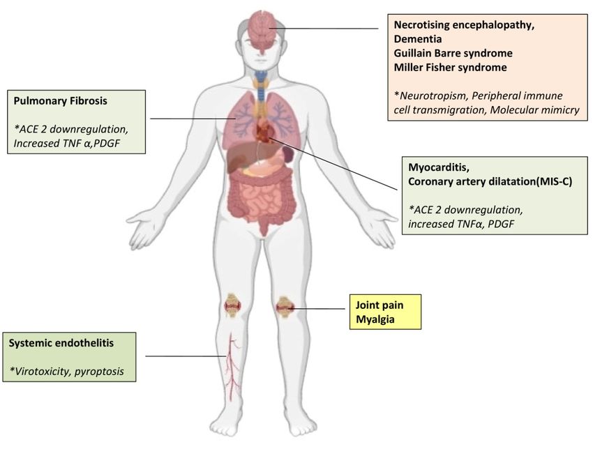
128
1. Introduction
The emergence of novel coronavirus strain SARS-COV-2 has resulted in the accumulation of
critically ill patients in hospital beds across the globe. Our understanding of the nature of the
infection has evolved dynamically and SARS-COV-2 infection which was perceived initially as
pneumonia of unknown etiology is currently proven to cause derangement of immunological and
endothelial physiology leading to prothrombotic tendencies and multiorgan dysfunction. The
exponential trajectory of the spread of the infection coupled with increased morbidity and mortality
have posed several unresolved clinical questions or dilemmas on the outcome of SARS-CoV-2
infection. Suspicions arise on lingering maladies in the patients who have recovered from COVID-19.
Reports of restricted lung function and decreased exercise capacity were reported in long term
survivors of SARS and MERS infections [1], the close allies of the causative virus of the current
pandemic. At a stage, any virus can become a powerful stimulator of inflammation to such an extent
that the inflammatory response gets stuck up in a sustained activated state even after the virus is
eliminated. This review aims to identify the potential immune pathways that may lead to the
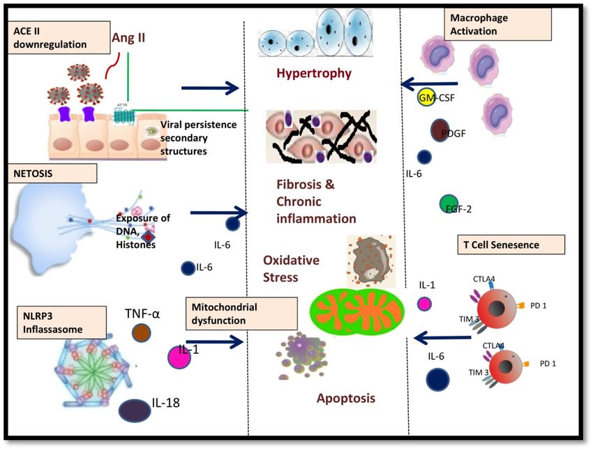
development of chronic systemic inflammatory diseases post SARS-Cov-2 infection (Figure 1).
Improved knowledge of the shared pathophysiology of COVID-19 and chronic immune diseases
may help in devising diagnostic algorithms and therapeutic strategies to prevent or dampen the
chronic complications of SARS-COV-2 infection in vulnerable patients.
Figure 1. SARS-COV-2 induced immune pathways postulated to play a role in
pathogenesis of chronic systemic inflammatory syndrome: Depicts the pathways
common between immunopathogenesis of COVID-19 and chronic immune mediated
inflammatory diseases.
AIMS Allergy and Immunology Volume 5, Issue 2, 127–134.
129

2.   Persistent antigenic stimulus and molecular mimicry

     SARS-CoV-2 viral RNA is known to be present in various clinical samples for quite some time,
and the durations reported in studies range from 33 days [2] to 82 days [3] from the onset of the
symptoms. The genome of SARS-CoV-2 is known to be remarkably structured enabling shielding
from host immunity. Presence of secondary structures throughout the viral RNA termed as
“GORS” (genome ordered RNA structures) enable decreased viral recognition leading to defects in
macrophage activation, T cell maturation and recruitment, thereby aiding viral persistence [4].
Molecular mimicry between viral proteins and host peptides may favor an aberrant activation of auto
reactive T cells or B cells, leading to autoimmune response. Sharing of DAB1, SURF1, and AIFM
peptides between the respiratory neurons in pre-Botzinger complex and SARS-CoV-2 viral proteins
is hypothesized as a cause of respiratory failure in severe SARS-CoV-2 infected patients [5]. Viral
proteins mimicking human peptides are also implicated in the pathogenesis of anemia, leucopenia,
and vascular damage in COVID-19 patients [6].

3.   ACE II downregulation

     Binding of SARS-CoV-2 receptor binding domain to the host ACE II receptor [7] leads to
internalization and shedding of ACE II. Downregulation of ACE II leads to a relative increase in
Ang II tilting the RAS axis to the proinflammatory state. Ang II is believed to have pro-inflammatory,
pro-oxidant, and pro-thrombotic, profibrotic properties [8] and interfere in intracellular insulin
signaling. Elevated levels of angiotensin II are documented in liver fibrosis, pulmonary arterial
hypertension, diabetes, obesity, and myocardial dysfunction. Increasing clinical evidence shows that
the most common co-morbidities observed in COVID-19 patients, that are associated with worse
prognosis and a higher rate of death, are systemic hypertension, diabetes, obesity, old age in which
ACE II deficiency is known to be a significant determinant [9,10].

4.   Dysregulated NLRP3 activation

     Rapid and dysregulated NLRP3 activation by SARS-CoV-2 ion channel proteins E, and
accessory proteins ORF 3a ORF 8b resulting in increased TNF-α, IL-1 and IL-18, is documented in
SARS-CoV-2 infection [11]. Constitutive activation of NLRP3 inflammasome is associated with
hereditary autoinflammatory diseases like familial cold urticaria syndrome (FCAS) [12], Muckle-Wells
syndrome (MWS) [13], and chronic infantile neurological cutaneous and articular (CINCA)
syndrome also known as neonatal onset multisystemic inflammatory disease (NOMID) [14].

5.   Alternate macrophage activation

      Pivotal role of macrophages in the pathogenesis of SARS-CoV-2 infection is demonstrated in
many studies [15]. Lio et al. in a study on bronchoalveolar fluid demonstrated evidence of activation
of M2 macrophages [16]. M2 macrophages possess a profibrotic profile and bring about
vasculogenesis, cell proliferation. A microenvironment rich in M2 macrophages plays an important
role in pulmonary fibrosis [17].

AIMS Allergy and Immunology                                                   Volume 5, Issue 2, 127–134.
130
6. Neutrophil extracellular traps formation (NETosis)
Sera of severely ill COVID-19 patients show elevated NET breakdown products [18]. The
intracellular components like DNA, MPO, histones externalized through NETosis have recognized
autoantigens and defective clearing of these extruded components may prolong the half-life of the
lattices leading to persistent inflammation and tissue damage [19]. Increased NET formation in blood
and tissue lesions is reported in chronic diseases like ANCA antibody-associated vasculitis,
rheumatoid arthritis, psoriasis, autoimmune pancreatitis, dermatomyositis, polymyositis, and
multiple sclerosis.
7. Senescent activated secretory phenotype of T cells
T cells play a major role in viral clearance. In SARS-Cov-2 infection the number of T cells is
reduced and the T cells are known to be in a senescent or functionally exhausted state, demonstrated
by the expression of PD1, TIM-3 [20], and CTLA-4 and TIGIT. In the inflammatory milieu, the
senescent cells are said to be in a hypo proliferative but functionally active state named as senescent
activated secretory phenotype (SSAP). The SSAP cells are known to secrete many cytokines, growth
factors, and proteases implicated in tissue injury [21]. T cell senescence is currently implicated in the
pathogenesis of inflammatory bowel diseases and rheumatoid arthritis [22].
8. Interleukins
Elevated levels of IL-1β, IL-6, IL-7, IL-8, IL-9, IL-10, FGF, GM-CSF, IFN-γ, IL-10, MCP-1,
PDGF, TNF-α, and VEGF are seen in COVID-19 patients compared to healthy adults. Table 1
discusses the relevant cytokines common between SARS-Cov-2 infection and chronic inflammation.
Table 1. Cytokines and their functions in relevance to both COVID-19 and
immune-mediated inflammatory diseases.
Cytokine Functions in chronic inflammation Diseases associated
IL-1 Tissue destruction, fibroblast proliferation, Alzheimer’s diseases, amyotrophic lateral sclerosis,
collagen deposition, perpetuation of atherosclerosis, gout, rheumatoid arthritis
inflammation through IFN-γ, IL-17, GM-CSF
IL-6 Activates the endothelial cell, induction of IL-8, Rheumatoid arthritis, systemic lupus erythematosis,
MCP-1, expression of adhesion molecules, psoriasis, Crohn’s disease
transition from acute to chronic inflammation
FGF-2 Mitogen for fibroblasts, cell proliferation, Asthma, COPD, chronic bronchitis
migration, apoptosis of airway epithelial cells
GMCSF Chemokine-17 induction and induction of Rheumatoid arthritis, Kawasaki disease,
fibroblasts and endothelial cells myocarditis
TNF-α Cytotoxicity, cell growth, NF-κB activation Rheumatoid arthritis, inflammatory bowel disease,
Amyotrophic lateral sclerosis, Alzheimers disease
PDGF Airway smooth muscle migration, fibroblast Bronchial asthma, pulmonary fibrosis, pulmonary
proliferation artery hypertension
AIMS Allergy and Immunology Volume 5, Issue 2, 127–134.131
9. Mitochondrial dysfunction
On evaluating ACE-II downregulation, NLRP3 inflassasome activation and inflammaging in the
context of their close association with mitochondria, the distinct immune-metabolic pathways in
SARS-Cov-2 infection, may be partially attributed to mitochondrial dysfunction. Mitochondrial
molecules like mitofusin-2 [23] and Mitochondrial Antiviral Signaling Proteins MAVS [24] interact
with NLRP3 during viral infections. Mitochondria act as scaffolds to NLRP3 inflassasome activation.
Thompson et al. [25] identified unique population of H3K27me3hiVDAC1hi T cells with upregulation
of mitochondrial protein voltage dependant anion channel (VDAC1). H3K27me3hiVDAC1hi T cells
were associated with increased T cell apoptosis, and lacked traditional activation response to TCR
stimulation. VDAC is a multifunctional mitochondrial membrane protein involved in release of
mtDNA, inflassasome activation, apoptosis, type I Interferon release and is known to be a promoter
of lupus like disease [26]. Increased VDAC3 is reported in SLE and VDACs are also reported to play
a key role in neurodegenerative diseases, cardiac injury and neoplastic diseases [27]. Mitochondrial
ROS and mtDNA release may have a role in initiation and upregulation of autoimmunity by
promoting NETosis, cell survival disruption and induction of type 1 interferon signature [28].
Mitochondrial dysfunction is also associated with ageing, obesity, diabetes the comorbidities
associated with severe COVID-19 [29]. Thus mitochondrial dysfunction may be a convergence point
of multiple mechanisms driving increased inflammation and predisposition to autoimmunity in
COVID-19 patients.
In line with the above discussions, there are definite intersections between the immune
pathways of SARS-Cov-2 infection and immune-mediated inflammatory diseases at multiple levels.
The outcome of the pandemic depends on the interaction between a genomic heterogeneous virus
and an unpredictable immune system attributed to the mosaic global population with a variegated
immune composition. The frenzied immune system may not shut off instantly but may linger in an
activated state or resolve to scar the organs involved. Thus it is reasonable for us to hypothesize that
COVID-19 may be the beginning of a new entity called a chronic viral systemic inflammatory
syndrome.
10. Evaluation of the hypothesis
Multisystem dissemination of SARS-Cov-2 is possible because of ACE 2 expression on
endothelial cells, smooth muscle cells, and perivascular pericytes in virtually all organs [30].
Autopsy studies show macrophage infiltration, peri-bronchial fibrometaplasia, thickened alveolar
walls, and fibrous proliferation in the lung parenchyma. Reports on possible chronic sequelae of
SARS-CoV-2 infections are surfacing recently. Figure 2 summarises the major chronic effects
reported and the postulated underlying mechanisms. Combet et al. reported aggressive pulmonary
fibrosis in a patient who recovered from the acute infection without mechanical ventilation [31].
Myocyte degeneration with interstitial hyperplasia and CD20 positive lymphocytic infiltrates were
also seen in the myocardium. A Kawasaki disease like presentation with a higher rate of cardiac
involvement and macrophage activation termed, multisystem inflammatory syndrome (MIS-C), is
reported in children with evidence of COVID antibodies in the serum [32]. In a study on German
patients cured of COVID-19, 60% had persistent ongoing inflammation in the myocardium and
pericardium, irrespective of the severity of the clinical presentation in the acute infection [33].
AIMS Allergy and Immunology Volume 5, Issue 2, 127–134.132
Figure 2. Chronic sequelae reported in COVID 19 survivors and the proposed
underlying mechanisms: Depicts the major post COVID chronic sequelae reported and
the underlying mechanisms proposed.
11. Conclusions
As of now owing to the remarkably high proportion of the global population affected, only the
acute effects of the pandemic are coming to light. It will take a few years to determine the chronic
effects of SARS-CoV-2 infection. The breadth of the current SARS-Cov-2 pandemic may require a
closer examination of the underlying mechanisms, preventive measures, diagnostic methods, and
interventions for post-viral systemic chronic inflammatory sequelae. Thus follow-up of COVID-19
survivors is indispensable to fully appreciate and mitigate a deleterious pile-up of patients with
chronic diseases precipitated by the pandemic.
Conflict of interest
All authors declare no conflicts of interest in this paper.
AIMS Allergy and Immunology Volume 5, Issue 2, 127–134.133
References
1. Ong KC, Ng AWK, Lee LSU, et al. (2004) Pulmonary function and exercise capacity in
survivors of severe acute respiratory syndrome. Eur Respir J 24: 436–442.
2. Gombar S, Chang M, Hogan CA, et al. (2020) Persistent detection of SARS-CoV-2 RNA in
patients and healthcare workers with COVID-19. J Clin Virol 129: 104477.
3. Li N, Wang X, Lv T (2020) Prolonged SARS-CoV-2 RNA shedding: Not a rare phenomenon. J
Med Virol 92: 2286–2287.
4. Simmonds P, Tuplin A, Evans DJ (2004) Detection of genome-scale ordered RNA structure
(GORS) in genomes of positive-stranded RNA viruses: implications for virus evolution and host
persistence. RNA 10: 1337–1351.
5. Lucchese G, Flöel A (2020) Molecular mimicry between SARS-CoV-2 and respiratory
pacemaker neurons. Autoimmun Rev 19: 102556.
6. Angileri F, Legare S, Gammazza AM, et al. (2020) Molecular mimicry may explain multi-organ
damage in COVID-19. Autoimmun Rev 19: 102591.
7. Gheblawi M, Wang K, Viveiros A, et al. (2020) Angiotensin-converting enzyme 2:
SARS-CoV-2 receptor and regulator of the renin-angiotensin system: celebrating the 20th
anniversary of the discovery of ACE2. Circ Res 126: 1456–1474.
8. Kamo T, Akazawa H, Komuro I (2015) Pleiotropic effects of angiotensin II receptor signaling in
cardiovascular homeostasis and aging. Int Heart J 56: 249–254.
9. Verdecchia P, Cavallini C, Spanevello A, et al. (2020) The pivotal link between ACE2
deficiency and SARS-CoV-2 infection. Eur J Intern Med 76: 14–20.
10. Tseng YH, Yang RC, Lu TS (2020) Two hits to the renin-angiotensin system may play a key role
in severe COVID-19. Kaohsiung J Med Sci 36: 389–392.
11. Nieto-Torres JL, Verdiá-Báguena C, Jimenez-Guardeño JM, et al. (2015) Severe acute
respiratory syndrome coronavirus E protein transports calcium ions and activates the NLRP3
inflammasome. Virology 485: 330–339.
12. Hoffman HM, Wanderer AA, Broide DH (2001) Familial cold autoinflammatory syndrome:
phenotype and genotype of an autosomal dominant periodic fever. J Allergy Clin Immun 108:
615–620.
13. Muckle TJ, Wells M (1962) Urticaria, deafness, and amyloidosis: a new heredo-familial
syndrome. QJM-Int J Med 31: 235–248.
14. Prieur AM, Griscelli C, Lampert F, et al. (1987) A chronic, infantile, neurological, cutaneous
and articular (CINCA) syndrome. A specific entity analysed in 30 patients. Scand J Rheumatol
16: 57–68.
15. Wang C, Xie J, Zhao L, et al. (2020) Alveolar macrophage dysfunction and cytokine storm in
the pathogenesis of two severe COVID-19 patients. EBioMedicine 57: 102833.
16. Liao M, Liu Y, Yuan J, et al. (2020) Single-cell landscape of bronchoalveolar immune cells in
patients with COVID-19. Nat Med 26: 842–844.
17. Hou J, Shi J, Chen L, et al. (2018) M2 macrophages promote myofibroblast differentiation of
LR-MSCs and are associated with pulmonary fibrogenesis. Cell Commun Signaling 16: 89.
18. Zuo Y, Yalavarthi S, Shi H, et al. (2020) Neutrophil extracellular traps (NETs) as markers of
disease severity in COVID-19. JCI Insight 5: e138999.
AIMS Allergy and Immunology Volume 5, Issue 2, 127–134.134
19. Gupta S, Kaplan MJ (2016) The role of neutrophils and NETosis in autoimmune and renal
diseases. Nat Rev Nephrol 12: 402–413.
20. Diao B, Wang C, Tan Y, et al. (2020) Reduction and functional exhaustion of T cells in patients
with coronavirus disease 2019 (COVID-19). Front Immunol 11: 827.
21. Lopes-Paciencia S, Saint-Germain E, Rowell MC, et al. (2019) The senescence-associated
secretory phenotype and its regulation. Cytokine 117: 15–22.
22. Weyand CM, Yang Z, Goronzy JJ (2014) T cell aging in rheumatoid arthritis. Curr Opin
Rheumatol 26: 93–100.
23. Ichinohe T, Yamazaki T, Koshiba T, et al. (2013) Mitochondrial protein mitofusin 2 is required
for NLRP3 inflammasome activation after RNA virus infection. P Natl Acad Sci USA 110:
17963–17968.
24. Park S, Juliana C, Hong S, et al. (2013) The mitochondrial antiviral protein MAVS associates
with NLRP3 and regulates its inflammasome activity. J Immunol 191: 4358–4366.
25. Thompson EA, Cascino K, Ordonez AA, et al. (2021) Metabolic programs define dysfunctional
immune responses in severe COVID-19 patients. Cell Rep 34: 108863.
26. Kim J, Gupta R, Blanco LP, et al. (2019) VDAC oligomers form mitochondrial pores to release
mtDNA fragments and promote lupus-like disease. Science 366: 1531–1536.
27. Camara AKS, Zhou Y, Wen PC, et al. (2017) Mitochondrial VDAC1: a key gatekeeper as
potential therapeutic target. Front Physiol 8: 460.
28. Lood C, Blanco LP, Purmalek MM, et al. (2016) Neutrophil extracellular traps enriched in
oxidized mitochondrial DNA are interferogenic and contribute to lupus-like disease. Nat Med 22:
146.
29. Vernochet C, Kahn CR (2012) Mitochondria, obesity and aging. Aging (Albany NY) 4: 859.
30. Hamming I, Timens W, Bulthuis MLC, et al. (2004) Tissue distribution of ACE2 protein, the
functional receptor for SARS coronavirus. A first step in understanding SARS pathogenesis. J
Pathol 203: 631–637.
31. Combet M, Pavot A, Savale L, et al. (2020) Rapid onset honeycombing fibrosis in
spontaneously breathing patient with Covid-19. Eur Respir J 56: 2001808.
32. Verdoni L, Mazza A, Gervasoni A, et al. (2020) An outbreak of severe Kawasaki-like disease at
the Italian epicentre of the SARS-CoV-2 epidemic: an observational cohort study. Lancet 395:
1771–1778.
33. Rajpal S, Tong MS, Borchers J, et al. (2021) Cardiovascular magnetic resonance findings in
competitive athletes recovering from COVID-19 infection. JAMA Cardiol 6: 116–118.
© 2021 the Author(s), licensee AIMS Press. This is an open access
article distributed under the terms of the Creative Commons
Attribution License (http://creativecommons.org/licenses/by/4.0)
AIMS Allergy and Immunology Volume 5, Issue 2, 127–134.You can also read