TRANSMISSION OF SARS-COV-2 FROM HUMAN TO DOMESTIC FERRET
←
→
Page content transcription
If your browser does not render page correctly, please read the page content below
DISPATCHES

   Transmission of SARS-CoV-2 from
      Human to Domestic Ferret
        Jožko Račnik, Ana Kočevar, Brigita Slavec, Miša Korva, Katarina Resman Rus, Samo Zakotnik,
           Tomaž Mark Zorec, Mario Poljak, Milan Matko, Olga Zorman Rojs, Tatjana Avšič Županc

 We report a case of natural infection with severe acute           >5% dehydrated with low body temperature (36.4°C,
 respiratory syndrome coronavirus 2 transmitted from an            reference range 37.8–40°C) and slow heart rate (180
 owner to a pet ferret in the same household in Slovenia.          beats/min, reference 200–400 beats/min). The body
 The ferret had onset of gastroenteritis with severe dehy-         condition of the ferret was good, with a bodyweight
 dration. Whole-genome sequencing of the viruses isolat-           of 1.3 kg. Several hematology and serum biochemis-
 ed from the owner and ferret revealed a 2-nt difference.          try results were elevated: red blood cell count (12.36
                                                                   × 106/µL, reference 7.01–9.65 × 10⁶/µL), hemoglobin
N     atural infections with severe acute respiratory
      syndrome coronavirus 2 (SARS-CoV-2) in do-
mestic animals living in infected households have
                                                                   concentration (21.2 g/dL, reference 12.2–16.5 g/dL),
                                                                   and hematocrit (0.57%, reference 0.36%–0.48%); blood
                                                                   urea nitrogen (>129.94 mg/dL, reference 18–32 mg/
been reported (1). Because of their increased popular-             dL), hyperproteinemia (8.5 g/dL, reference 4.5–6.2 g/
ity as a pet (2), domestic ferrets (Mustela putorius furo)         dL), hyperglobulinemia (4.4 g/dL, reference 2.8–3.6
pose a high risk for transmitting anthropozoonotic                 g/dL), and borderline hyperalbuminemia (4.0 g/
infections. A recent study in Spain showed that natu-              dL, reference 2.5–4.0 g/dL) were consistent with
ral SARS-CoV-2 infections can occur in ferrets kept                dehydration and possible infection. The results of
as working animals for rabbit hunting, especially if a             all other hematologic and biochemical values were
high viral circulation is present in the human popula-             within reference ranges. Whole-body radiographs
tion (3). Further, ferrets are common laboratory ani-              (Appendix Figure, https://wwwnc.cdc.gov/EID/
mal models and, at least in experimental conditions,               article/27/9/21-0774-App1.pdf) showed spleno-
have been shown to be highly susceptible to SARS-                  megaly and gas accumulation in intestinal loops. In-
CoV-2 infection and likely to transmit the virus to                terstitial and alveolar patterns of cranial lung lobes
other ferrets without apparent clinical signs (4).                 were present, indicating possible lobar pneumonia.
                                                                   The ferret was hospitalized and initially given fluid
The Study                                                          therapy, amoxicillin, esomeprazole, maropitant, and
On November 20, 2020, a 5-year-old neutered male                   dexamethasone. Three days later, the clinical status
domestic ferret had signs of acute gastroenteritis, in-            of the ferret improved, hematologic and biochemical
cluding apathy, anorexia, vomiting, and profuse mu-                values normalized, and the ferret was scheduled for
cous diarrhea. Another ferret in the same household                discharge. However, on the same day, the owner in-
appeared healthy. Because the ferret’s condition did               formed the veterinary hospital of having positive re-
not improve, the owner took it to a veterinary hospi-              sults for SARS-CoV-2 RNA tested on November 24,
tal for clinical examination on November 23. The fer-              after 9 days of malaise. Additional rectal and oropha-
ret was lethargic and, on the basis of skin turgor, was            ryngeal swab specimens and blood samples were tak-
                                                                   en from the ferret for further diagnostic procedures,
Author affiliations: University of Ljubljana Faculty of Veterinary   and the ferret was discharged from the hospital and
Medicine, Ljubljana, Slovenia (J. Račnik, B. Slavec,               put into isolation at the owner’s home. Samples were
O. Zorman Rojs); Toplica Veterinary Hospital, Topolšica,           not taken from the other pet ferret at the residence,
Slovenia (A. Kočevar, M. Matko); University of Ljubljana Faculty   but the rest of household members tested negative for
of Medicine, Ljubljana (M. Korva, K. Resman Rus, S. Zakotnik,      SARS-CoV-2 RNA on November 25.
T.M. Zorec, M. Poljak, T. Avšič Županc)                                 We tested the ferret’s samples for SARS-
DOI: https://doi.org/10.3201/eid2709.210774                        CoV-2 RNA (Appendix) and ferret-specific enteric

2450                       Emerging Infectious Diseases • www.cdc.gov/eid • Vol. 27, No. 9, September 2021
Transmission of SARS-CoV-2 from Human to Ferret

coronavirus (FERCV) (5) by real-time reverse tran-             and supportive care with antibiotics, antacids, anti-
scription PCR; influenza A and B viruses (6) by reverse        emetics, and parenteral dexamethasone. The ferret re-
transcription PCR; and herpesviruses (7), adenovirus-          sponded to the therapy promptly and fully recovered
es (8), and circoviruses (9) and by PCR. The only posi-        in 3 days. Acute epizootic catarrhal enteritis caused
tive result was the detection of SARS-CoV-2 RNA in
the rectal and oropharyngeal swab specimens. In the
oropharyngeal swab specimen, all 3 targeted genes
(envelope, cycle threshold [Ct] 27.7; RNA dependent
RNA polymerase, Ct 28.5; and nucleocapsid, Ct 32.1)
were detected, and in the rectal swab specimen only
envelope gene (Ct 35.6) was detected, a finding proba-
bly attributable to a lower virus concentration. To com-
pare the SARS-CoV-2 detected in the owner and the
ferret, we conducted whole-genome sequencing on Il-
lumina MiSeq (Illumina, https://www.illumina.com)
on the basis of the ARTIC protocol (https://artic.net-
work/ncov-2019/ncov2019-bioinformatics-sop.html).
The complete genome sequences were deposited in
the GISAID database (https://www.gisaid.org; acces-
sion nos. EPI_ISL_1490186 and EPI_ISL_1490187). Ac-
cording to the pangolin nomenclature, the sequences
belonged to the B.1.258 Pango lineage, which was on
the rise in Slovenia in November 2020 (Figure 1). The
comparison of both sequences showed ≈100% identi-
ty, differing by 2 nucleotides (position/owner/ferret:
2,097/G/T; 22,832/C/A).
     We also confirmed the SARS-CoV-2 infection in
the ferret on the basis of SARS-CoV-2 IgG serocon-
version and development of neutralizing antibod-
ies. We tested the ferret’s acute and convalescent
serum samples with an in-house immunofluores-
cent assay (Appendix). The first serum sample ob-
tained on day 6 after disease onset tested negative;
however, seroconversion was observed on day 19,
when the IgG titer was 1:1,024 (Figure 2, panels
A, B). In addition, we detected a high neutralizing
antibody titer of 1:320 in the second serum sample
(Figure 2, panel C).

Conclusions
SARS-CoV-2 originated in animals, jumped into hu-
mans, and is now easily transmitted among humans.
In addition to spreading from animals to humans,
                                                               Figure 1. Phylogenetic sequence context consisted of high-quality
the virus can be transmitted back into animals, as             complete severe acute respiratory syndrome coronavirus 2 genome
observed in farmed mink (Neovison vison) (10). Most            sequences from a domestic ferret, Slovenia, corresponding to Pango
experimentally infected ferrets do not exhibit clinical        lineage B.1.258. The context sequences were downloaded from
signs or have only mild fever, lethargy, loss of appe-         GISAID (https://www.gisaid.org) and subsampled to 62 sequences
tite, and occasional cough (4,11). Also, among work-           and National Center for Biotechnology Information reference
                                                               sequence NC_045512.2. The phylogenetic reconstruction using a
ing ferrets naturally infected with SARS-CoV-2 in              general time-reversible plus F plus R4 substitution model was built in
Spain, no signs of illness were reported (3).                  Figtree (Evomics, http://evomics.org) with 1,000 bootstrap replicates.
     In our study, the infected ferret had onset of se-        The reference sequence was used as an outgroup to root the
vere disease with gastroenteritis, pneumonia, and              phylogenetic tree. GISAID accession numbers and isolation dates
dehydration and required aggressive fluid therapy              are provided. Scale bar indicates substitutions per site.

                      Emerging Infectious Diseases • www.cdc.gov/eid • Vol. 27, No. 9, September 2021                           2451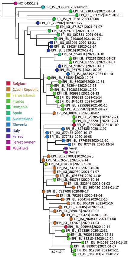
DISPATCHES
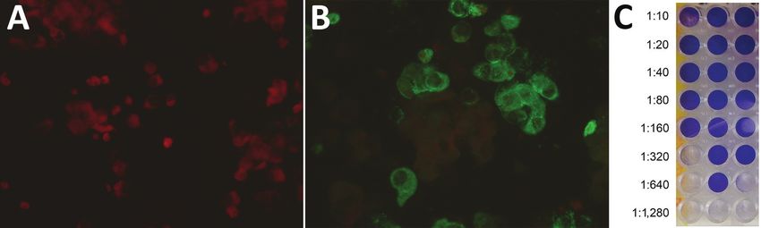
Figure 2. Serological response to infection with severe acute respiratory syndrome coronavirus 2 in a domestic ferret, Slovenia.
Immunofluorescent tests showed a negative result in the ferret’s acute serum sample obtained on day 6 after disease onset (A) and a
positive reaction at titer 1:64 in the ferret’s convalescent serum sample obtained on day 19 (B). The neutralization test (C) showed the
highest dilution of the ferret’s convalescent serum sample that inhibited a cytopathic effect in >2 of 3 wells to be 1:320.
by FERCV was one of the plausible differential diag- ferret revealed only a 2-nt difference, and neither of
noses in the initial treatment plan for the ferret. For those was present in the spike protein gene. None-
this reason, dexamethasone was used parenterally theless, retaining the One Health approach is cru-
because additional treatment with a short course of cial for early detection and monitoring of emerging
steroids might speed the recovery and reduce future zoonoses in humans.
problems of malabsorption attributable to villi de-
struction caused by fulminate FERCV infection (12). Acknowledgments
In humans, the effective drugs against coronavirus We thank all members of the COVID-19 Next
disease are poorly defined, yet dexamethasone in Generation Sequencing team for great technical
combination with supportive therapy is frequently assistance in sequencing SARS-CoV-2 genomes and
used (13). However, the risk for unnecessary use and the staff at the Toplica Veterinary Hospital for caring for
adverse effects must be considered before treatment the ferret. We thank the staff at the Institute for Poultry,
attempts with corticosteroids. Birds, Small Mammals, and Reptiles for their assistance
We assume that SARS-CoV-2 likely spread from the in performing PCR assays. Thanks to the owners of the
infected owner to the ferret living in the same household. ferret for their kind support and for allowing publication
Symptoms appeared in the owner 4 days before the fer- of this report.
ret became ill. All other household members tested nega- This work was supported by the Slovenian Research
tive for SARS-COV-2 RNA, ruling out asymptomatic in- Agency (grant nos. P3-0083 and V3-2034 at the Institute
fected persons in the family. Another close contact ferret of Microbiology and Immunology, Faculty of Medicine,
in the same household appeared healthy. Likewise, no University of Ljubljana and P4-0092 at the Faculty of
disease among staff or animals at the veterinary hospital Veterinary Medicine, University of Ljubljana) and by the
was reported during or after the hospitalization of the European Virus Archive Global project, which received
ferret. Nevertheless, ferrets as laboratory models were funding from the European Union’s Horizon 2020 research
shown to shed SARS-CoV-2 up to 8 days postinfection in and innovation program under grant no. 871029.
nasal swab, saliva, urine, and fecal samples. Ferrets can
effectively transmit the infection to other animals (14) or About the Author
possibly humans, thus highlighting the importance of Dr. Račnik is an associate professor at the Faculty of
recognizing the infection in pets early to prevent spread Veterinary Medicine, University of Ljubljana, Slovenia, and
to other animals or humans in the same household diplomate of the European College of Zoological Medicine,
or elsewhere (15). wildlife population health specialty. His primary research
In the mink farm outbreak in Denmark, SARS- interests include clinical veterinary medicine and emerging
CoV-2 transmission was shown to spill over from diseases of exotic pets and wild birds.
minks to humans accumulating mutations that
are resistant to neutralizing antibodies or vaccines
References
along the way (10). In our study, whole-genome 1. Patterson EI, Elia G, Grassi A, Giordano A, Desario C,
sequencing of the virus detected in the owner and Medardo M, et al. Evidence of exposure to SARS-CoV-2
2452 Emerging Infectious Diseases • www.cdc.gov/eid • Vol. 27, No. 9, September 2021
Transmission of SARS-CoV-2 from Human to Ferret

      in cats and dogs from households in Italy. Nat Commun.

                                                                                 EID Podcast
      2020;11:6231. https://doi.org/10.1038/s41467-020-20097-0
 2.   Bixler H, Ellis C. Ferret care and husbandry. Vet Clin North
      Am Exot Anim Pract. 2004;7:227–55, v. https://doi.org/

 3.
      10.1016/j.cvex.2004.02.002
      Gortázar C, Barroso-Arévalo S, Ferreras-Colino E, Isla J,                 Developing
      de la Fuente G, Rivera B, et al. Natural SARS-CoV-2 infection
      in kept ferrets, Spain. Emerg Infect Dis. 2021;27(7):1994-1996.      Biological Reference
 4.
      https://doi.org/10.3201/eid2707.210096
      Shi J, Wen Z, Zhong G, Yang H, Wang C, Huang B, et al.               Materials to Prepare
                                                                              for Epidemics
      Susceptibility of ferrets, cats, dogs, and other domesticated
      animals to SARS-coronavirus 2. Science. 2020;368:1016–20.
      https://doi.org/10.1126/science.abb7015
 5.   Muradrasoli S, Mohamed N, Hornyák A, Fohlman J, Olsen B,
      Belák S, et al. Broadly targeted multiprobe QPCR for detection
      of coronaviruses: coronavirus is common among mallard
      ducks (Anas platyrhynchos). J Virol Methods. 2009;159:277–87.
      https://doi.org/10.1016/j.jviromet. 2009.04.022
 6.   Boonsuk P, Payungporn S, Chieochansin T,
      Samransamruajkit R, Amonsin A, Songserm T, et al.
      Detection of influenza virus types A and B and type A
      subtypes (H1, H3, and H5) by multiplex polymerase
      chain reaction. Tohoku J Exp Med. 2008;215:247–55.
      https://doi.org/10.1620/tjem.215.247
 7.   VanDevanter DR, Warrener P, Bennett L, Schultz ER,
      Coulter S, Garber RL, et al. Detection and analysis of diverse
      herpesviral species by consensus primer PCR. J Clin
      Microbiol. 1996;34:1666–71. https://doi.org/10.1128/
      jcm.34.7.1666-1671.1996
 8.   Wellehan JF, Johnson AJ, Harrach B, Benkö M, Pessier AP,
      Johnson CM, et al. Detection and analysis of six lizard
      adenoviruses by consensus primer PCR provides further
      evidence of a reptilian origin for the atadenoviruses.
      J Virol. 2004;78:13366–9. https://doi.org/10.1128/
      JVI.78.23.13366-13369.2004
 9.   Halami MY, Nieper H, Müller H, Johne R. Detection of a
      novel circovirus in mute swans (Cygnus olor) by using nested
      broad-spectrum PCR. Virus Res. 2008;132:208–12.
      https://doi.org/10.1016/j.virusres.2007.11.001
10.   Koopmans M. SARS-CoV-2 and the human-animal interface:
                                                                          Having standard biological reference ma-
      outbreaks on mink farms. Lancet Infect Dis. 2021;21:18–9.
      https://doi.org/10.1016/S1473-3099(20)30912-9                       terials, such as antigens and antibodies, is
11.   Abdel-Moneim AS, Abdelwhab EM. Evidence for                         crucial for developing comparable research
      SARS-CoV-2 infection in animal hosts. Pathogens. 2020;9:529.        across international institutions. However,
      https://doi.org/10.3390/pathogens9070529
                                                                          the process of developing a standard can
12.   Perpiñán D, Johnson Delaney CA. Disorders of the
      digestive system and liver. In: Johnson Delaney CA, editor.         be long and difficult.
      Ferret medicine and surgery. Boca Raton (FL): CRC Press;
      2017. p. 159–90.                                                    In this EID podcast, Dr. Tommy Rampling,
13.   Ledford H. Coronavirus breakthrough: dexamethasone is
                                                                          a clinician and academic fellow at the Hos-
      first drug shown to save lives. Nature. 2020;582:469.
      https://doi.org/10.1038/d41586-020-01824-5                          pital for Tropical Diseases and University
14.   Kim Y-I, Kim S-G, Kim S-M, Kim E-H, Park S-J, Yu K-M,               College in London, explains the intricacies
      et al. Infection and rapid transmission of SARS-CoV-2 in ferrets.   behind the development and distribution
      Cell Host Microbe. 2020;27:704–709.e2. https://doi.org/
                                                                          of biological reference materials.
      10.1016/j.chom.2020.03.023
15.   United Kingdom Animal and Plant Health Agency.
      Preventative measures regarding SARS-CoV-2 and ferrets in           Visit our website to listen:
      the UK. 2020 [cited 2021 May 8]. http://apha.defra.gov.uk/                https://go.usa.gov/xyfJX
      documents/guidance-sars-cov-2-ferrets.pdf

Address for correspondence: Jožko Račnik, Institute for Poultry,
Birds, Small Mammals, and Reptiles, Faculty of Veterinary
Medicine, University of Ljubljana, Gerbičeva 60, 1000 Ljubljana,
Slovenia; email: josko.racnik@vf.uni-lj.si

                             Emerging Infectious Diseases • www.cdc.gov/eid • Vol. 27, No. 9, September 2021           2453
Article DOI: https://doi.org/10.3201/eid2709.210774
Transmission of SARS-CoV-2 from
Human to Domestic Ferret
Appendix
Materials and Methods
SARS-CoV-2 real-time reverse transcription—polymerase chain reaction (rRT-PCR)
Viral RNA was isolated with EZ1 Advanced XL using Virus Mini Kit v2.0 (Qiagen)
following the manufacturer’s instructions. For real-time RT-PCR amplification, we used
LightMix® Modular SARS-CoV E-, RdRp-, and N-gene (Tib-Molbol, Berlin Germany) in
combination with TaqMan® Fast Virus 1-Step MasterMix (Thermo Fisher Scientific, Grand
Island, NY, USA) on QuantStudio 7 Pro Real-Time PCR System (Applied Biosystems,
Thermo Fisher Scientific). The cycling conditions were as follows: reverse transcription at
50°C for 5 min and 95°C for 20 sec, followed by 40 cycles of PCR at 95°C for 3 sec and 60°C
for 30 sec.
Whole genome sequencing
RNA was used for cDNA synthesis and PCR amplicon generation according to the
nCoV-2019 sequencing protocol designed by Artic Network (https://artic.network/ncov-
2019). One ng of cleaned PCR amplicon was used for synthesis of the NGS library with the
Nextera XT DNA Library Preparation Kit (Illumina) following the manufacturer’s
instructions with an increasing number of PCR cycles in step Amplify Libraries from 12
(instructions) to 14. The NGS libraries were cleaned using a 1.8 ratio sample: Agencourt
ampure XP beads (Beckman Coulter) as per instructions for short amplicons. We measured
the NGS libraries’ concentration with Qubit 3 (Thermo Scientific) using the Qubit dsDNA HS
Assay. We measured the average fragment size and checked the NGS libraries for primer
dimers with Agilent 2100 Bioanalyzer (Agilent) using the Agilent High Sensitivity DNA Kit
(Agilent). NGS libraries were normalized following the manufacturer’s instructions for
standard normalization for the MiSeq system. In brief, all libraries were diluted to 4 nM with
nuclease-free water (Qiagen), and 10 μL of each NGS library was pooled together. The
pooled NGS libraries were vortexed, and 5 μL of pool was used for library denaturation and
sequencing.
Page 1 of 3Immunofluorescent test
The sera were diluted from 1:16 to 1:1,024 and tested using indirect
immunofluorescence assay on spot slides containing Vero E6 cells infected with SARS-CoV-
2 (strain Slovenia/SI-4265/20, D614G; EVA GLOBAL Ref-SKU: 005V-03961). In brief,
serum dilutions were dispensed on the antigen spot on the slide. The slide was incubated for
30 min at room temperature and washed in phosphate-buffered saline (PBS), and afterward
goat anti-ferret IgG (H+L) FITC conjugate (KPL, USA) dilution at a ratio of 1:10 was added.
Another incubation at room temperature followed. Then the slide was washed in PBS and
dried again. The fluorescence was examination under a fluorescent microscope (Eclipse 80i;
Nikon).
Neutralization test
VERO E6 cells were seeded on a 96-well plate in a concentration of 105 cells/mL 1
day before the neutralization test was performed. The plasma sample was incubated at 56°C
for 30 min. The sample was diluted from 1:10 to 1:1,280 in DMEM with 1% FBS. SARS
CoV-2 virus (strain Slovenia/SI-4265/20, D614G; EVA GLOBAL Ref-SKU: 005V-03961;
100 TCID50/mL) was added to serial dilutions in a ratio of 1:1 and incubated for 1 hour. Then
50 µL of a mixture of diluted plasma and virus was inoculated into the VERO E6 cells. The
inoculated cells were incubated for 4 days at 37°C and 5% CO2. On day 5, the cells were
observed for CPE and the antibody titer was detected as the highest dilution that CPE was
inhibited on for at least 2 of 3 wells.
Page 2 of 3Appendix Figure. Whole-body radiograph in laterolateral (A) and ventrodorsal (B) projections of a
ferret presenting splenomegaly and gas accumulation in intestinal loops. Moreover, interstitial and
alveolar patterns of cranial lung lobes are present, indicating possible lobar pneumonia.
Page 3 of 3You can also read