Intestinal Protothecosis in a Young Bengal Cat
←
→
Page content transcription
If your browser does not render page correctly, please read the page content below
Open Journal of Veterinary Medicine, 2021, 11, 157-164
                                                                                                 https://www.scirp.org/journal/ojvm
                                                                                                             ISSN Online: 2165-3364
                                                                                                               ISSN Print: 2165-3356

Intestinal Protothecosis in a Young Bengal Cat

Sara Manfredini1, Luca Formaggini1, Michele Marino2, Luigi Venco1*

Clinica Veterinaria Lago Maggiore, Dormelletto (NO), Italy
1

Laboratorio Analisi Veterinarie La Vallonea, Passirana di Rho (MI), Italy
2

How to cite this paper: Manfredini, S.,         Abstract
Formaggini, L., Marino, M. and Venco, L.
(2021) Intestinal Protothecosis in a Young      Background: Intestinal protothecosis is an uncommon and insidious mycotic
Bengal Cat. Open Journal of Veterinary          disease. Only one human case and a few rare cases in dogs have been reported.
Medicine, 11, 157-164.
                                                To the authors’ knowledge, intestinal protothecosis has never been reported
https://doi.org/10.4236/ojvm.2021.115011
                                                in cats. Case description: This paper describes a case of intestinal prototheco-
Received: March 19, 2021                        sis in a nine-month-old male, Bengal cat. The cat presented because of onset
Accepted: May 15, 2021                          of haemorrhagic diarrhoea. Investigations allowed diagnosis of intestinal pro-
Published: May 18, 2021
                                                tothecosis, confirmed by PCR test on faeces. Treatment with itraconazole did
Copyright © 2021 by author(s) and               not improve the clinical signs. Treatment with nystatin was prescribed and
Scientific Research Publishing Inc.             caused improvement in the clinical signs and decreased number of pathogens
This work is licensed under the Creative
                                                seen on faecal cytology. PCR on faecal samples was negative two months after
Commons Attribution International
License (CC BY 4.0).                            treatment, with complete resolution of symptoms. Conclusion: Infection with
http://creativecommons.org/licenses/by/4.0/     Prototheca should be part of the list of differential diagnoses for diarrhoea in
               Open Access                      cats. nystatin was effective in treating the infection in this case; this drug should
                                                be considered as a first-line treatment in cats as well as in dogs, in which
                                                protothecosis appears to have a poor prognosis. Although protothecosis is not
                                                considered a zoonotic disease, cases of algal infections in companion animals
                                                might be considered indicators of environmental risks for humans.

                                                Keywords
                                                Chronic Diarrhoea, Haemorrhagic Colitis, Feline Medicine,
                                                Intestinal Protothecosis, Prototheca Infection

                                              1. Introduction
                                              Protothecosis is an uncommon cutaneous or systemic disease caused by Proto-
                                              theca spp., which are unicellular algae [1]. These algae lack chlorophyll, have a
                                              saprophytic life cycle, with a worldwide distribution except Antarctica [2]. They
                                              favour warm, humid climates where there is abundant organic matter with high
                                              water content [1]. Prototheca cells appear ovoid or oblong in tissue section and

DOI: 10.4236/ojvm.2021.115011 May 18, 2021                         157                           Open Journal of Veterinary Medicine
S. Manfredini et al.

                                spherical in suspension, with diameters ranging from 1.5 to 30 μm, granular-
                                basophilic cytoplasm and a thick cell wall [3] [4]. Currently recognized Proto-
                                theca species include: P. zopfii, P. wickerhamii, P. blaschkeae, P. stagnora, P.
                                ulmea and P. cutis. Only P. zopfii and P. wickerhamii cause disease in dogs and
                                cats [1]. Clinical presentations of human protothecosis include localized infec-
                                tions such as cutaneous or subcutaneous infections or bursitis, which occur in
                                immunocompetent hosts and usually result from traumatic inoculation. Disse-
                                minated infections can occur in immunocompromised hosts [4]. One case of hu-
                                man intestinal protothecosis has been reported [5]. Fewer than 50 cases have
                                been reported in dogs, with most relating to single cases from North America
                                and Australia [6] [7]. Interestingly, even fewer confirmed cases have been do-
                                cumented in Europe, from Germany, Italy, Poland, Spain and the United King-
                                dom [8]. In dogs, protothecosis is usually a serious disseminated disease, but lo-
                                calized cutaneous disease occurs occasionally [1]. Most affected dogs do not have a
                                history of immunosuppressive drug therapy or illness. Boxer and Collies breeds
                                may be predisposed, possibly secondary to an underlying genetic immunodefi-
                                ciency, although a variety of other small and large breed dogs also can be af-
                                fected [8]. Systemic invasion probably occurs after ingestion of a large number
                                of microorganisms and colonization of the colon and then the rectus. Haemato-
                                genic spread, especially in the setting of concurrent ulcerative disease, e.g. gra-
                                nulomatous colitis of Boxer dogs [2], can lead to ocular and central nervous sys-
                                tem involvement [9]. Protothecosis is very rare in cats because of either natural
                                resistance to infection or avoidance of environmental niches where algae are
                                typically found [9]. The few published cases were all described in clinically healthy
                                adult cats with firm, non-ulcerated, cutaneous or subcutaneous masses on the
                                forehead, distal limbs, tail base, nose, or pinnae. Affected cats were typically FIV
                                and FeLV negative were otherwise in good health and aged from 3 to 16 years
                                [10]. The absence of regional lymphadenomegaly and the lack of any clinical
                                signs of systemic disease in these cats suggest that the infections were localized.
                                One cat developed new, distant nodules several months after excisional biopsy of
                                an original solitary lesion [11]; systemic disease has not been reported.
                                   We present a case of a cat affected by intestinal protothecosis diagnosed in
                                North-West Italy in December 2018.

                                2. Case Details
                                A six-month-old Bengal male cat of 1.2 kg bodyweight was presented to our
                                clinic because of acute onset of hemorrhagic large bowel diarrhoea in the pre-
                                vious 5 days. The cat was one of five; none of the other litter mates had gastroin-
                                testinal signs and they did not outdoor access. The cat was adopted by a private
                                owner from Turin (Italy) and lived exclusively indoors. Appetite was normal. On
                                clinical examination, the patient had a body condition score of 2/5 and appeared
                                dehydrated (estimated dehydration of 7%). The level of consciousness was nor-
                                mal. The feces were soft with mucous and streaked with fresh blood. A flotation
                                faecal examination test was negative for roundworms, tapeworms and coccids. A

DOI: 10.4236/ojvm.2021.115011                        158                          Open Journal of Veterinary Medicine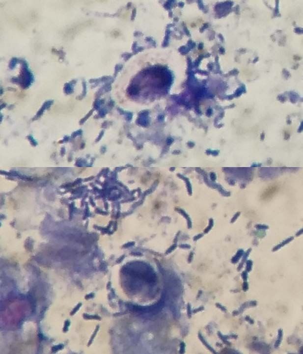
S. Manfredini et al.

                                prescription diet (Hill’s® w/d) and probiotics (Microbiotal®; NBF Lanes) were
                                initially prescribed, however, there was no improvement in the clinical signs
                                after three weeks of treatment. Blood work including haematology and bio-
                                chemistry, serum folate and B12 vitamin were performed and were all within
                                normal limits. ELISA test for feline immunodeficiency virus and feline leukemia
                                virus (Witness FeLV-FIV; Zoetis) were negative. Abdominal ultrasound revealed
                                presence of liquid faeces in the gastrointestinal tract. PCR tests for Tritrichomo-
                                nas foetus and Cryptosporidium spp. were negative. Flotation faecal examina-
                                tion test was repeated yielding negative results. Cytology on a faecal specimen
                                collected by gentle rectal scraping with a cotton swab revealed the presence
                                of oval to rounded organisms with basophilic internal structure surrounded by a
                                clear capsule (Figure 1, Figure 2). Yeast-sustained diarrhoea was suspected and
                                PCR for Prototheca spp. on faeces was performed. Total genomic DNA was iso-
                                lated from faecal material by QIAsymphony SP instrument (Qiagen, Milan, It-
                                aly), using the DSP Virus/Pathogen Mini kit and following the manufacturer’s
                                instructions. PCR was performed in a final reaction volume of 50 µL, in dupli-
                                cate, containing 25 µL of 2× HotStarTaq Master Mix (Qiagen, Milan, Italy), 0.3
                                µM of each primer and 5 µL of DNA template; the reaction was brought to the
                                final volume of 50 µL with PCR Water. The sequence of the primers N476-F
                                (5'-TCGGAGTTAGCTGGTTCTCC-3') and N476-R
                                (5'-ATTTTGGGGCCTTAACTGGT-3') to detect all Prototheca spp. was pre-
                                viously described [12], to produce a 216 bp amplicon. The cycling conditions
                                were: initial activation step at 95˚C for 15 minutes; following by 45 cycles of de-
                                naturation at 95˚C for 30 seconds, annealing at 60˚C for 30 seconds and exten-
                                sion at 72˚C for 60 seconds, followed by a final extension for 15 minutes at 72˚C.
                                Negative control, with distilled water, was included in the PCR reactions. The
                                PCR products were separated by capillary gel electrophoresis using the QIAxcel
                                Advanced (Qiagen, Milan, Italy) and represented as electropherograms by the
                                QIAxcel ScreenGel Software, version 1.5 (Qiagen).

                                Figure 1. Cytology from a faecal sample collected by rectal scraping revealed the presence
                                (arrowheads) of yeast-like oval to rounded organisms with basophilic internal structure
                                surrounded by clear capsule (Romanowsky staining, 1000× magnification).

DOI: 10.4236/ojvm.2021.115011                         159                            Open Journal of Veterinary Medicine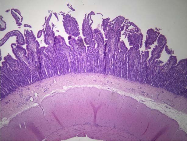
S. Manfredini et al.
Figure 2. Close up picture of the organisms.
PCR was positive for Prototheca (Figure 3). Itraconazole treatment (Itrafun-
gol®; Elanco) at a dose of 5 mg/kg SID per os was prescribed. The stool quality
worsened and the faeces became liquid with undigested food. A hypoallergenic
diet (Hill’s® z/d) and prednisolone (Medrol® VET; Zoetis) at the dose of 0.5 mg/kg
BID per os were added to medical treatment with itraconazole. During the fol-
lowing month the general condition of the cat deteriorated, with reduction in
body weight and progressive worsening of the diarrhoea; repeated episodes of
rectal prolapse also occurred. Medical management with purse-string sutures
were attempted three times. Finally, colopexy was performed and multiple
full-thickness intestinal biopsies were obtained. Abdominal lymphadenopathy
was observed intraoperatively, however sampling of the abnormal-looking
lymph nodes was not performed. Histopathology of duodenum and jejunum in-
testine showed evidence of chronic mild diffuse neutrophilic and lymphoplas-
macellular enteritis and the colon biopsies confirmed chronic mild multifocal
lymphoplasmacellular colitis (Figure 4). Prototheca organisms were detected on
repeated cytological tests of faecal samples. Treatment with nystatin (Mycostatin
100,000 UI/mL; Sanofi) at the dose of 100,000 UI per os every six hours [13] was
introduced, in place of itraconazole, following the owner’s consent for the pro-
posed off-label treatment. Several days after introduction of nystatin, clinical
signs and faecal characteristics improved. Prednisolone dosage was gradually
reduced and discontinued after two weeks. The presence of Prototheca was mo-
nitored by performing weekly cytological tests on faecal samples obtained by
gentle rectal scraping. After two months of treatment with nystatin 100,000 UI
per os every six hours, cytological tests were negative. PCR on faeces was re-
peated two weeks after the last negative cytological fecal test and it was negative.
Nystatin treatment was then interrupted. On follow-up two weeks after the dis-
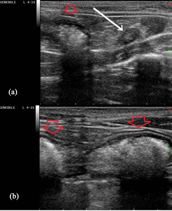
continuation of medication, there was no recurrence of the clinical signs. Ab-
dominal ultrasound confirmed resolution of the previously detected lymphade-
nomegaly (Figure 5). The patient continued to gain weight and was in good
body condition at follow-up, one and two years later. Two years after resolution
DOI: 10.4236/ojvm.2021.115011 160 Open Journal of Veterinary Medicine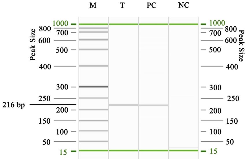
S. Manfredini et al.

                                Figure 3. PCR product of the Prototheca strain object of this study, separated by
                                High-resolution capillary electrophoresis. M: QIAxcel DNA Size Marker, 50 - 800 bp
                                Ladder (Qiagen, Milan, Italy). T: DNA template. PC: Positive control. NC: Negative con-
                                trol.

                                Figure 4. Histopathologic section of jejunum intestine that shows a chronic mild diffuse
                                neutrophilic and lymphoplasmacellular enteritis.

                                Figure 5. Abdominal ultrasonography (10 MHz linear probe) of the caudal portion of the
                                abdomen. Mild regional lymphadenomegaly (white arrow) of the colon (red arrowhead)
                                at the beginning of nystatin treatment (a) that disappears at the end of the treatment (b).

DOI: 10.4236/ojvm.2021.115011                          161                           Open Journal of Veterinary Medicine
S. Manfredini et al.
of the illness, a new female kitten was introduced in the same domestic environ-
ment. At the time of writing the new kitten remained healthy without intestinal
symptoms. The owners have never shown anyclinical signs.
3. Discussion
Intestinal protothecosis has been described in one human patient [5] and in dogs
[9]. In cats, this disease is typically confined to skin nodules and is sensible to
local treatment. To the authors’ knowledge this is the first case report of intes-
tinal protothecosis in a cat with a positive outcome.
Haemorrhagic colitis is the most common complaint in dogs with protothe-
cosis [7]. Breed predisposition to the disease has recently been suggested for
Boxers and Collies, possibly secondary to underlying genetic immunodeficiency
[8]. The common progression of the disease features weight loss and progres-
sively worsening chronic diarrhea, with subsequent dissemination of the algae to
other sites, with ocular and neurological involvement [8]. Death in dogs is due to
generalization of the infection usually within days or weeks [6]. Infection with
Prototheca spp. does not cause pathognomonic laboratory and imaging abnor-
malities [6] [7]. To date, there are no guidelines for the treatment of canine pro-
tothecosis [8].
Similar to previous case reports in dogs, intestinal protothecosis caused hae-
morrhagic colitis with progressive worsening in the general condition in the cat
of the present case report. No relevant laboratory abnormalities were detected.
The cat was FIV-FeLV negative and no comorbidity was found. In this case there
was no generalized dissemination of the infection; this can be explained by the
natural resistance of the feline species to infection [9], alongside the beneficial
treatment with nystatin. This drug is a polyene antibiotic with antifungal activi-
ty, with a spectrum of activity and mechanism of action similar to its novel se-
misynthetic derivative Amphotericin B and is relatively inexpensive. Nystatin is
not absorbed after oral administration and it is almost entirely excreted un-
changed in the faeces [14]. Orally administered Nystatin is used primarily for the
treatment of oral or gastrointestinal Candida infections in dogs, cats, and birds;
it has been used less commonly in other species for the same indications [14].
Nystatin binds to sterols in the membrane of the fungal cell, altering the per-
meability of the membrane and allowing intracellular potassium and other cel-
lular constituents to “leak out” [14]. In vitro studies have shown that Prototheca
spp. is sensitive to Amphotericin B, azoles and a wide range of antibacterial agents
[15]. However, none of these compounds have shown convincing efficacy in
dogs [7] [16]. When itraconazole was used, alone or together with enrofloxacin,
it induced only partial and temporary remission of clinical signs in dogs [8]. In
this case report treatment with itraconazole did not improve clinical signs nor
reduced the number of Prototheca organisms in repeated fecal cytological ex-
ams. On the other hand, nystatin alone appeared to be effective. It is the authors’
opinion that treatment with nystatin played a key role in the resolution of the
infection sustained by Prototheca in the present case.
DOI: 10.4236/ojvm.2021.115011 162 Open Journal of Veterinary MedicineS. Manfredini et al.
Interestingly, the patient was an exclusively indoor cat. The source of the in-
fection is not known, as none of the other kittens or the owners had symptoms.
The potential focus in the domestic environment was not evaluated. The cat’s
owners did not report any similar symptoms. Protothecosis is not considered a
zoonosis, but cases in veterinary patients should be considered as a potential for
environmental risk for humans.
The cat is a Bengal cat. Further studies should aim at identifying genetic or
breed-predisposing factors in cats, similar to those suspected in Boxer and Collie
breeds, as well as the role of the environment in being a favorable condition for
infection.
Limitations of the present study are the lack of typing of Prototheca isolated
from the intestinal biopsies. Additionally, histopathology of mesenteric lymph
nodes was not performed; this would have allowed diagnosis of an inflammatory
lymphadenopathy or a potential progression and dissemination of the infectious
disease.
This case report suggests that protothecosis should be listed in the differential
diagnosis of colitis and diarrhoea in cats. Prototheca infection can be suspected
with a simple, inexpensive cytology smears from rectal scrapings. In the authors’
opinion nystatin should be considered for the treatment for intestinal protothe-
cosis in cats as well as in dogs. In fact, in dogs the disease appears to be more
aggressive than in cats, and no known effective treatment is currently available.
Authors declare to have obtained an informed consent from the cat’s owners to
report this case.
Conflicts of Interest
The Authors declare that there is no conflict of interest.
Authors’ Contributions
Dr. Sara Manfredini was the clinician responsible for the case management. Dr.
Luigi Venco performed cytology on faeces, requested PCR tests for Prototheca
and introduced the treatment with nystatin. He also performed abdominal ul-
trasounds for the case. Dr. Luca Formaggini was responsible for the surgery. Dr.
Michele Marino was the consultant for the laboratory PCR test. All four authors
contributed to write and edit the manuscript.
The authors thank Prof Laura Kramer, Ph.D., Dip. EVPC, EBVSTM President,
for revising the English language of the manuscript.
References
[1] Sykes, J.E. (2014) Chapter 70—Protothecosis. In: Sykes, J.E., Eds., Canine and Feline
Infectious Diseases, Elsevier, Saunders Inc., St. Louis, USA, 679-685.
https://doi.org/10.1016/B978-1-4377-0795-3.00070-3
[2] Danesi, P., Falcaro, C., Binanti, D., et al. (2018) Abstract No: S2.6c. In: Congress
Mycology. https://www.isham2018.org/ (Accessed 15 March 2019)
[3] Pfaller, M.A. and Diekema, D.J. (2005) Unusual Fungal and Pseudofungal Infec-
DOI: 10.4236/ojvm.2021.115011 163 Open Journal of Veterinary MedicineS. Manfredini et al.
tions of Humans. Journal of Clinical Microbiology, 43, 1495-1504.
https://doi.org/10.1128/JCM.43.4.1495-1504.2005
[4] Lass-Flor, C. and Mayr, A. (2007) Human Protothecosis. Journal of Clinical Micro-
biology, 20, 230-242. https://doi.org/10.1128/CMR.00032-06
[5] Konzi, K., et al. (2019) A Case of Intestinal Protothecosis. Médecine et Maladies
Infectieuses, 49, 621-623. https://doi.org/10.1016/j.medmal.2019.09.002
[6] Pressler, B.M., Gookin, J.L., Sykes, J.E., et al. (2005) Urinary Tract Manifestations of
Protothecosis in Dogs. Journal of Veterinary Internal Medicine, 19, 115-119.
https://doi.org/10.1111/j.1939-1676.2005.tb02669.x
[7] Stenner, V.J., Mackay, B., King, T., et al. (2007) Protothecosis in 17 Australian Dogs
and a Review of the Canine Literature. Medical Mycology, 45, 249-266.
https://doi.org/10.1080/13693780601187158
[8] Bottero, E., Mercuriali, E., Abramo, F., et al. (2016) Fatal Protothecosis in Four
Dogs with Large Bowel Disease in Italy. Wiener Tierärztliche Monatsschrift-Vete-
rinary Medicine Austria, Wien.
[9] Nardoni, S. and Mancianti, F. (2018) Micologia in Traversa D. In: Venco, L., Eds.,
Parassitologia del cane e del gatto, Point Veterinaire Italie, Milano, Italy, 351-376
[10] Sykes, J.E. and Greene, E.G. (2012) Infectious Diseases of the Dog and Cat. 4th Edi-
tion, Saunders, Elsevier Inc., St. Louis, USA.
[11] Mahendra, P., Ashebr, A., Tanvir, R., et al. (2014) Protothecosis: An Emerging Algal
disease of Humans and Animals. International Journal of Life science and Pharma
Research, 3, 16 p.
[12] Capra, E., Cremonesi, P., Cortimiglia, C., Bignoli, G., Ricchi, M., Moroni, P., Pesce,
A., Luini, M. and Castiglioni, B. (2014) Simultaneous Identification by Multiplex
PCR of Major Prototheca spp. Isolated From Bovine and Buffalo Intramammary
Infection and Bulk Tank. Letters in Applied Microbiology, 59, 642-647.
https://doi.org/10.1111/lam.12326
[13] Kirk, R.W. (1989) Current Veterinary Therapy X: Small Animal Practice. W.B.
Saunders, Philadelphia.
[14] Plumb, D.C. and Pharm, D. (2011) Veterinary Drug Handbook. 7th Edition, Phar-
maVet Inc., Stockholm, Wisconsin, USA, 1890-1893.
[15] Sapierzynski, R. and Jaworska, O. (2008) Protothecosis as a Cause of Chronic Di-
arrhoea in a Dog. Polish Journal of Veterinary Sciences, 11, 225-229.
[16] Sobukawa, H., Kano, R., Ito, T., et al. (2011) In vitro Susceptibility of Prototheca zopfii
Genotypes 1 and 2. Journal of Medical Mycology, 49, 222-224.
https://doi.org/10.3109/13693786.2010.511285
DOI: 10.4236/ojvm.2021.115011 164 Open Journal of Veterinary MedicineYou can also read