Case Report Atypical Salmonellosis in a Horse: Implications for Hospital Safety
←
→
Page content transcription
If your browser does not render page correctly, please read the page content below
Hindawi
Case Reports in Veterinary Medicine
Volume 2020, Article ID 7062408, 5 pages
https://doi.org/10.1155/2020/7062408

Case Report
Atypical Salmonellosis in a Horse: Implications for Hospital Safety

          Kristina L. Rothers,1 Eileen S. Hackett,1 Gary L. Mason,2 and Brad B. Nelson                                   1

          1
              Department of Clinical Sciences, Colorado State University, 300 West Drake Road, Fort Collins, CO 80523, USA
          2
              Veterinary Diagnostic Laboratory, Colorado State University, 300 West Lake Road, Fort Collins, CO 80523, USA

          Correspondence should be addressed to Brad B. Nelson; brad.nelson@colostate.edu

          Received 4 March 2020; Revised 13 May 2020; Accepted 16 May 2020; Published 4 June 2020

          Academic Editor: Giuseppe Mazzullo

          Copyright © 2020 Kristina L. Rothers et al. This is an open access article distributed under the Creative Commons Attribution
          License, which permits unrestricted use, distribution, and reproduction in any medium, provided the original work is
          properly cited.

          A 17-year-old Quarter Horse mare was evaluated for colic of 24-hour duration. Clinical signs and diagnostic evaluation were
          consistent with duodenitis-proximal jejunitis. The horse’s clinical condition deteriorated despite medical treatment and was
          euthanized. Aerobic culture collected from small intestinal ingesta was positive for Salmonella enterica subsp. enterica serovar
          Hadar. Salmonella sp. is commonly implicated in nosocomial infections in equine veterinary hospitals usually through feces
          containing the organism. Considering Salmonella sp. was cultured from the jejunal luminal contents and the large volume of
          nasogastric reflux that was evacuated in this case, a perceived risk of Salmonella sp. transmission from infected gastric reflux to
          other hospitalized cases was realized. Infectious agent biosecurity precautions should be undertaken in horses with nasogastric
          reflux to prevent hospital-acquired transmission.

1. Introduction                                                          horse with large quantities of gastrointestinal reflux, fever,
                                                                         and a diagnosis of enteritis attributed to an atypical Salmo-
Salmonella sp. infection in adult horses is typically associated         nella enterica serovar. The clinical and laboratory findings
with enterocolitis manifesting with diarrhea and hypopro-                highlight the potential risk of disease transmission from
teinemia [1]. Atypical presentations of salmonellosis can                sources other than feces and emphasize the importance of
include signs such as voluminous gastric reflux and small                 considering alternate sources of Salmonella sp. transmission.
intestinal ileus without diarrhea [1]. Horses can also be
asymptomatic carriers, thereby shedding Salmonella spp.                  2. Case Presentation
into the environment without any clinical signs of disease
[2]. Nosocomial infections from Salmonella spp. are the most             A 17-year-old Quarter Horse mare was referred to the Colo-
common source of disease transmission in equine hospitals                rado State University Veterinary Teaching Hospital for colic
[3, 4]. Horses presenting with fever, loose feces, and neutro-           of 24-hour duration. The horse was anorexic and lying down
penia or with presumptive diagnoses of anterior enteritis or             frequently the day prior to presentation. Findings from the
colitis warrant the implementation of biosecurity protocols              referring veterinarian at that time were rectal temperature
to minimize the risk of hospital-acquired infections [3–6].              of 102°F (38.9°C), heart rate of 50 beats/minute (bpm),
Salmonella sp. is most commonly cultured from feces of                   reduced borborygmi in all four quadrants, and 6 liters of
horses with colic, and the potential risk of infection is usually        net nasogastric reflux. Flunixin meglumine (1.1 mg/kg body
attributed to fecal contamination disseminating throughout               weight (BW) IV) administration resulted in temporary
the facility [3, 4]. This identified risk has prompted routine            improvement in clinical signs. Within 12 hours, the horse
surveillance fecal culturing as a method to detect shedding              began showing mild signs of colic and was referred for fur-
of the organism and enables the ability to segregate and                 ther evaluation and treatment.
maintain barriers between Salmonella sp. shedding horses                     On hospital admission, the mare weighed 530 kg and had
and the general population [3, 7]. This report describes a               a rectal temperature of 102°F (38.9°C), heart rate of 54 bpm,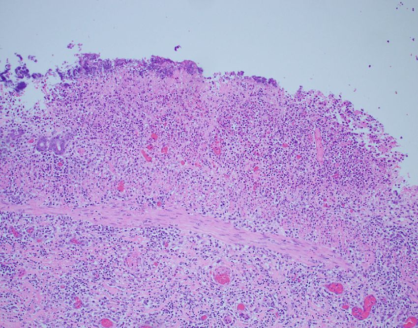
2                                                                                             Case Reports in Veterinary Medicine

Figure 1: Gross appearance of the jejunal mucosa on cut section. Note the prominent edema, mural thickening, and reddening visible. The
inset shows the jejunal mucosa magnified.

reduced borborygmi in all four quadrants, and hyperemic              esis fluid was collected and was grossly serosanguinous. The
mucous membranes. Following nasogastric intubation, 10               peritoneal fluid had a total protein of 38 g/L and a total
liters of net reflux was obtained. Transabdominal ultrasound          nucleated cell count of 1470 cells/μL, with differential cell
examination revealed hypomotile, nondilated loops of the             proportions of 40% nondegenerate neutrophils, 40% large
small intestine with 6-8 mm wall thickness and minimal peri-         mononuclear cells, and 20% lymphocytes. Despite the con-
toneal fluid in the ventral abdomen. Complete blood cell              tinued medical therapies described previously, clinical signs
count examination identified leukopenia (4:6 × 10⁹ /L; refer-         did not improve over the next 30 hours of treatment. After
ence interval (RI): 5.5–10:5 × 10⁹ /L) characterized by neutro-      discussions of case progression and minimal response to
penia (2:6 × 10⁹ /L; RI: 3.0–7:0 × 109 /L) with a left shift         medical treatments, the owner elected euthanasia.
(0:2 × 10⁹ /L; RI: 0.0–0:1 × 10⁹ /L) and slight toxic changes, as         Postmortem examination revealed diffuse edema and
well as hyperfibrinogenemia (7.0 g/L; RI: 1.0–4.0 g/L). Serum         mural thickening of the jejunum and ileum (Figure 1). The
biochemistry revealed hypophosphatemia (1.1 mmol/L; RI:              mucosa had a dull reddish discoloration, and the mesenteric
1.7–4.5 mmol/L), hypocalcaemia (1.09 mmol/L; RI: 1.15–               lymph nodes were hemorrhagic. There was also a mild red
1.40 mmol/L), hypomagnesemia (1.4 mmol/L; RI: 1.6–                   discoloration on the serosal surfaces of the duodenum to
2.2 mmol/L), hyperproteinemia (80 g/L; RI: 58–74 g/L), hyper-        the cecum. The tissues were processed routinely for histopa-
globulinemia (5.1 mmol/L; RI: 2.5–4.5 mmol/L), and increased         thology. Histopathologic findings revealed suppurative gas-
creatine kinase (6.6 mmol/L; RI: 1.0–4.7 mmol/L), aspartate          tritis with ulceration, acute erosive necrotizing enteritis
aminotransferase (3.97 mmol/L; RI: 1.85–3.75 mmol/L) and             with Paneth cell metaplasia (Figure 2), colonic arteritis with
gamma-glutamyl transferase (28 U/L; RI: 10–25 IU/L).                 thrombosis, and portal to centrilobular bacterial hepatitis.
     An intravenous jugular catheter was placed, and an initial      Jejunal luminal contents as well as fecal samples were submit-
bolus of 10 liters (20 mL/kg BW) of isotonic crystalloid fluids       ted for aerobic culture. Samples were added to tetrathionate
(Veterinary Plasma-Lyte A, Abbott, North Chicago, Illinois,          broth supplemented with iodine incubated at 42 degrees C
USA) was administered. Crystalloid fluids were then contin-           overnight. Tetrathionate was subcultured to Xylose Lysine
ued at a rate of 3 L/hr (approx. 5.7 mL/kg BW/hr), with cal-         Tergitol 4 (XLT4) agar and incubated at 35 degrees C over-
cium gluconate (5.6 g/L) and magnesium sulfate (400 mg/L)            night. A representative colony was identified as Salmonella
supplementation. Additional treatments consisted of lido-            spp. Using triple sugar iron agar and agglutination in poly-
caine (50 μg/kg BW/min), polymyxin B (1,000 IU/kg BW                 O antisera, the isolate agglutinated in C2 antisera. Serotyping
IV q 12 hr), ranitidine (1.1 mg/kg BW IV q 8 hr), and flunixin        was performed by a reference laboratory (National Veteri-
meglumine (0.7 mg/kg BW IV q 12 hr). Gastric decompres-              nary Services Laboratories, Ames, IA, USA), which identified
sion was performed q 2 hr and resulted in approximately              Salmonella enterica subsp. enterica, group C2, serovar Hadar
4 L net reflux per hr.                                                (Salmonella Hadar). Growth of Clostridium perfringens was
     After 18 hrs of medical treatment, rectal temperature was       also isolated from the small intestinal contents.
102.5°F (39.2°C), and there was decreased borborygmi,
hyperemic mucous membranes, and persistent colic charac-             3. Discussion
terized by pacing and muscle fasciculations. A venous blood
gas revealed mild acidosis (pH 7.3) and decreased ionized cal-       This report describes a horse with fever, nasogastric reflux,
cium (1.2 mmol/L; RI: 1.3–1.9 mmol/L). Repeat ultrasound             and a suspected diagnosis of duodenitis-proximal jejunitis
examination revealed 5-6 cm dilated and hypomotile small             (DPJ) with a positive anaerobic culture of Salmonella Hadar
intestinal loops with 3-6 mm wall thickness. Abdominocent-           from small intestinal ingesta. Gross pathological features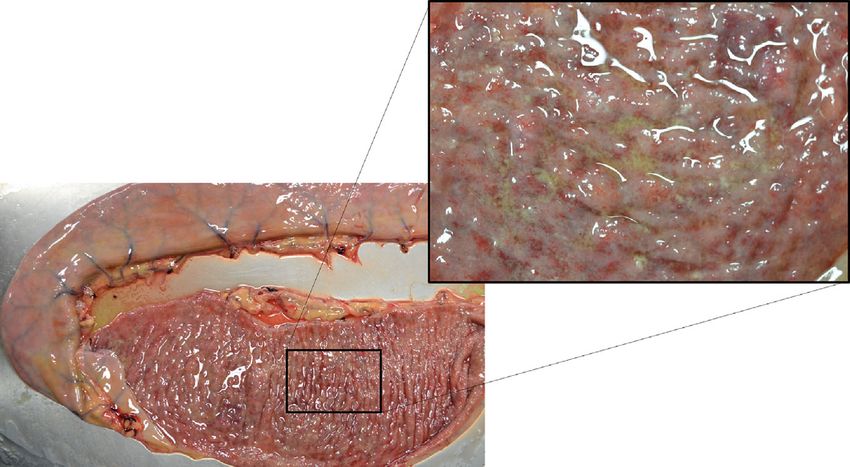
Case Reports in Veterinary Medicine 3
(a) (b)
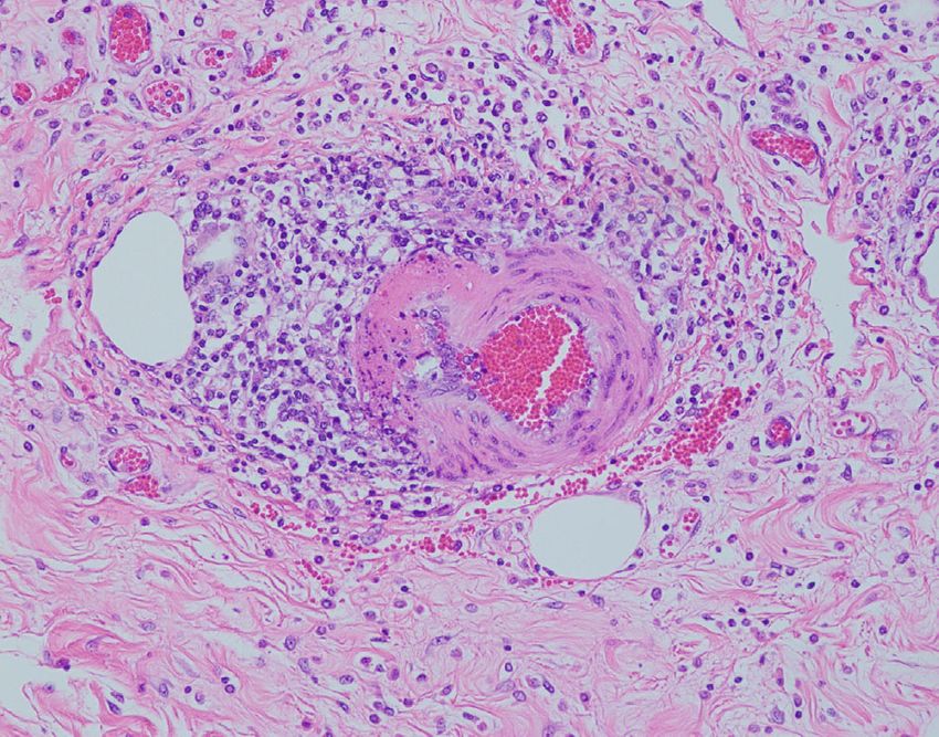
Figure 2: (a) Photomicrograph of the small intestinal mucosa and submucosa. The lamina propria and submucosa are expanded by chronic
nonsuppurative inflammation with vascular congestion, acute erosive necrotizing enteritis, and bacterial colonization of necrotic surface
debris. Hematoxylin and eosin, ×100 magnification. (b) Photomicrograph of the small intestinal submucosa. Periarterial nonsuppurative
inflammation is visible, with acute changes including lymphatic dilation, edema, and segmental leukocytoclastic necrotizing arteritis.
Hematoxylin and eosin, ×100 magnification.
consistent with salmonellosis included diffuse fibrinous and [14]. Diagnosis is usually confirmed surgically or at necropsy
hemorrhagic inflammation of the jejunum and ileum. Histo- with hyperemia, edema, hemorrhage, and necrosis involving
logical assessment further characterized the intestinal lesions the affected intestinal segments [9]. The horse in this case had
as acute erosive necrotizing enteritis with chronic inflamma- lesions in the jejunum and ileum, consisting of edema and
tion in the lamina propria and submucosa in conjunction mural thickening. Culture of the affected small intestinal
with vascular congestion and bacterial colonization of contents revealed Salmonella Hadar and Clostridium perfrin-
necrotic surface debris [8]. While many of the features gens, while the fecal culture was positive for Salmonella
observed in this case are consistent with DPJ, the apparent Hadar. Additionally, there was hemorrhage in the mesenteric
lack of involvement of the duodenum is uncharacteristic of lymph nodes as well as Paneth cell metaplasia consistent with
the disease [1, 9, 10]. a nonacute disease process [15].
The horse in this case presented with signs typical of DPJ Salmonellosis typically manifests in adult horses as
including fever, voluminous nasogastric reflux, and colic enterocolitis with acute severe diarrhea and protein-losing
signs that resolved shortly following gastric decompression. enteropathy [1]. However, horses may also be latent subclin-
Clinicopathologic variables commonly encountered in cases ical carriers that shed during stress or present to the hospital
with DPJ were also observed, though these electrolyte abnor- as neonatal foals with bacteremia [1]. Salmonella enterica ssp.
malities can be variable depending on the stage of the disease enterica accounts for approximately 60% of all Salmonella
process [9]. The peritoneal total protein was increased with- subspecies and approximately 99% of the clinical and sub-
out elevation in the total nucleated cell count, and elevated clinical infections in warm-blooded animals [16]. Considered
peritoneal total protein has been associated with increased an opportunistic pathogen, Salmonella enterica sp. is trans-
mortality in horses with DPJ [11]. The cause of DPJ in horses mitted by fecal-oral routes and colonizes sections of the gas-
remains elusive [9]. Bacterial agents including Clostridium trointestinal tract, disrupting normal physiologic processes
difficile, Clostridium perfringens, and Salmonella spp., as well of absorption and secretion [1, 8]. Commonly affecting the
as parasitic infections and toxins including mycotoxins colon in horses, the disruption of the colonic wall permits
(fumonisin B1), have all been implicated as potential etiolo- protein loss and the inability to reabsorb water, leading to
gies based on their isolation from affected cases [9]. Though diarrhea. While this horse did have a neutropenia with a left
Clostridium difficile and its toxins are commonly implicated shift likely due to neutrophil migration into the affected
in the pathogenesis of DPJ [1, 12, 13], data to confirm this intestinal tissues as is common with salmonellosis, there
hypothesis is lacking [9]. was no diarrhea or hypoproteinemia observed. A majority
Subsequent inflammation of the duodenum and proxi- of equine salmonellosis cases are associated with Salmonella
mal jejunum is characteristic of DPJ [1, 9, 10]. The inflamma- enterica serovars Typhimurium, Newport, Javiana, Braen-
tion reduces intestinal absorption while increasing intestinal derup, Anatum, Infantis, Muenchen, and Mbandaka [3, 6,
secretions into the lumen causing distension, thereby 17, 18]. Salmonella Hadar is a rare serovar encountered in
compromising intestinal peristalsis and culminating in ileus. horses with intestinal disease and to our knowledge has not
Varying degrees of dehydration and endotoxemia are present been detected in horses outside of the Netherlands [19, 20].
and reflect the severity and chronicity of the stage of disease More commonly, Salmonella Hadar is associated with poultry4 Case Reports in Veterinary Medicine
and foodborne illness outbreaks in humans [21, 22]. None- Data Availability
theless, a clinical disease outbreak has occurred in a human
maternity and neonatal hospital ward in the United King- There are no supplementary data for this article.
dom [23]. Salmonella sp. has zoonotic potential, and contam-
inated fluids and excretions from infected horses should be Conflicts of Interest
avoided, though it commonly does not impact humans out-
side of those with compromised immune systems. The authors declare that there is no conflict of interest
Salmonella sp. outbreaks are a common source of noso- regarding the publication of this case report. The authors
comial infections in equine hospitals [3, 4, 6]. Therefore, bio- are all employed by Colorado State University, and work
security protocols should be in place when an atypical was performed under this employment.
presentation occurs. Monitoring equine hospital patient
safety requires education, implementation of effective biose- Acknowledgments
curity protocols, and surveillance for contagious pathogens
[3]. Using routine surveillance and raising awareness of cases We acknowledge Dr. Alexander Daniel and the technical staff
that potentially shed Salmonella spp. are critical to prevent at the Equine Veterinary Teaching Hospital at Colorado State
hospital-acquired infections. Once a case is identified, the University for medical care of this horse.
patient can be isolated from the general population and addi-
tional precautions to prevent disease transmission are imple- References
mented. Identified risk factors of nosocomial colonization
include exposure to hospitalized horses shedding Salmonella [1] D. J. Feary and D. M. Hassel, “Enteritis and colitis in horses,”
spp., either with direct contact or with environmental con- Veterinary Clinics: Equine Practice, vol. 22, no. 2, pp. 437–
tamination, high ambient temperatures, treatment with anti- 479, 2006.
microbial agents, concurrent gastrointestinal tract disease, [2] A. Merritt, J. Robbins, and B. Brewer, “Is Salmonella infection
changes in diet, and use of contaminated equipment amongst a cause of the acute gastric dilitation/ileus syndrome in
patients (rectal thermometers, nasogastric tubes) [17]. An horses?,” Proceedings of the Equine Colic Research Symposium:
1982, pp. 119–124, Athens, GA, 1982.
awareness of these risk factors will help guide individual bio-
security protocols to address these potential risks. In the [3] B. A. Burgess and P. S. Morley, “Managing Salmonella in
equine populations,” The Veterinary Clinics of North America.
report of Salmonella Hadar transmission in a human mater-
Equine Practice, vol. 30, no. 3, pp. 623–640, 2014.
nity ward [23], the index case was the mother of an admitted
[4] B. A. Burgess and P. S. Morley, “Risk factors for shedding of
child with diarrhea and subsequent disease transmission was
Salmonella enterica among hospitalized large animals over a
spread to 11 neonates over 3 months. Though predominantly 10-year period in a veterinary teaching hospital,” Journal of
neonates were infected, the duration of the outbreak sug- Veterinary Internal Medicine, vol. 33, no. 5, pp. 2239–2248,
gested a difficulty in controlling disease propagation and a 2019.
potential increased survivability of Salmonella Hadar in the [5] B. A. Burgess, K. Bauknecht, N. M. Slovis, and P. S. Morley,
environment despite routine hospital cleaning protocols [23]. “Factors associated with equine shedding of multi-drug-
Clinically normal horses or horses without diarrhea may resistant Salmonella enterica and its impact on health out-
have the organism in their feces and pose an infection risk to comes,” Equine Veterinary Journal, vol. 50, no. 5, pp. 616–
susceptible animals. Using barrier precautions, disinfecting 623, 2018.
common equipment, and being careful of disposal of feces [6] B. L. Dallap Schaer, H. Aceto, and S. C. Rankin, “Outbreak of
and as this case suggests gastric reflux fluid can help reduce salmonellosis caused by Salmonella enterica serovar Newport
exposure to the general hospital population. The positive cul- MDR-AmpC in a large animal veterinary teaching hospital,”
ture of Salmonella Hadar from the ingesta of the small intes- Journal of Veterinary Internal Medicine, vol. 24, no. 5,
tine highlights the potential of recovered gastric reflux to be a pp. 1138–1146, 2010.
source of Salmonella sp. transmission, especially when large [7] B. A. Burgess, N. R. Noyes, D. S. Bolte, D. R. Hyatt, D. C. van
volumes of gastric reflux are evacuated. In these patients Metre, and P. S. Morley, “Rapid Salmonella detection in exper-
imentally inoculated equine faecal and veterinary hospital
without diarrhea, this gastric fluid could serve as a conduit
environmental samples using commercially available lateral
for nosocomial infections. Thus, biosecurity protocols and flow immunoassays,” Equine Veterinary Journal, vol. 47,
procedures should not neglect the potential transmission no. 1, pp. 119–122, 2015.
routes of infectious disease in horses without diarrhea. [8] W. J. Saville, K. W. Hinchcliff, B. R. Moore et al., “Necrotizing
This case illustrates the importance of considering an enterocolitis in horses: a retrospective study,” Journal of Veter-
infectious agent, namely, Salmonella sp., as an etiology for inary Internal Medicine, vol. 10, no. 4, pp. 265–270, 1996.
atypical clinical presentations of gastrointestinal disease. [9] L. G. Arroyo, D. E. Gomez, and C. Martins, “Equine
Despite the clinical signs being typical of DPJ, a diagnosis duodenitis-proximal jejunitis: a review,” The Canadian veteri-
of enteritis with Salmonella Hadar infection was detected. nary journal=La revue veterinaire canadienne, vol. 59, no. 5,
The atypical presentation of Salmonella spp. detected in the pp. 510–517, 2018.
jejunal fluid coupled with the large volume of reflux retrieved [10] D. E. Freeman, “Duodenitis-proximal jejunitis,” Equine Veter-
during gastric decompression highlights the importance of inary Education, vol. 12, no. 6, pp. 322–332, 2000.
considering alternative nosocomial transmission pathways [11] T. L. Seahorn, J. L. Cornick, and N. D. Cohen, “Prognostic
to protect the safety of hospital patients. indicators for horses with duodenitis-proximal Jejunitis 75Case Reports in Veterinary Medicine 5
horses (1985-1989),” Journal of Veterinary Internal Medicine,
vol. 6, no. 6, pp. 307–311, 1992.
[12] L. G. Arroyo, H. Staempfli, J. D. Rousseau et al., “Culture
evaluation of Clostridium spp. in the nasogastric reflux of
horses with duodenitis proximal jejunitis,” Proceedings of the
Eighth International Equine Colic Research Symposium: 2005,
pp. 51-52, Lexington (KY), 2005.
[13] L. G. Arroyo, M. C. Costa, B. B. Guest, B. L. Plattner, B. N. Lil-
lie, and J. S. Weese, “Duodenitis-proximal jejunitis in horses
after experimental administration of Clostridium difficile
toxins,” Journal of Veterinary Internal Medicine, vol. 31,
no. 1, pp. 158–163, 2017.
[14] J. Schumacher, J. F. Edwards, and N. D. Cohen, “Chronic idi-
opathic inflammatory bowel diseases of the horse,” Journal of
Veterinary Internal Medicine, vol. 14, no. 3, pp. 258–265, 2000.
[15] N. Gassler, “Paneth cells in intestinal physiology and patho-
physiology,” World J Gastrointest Pathophysiol, vol. 8, no. 4,
pp. 150–160, 2017.
[16] R. Lan, P. R. Reeves, and S. Octavia, “Population structure, ori-
gins and evolution of major Salmonella enterica clones,” Infec-
tion, Genetics and Evolution, vol. 9, no. 5, pp. 996–1005, 2009.
[17] H. C. Schott II, S. L. Ewart, R. D. Walker et al., “An outbreak of
salmonellosis among horses at a veterinary teaching hospital,”
Journal of the American Veterinary Medical Association,
vol. 218, no. 7, pp. 1152–1159, 2001.
[18] B. A. Burgess, C. B. Weller, K. L. Pabilonia, D. S. Bolte, D. C.
Van Metre, and P. S. Morley, “Detection of different serotypes
of Salmonella enterica in experimentally inoculated equine
fecal samples by commercially available rapid tests,” Journal
of Veterinary Internal Medicine, vol. 28, no. 6, pp. 1853–
1859, 2014.
[19] E. van Duijkeren, M. S. van Oldruitenborgh-Oosterbaan,
D. Houwers, W. van Leeuwen, and H. Kalsbeek, “Equine sal-
monellosis in a Dutch veterinary teaching hospital,” Veteri-
nary Record, vol. 135, no. 11, pp. 248–250, 1994.
[20] E. van Duijkeren, B. van Klingeren, and A. G. Vulto, “In vitro
susceptibility to antimicrobial drugs of 62 Salmonella strains
isolated from horses in the Netherlands,” Veterinary Microbi-
ology, vol. 45, no. 1, pp. 19–26, 1995.
[21] F. Sampedro, S. J. Wells, J. B. Bender, and C. W. Hedberg,
“Developing a risk management framework to improve public
health outcomes by enumeratingSalmonellain ground turkey,”
Epidemiology and Infection, vol. 147, 2019.
[22] B. R. Jackson, P. M. Griffin, D. Cole, K. A. Walsh, and S. J.
Chai, “Outbreak-associated Salmonella enterica serotypes
and food commodities, United States, 1998-2008,” Emerging
Infectious Diseases, vol. 19, no. 8, pp. 1239–1244, 2013.
[23] A. Deshpande, E. T. Curran, S. Jamdar, T. Inkster, and B. L.
Jones, “Historical outbreak of Salmonella Hadar,” The Journal
of Hospital Infection, vol. 91, no. 2, pp. 171–175, 2015.You can also read